1323 E InCeram - Panadent · VITA In-Ceram ALUMINA 40 VITA In-Ceram SPINELL 41 VITA In-Ceram...
88
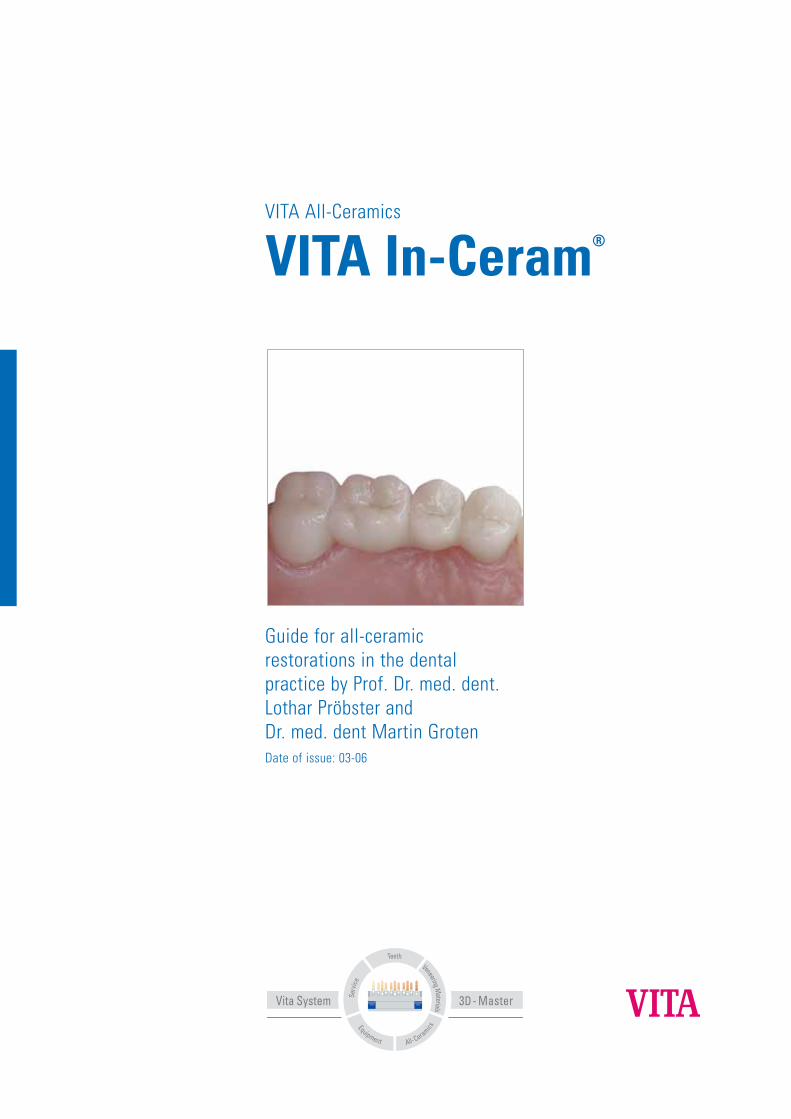
3D - Master Vita System E quip m e nt A ll - C e r a m ic s S e r v ic e V e n e e r i n g M a t e r ia l s T eet h VITA In-Ceram ® VITA All-Ceramics Guide for all-ceramic restorations in the dental practice by Prof. Dr. med. dent. Lothar Pröbster and Dr. med. dent Martin Groten Date of issue: 03-06
-
Upload
truongtuyen -
Category
Documents
-
view
226 -
download
0
Transcript of 1323 E InCeram - Panadent · VITA In-Ceram ALUMINA 40 VITA In-Ceram SPINELL 41 VITA In-Ceram...
3D - MasterVita System
Equipment All-Ceramics
Serv
ice
Veneering Materials
Teeth
VITA In-Ceram®
VITA All-Ceramics
Guide for all-ceramic restorations in the dental practice by Prof. Dr. med. dent.Lothar Pröbster andDr. med. dent Martin Groten Date of issue: 03-06
Professor Dr. med. dent. Lothar Pröbster
Lothar Pröbster, born in 1958, was an assistant in a practiceafter completing his dental examination in 1983; from 1985 to1997 he worked as a scientific staff member for Professor Dr. E.Körber and Professor Dr. H. Weber and the policlinics for DentalProsthodontics of the University of Tübingen. During his time atthe university he was preclinic director, one of the projectmanagers of the special research field of implantology andclinical senior dentist. He habilitated in 1995 and in 1997 hewas appointed specialist for prosthodontics of the GermanAssociation of Prosthodontics and Dental Materials. Since 1997he has been co-owner of a dental practice in Wiesbaden. In2001 he was appointed associate professor by the Universityof Tübingen.
His fields of activities include adhesive and implant restorations,material science and, above all, all-ceramic restoration systems.Up until today he has been dealing closely with all-ceramicprocedures, which in 2001 resulted in the co-authorship of thescientific statement of the German Society of Dental Oral andCraniomandibular Sciences on the scientific recognition ofall-ceramic crowns and bridges. So far his clinical-scientificactivities comprise more than 100 publications, a book andapprox. 300 speeches, seminars and workshops. Prof. Pröbsteris a member in some professional associations and works as anexpert/advisor for Deutsche Forschungsgemeinschaft (GermanResearch Foundation) and for dental magazines.
Dr. med. dent. Martin Groten
Martin Groten, born in 1965, is senior dentist at the policlinicsfor Dental Prosthodontics and the Department of MedicalMaterials and Technology (Medical Director: Prof. Dr. HeinerWeber) at the Center for Dental, Oral and MaxillofacialMedicine of the University of Tübingen. His main activities atthe clinic include the areas of fixed, combined and implant-sup-ported restorations, all-ceramic restorations and the use ofminimally invasive and adhesive techniques in prosthodontics.Additionally, he provides scientific and photographic documen-tation of clinical treatment procedures.
As clinical examiner in charge, he has been dealing withplanning, design, performance and evaluation of clinicalstudies and the rules of clinical tests of medical products. Asassistant director of the Steinbeis-Transferzentrum DentalProducts/Clinical Testing/Certification (STZ-DCZC) he has beensupervising its accreditation as medical institute for clinicaltesting of medical products according to the Directive93/42/EWG for Medical Devices (MDD), EN ISO 17025 andthe ICH E6 guidelines for Good Clinical Practice (GCP). Hegives numerous speeches in Germany and in foreign countriesand is the author or co-author on a series of publications anda manual on clinical testing of medical devices in English.Since 1993 he has been training students and involved in thedevelopment of modern teaching concepts. He has been incharge of preclinical education of dental students at theUniversity of Tübingen since 1999.
Preface
Currently, the fabrication of all-ceramic restorationsreflects the strongest trend in restorative dentistry. Basedon procedural and material-scientific innovations, inlays,crowns, bridges, primary telescopes, implant suprastructuresand even implants can be produced exclusively from ceramic,that is without any metal affecting aesthetics or biocompatibili-ty. These materials allow to restore "white esthetics" of teethtrue to nature.
VITA Zahnfabrik has gathered experience with all-ceramicmaterials over decades and become one of the leadingmanufacturers worldwide.
With this brochure we intend to provide a description of thewide application range of the VITA In-Ceram product rangeand show the application possibilities for the daily practice.
We wish all readers success in working with the highlyaesthetic and proven VITA In-Ceram materials.
Wiesbaden and Tübingen, June 2006
Prof. Dr. Lothar Pröbster
Schöne Aussicht 18
65193 Wiesbaden
Dr. Martin Groten
Universitätsklinikum
Zentrum für Zahn-, Mund- und
Kieferheilkunde
Osianderstraße 2-8
72076 Tübingen
Acknowledgement
The authors would like to use the publication of the new VITAIn-Ceram brochure to thank the numerous persons who havecontributed to our results with the VITA In-Ceram system overthe years.
First of all we would like to thank the active and former dentaltechnicians at the Center for Dental, Oral and MaxillofacialMedicine of the University of Tübingen for numerous years ofcooperation and the fabrication of the VITA In-Ceram restora-tions: Bettina Vogel, Susanne Deiser, Karina Schmidt, JochenDiel, Volker Scheer and Ekkehardt Kröverath. We would also like to thank our dental assistants for theirdependability and help and especially for their patience duringthe production of the clinical photos: Patricia Scholze, ZizaGhaxeri and Silke Saile.
We would like to thank Sonja Ganz and Kurt Reichel, ReichelZahntechnik in Hermeskeil, for the fabrication of the VITAIn-Ceram YZ restorations and for providing the photos illustratingthe respective dental-technical steps.
We also owe our thanks to all staff members and colleagueswhose clinical and scientific commitment was an indispensablecontribution to the collection of our clinical data, documenta-tion and experience. Among these persons were Dr. StephanGirthofer, Dr. Steffen Obergfell and Dr. Corinna Walter.
Last not least we would like to thank all manufacturers andcompanies who have supported us or provided illustratedmaterial about their systems: VITA Zahnfabrik, Sirona DentalSystems GmbH, Mikrona Technologie AG, Straumann GmbH,DCS Dental AG, C. Hafner GmbH & Co. KG, Amann-GirrbachDental GmbH and TeamZiereis GmbH.
VITA All-CeramicsVITA In-Ceram®
Guide for all-ceramic restorations in the dental practice
Table of contents
Material properties of all-ceramics 8
All-ceramic restorations made from VITA In-Ceram 9
Characteristics of all-ceramic systems 11
All-ceramics versus metal ceramics 12
Materials science and characteristics 14
Fabrication - VITA In-Ceram 19Initial clinical situation 19Slip techniques 20
VITA In-Ceram 20VITA In-Ceram sprint 28WOL-CERAM 28CeHa White ECS 29
Milling techniques 30Copy milling 30
CELAY 30CAD/CAM techniques 32
CEREC / inLab 32DCS PRECIDENT 33Digident 33
Special application 34synOcta In-Ceram blank 34
Fabrication - VITA In-Ceram 36VITA In-Ceram YZ 36VITA In-Ceram AL 39
Indications for the VITA In-Ceram system 40VITA In-Ceram ALUMINA 40VITA In-Ceram SPINELL 41VITA In-Ceram ZIRCONIA 41VITA In-Ceram YZ 42VITA In-Ceram AL 42Indications with experimental character 43
Clinical preparation techniques 45Fundamentals 45Preparation depths 47Preparation types 48
Cementing VITA In-Ceram restorations 56 Conventional cementing 56Adhesive cementing 57
Clinical experience with VITA In-Ceram restorations 64VITA In-Ceram ALUMINA Crowns 67VITA In-Ceram ALUMINA Bridges 67VITA In-Ceram SPINELL Crowns 67VITA In-Ceram ZIRCONIA Restorations 68Own clinical experience with VITA In-Ceram 68
Clinical documentations 74
Literature 83
VITA In-Ceram® – VITA All-Ceramics
VITA In-Ceram® – What´s behind all-ceramics?
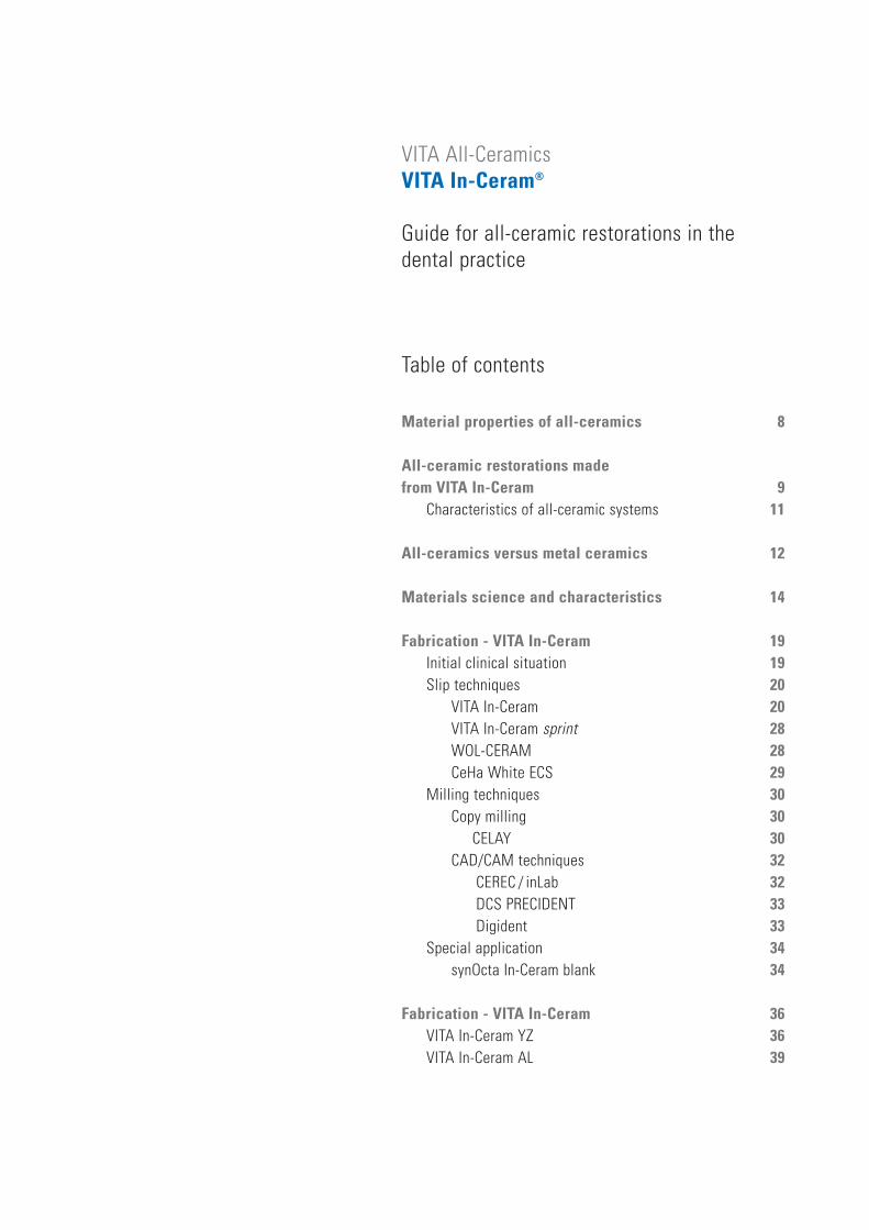
What´s behind of all-ceramics?Ceramics comprise a large family of inorganic materials within thegroup of non-metals. They are divided into three subcategories:silicate ceramic, oxide ceramic and non-oxide ceramic materials.The silicate ceramic materials have the same base materials incommon: quartz and feldspar, naturally occurring minerals resultingin a material which consists of silicates (fig. 1):
Fig. 1 Micrograph of a ceramic veneer made from a silicate ceramic material. Translucency and refraction behavior are determined by the crystals embedded in the silicate glass matrix (SiO2) and resemble the properties of natural enamel.
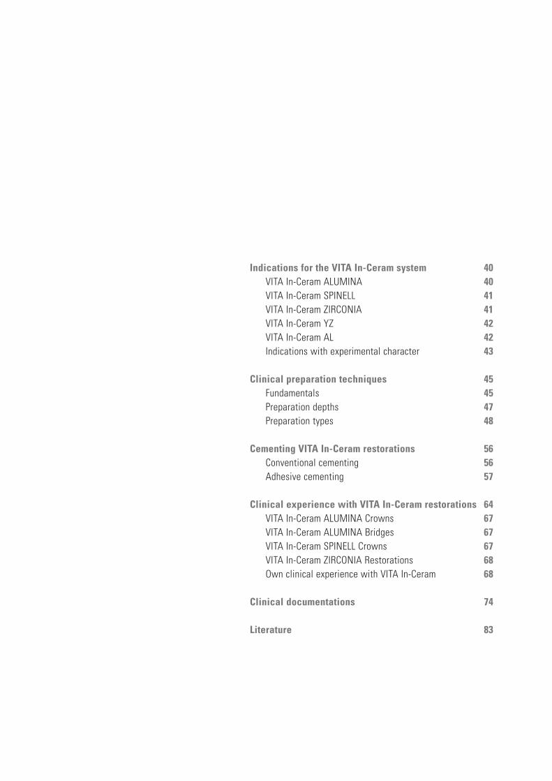
Silicate ceramic materials, however, can also be synthesized frominorganic pure substances (lithium disilicate ceramic Empress 2 / IPSby Ivoclar Vivadent AG). The term oxide ceramics is used forceramic materials consisting of simple oxides such as aluminiumoxide and zirconium oxide and for complex oxides such as spinels.In the narrower sense oxide ceramics are polycrystalline materialsbased only on the respective oxides. Glass-infiltrated ceramics suchas VITA In-Ceram with high oxide content but also glass contentdue to the infiltration process (fig. 2) take an intermediate positionbetween silicate ceramics and polycrystalline oxide ceramics.
Fig. 2 Coping made from VITA In-Ceram ALUMINA immediatelyafter glass infiltration firing and prior to removal of excessglass. The pores and cavities between the ALUMINA particles(aluminium oxide) connected with sintered bridges are filled with a lanthanum glass. This glass matrix accounts for only a very small part of the structure, however, it dominates withits optical properties so that the coping is translucent.
Non-oxide ceramics ("special ceramics") are compounds suchas nitrides and carbides which do not play a significant role asrestorative materials but are used for "tungsten carbide burs"and polishing agents for the daily dental and dental-technicaluse. The dental ceramic materials themselves are just a smallgroup within the entire range of ceramics. Owing to the differentindications of the individual ceramics, however, a basic under-standing of materials science of ceramics is indispensable fordentists in order to ensure correct classification of a dentalceramic system and to be able to use it successfully.
page 8
1
2
SEM-photo of an etched VITA VM 7 surfaceMagnification x 5000
page 9VITA In-Ceram® – VITA All-Ceramics
VITA In-Ceram® – All-ceramic restorations made from VITA In-Ceram
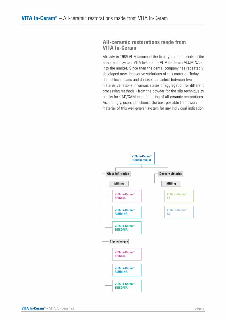
All-ceramic restorations made from VITA In-CeramAlready in 1989 VITA launched the first type of materials of theall-ceramic system VITA In-Ceram - VITA In-Ceram ALUMINA -into the market. Since then the dental company has repeatedlydeveloped new, innovative variations of this material. Todaydental technicians and dentists can select between five material variations in various states of aggregation for differentprocessing methods - from the powder for the slip technique toblocks for CAD/CAM manufacturing of all-ceramic restorations.Accordingly, users can choose the best possible frameworkmaterial of this well-proven system for any individual indication.
VITA In-Ceram®
(Oxidkeramik)
Glass infiltration Densely sintering
Milling
Slip technique
Milling
VITA In-Ceram®
SPINELL
VITA In-Ceram®
ALUMINA
VITA In-Ceram®
YZ
VITA In-Ceram®
AL
VITA In-Ceram®
ZIRCONIA
VITA In-Ceram®
SPINELL
VITA In-Ceram®
ALUMINA
VITA In-Ceram®
ZIRCONIA
VITA In-Ceram® – VITA All-Ceramics
VITA In-Ceram® – All-ceramic restorations made from VITA In-Ceram
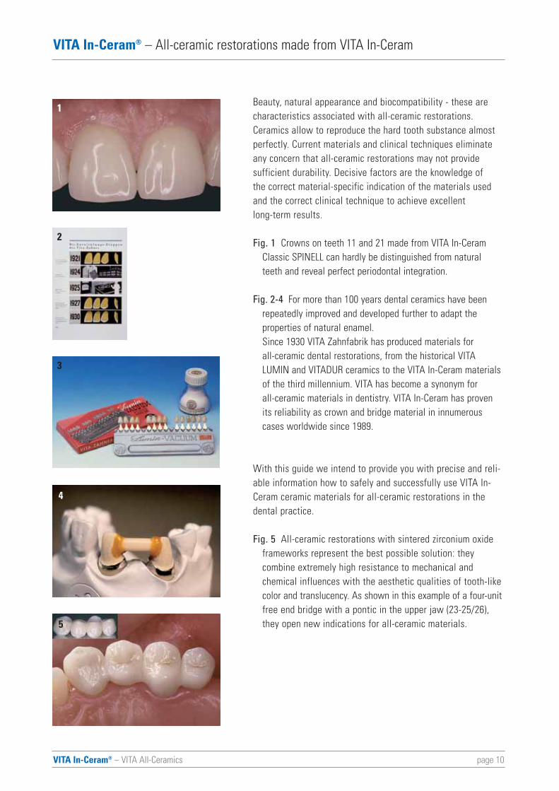
Beauty, natural appearance and biocompatibility - these arecharacteristics associated with all-ceramic restorations.Ceramics allow to reproduce the hard tooth substance almostperfectly. Current materials and clinical techniques eliminateany concern that all-ceramic restorations may not provide sufficient durability. Decisive factors are the knowledge of the correct material-specific indication of the materials usedand the correct clinical technique to achieve excellent long-term results.
Fig. 1 Crowns on teeth 11 and 21 made from VITA In-Ceram Classic SPINELL can hardly be distinguished from natural teeth and reveal perfect periodontal integration.
Fig. 2-4 For more than 100 years dental ceramics have been repeatedly improved and developed further to adapt the properties of natural enamel. Since 1930 VITA Zahnfabrik has produced materials for all-ceramic dental restorations, from the historical VITA LUMIN and VITADUR ceramics to the VITA In-Ceram materials of the third millennium. VITA has become a synonym for all-ceramic materials in dentistry. VITA In-Ceram has proven its reliability as crown and bridge material in innumerous cases worldwide since 1989.
With this guide we intend to provide you with precise and reli-able information how to safely and successfully use VITA In-Ceram ceramic materials for all-ceramic restorations in thedental practice.
Fig. 5 All-ceramic restorations with sintered zirconium oxide frameworks represent the best possible solution: they combine extremely high resistance to mechanical and chemical influences with the aesthetic qualities of tooth-like color and translucency. As shown in this example of a four-unitfree end bridge with a pontic in the upper jaw (23-25/26), they open new indications for all-ceramic materials.
page 10
4
3
2
1
5
page 11VITA In-Ceram® – VITA All-Ceramics
VITA In-Ceram® – All-ceramic restorations made from VITA In-Ceram
Characteristics of all-ceramic systems
All-ceramic substructures are characterized by the fact that ametal substructure is omitted and the restoration is exclusivelymade from ceramic material. Consequently, an opaque metalframework does not have to be masked and therefore a morenatural reconstruction of the tooth is possible. The all-ceramicrestoration systems available today can be classified accordingto several aspects. They can be distinguished on the basis ofthe material-scientific composition, the manufacturing method,the clinical use or the cementation technique. Some of the fab-rication systems available also allow processing of variousceramic materials for different clinical applications.
The optical and physical properties are essential for dentistry.Ceramics with a high oxide content (aluminium oxide, zirconiumoxide) exhibit very high values of strength but are also lesstranslucent or partly entirely opaque so that these materials(e.g. glass infiltrated oxide ceramic and polycrystalline oxideceramic) can only be used as core materials which need to beveneered with silicate ceramic materials to achieve the desiredaesthetic result.
Silicate ceramic materials in turn feature excellent opticalproperties to achieve unsurpassed aesthetic results. Due totheir lower strength, however, they require adhesive cementationto provide sufficient stability as a tooth-restoration bondingsystem. Additionally, they are used as veneering material forall-ceramic and metal frameworks.
VITA In-Ceram® – VITA All-Ceramics
VITA In-Ceram® – All-ceramics versus metal ceramics
All-ceramics versus metal ceramics
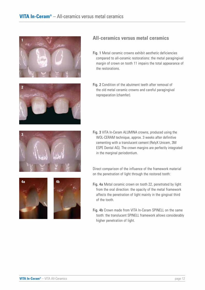
Fig. 1 Metal ceramic crowns exhibit aesthetic deficiencies compared to all-ceramic restorations: the metal paragingival margin of crown on tooth 11 impairs the total appearance of the restorations.
Fig. 2 Condition of the abutment teeth after removal of the old metal ceramic crowns and careful paragingival repreparation (chamfer).
Fig. 3 VITA In-Ceram ALUMINA crowns, produced using the WOL-CERAM technique, approx. 3 weeks after definitive cementing with a translucent cement (RelyX Unicem, 3M ESPE Dental AG). The crown margins are perfectly integrated in the marginal periodontium.
Direct comparison of the influence of the framework materialon the penetration of light through the restored tooth:
Fig. 4a Metal ceramic crown on tooth 22, penetrated by light from the oral direction: the opacity of the metal framework affects the penetration of light mainly in the gingival third of the tooth.
Fig. 4b Crown made from VITA In-Ceram SPINELL on the same tooth: the translucent SPINELL framework allows considerablyhigher penetration of light.
page 12
1
2
3
4a 4b
page 13VITA In-Ceram® – VITA All-Ceramics
VITA In-Ceram® – All-ceramics versus metal ceramics
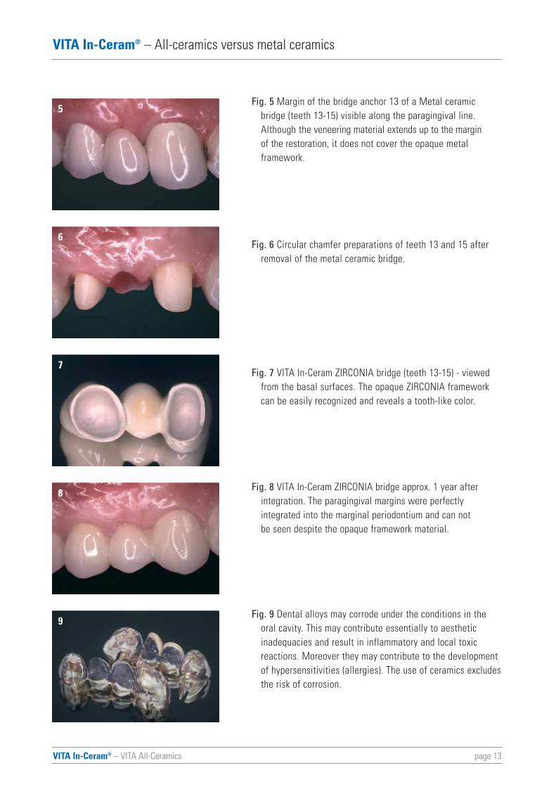
Fig. 5 Margin of the bridge anchor 13 of a Metal ceramic bridge (teeth 13-15) visible along the paragingival line. Although the veneering material extends up to the margin of the restoration, it does not cover the opaque metal framework.
Fig. 6 Circular chamfer preparations of teeth 13 and 15 after removal of the metal ceramic bridge.
Fig. 7 VITA In-Ceram ZIRCONIA bridge (teeth 13-15) - viewed from the basal surfaces. The opaque ZIRCONIA framework can be easily recognized and reveals a tooth-like color.
Fig. 8 VITA In-Ceram ZIRCONIA bridge approx. 1 year after integration. The paragingival margins were perfectly integrated into the marginal periodontium and can not be seen despite the opaque framework material.
Fig. 9 Dental alloys may corrode under the conditions in the oral cavity. This may contribute essentially to aesthetic inadequacies and result in inflammatory and local toxic reactions. Moreover they may contribute to the development of hypersensitivities (allergies). The use of ceramics excludesthe risk of corrosion.
5
6
7
8
9
VITA In-Ceram® – VITA All-Ceramics
VITA In-Ceram® – Materials science and characteristics
Materials science and characteristics
In the VITA In-Ceram product family two different types ofceramics must be distinguished:• glass infiltrated oxide ceramics:
VITA In-Ceram SPINELL, VITA In-Ceram ALUMINA and VITA In-Ceram ZIRCONIA: oxide reinforced.
• polycrystalline oxide ceramics: VITA In-Ceram YZ and VITA In-Ceram AL.
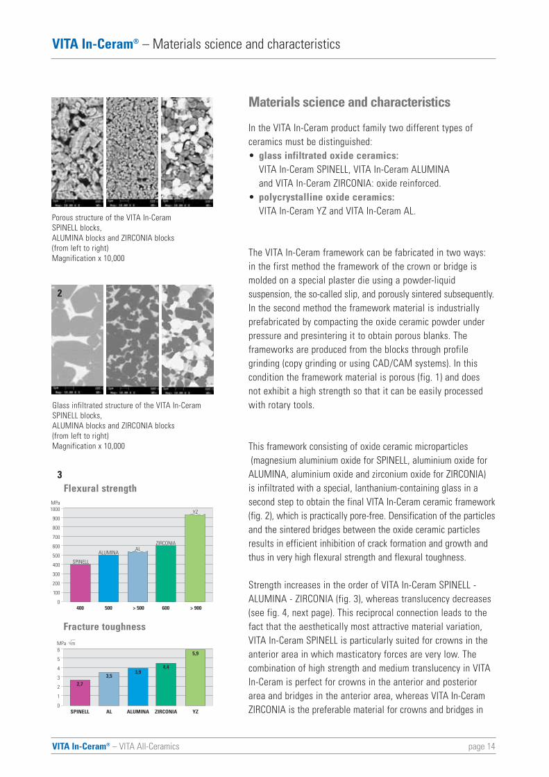
The VITA In-Ceram framework can be fabricated in two ways:in the first method the framework of the crown or bridge ismolded on a special plaster die using a powder-liquid suspension, the so-called slip, and porously sintered subsequently.In the second method the framework material is industriallyprefabricated by compacting the oxide ceramic powder underpressure and presintering it to obtain porous blanks. The frameworks are produced from the blocks through profile grinding (copy grinding or using CAD/CAM systems). In thiscondition the framework material is porous (fig. 1) and does not exhibit a high strength so that it can be easily processedwith rotary tools.
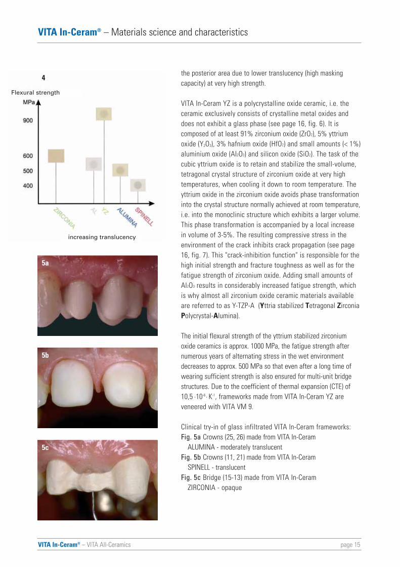
This framework consisting of oxide ceramic microparticles(magnesium aluminium oxide for SPINELL, aluminium oxide for
ALUMINA, aluminium oxide and zirconium oxide for ZIRCONIA)is infiltrated with a special, lanthanium-containing glass in asecond step to obtain the final VITA In-Ceram ceramic framework(fig. 2), which is practically pore-free. Densification of the particlesand the sintered bridges between the oxide ceramic particlesresults in efficient inhibition of crack formation and growth andthus in very high flexural strength and flexural toughness.
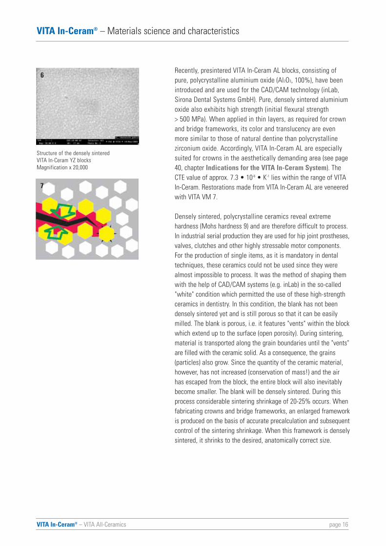
Strength increases in the order of VITA In-Ceram SPINELL -ALUMINA - ZIRCONIA (fig. 3), whereas translucency decreases(see fig. 4, next page). This reciprocal connection leads to thefact that the aesthetically most attractive material variation,VITA In-Ceram SPINELL is particularly suited for crowns in theanterior area in which masticatory forces are very low. Thecombination of high strength and medium translucency in VITAIn-Ceram is perfect for crowns in the anterior and posteriorarea and bridges in the anterior area, whereas VITA In-CeramZIRCONIA is the preferable material for crowns and bridges inSPINELL ALUMINA ZIRCONIA YZ
6
5
4
3
2
1
0
MPa · m
2,7
3,94,4
5,9
AL
3,5
400 500 600 > 900
1000
900
800
700
600
500
400
300
200
100
0
MPa
SPINELL
ALUMINA
ZIRCONIA
YZ
> 500
AL
page 14
3
1
Flexural strength
Fracture toughness
Porous structure of the VITA In-Ceram SPINELL blocks,ALUMINA blocks and ZIRCONIA blocks(from left to right)Magnification x 10,000
Glass infiltrated structure of the VITA In-CeramSPINELL blocks,ALUMINA blocks and ZIRCONIA blocks(from left to right)Magnification x 10,000
2
VITA In-Ceram® – VITA All-Ceramics
VITA In-Ceram® – Materials science and characteristics
4
5a
5b
5c
the posterior area due to lower translucency (high maskingcapacity) at very high strength.
VITA In-Ceram YZ is a polycrystalline oxide ceramic, i.e. theceramic exclusively consists of crystalline metal oxides anddoes not exhibit a glass phase (see page 16, fig. 6). It is composed of at least 91% zirconium oxide (ZrO2), 5% yttriumoxide (Y2O3), 3% hafnium oxide (HfO2) and small amounts (< 1%)aluminium oxide (Al2O3) and silicon oxide (SiO2). The task of thecubic yttrium oxide is to retain and stabilize the small-volume,tetragonal crystal structure of zirconium oxide at very high temperatures, when cooling it down to room temperature. Theyttrium oxide in the zirconium oxide avoids phase transformationinto the crystal structure normally achieved at room temperature,i.e. into the monoclinic structure which exhibits a larger volume.This phase transformation is accompanied by a local increasein volume of 3-5%. The resulting compressive stress in theenvironment of the crack inhibits crack propagation (see page16, fig. 7). This "crack-inhibition function" is responsible for thehigh initial strength and fracture toughness as well as for thefatigue strength of zirconium oxide. Adding small amounts ofAl2O3 results in considerably increased fatigue strength, whichis why almost all zirconium oxide ceramic materials availableare referred to as Y-TZP-A (Yttria stabilized Tetragonal ZirconiaPolycrystal-Alumina).
The initial flexural strength of the yttrium stabilized zirconiumoxide ceramics is approx. 1000 MPa, the fatigue strength afternumerous years of alternating stress in the wet environmentdecreases to approx. 500 MPa so that even after a long time ofwearing sufficient strength is also ensured for multi-unit bridgestructures. Due to the coefficient of thermal expansion (CTE) of10,5 ·10-6 · K-1, frameworks made from VITA In-Ceram YZ areveneered with VITA VM 9.
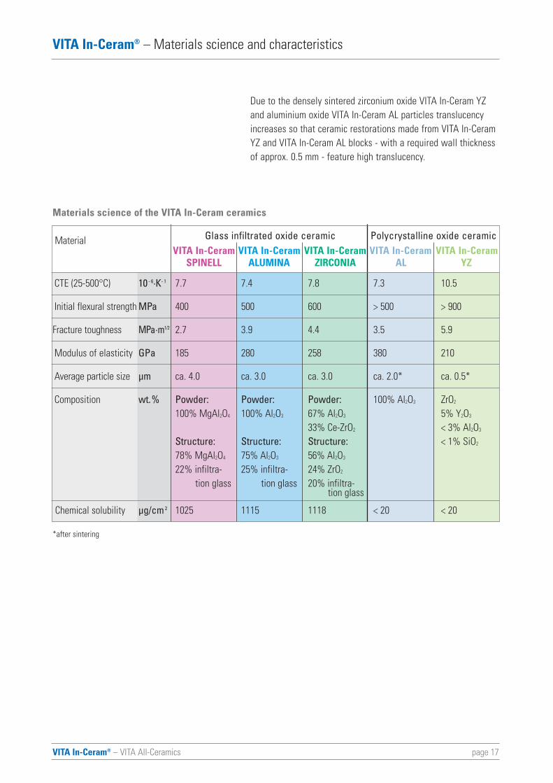
Clinical try-in of glass infiltrated VITA In-Ceram frameworks: Fig. 5a Crowns (25, 26) made from VITA In-Ceram
ALUMINA - moderately translucentFig. 5b Crowns (11, 21) made from VITA In-Ceram
SPINELL - translucentFig. 5c Bridge (15-13) made from VITA In-Ceram
ZIRCONIA - opaque
page 15
Flexural strength
increasing translucency
VITA In-Ceram® – VITA All-Ceramics
VITA In-Ceram® – Materials science and characteristics
Recently, presintered VITA In-Ceram AL blocks, consisting ofpure, polycrystalline aluminium oxide (Al2O3, 100%), have beenintroduced and are used for the CAD/CAM technology (inLab,Sirona Dental Systems GmbH). Pure, densely sintered aluminiumoxide also exhibits high strength (initial flexural strength > 500 MPa). When applied in thin layers, as required for crownand bridge frameworks, its color and translucency are evenmore similar to those of natural dentine than polycrystalline zirconium oxide. Accordingly, VITA In-Ceram AL are especiallysuited for crowns in the aesthetically demanding area (see page40, chapter Indications for the VITA In-Ceram System). TheCTE value of approx. 7.3 • 10-6 • K-1 lies within the range of VITAIn-Ceram. Restorations made from VITA In-Ceram AL are veneeredwith VITA VM 7.
Densely sintered, polycrystalline ceramics reveal extreme hardness (Mohs hardness 9) and are therefore difficult to process.In industrial serial production they are used for hip joint prostheses,valves, clutches and other highly stressable motor components.For the production of single items, as it is mandatory in dentaltechniques, these ceramics could not be used since they werealmost impossible to process. It was the method of shaping themwith the help of CAD/CAM systems (e.g. inLab) in the so-called"white" condition which permitted the use of these high-strengthceramics in dentistry. In this condition, the blank has not beendensely sintered yet and is still porous so that it can be easilymilled. The blank is porous, i.e. it features "vents" within the blockwhich extend up to the surface (open porosity). During sintering,material is transported along the grain boundaries until the "vents"are filled with the ceramic solid. As a consequence, the grains (particles) also grow. Since the quantity of the ceramic material,however, has not increased (conservation of mass!) and the airhas escaped from the block, the entire block will also inevitablybecome smaller. The blank will be densely sintered. During thisprocess considerable sintering shrinkage of 20-25% occurs. Whenfabricating crowns and bridge frameworks, an enlarged frameworkis produced on the basis of accurate precalculation and subsequentcontrol of the sintering shrinkage. When this framework is denselysintered, it shrinks to the desired, anatomically correct size.
page 16
7
6
Structure of the densely sintered VITA In-Ceram YZ blocksMagnification x 20,000
page 17VITA In-Ceram® – VITA All-Ceramics
VITA In-Ceram® – Materials science and characteristics
Due to the densely sintered zirconium oxide VITA In-Ceram YZand aluminium oxide VITA In-Ceram AL particles translucencyincreases so that ceramic restorations made from VITA In-CeramYZ and VITA In-Ceram AL blocks - with a required wall thicknessof approx. 0.5 mm - feature high translucency.
Materials science of the VITA In-Ceram ceramics
Glass infiltrated oxide ceramic Polycrystalline oxide ceramicVITA In-Ceram VITA In-Ceram VITA In-Ceram VITA In-Ceram VITA In-Ceram
SPINELL ALUMINA ZIRCONIA AL YZ
CTE (25-500°C) 10 - 6·K- 1 7.7 7.4 7.8 7.3 10.5
Initial flexural strength MPa 400 500 600 > 500 > 900
Fracture toughness MPa·m1/2 2.7 3.9 4.4 3.5 5.9
Modulus of elasticity GPa 185 280 258 380 210
Average particle size μm ca. 4.0 ca. 3.0 ca. 3.0 ca. 2.0* ca. 0.5*
Composition wt.% Powder: Powder: Powder: 100% Al2O3 ZrO2
100% MgAl2O4 100% Al2O3 67% Al2O3 5% Y2O3
33% Ce-ZrO2 < 3% Al2O3
Structure: Structure: Structure: < 1% SiO2
78% MgAl2O4 75% Al2O3 56% Al2O3
22% infiltra- 25% infiltra- 24% ZrO2
tion glass tion glass 20% infiltra-tion glass
Chemical solubility μg/cm 2 1025 1115 1118 < 20 < 20
Material
*after sintering
page 18VITA In-Ceram® – VITA All-Ceramics
VITA In-Ceram® – Materials science and characteristics
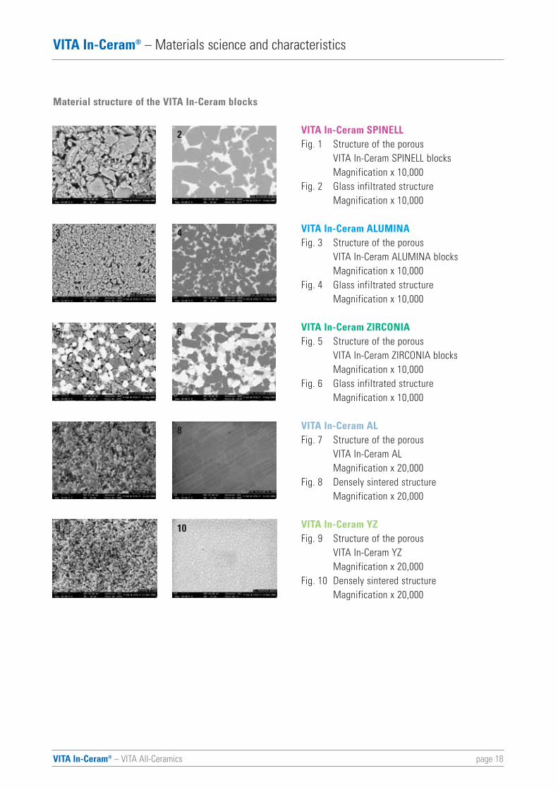
Material structure of the VITA In-Ceram blocks
VITA In-Ceram SPINELL Fig. 1 Structure of the porous
VITA In-Ceram SPINELL blocksMagnification x 10,000
Fig. 2 Glass infiltrated structureMagnification x 10,000
VITA In-Ceram ALUMINA Fig. 3 Structure of the porous
VITA In-Ceram ALUMINA blocksMagnification x 10,000
Fig. 4 Glass infiltrated structureMagnification x 10,000
VITA In-Ceram ZIRCONIA Fig. 5 Structure of the porous
VITA In-Ceram ZIRCONIA blocksMagnification x 10,000
Fig. 6 Glass infiltrated structureMagnification x 10,000
VITA In-Ceram ALFig. 7 Structure of the porous
VITA In-Ceram ALMagnification x 20,000
Fig. 8 Densely sintered structureMagnification x 20,000
VITA In-Ceram YZ Fig. 9 Structure of the porous
VITA In-Ceram YZMagnification x 20,000
Fig. 10 Densely sintered structureMagnification x 20,000
1
3
5
7
2
4
6
8
9 10
page 19VITA In-Ceram® – VITA All-Ceramics
VITA In-Ceram® – The fabrication - Infiltration ceramics
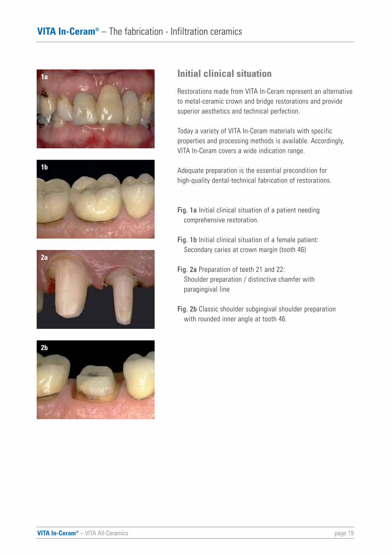
Initial clinical situation
Restorations made from VITA In-Ceram represent an alternativeto metal-ceramic crown and bridge restorations and providesuperior aesthetics and technical perfection.
Today a variety of VITA In-Ceram materials with specific properties and processing methods is available. Accordingly,VITA In-Ceram covers a wide indication range.
Adequate preparation is the essential precondition for high-quality dental-technical fabrication of restorations.
Fig. 1a Initial clinical situation of a patient needing comprehensive restoration.
Fig. 1b Initial clinical situation of a female patient:Secondary caries at crown margin (tooth 46)
Fig. 2a Preparation of teeth 21 and 22:Shoulder preparation / distinctive chamfer with paragingival line
Fig. 2b Classic shoulder subgingival shoulder preparation with rounded inner angle at tooth 46.
1a
1b
2a
2b
page 20VITA In-Ceram® – VITA All-Ceramics
VITA In-Ceram® – The fabrication - Infiltration ceramics
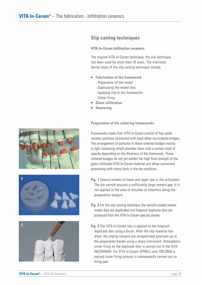
Slip casting techniques
VITA In-Ceram Infiltration ceramics
The original VITA In-Ceram technique, the slip technique, has been used for more than 16 years. The individual dental steps of the slip casting technique include
• Fabrication of the framework- Preparation of the model- Duplicating the model dies- Applying slip to the frameworks- Sinter firing
• Glass infiltration• Veneering
Preparation of the sintering frameworks
Frameworks made from VITA In-Ceram consist of fine oxideceramic particles connected with each other via sintered bridges.The arrangement of particles in these sintered bridges results in light scattering which provides them with a certain level ofopacity depending on the thickness of the framework. These sintered bridges do not yet exhibit the high final strength of theglass infiltrated VITA In-Ceram material and allow convenientprocessing with rotary tools in the dry condition.
Fig. 1 Sawcut models of lower and upper jaw in the articulator.The die varnish ensures a sufficiently large cement gap. It is not applied in the area of shoulder or chamfers along the preparation margins.
Fig. 2 For the slip casting technique the varnish-coated master model dies are duplicated and fireproof duplicate dies are produced from the VITA In-Ceram special plaster.
Fig. 3 The VITA In-Ceram slip is applied to the fireproof duplicate dies using a brush. After the slip material has dried, the coping margins are scraped back precisely up to the preparation border using a sharp instrument. Atmospheric sinter firing on the duplicate dies is carried out in the VITA INCERAMAT. For VITA In-Ceram SPINELL and ZIRCONIA a second sinter firing process is subsequently carried out on firing pad.
1
2
3
page 21VITA In-Ceram® – VITA All-Ceramics
VITA In-Ceram® – The fabrication - Infiltration ceramics
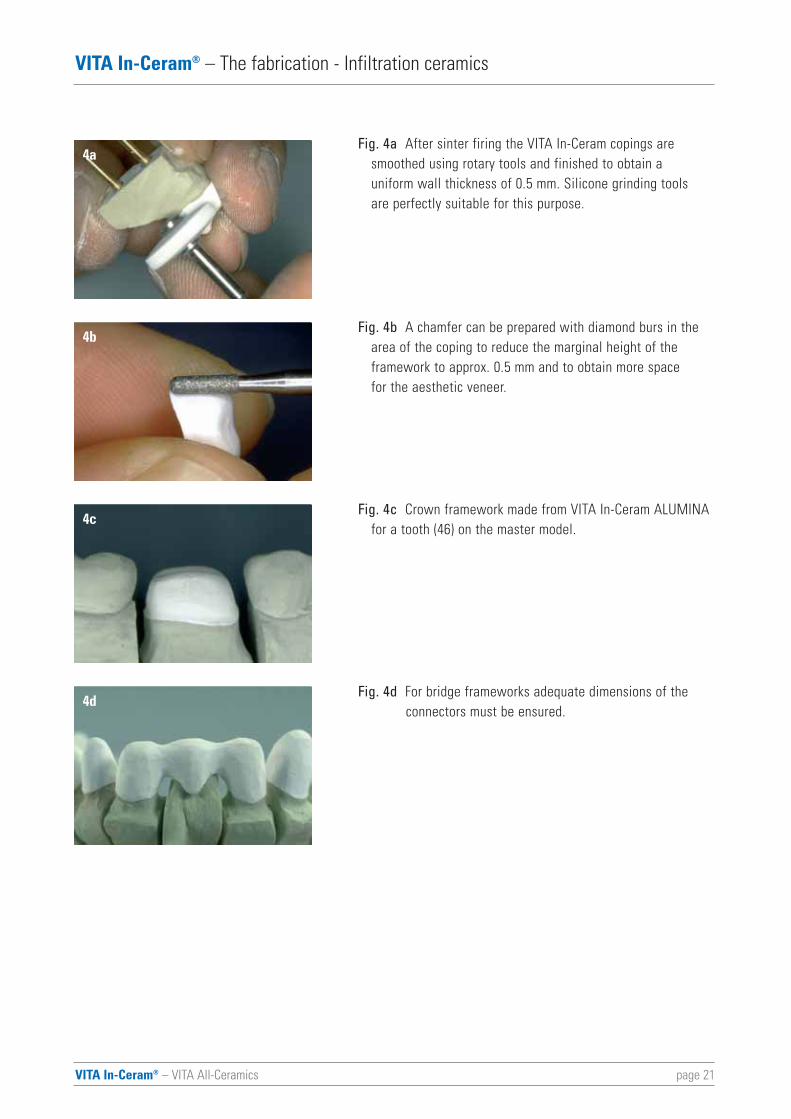
Fig. 4a After sinter firing the VITA In-Ceram copings are smoothed using rotary tools and finished to obtain a uniform wall thickness of 0.5 mm. Silicone grinding tools are perfectly suitable for this purpose.
Fig. 4b A chamfer can be prepared with diamond burs in the area of the coping to reduce the marginal height of the framework to approx. 0.5 mm and to obtain more space for the aesthetic veneer.
Fig. 4c Crown framework made from VITA In-Ceram ALUMINA for a tooth (46) on the master model.
Fig. 4d For bridge frameworks adequate dimensions of the connectors must be ensured.
4a
4b
4c
4d
VITA In-Ceram® – VITA All-Ceramics page 22
VITA In-Ceram® – The fabrication - Infiltration ceramics
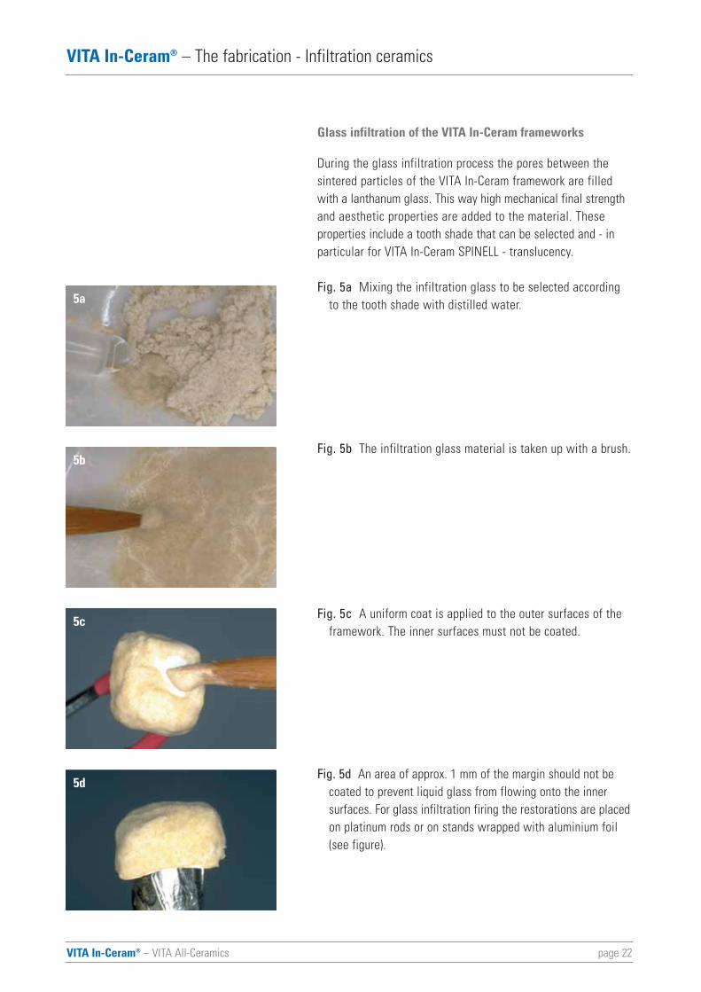
Glass infiltration of the VITA In-Ceram frameworks
During the glass infiltration process the pores between the sintered particles of the VITA In-Ceram framework are filledwith a lanthanum glass. This way high mechanical final strengthand aesthetic properties are added to the material. These properties include a tooth shade that can be selected and - inparticular for VITA In-Ceram SPINELL - translucency.
Fig. 5a Mixing the infiltration glass to be selected according to the tooth shade with distilled water.
Fig. 5b The infiltration glass material is taken up with a brush.
Fig. 5c A uniform coat is applied to the outer surfaces of the framework. The inner surfaces must not be coated.
Fig. 5d An area of approx. 1 mm of the margin should not be coated to prevent liquid glass from flowing onto the inner surfaces. For glass infiltration firing the restorations are placedon platinum rods or on stands wrapped with aluminium foil (see figure).
5a
5b
5c
5d
page 23VITA In-Ceram® – VITA All-Ceramics
VITA In-Ceram® – The fabrication - Infiltration ceramics
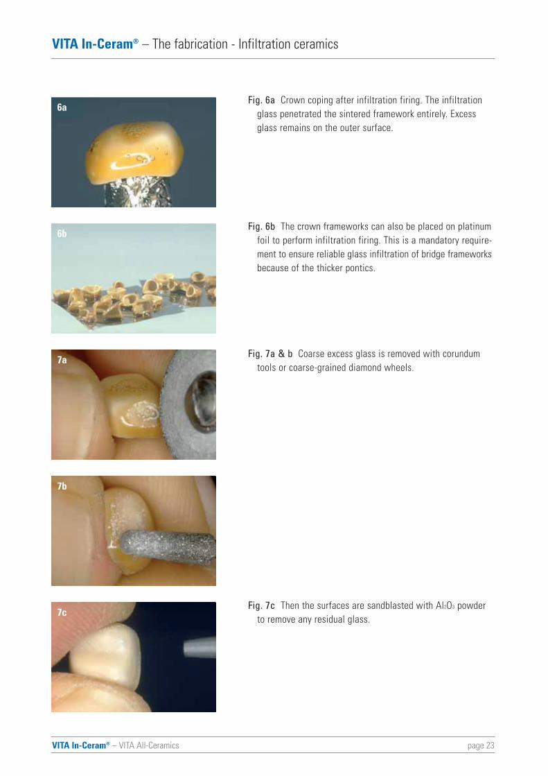
Fig. 6a Crown coping after infiltration firing. The infiltration glass penetrated the sintered framework entirely. Excess glass remains on the outer surface.
Fig. 6b The crown frameworks can also be placed on platinum foil to perform infiltration firing. This is a mandatory require-ment to ensure reliable glass infiltration of bridge frameworksbecause of the thicker pontics.
Fig. 7a & b Coarse excess glass is removed with corundum tools or coarse-grained diamond wheels.
Fig. 7c Then the surfaces are sandblasted with Al2O3 powderto remove any residual glass.
6a
6b
7a
7b
7c
VITA In-Ceram® – VITA All-Ceramics page 24
VITA In-Ceram® – The fabrication - Infiltration ceramics
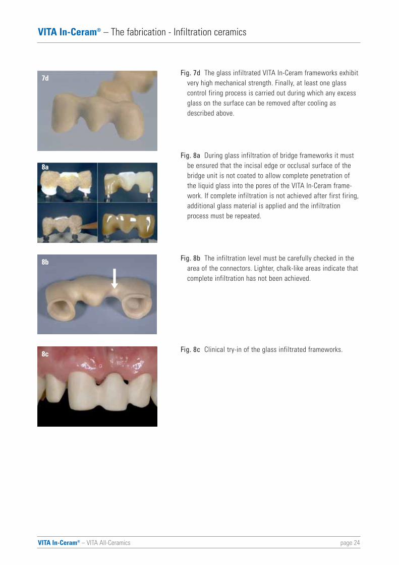
Fig. 7d The glass infiltrated VITA In-Ceram frameworks exhibitvery high mechanical strength. Finally, at least one glass control firing process is carried out during which any excess glass on the surface can be removed after cooling as described above.
Fig. 8a During glass infiltration of bridge frameworks it must be ensured that the incisal edge or occlusal surface of the bridge unit is not coated to allow complete penetration of the liquid glass into the pores of the VITA In-Ceram frame-work. If complete infiltration is not achieved after first firing,additional glass material is applied and the infiltration process must be repeated.
Fig. 8b The infiltration level must be carefully checked in the area of the connectors. Lighter, chalk-like areas indicate thatcomplete infiltration has not been achieved.
Fig. 8c Clinical try-in of the glass infiltrated frameworks.
7d
8a
8b
8c
9c
page 25VITA In-Ceram® – VITA All-Ceramics
VITA In-Ceram® – The fabrication - Infiltration ceramics
Veneering the glass infiltrated VITA In-Ceram frameworks
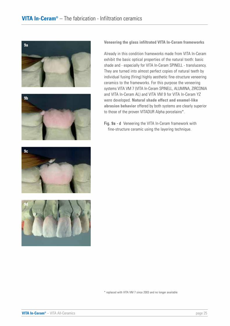
Already in this condition frameworks made from VITA In-Ceramexhibit the basic optical properties of the natural tooth: basicshade and - especially for VITA In-Ceram SPINELL - translucency.They are turned into almost perfect copies of natural teeth byindividual fusing (firing) highly aesthetic fine-structure veneeringceramics to the frameworks. For this purpose the veneeringsystems VITA VM 7 (VITA In-Ceram SPINELL, ALUMINA, ZIRCONIAand VITA In-Ceram AL) and VITA VM 9 for VITA In-Ceram YZwere developed. Natural shade effect and enamel-likeabrasion behavior offered by both systems are clearly superiorto those of the proven VITADUR Alpha porcelains*.
Fig. 9a - d Veneering the VITA In-Ceram framework with fine-structure ceramic using the layering technique.
* replaced with VITA VM 7 since 2003 and no longer available
9a
9b
9c
9d
VITA In-Ceram® – VITA All-Ceramics page 26
VITA In-Ceram® – The fabrication - Infiltration ceramics
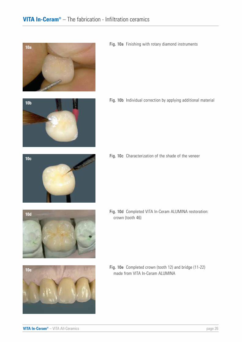
Fig. 10a Finishing with rotary diamond instruments
Fig. 10b Individual correction by applying additional material
Fig. 10c Characterization of the shade of the veneer
Fig. 10d Completed VITA In-Ceram ALUMINA restoration: crown (tooth 46)
Fig. 10e Completed crown (tooth 12) and bridge (11-22) made from VITA In-Ceram ALUMINA
10a
10b
10c
10d
10e
page 27VITA In-Ceram® – VITA All-Ceramics
VITA In-Ceram® – The fabrication - Infiltration ceramics
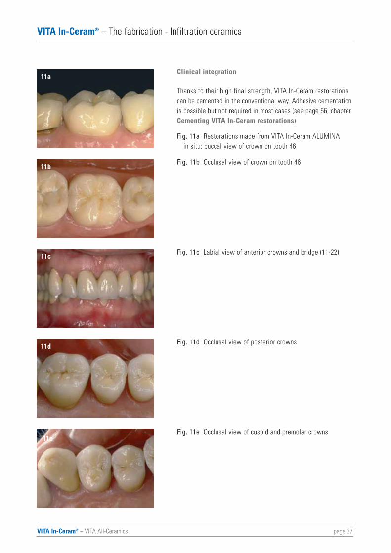
Clinical integration
Thanks to their high final strength, VITA In-Ceram restorationscan be cemented in the conventional way. Adhesive cementationis possible but not required in most cases (see page 56, chapterCementing VITA In-Ceram restorations)
Fig. 11a Restorations made from VITA In-Ceram ALUMINA in situ: buccal view of crown on tooth 46
Fig. 11b Occlusal view of crown on tooth 46
Fig. 11c Labial view of anterior crowns and bridge (11-22)
Fig. 11d Occlusal view of posterior crowns
Fig. 11e Occlusal view of cuspid and premolar crowns
11a
11b
11c
11d
11e
VITA In-Ceram® – VITA All-Ceramics page 28
VITA In-Ceram® – The fabrication - Infiltration ceramics
VITA In-Ceram sprint
The VITA In-Ceram sprint technique allows the fabrication ofindividual posterior and anterior crowns made from VITA In-Ceram ALUMINA and VITA In-Ceram ZIRCONIA in a convention-al vacuum furnace at reduced process times. Based on thistechnique, the fabrication of VITA In-Ceram ALUMINA and VITAIn-Ceram ZIRCONIA frameworks takes only a third of the timerequired for the conventional slip technique.
In contrast to the classic slip technique, the duplicate dies aremanufactured from VITA In-Ceram special plaster and the slip-coated crown copings are heated in the furnace at 130° and160° for 20 min. before sinter firing. After cooling, the copingcan be removed and sintered without the plaster die. Thiswill reduce the time for sinter firing up to approx. 70%.
Glass infiltration and ceramic veneering are performed in thesame way as in the conventional slip technique.
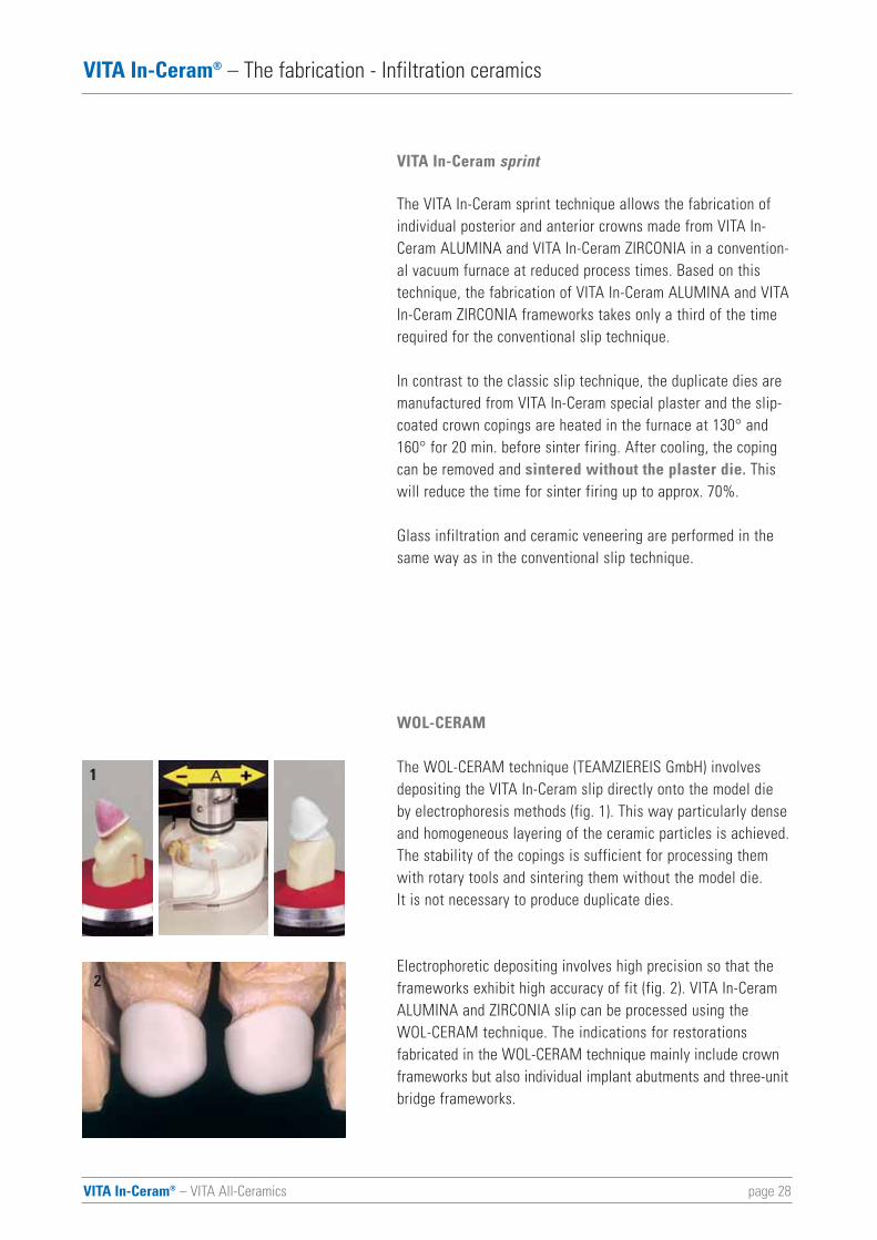
WOL-CERAM
The WOL-CERAM technique (TEAMZIEREIS GmbH) involvesdepositing the VITA In-Ceram slip directly onto the model dieby electrophoresis methods (fig. 1). This way particularly denseand homogeneous layering of the ceramic particles is achieved.The stability of the copings is sufficient for processing themwith rotary tools and sintering them without the model die. It is not necessary to produce duplicate dies.
Electrophoretic depositing involves high precision so that theframeworks exhibit high accuracy of fit (fig. 2). VITA In-CeramALUMINA and ZIRCONIA slip can be processed using the WOL-CERAM technique. The indications for restorations fabricated in the WOL-CERAM technique mainly include crownframeworks but also individual implant abutments and three-unitbridge frameworks.
1
2
page 29VITA In-Ceram® – VITA All-Ceramics
VITA In-Ceram® – The fabrication - Infiltration ceramics
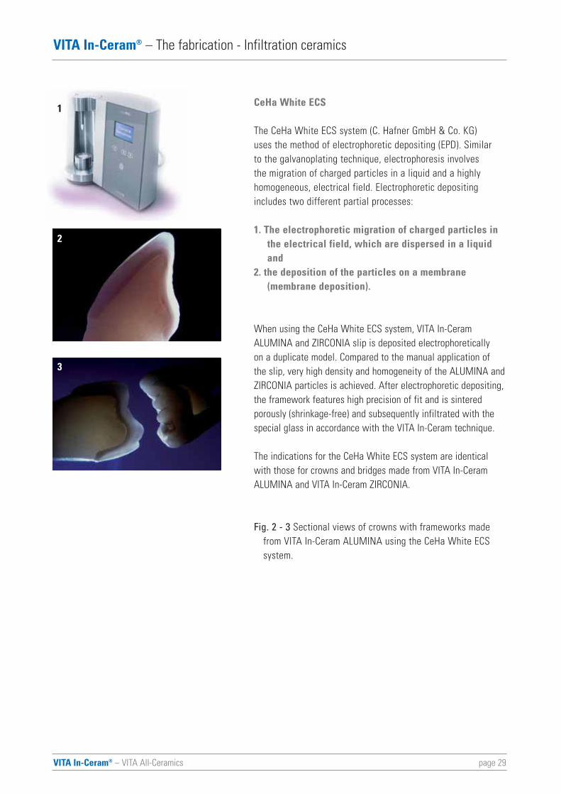
CeHa White ECS
The CeHa White ECS system (C. Hafner GmbH & Co. KG) uses the method of electrophoretic depositing (EPD). Similar to the galvanoplating technique, electrophoresis involves the migration of charged particles in a liquid and a highlyhomogeneous, electrical field. Electrophoretic depositingincludes two different partial processes:
1. The electrophoretic migration of charged particles in the electrical field, which are dispersed in a liquid and
2. the deposition of the particles on a membrane (membrane deposition).
When using the CeHa White ECS system, VITA In-Ceram ALUMINA and ZIRCONIA slip is deposited electrophoretically on a duplicate model. Compared to the manual application of the slip, very high density and homogeneity of the ALUMINA andZIRCONIA particles is achieved. After electrophoretic depositing,the framework features high precision of fit and is sinteredporously (shrinkage-free) and subsequently infiltrated with thespecial glass in accordance with the VITA In-Ceram technique.
The indications for the CeHa White ECS system are identicalwith those for crowns and bridges made from VITA In-CeramALUMINA and VITA In-Ceram ZIRCONIA.
Fig. 2 - 3 Sectional views of crowns with frameworks made from VITA In-Ceram ALUMINA using the CeHa White ECS system.
1
2
3
VITA In-Ceram® – VITA All-Ceramics page 30
VITA In-Ceram® – The fabrication - Infiltration ceramics
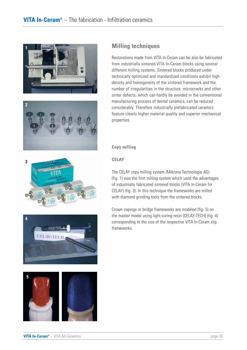
Milling techniques
Restorations made from VITA In-Ceram can be also be fabricatedfrom industrially sintered VITA In-Ceram blocks using severaldifferent milling systems. Sintered blocks produced under technically optimized and standardized conditions exhibit highdensity and homogeneity of the sintered framework and thenumber of irregularities in the structure, microcracks and othersinter defects, which can hardly be avoided in the conventionalmanufacturing process of dental ceramics, can be reduced considerably. Therefore industrially prefabricated ceramics feature clearly higher material quality and superior mechanicalproperties.
Copy milling
CELAY
The CELAY copy milling system (Mikrona Technologie AG) (fig. 1) was the first milling system which used the advantagesof industrially fabricated sintered blocks (VITA In-Ceram forCELAY) (fig. 3). In this technique the frameworks are milled with diamond grinding tools from the sintered blocks.
Crown copings or bridge frameworks are modeled (fig. 5) on the master model using light-curing resin (CELAY-TECH) (fig. 4)corresponding to the size of the respective VITA In-Ceram slipframeworks.
1
2
3
4
5
page 31VITA In-Ceram® – VITA All-Ceramics
VITA In-Ceram® – The fabrication - Infiltration ceramics
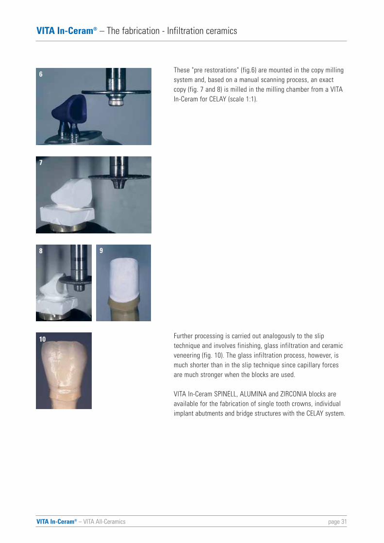
These "pre restorations" (fig.6) are mounted in the copy millingsystem and, based on a manual scanning process, an exactcopy (fig. 7 and 8) is milled in the milling chamber from a VITAIn-Ceram for CELAY (scale 1:1).
Further processing is carried out analogously to the slip technique and involves finishing, glass infiltration and ceramicveneering (fig. 10). The glass infiltration process, however, ismuch shorter than in the slip technique since capillary forcesare much stronger when the blocks are used.
VITA In-Ceram SPINELL, ALUMINA and ZIRCONIA blocks areavailable for the fabrication of single tooth crowns, individualimplant abutments and bridge structures with the CELAY system.
6
7
8
10
9
VITA In-Ceram® – VITA All-Ceramics page 32
VITA In-Ceram® – The fabrication - Infiltration ceramics
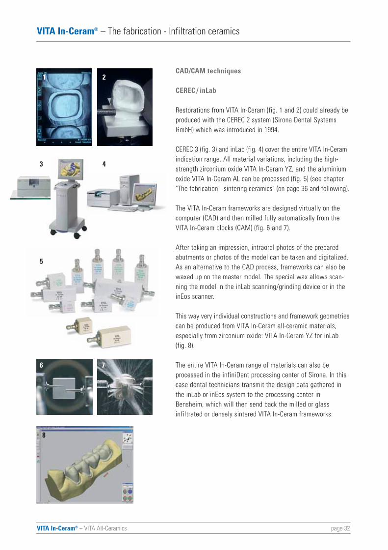
CAD/CAM techniques
CEREC / inLab
Restorations from VITA In-Ceram (fig. 1 and 2) could already beproduced with the CEREC 2 system (Sirona Dental SystemsGmbH) which was introduced in 1994.
CEREC 3 (fig. 3) and inLab (fig. 4) cover the entire VITA In-Ceramindication range. All material variations, including the high-strength zirconium oxide VITA In-Ceram YZ, and the aluminiumoxide VITA In-Ceram AL can be processed (fig. 5) (see chapter"The fabrication - sintering ceramics" (on page 36 and following).
The VITA In-Ceram frameworks are designed virtually on thecomputer (CAD) and then milled fully automatically from theVITA In-Ceram blocks (CAM) (fig. 6 and 7).
After taking an impression, intraoral photos of the preparedabutments or photos of the model can be taken and digitalized.As an alternative to the CAD process, frameworks can also bewaxed up on the master model. The special wax allows scan-ning the model in the inLab scanning/grinding device or in theinEos scanner.
This way very individual constructions and framework geometriescan be produced from VITA In-Ceram all-ceramic materials,especially from zirconium oxide: VITA In-Ceram YZ for inLab(fig. 8).
The entire VITA In-Ceram range of materials can also beprocessed in the infiniDent processing center of Sirona. In thiscase dental technicians transmit the design data gathered inthe inLab or inEos system to the processing center inBensheim, which will then send back the milled or glass infiltrated or densely sintered VITA In-Ceram frameworks.
1 2
5
6 7
8
3 4

VITA In-Ceram® – VITA All-Ceramics
VITA In-Ceram® – The fabrication - Infiltration ceramics
DCS PRECIDENT
VITA In-Ceram blocks and VITA In-Ceram ZIRCONIA blocks areavailable for the DCS PRECIDENT system (DCS Dental AG)which was introduced in 1989 and comprises a scanner and amilling unit (fig. 1). The indication range includes crown andthree-unit bridge frameworks. Perfect processing of the block isensured thanks to a recognition system for blanks.
Fig. 3 shows the DCS Dentform software displaying a sectionof a crowns and a scanned wax-up on the monitor.
Digident®
Another CAD/CAM system for processing VITA In-Ceram materials is the Digident system (Digident GmbH) (fig. 1). A stripe light scanner scans the preparations based on a master model and digitalizes them (fig. 2).
The CAD/CAM reconstructions (fig. 3) cover the indications of anterior and posterior crowns and three-unit bridges withVITA In-Ceram ALUMINA blocks and VITA In-Ceram ZIRCONIAblocks (fig. 4).
page 33
3
1 2
2
3
4
1
page 34VITA In-Ceram® – VITA All-Ceramics
VITA In-Ceram® – The fabrication - Infiltration ceramics
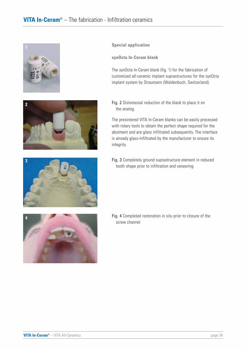
Special application
synOcta In-Ceram blank
The synOcta In-Ceram blank (fig. 1) for the fabrication of customized all-ceramic implant suprastructures for the synOctaimplant system by Straumann (Waldenbuch, Switzerland).
Fig. 2 Distomesial reduction of the blank to place it on the analog.
The presintered VITA In-Ceram blanks can be easily processedwith rotary tools to obtain the perfect shape required for theabutment and are glass infiltrated subsequently. The interface is already glass-infiltrated by the manufacturer to ensure itsintegrity.
Fig. 3 Completely ground suprastructure element in reduced tooth shape prior to infiltration and veneering
Fig. 4 Completed restoration in situ prior to closure of the screw channel
1
2
3
4
page 35VITA In-Ceram® – VITA All-Ceramics
VITA In-Ceram® – The fabrication - Infiltration ceramics
Duplicating the models
Fabrication of the framework
Glass infiltration
Ceramic veneering
Model fabrication
SPINELL, ALUMINA1), ALUMINA, SPINELL, ALUMINA, SPINELL, ALUMINA, ALUMINA,ZIRCONIA1) ZIRCONIA ZIRCONIA ZIRCONIA ZIRCONIA
not requirednot required
CAD process or scan CAD process ModellingElectrophoresis
Applying the slip
Sinter firing Copy milling CAM milling CAM milling
Table: Systems and techniques for the fabrication of VITA In-Ceram restorations
Manual Electrophoresis Milling and CAD/CAM techniquesslip technique WOL-CERAM CELAY CEREC/ DCS PRECIDENTVITA In-Ceram CeHa White ECS inLab Digident
optical impression Model scan
1) Also possible in the VITA In-Ceram sprint technique 2) Additional systems for the use: etkon, Hint-Els Denta CAD, Cynovad Neo (Dentaurum)
Digident
Digident
CeHa White ECS
Digident
VITA In-Ceram® – VITA All-Ceramics page 36
VITA In-Ceram® – The fabrication - Sintering ceramics
VITA In-Ceram sintering ceramicsVITA In-Ceram YZ for inLab
VITA In-Ceram YZ for inLab (fig. 1) are presintered zirconiumoxide blocks partially stabilized with yttrium oxide. The presintered zirconium oxide can be perfectly processed withrotary tools and shaped to obtain a framework for all-ceramiccrown and bridge structures. During subsequent sinter firingthe framework shrinks to approximately 25% of its original size(fig. 2). When planning and preparing the ceramic models, sinter shrinkage must be accounted for. Accordingly, CAD/CAMtechnology is used for processing VITA In-Ceram YZ.
Prior to grinding (milling) the CAD/CAM system calculates theframework geometry of the VITA In-Ceram YZ blocks to ensurethat the CAM framework to be milled exhibits the requiredenlarged size. The necessary information is included in a barcode printed on every YZ block and can be scanned by the system.
Currently, the inLab system by Sirona Dental Systems GmbH is the only approved CAD/CAM system for processing VITA In-Ceram YZ.
The guidelines for the fabrication of frameworks from the YZ blocks in the inLab system are based on those for VITA In-Ceram ZIRCONIA. Two methods can be employed:
• Waxing up the framework on the master model and subse-quent scanning and digitalizing the framework model (seepage 36-37, fig. 3-5).
• CAD construction of the framework after optical impressionor scanning for digitalizing the master model die (see fig. 5).
The attachment-supported bridge in this example (see page 35,fig.4 to page 38, fig. 14) describes an experimental indication.
Fig. 5 Scanning the models allows to fabricate individual constructions and framework geometries. Their purely CAD-based creation could only be done using complex software.
1
2
3
4
5
page 37VITA In-Ceram® – VITA All-Ceramics
VITA In-Ceram® – The fabrication - Sintering ceramics
Fig. 6 Completely milled crown with attached patrix made fromVITA In-Ceram YZ for inLab.
Fig. 7 If required, the milled frameworks made from VITA In-Ceram can be colored (entirely or partly) using COLORING liquid prior to sinter firing. The COLORING LIQUID is available in the five lightness levels (LL1 - LL5) of VITA SYSTEM 3D-MASTER.
Fig. 8 After sinter firing in the VITA ZYrcomat or Thermo-Star high-temperature furnaces at 1530° C the VITA In-Ceram YZ frameworks exhibit tooth-shaded translucent properties and very high strength of > 900 MPa.
Fig. 9 Checking the fit of the frameworks on the master model.
Fig. 10 Only minor contour corrections should be made and must be performed with fine-grain diamond tools whilst cooling with water.
6
7
8
9
10
VITA In-Ceram® – VITA All-Ceramics page 38
VITA In-Ceram® – The fabrication - Sintering ceramics
Fig. 11 - 13 The frameworks made from VITA In-Ceram YZ for inLab are veneered with VITA VM 9 - the veneering material of the VITA VM system which was developed especially for zirconium oxide frameworks in the CTE range of approx. 10.5.
Fig. 14 Restorations made from VITA In-Ceram YZ blocks are suitable for conventional and for adhesive cementation as well (bridges 14-17, 45-48).
11
12
13
14
page 39VITA In-Ceram® – VITA All-Ceramics
VITA In-Ceram® – The fabrication - Sintering ceramics
VITA In-Ceram AL for inLab
VITA In-Ceram AL for inLab are presintered blocks consisting ofpure aluminium oxide. They are milled with the inLab system and dense sintered justlike VITA In-Ceram YZ for inLab in the VITA ZYrcomat or Thermo-Star high-temperature furnaces. The resulting polycrystallineAl2O3 framework is somewhat more translucent than VITA In-Ceram YZ and its color is more similar to natural dentine.
Just like VITA In-Ceram infiltration ceramics, the frameworksmade from VITA In-Ceram AL are veneered with VITA VM 7 inaccordance with the CTE value of aluminium oxide.
Frameworks made from VITA In-Ceram AL are suitable for conventional cementation. If required, adhesive cementing is also possible.
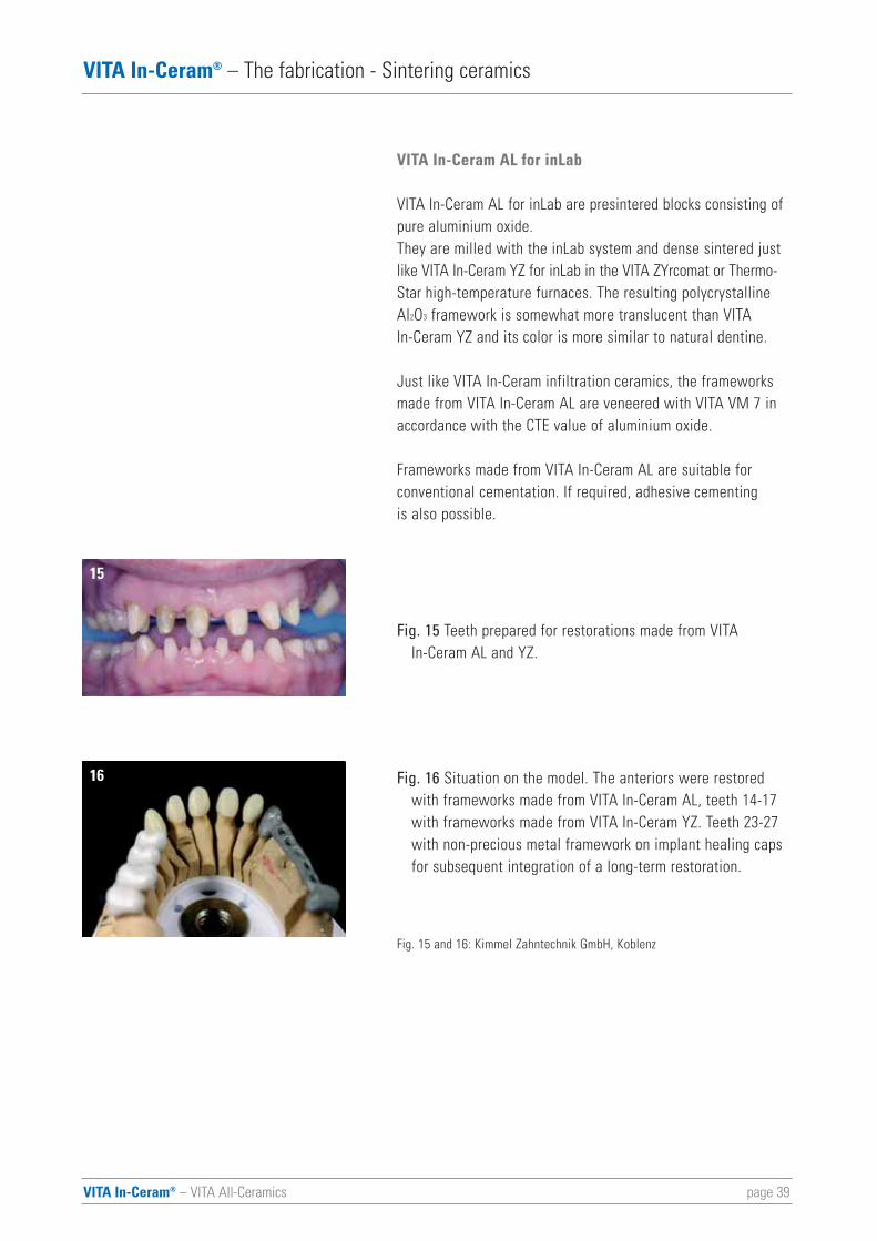
Fig. 15 Teeth prepared for restorations made from VITA In-Ceram AL and YZ.
Fig. 16 Situation on the model. The anteriors were restored with frameworks made from VITA In-Ceram AL, teeth 14-17 with frameworks made from VITA In-Ceram YZ. Teeth 23-27 with non-precious metal framework on implant healing caps for subsequent integration of a long-term restoration.
Fig. 15 and 16: Kimmel Zahntechnik GmbH, Koblenz
15
16
VITA In-Ceram® – VITA All-Ceramics page 40
VITA In-Ceram® – Indications for the VITA In-Ceram system
Indications for the VITA In-Ceram system
Restorations made from VITA In-Ceram account for the majorityof standard prosthetic indications for crowns and bridges.Preconditions for their long-term clinical success are
• putting the preparation guidelines into practice and adhering to them and
• selecting the suitable materials of the VITA In-Ceram system in accordance with the aesthetic and functional requirements.
In situations considered to be difficult from the prosthetic pointof view, e.g. insufficient space in patients with short clinicalcrowns or high functional stress in bruxists, the indication mustbe considered very carefully, especially for all-ceramic bridges.
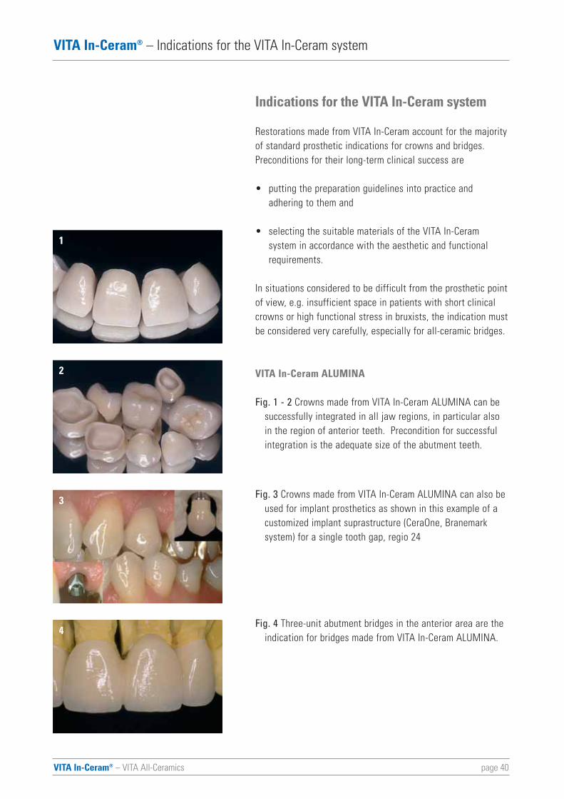
VITA In-Ceram ALUMINA
Fig. 1 - 2 Crowns made from VITA In-Ceram ALUMINA can be successfully integrated in all jaw regions, in particular also in the region of anterior teeth. Precondition for successful integration is the adequate size of the abutment teeth.
Fig. 3 Crowns made from VITA In-Ceram ALUMINA can also be used for implant prosthetics as shown in this example of a customized implant suprastructure (CeraOne, Branemark system) for a single tooth gap, regio 24
Fig. 4 Three-unit abutment bridges in the anterior area are the indication for bridges made from VITA In-Ceram ALUMINA.
1
2
3
4
VITA In-Ceram® – VITA All-Ceramics page 41
VITA In-Ceram® – Indications for the VITA In-Ceram system
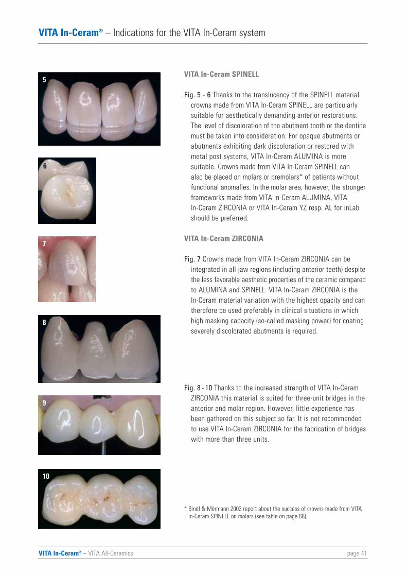
VITA In-Ceram SPINELL
Fig. 5 - 6 Thanks to the translucency of the SPINELL material crowns made from VITA In-Ceram SPINELL are particularly suitable for aesthetically demanding anterior restorations. The level of discoloration of the abutment tooth or the dentinemust be taken into consideration. For opaque abutments or abutments exhibiting dark discoloration or restored with metal post systems, VITA In-Ceram ALUMINA is more suitable. Crowns made from VITA In-Ceram SPINELL can also be placed on molars or premolars* of patients without functional anomalies. In the molar area, however, the strongerframeworks made from VITA In-Ceram ALUMINA, VITA In-Ceram ZIRCONIA or VITA In-Ceram YZ resp. AL for inLab should be preferred.
VITA In-Ceram ZIRCONIA
Fig. 7 Crowns made from VITA In-Ceram ZIRCONIA can be integrated in all jaw regions (including anterior teeth) despitethe less favorable aesthetic properties of the ceramic comparedto ALUMINA and SPINELL. VITA In-Ceram ZIRCONIA is the In-Ceram material variation with the highest opacity and can therefore be used preferably in clinical situations in which high masking capacity (so-called masking power) for coating severely discolorated abutments is required.
Fig. 8 - 10 Thanks to the increased strength of VITA In-Ceram ZIRCONIA this material is suited for three-unit bridges in the anterior and molar region. However, little experience has been gathered on this subject so far. It is not recommended to use VITA In-Ceram ZIRCONIA for the fabrication of bridgeswith more than three units.
* Bindl & Mörmann 2002 report about the success of crowns made from VITAIn-Ceram SPINELL on molars (see table on page 66).
5
6
7
8
9
10
page 42
14
15
VITA In-Ceram® – VITA All-Ceramics
VITA In-Ceram® – Indications for the VITA In-Ceram system
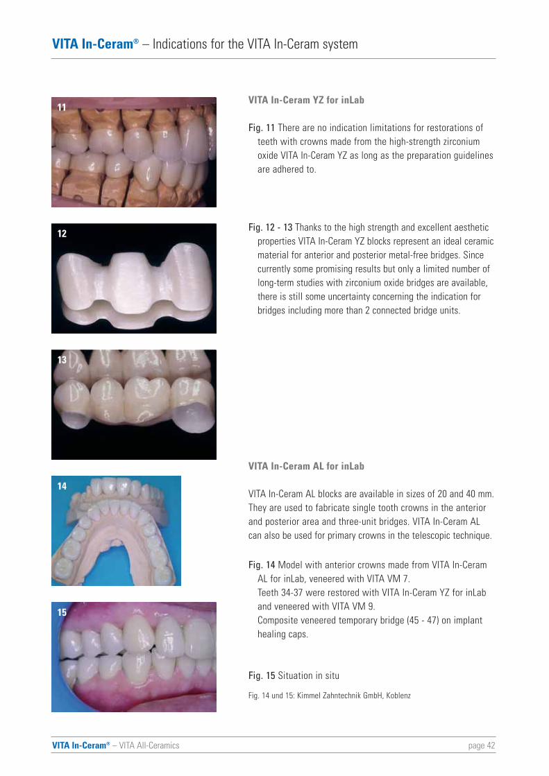
VITA In-Ceram YZ for inLab
Fig. 11 There are no indication limitations for restorations of teeth with crowns made from the high-strength zirconium oxide VITA In-Ceram YZ as long as the preparation guidelinesare adhered to.
Fig. 12 - 13 Thanks to the high strength and excellent aesthetic properties VITA In-Ceram YZ blocks represent an ideal ceramicmaterial for anterior and posterior metal-free bridges. Since currently some promising results but only a limited number of long-term studies with zirconium oxide bridges are available, there is still some uncertainty concerning the indication for bridges including more than 2 connected bridge units.
VITA In-Ceram AL for inLab
VITA In-Ceram AL blocks are available in sizes of 20 and 40 mm.They are used to fabricate single tooth crowns in the anteriorand posterior area and three-unit bridges. VITA In-Ceram ALcan also be used for primary crowns in the telescopic technique.
Fig. 14 Model with anterior crowns made from VITA In-Ceram AL for inLab, veneered with VITA VM 7. Teeth 34-37 were restored with VITA In-Ceram YZ for inLaband veneered with VITA VM 9. Composite veneered temporary bridge (45 - 47) on implant healing caps.
Fig. 15 Situation in situ
Fig. 14 und 15: Kimmel Zahntechnik GmbH, Koblenz
11
12
13
VITA In-Ceram® – VITA All-Ceramics page 43
VITA In-Ceram® – Indications for the VITA In-Ceram system
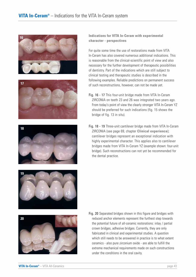
Indications for VITA In-Ceram with experimental character - perspectives
For quite some time the use of restorations made from VITA In-Ceram has also covered numerous additional indications. This is reasonable from the clinical-scientific point of view and alsonecessary for the further development of therapeutic possibilitiesof dentistry. Part of the indications which are still subject to clinical testing and therapeutic studies is described in the following examples. Reliable predictions on permanent success of such reconstructions, however, can not be made yet.
Fig. 16 - 17 This four-unit bridge made from VITA In-Ceram ZIRCONIA on teeth 23 and 26 was integrated two years ago. From today's point of view the clearly stronger VITA In-Ceram YZ should be preferred for such indications (fig. 15 shows the bridge of fig. 13 in situ).
Fig. 18 - 19 Three-unit cantilever bridge made from VITA In-CeramZIRCONIA (see page 69, chapter Clinical experience);cantilever bridges represent an exceptional indication with highly experimental character. This applies also to cantilever bridges made from VITA In-Ceram YZ (example shown: four-unitbridge). Such reconstructions can not yet be recommended for the dental practice.
Fig. 20 Separated bridges shown in this figure and bridges with reduced anchor elements represent the furthest step towards the potential future of all-ceramic restorations: inlay / partial crown bridges, adhesive bridges. Currently, they are only fabricated in clinical and experimental studies. A question which still needs to be answered in practice is to what extent ceramics - also pure zirconium oxide - are able to fulfill the extreme mechanical requirements made on such constructions under the conditions in the oral cavity.
16
17
18
19
20
page 44VITA In-Ceram® – VITA All-Ceramics
VITA In-Ceram® – Indications for the VITA In-Ceram system
When looking at the development of the two lastdecades which brought continuous further developmentof all-ceramic restoration techniques and the VITA In-Ceram system, additional indications for VITA In-Ceram and all-ceramic reconstructions can beassumed.
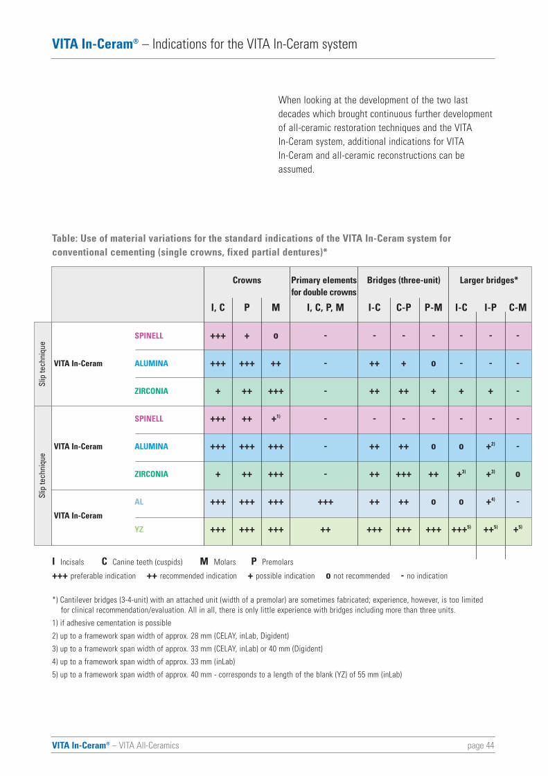
Table: Use of material variations for the standard indications of the VITA In-Ceram system for conventional cementing (single crowns, fixed partial dentures)*
Crowns Primary elements Bridges (three-unit) Larger bridges*for double crowns
I, C P M I, C, P, M I-C C-P P-M I-C I-P C-M
+++ + o - - - - - - -
+++ +++ ++ - ++ + o - - -
+ ++ +++ - ++ ++ + + + -
+++ ++ +1) - - - - - - -
+++ +++ +++ - ++ ++ o o +2) -
+ ++ +++ - ++ +++ ++ +3) +3) o
+++ +++ +++ +++ ++ ++ o o +4) -
+++ +++ +++ ++ +++ +++ +++ +++5) ++5) +5)
SPINELL
VITA In-Ceram ALUMINA
ZIRCONIA
SPINELL
VITA In-Ceram ALUMINA
ZIRCONIA
AL
VITA In-Ceram
YZ
I Incisals C Canine teeth (cuspids) M Molars P Premolars
+++ preferable indication ++ recommended indication + possible indication o not recommended - no indication
*) Cantilever bridges (3-4-unit) with an attached unit (width of a premolar) are sometimes fabricated; experience, however, is too limited for clinical recommendation/evaluation. All in all, there is only little experience with bridges including more than three units.
1) if adhesive cementation is possible
2) up to a framework span width of approx. 28 mm (CELAY, inLab, Digident)
3) up to a framework span width of approx. 33 mm (CELAY, inLab) or 40 mm (Digident)
4) up to a framework span width of approx. 33 mm (inLab)
5) up to a framework span width of approx. 40 mm - corresponds to a length of the blank (YZ) of 55 mm (inLab)
Slip
tech
niqu
eSl
ip te
chni
que
VITA In-Ceram® – VITA All-Ceramics page 45
VITA In-Ceram® – Clinical tooth preparation techniques
Clinical tooth preparation
Fundamentals
Clinical preparation for prosthetic treatment with restorationsmade from VITA In-Ceram is based on the main principle ofdental tooth preparation:
As much as necessary - as little as possible
Each preparation should offer
• a form of retention and stability for the restoration and
• a form of resistance for the abutment tooth
but also ensure
• sufficient space for functional design and structural durability of the restoration
• whilst ensuring removal of substance in accordance with proper axial alignment and the anatomical tooth shape
and
• reveal a clearly defined preparation margin.
The axial preparation angle should be 5 to 10°. Owing to thesensitivity of the ceramic to any tensile forces friction on theprepared abutment is renounced for all-ceramic crowns (in thenon-cemented condition).
page 46VITA In-Ceram® – VITA All-Ceramics
VITA In-Ceram® – Clinical tooth preparation techniques
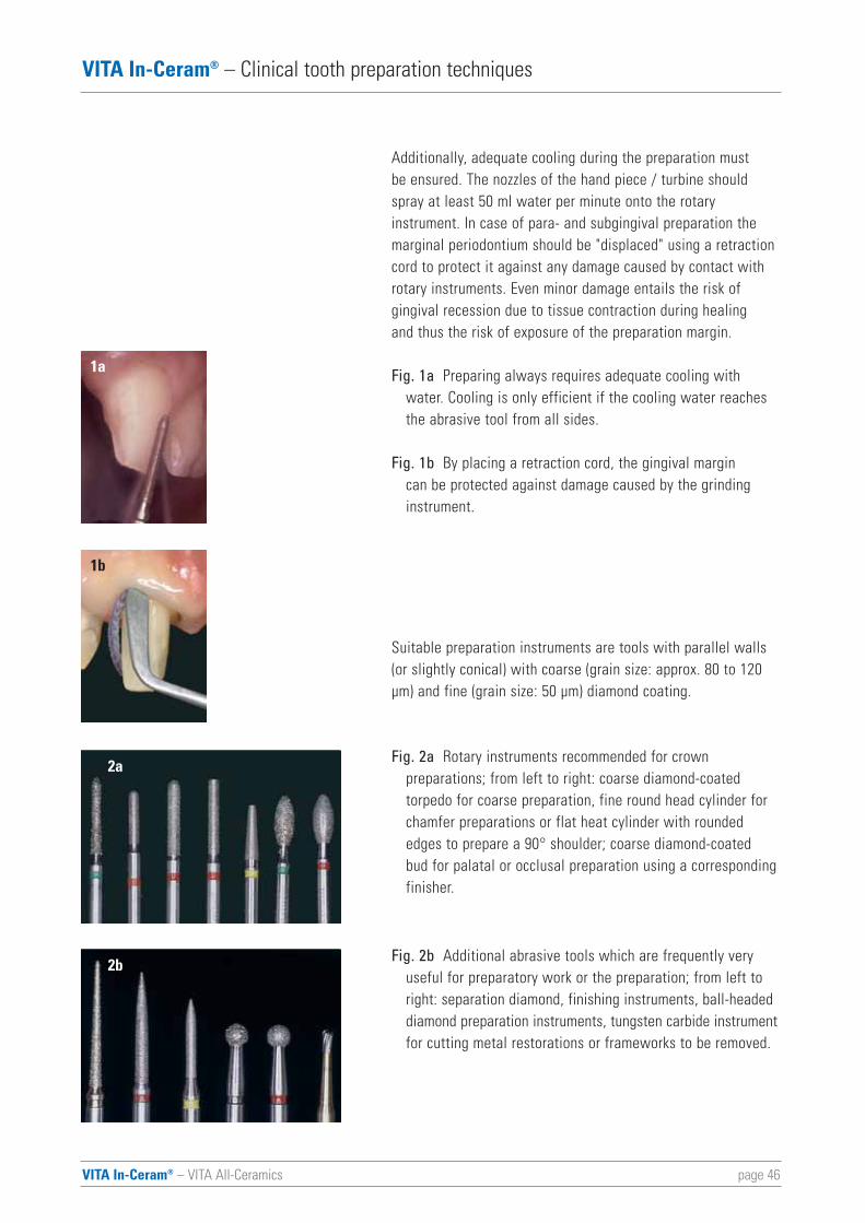
Additionally, adequate cooling during the preparation must be ensured. The nozzles of the hand piece / turbine shouldspray at least 50 ml water per minute onto the rotary instrument. In case of para- and subgingival preparation themarginal periodontium should be "displaced" using a retractioncord to protect it against any damage caused by contact withrotary instruments. Even minor damage entails the risk of gingival recession due to tissue contraction during healing and thus the risk of exposure of the preparation margin.
Fig. 1a Preparing always requires adequate cooling with water. Cooling is only efficient if the cooling water reaches the abrasive tool from all sides.
Fig. 1b By placing a retraction cord, the gingival margin can be protected against damage caused by the grinding instrument.
Suitable preparation instruments are tools with parallel walls(or slightly conical) with coarse (grain size: approx. 80 to 120μm) and fine (grain size: 50 μm) diamond coating.
Fig. 2a Rotary instruments recommended for crown preparations; from left to right: coarse diamond-coated torpedo for coarse preparation, fine round head cylinder for chamfer preparations or flat heat cylinder with rounded edges to prepare a 90° shoulder; coarse diamond-coated bud for palatal or occlusal preparation using a correspondingfinisher.
Fig. 2b Additional abrasive tools which are frequently very useful for preparatory work or the preparation; from left to right: separation diamond, finishing instruments, ball-headeddiamond preparation instruments, tungsten carbide instrumentfor cutting metal restorations or frameworks to be removed.
1a
1b
2a
2b

VITA In-Ceram® – VITA All-Ceramics page 47
VITA In-Ceram® – Clinical tooth preparation techniques
Preparation depths
For the required preparation depth the minimum wall thicknessof 0.5 mm for the ceramic substructure (coping) made fromVITA In-Ceram must be adhered to in order to avoid the risk offracture when exposing the restoration to masticatory load.
Depending on the functional situation, aesthetic requirementsand location of the clinical tooth crown, additional space of 0.5to 1.0 mm must be created for veneering with feldspar ceramic.Accordingly, an axial preparation depth of 1 to 1.5 mm (removalof tooth substance) is obtained. In the occlusal or incisal areasubstance equal to 1.5 to 2 mm must be removed. These preparation depths are very similar to those required for metalceramic restorations.
Fig. 3 Checking the preparation depth for the preparation of a central upper incisor for a VITA In-Ceram YZ bridge with a silicone key and a PA probe. In the labial area slightly more space should be created (previously 1.0 mm) during finishing.It is not necessarily required to achieve a preparation depth of 1.5 mm due to the natural dentine color and translucency of the VITA In-Ceram YZ framework. An incisal preparation depth of approx. 2 mm is already sufficient.
Any necessary reduction of the recommended cutting depthsmust not affect the thickness of the framework (coping) wall.The VITA In-Ceram framework, however, must not be exposed. It should be at least coated with a layer of glaze material1.
1) This requirement applies only to frameworks made from VITA In-Ceram SPINELL, ALUMINA and ZIRCONIA.
3
page 48VITA In-Ceram® – VITA All-Ceramics
VITA In-Ceram® – Clinical tooth preparation techniques
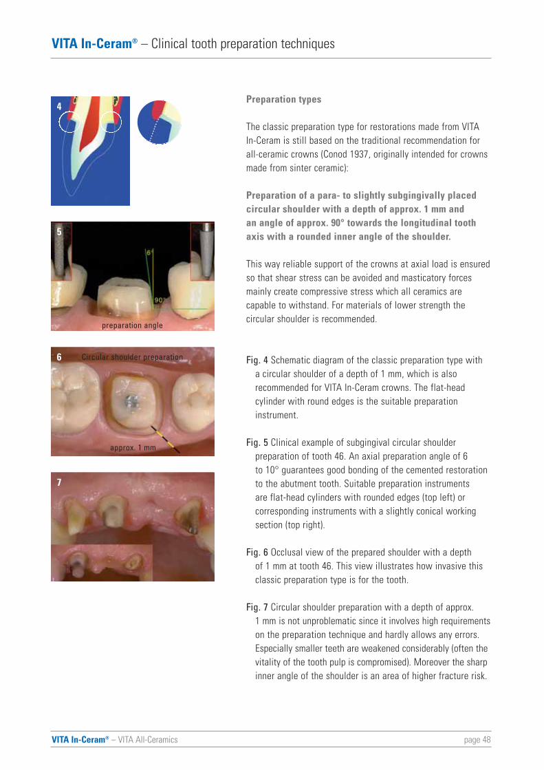
Preparation types
The classic preparation type for restorations made from VITAIn-Ceram is still based on the traditional recommendation forall-ceramic crowns (Conod 1937, originally intended for crownsmade from sinter ceramic):
Preparation of a para- to slightly subgingivally placed circular shoulder with a depth of approx. 1 mm and an angle of approx. 90° towards the longitudinal toothaxis with a rounded inner angle of the shoulder.
This way reliable support of the crowns at axial load is ensuredso that shear stress can be avoided and masticatory forcesmainly create compressive stress which all ceramics are capable to withstand. For materials of lower strength the circular shoulder is recommended.
Fig. 4 Schematic diagram of the classic preparation type with a circular shoulder of a depth of 1 mm, which is also recommended for VITA In-Ceram crowns. The flat-head cylinder with round edges is the suitable preparation instrument.
Fig. 5 Clinical example of subgingival circular shoulder preparation of tooth 46. An axial preparation angle of 6 to 10° guarantees good bonding of the cemented restoration to the abutment tooth. Suitable preparation instruments are flat-head cylinders with rounded edges (top left) or corresponding instruments with a slightly conical working section (top right).
Fig. 6 Occlusal view of the prepared shoulder with a depth of 1 mm at tooth 46. This view illustrates how invasive this classic preparation type is for the tooth.
Fig. 7 Circular shoulder preparation with a depth of approx. 1 mm is not unproblematic since it involves high requirementson the preparation technique and hardly allows any errors. Especially smaller teeth are weakened considerably (often thevitality of the tooth pulp is compromised). Moreover the sharpinner angle of the shoulder is an area of higher fracture risk.
4
5
6
7
approx. 1 mm
Circular shoulder preparation
preparation angle
VITA In-Ceram® – VITA All-Ceramics page 49
VITA In-Ceram® – Clinical tooth preparation techniques
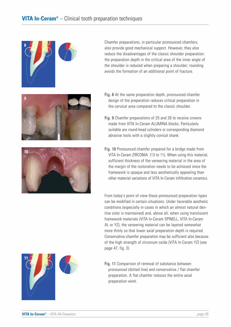
Chamfer preparations, in particular pronounced chamfers, also provide good mechanical support. However, they alsoreduce the disadvantages of the classic shoulder preparation:the preparation depth in the critical area of the inner angle ofthe shoulder is reduced when preparing a shoulder; roundingavoids the formation of an additional point of fracture.
Fig. 8 At the same preparation depth, pronounced chamfer design of the preparation reduces critical preparation in the cervical area compared to the classic shoulder.
Fig. 9 Chamfer preparations of 25 and 26 to receive crowns made from VITA In-Ceram ALUMINA blocks. Particularly suitable are round-head cylinders or corresponding diamondabrasive tools with a slightly conical shank.
Fig. 10 Pronounced chamfer prepared for a bridge made from VITA In-Ceram ZIRCONIA (13 to 11). When using this material,sufficient thickness of the veneering material in the area of the margin of the restoration needs to be achieved since the framework is opaque and less aesthetically appealing than other material variations of VITA In-Ceram infiltration ceramics.
From today's point of view these pronounced preparation typescan be modified in certain situations. Under favorable aestheticconditions (especially in cases in which an almost natural den-tine color is maintained) and, above all, when using translucentframework materials (VITA In-Ceram SPINELL, VITA In-CeramAL or YZ), the veneering material can be layered somewhatmore thinly so that lower axial preparation depth is required.Conservative chamfer preparation may be sufficient also becauseof the high strength of zirconium oxide (VITA In-Ceram YZ) (seepage 47, fig. 3).
Fig. 11 Comparison of removal of substance between pronounced (dotted line) and conservative / flat chamfer preparation. A flat chamfer reduces the entire axial preparation work.
8
9
10
11
page 50VITA In-Ceram® – VITA All-Ceramics
VITA In-Ceram® – Clinical tooth preparation techniques
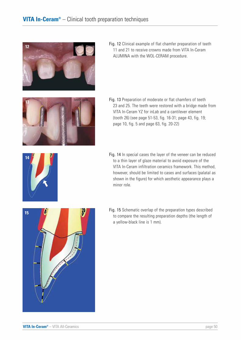
Fig. 12 Clinical example of flat chamfer preparation of teeth 11 and 21 to receive crowns made from VITA In-Ceram ALUMINA with the WOL-CERAM procedure.
Fig. 13 Preparation of moderate or flat chamfers of teeth 23 and 25. The teeth were restored with a bridge made from VITA In-Ceram YZ for inLab and a cantilever element (tooth 26) (see page 51-53, fig. 16-31; page 43, fig. 19; page 10, fig. 5 and page 63, fig. 20-22)
Fig. 14 In special cases the layer of the veneer can be reduced to a thin layer of glaze material to avoid exposure of the VITA In-Ceram infiltration ceramics framework. This method, however, should be limited to cases and surfaces (palatal as shown in the figure) for which aesthetic appearance plays a minor role.
Fig. 15 Schematic overlap of the preparation types described to compare the resulting preparation depths (the length of a yellow-black line is 1 mm).
12
13
14
15
tooth axis
VITA In-Ceram® – VITA All-Ceramics page 51
VITA In-Ceram® – Clinical tooth preparation techniques
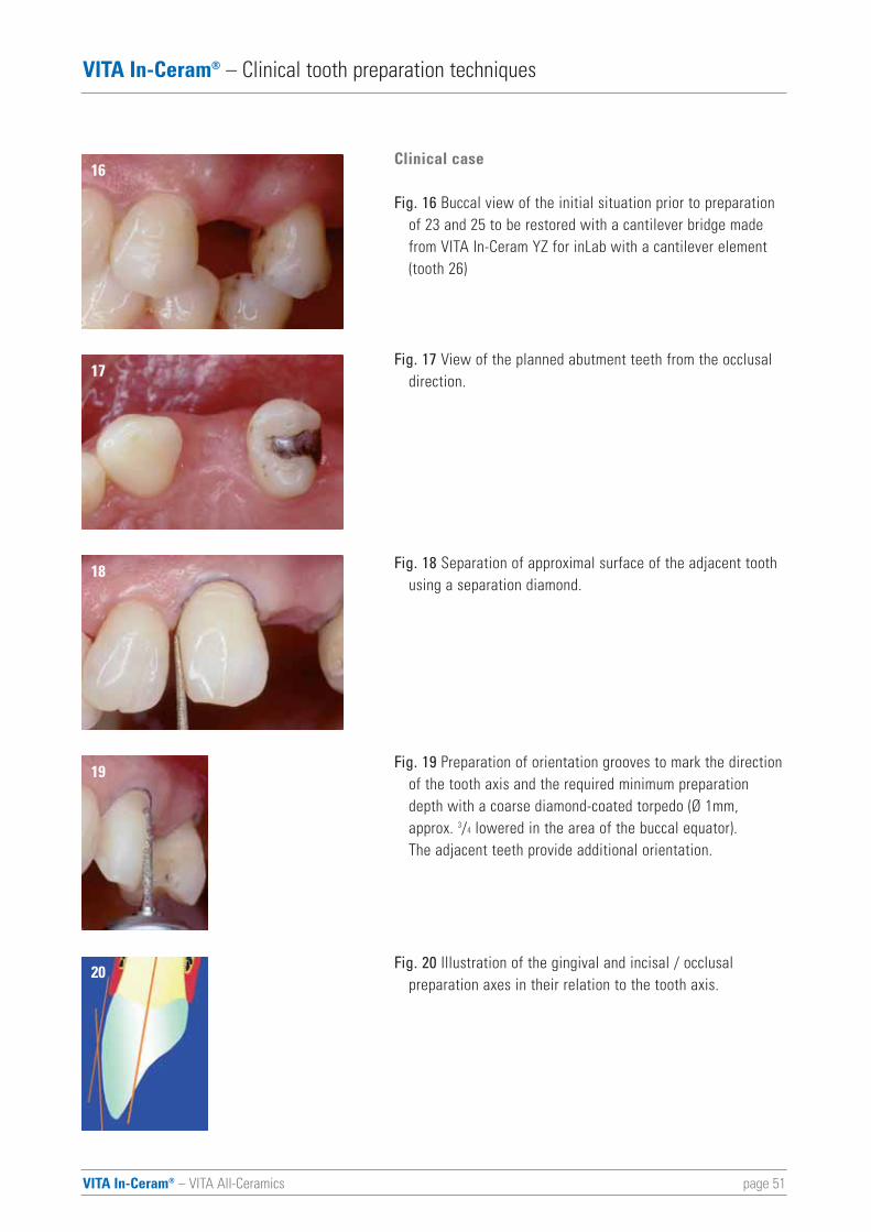
Clinical case
Fig. 16 Buccal view of the initial situation prior to preparation of 23 and 25 to be restored with a cantilever bridge made from VITA In-Ceram YZ for inLab with a cantilever element (tooth 26)
Fig. 17 View of the planned abutment teeth from the occlusal direction.
Fig. 18 Separation of approximal surface of the adjacent tooth using a separation diamond.
Fig. 19 Preparation of orientation grooves to mark the directionof the tooth axis and the required minimum preparation depth with a coarse diamond-coated torpedo (Ø 1mm, approx. 3/4 lowered in the area of the buccal equator). The adjacent teeth provide additional orientation.
Fig. 20 Illustration of the gingival and incisal / occlusal preparation axes in their relation to the tooth axis.
16
17
18
19
20
page 52VITA In-Ceram® – VITA All-Ceramics
VITA In-Ceram® – Clinical tooth preparation techniques
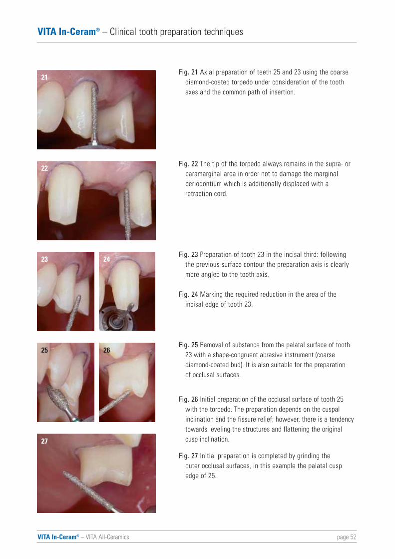
Fig. 21 Axial preparation of teeth 25 and 23 using the coarse diamond-coated torpedo under consideration of the tooth axes and the common path of insertion.
Fig. 22 The tip of the torpedo always remains in the supra- or paramarginal area in order not to damage the marginal periodontium which is additionally displaced with a retraction cord.
Fig. 23 Preparation of tooth 23 in the incisal third: following the previous surface contour the preparation axis is clearly more angled to the tooth axis.
Fig. 24 Marking the required reduction in the area of the incisal edge of tooth 23.
Fig. 25 Removal of substance from the palatal surface of tooth 23 with a shape-congruent abrasive instrument (coarse diamond-coated bud). It is also suitable for the preparation of occlusal surfaces.
Fig. 26 Initial preparation of the occlusal surface of tooth 25 with the torpedo. The preparation depends on the cuspal inclination and the fissure relief; however, there is a tendency towards leveling the structures and flattening the original cusp inclination.
Fig. 27 Initial preparation is completed by grinding the outer occlusal surfaces, in this example the palatal cusp edge of 25.
21
22
23 24
2625
27
VITA In-Ceram® – VITA All-Ceramics page 53
VITA In-Ceram® – Clinical tooth preparation techniques
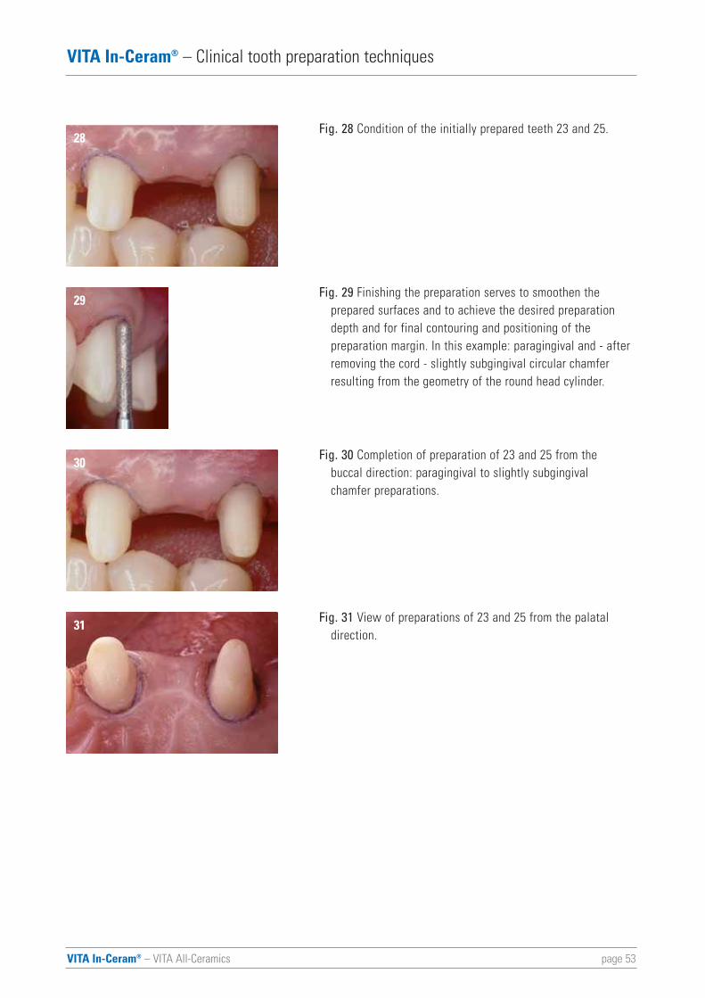
Fig. 28 Condition of the initially prepared teeth 23 and 25.
Fig. 29 Finishing the preparation serves to smoothen the prepared surfaces and to achieve the desired preparation depth and for final contouring and positioning of the preparation margin. In this example: paragingival and - after removing the cord - slightly subgingival circular chamfer resulting from the geometry of the round head cylinder.
Fig. 30 Completion of preparation of 23 and 25 from the buccal direction: paragingival to slightly subgingival chamfer preparations.
Fig. 31 View of preparations of 23 and 25 from the palatal direction.
28
29
30
31
page 54VITA In-Ceram® – VITA All-Ceramics
VITA In-Ceram® – Clinical tooth preparation techniques
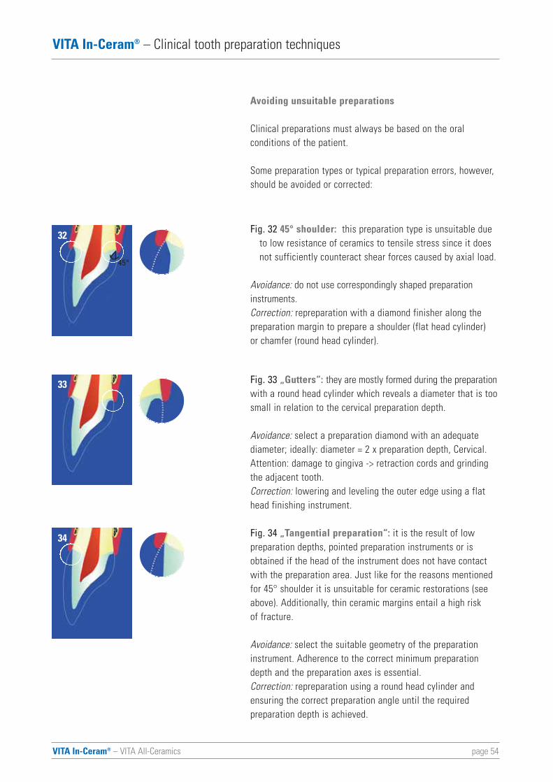
Avoiding unsuitable preparations
Clinical preparations must always be based on the oral conditions of the patient.
Some preparation types or typical preparation errors, however,should be avoided or corrected:
Fig. 32 45° shoulder: this preparation type is unsuitable due to low resistance of ceramics to tensile stress since it does not sufficiently counteract shear forces caused by axial load.
Avoidance: do not use correspondingly shaped preparationinstruments.Correction: repreparation with a diamond finisher along thepreparation margin to prepare a shoulder (flat head cylinder) or chamfer (round head cylinder).
Fig. 33 „Gutters“: they are mostly formed during the preparationwith a round head cylinder which reveals a diameter that is toosmall in relation to the cervical preparation depth.
Avoidance: select a preparation diamond with an adequatediameter; ideally: diameter = 2 x preparation depth, Cervical.Attention: damage to gingiva -> retraction cords and grindingthe adjacent tooth. Correction: lowering and leveling the outer edge using a flathead finishing instrument.
Fig. 34 „Tangential preparation“: it is the result of lowpreparation depths, pointed preparation instruments or isobtained if the head of the instrument does not have contactwith the preparation area. Just like for the reasons mentionedfor 45° shoulder it is unsuitable for ceramic restorations (seeabove). Additionally, thin ceramic margins entail a high risk of fracture.
Avoidance: select the suitable geometry of the preparationinstrument. Adherence to the correct minimum preparationdepth and the preparation axes is essential.Correction: repreparation using a round head cylinder andensuring the correct preparation angle until the required preparation depth is achieved.
32
33
34
45°
VITA In-Ceram® – VITA All-Ceramics page 55
VITA In-Ceram® – Clinical tooth preparation techniques
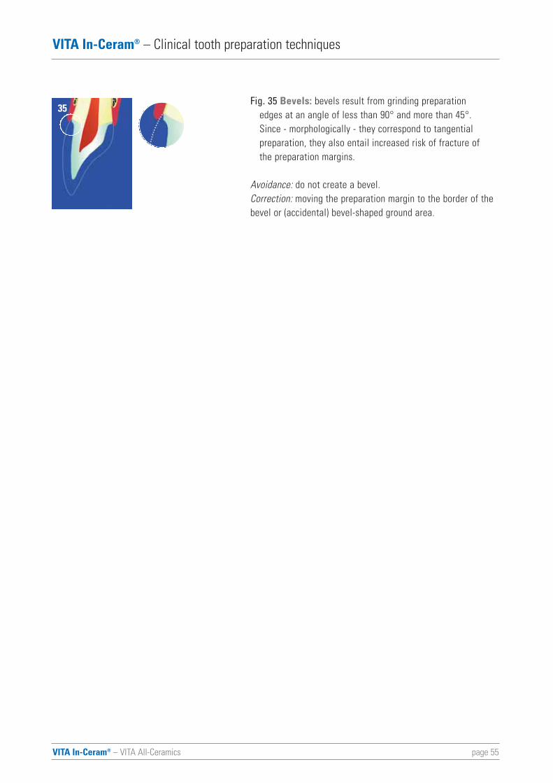
Fig. 35 Bevels: bevels result from grinding preparation edges at an angle of less than 90° and more than 45°. Since - morphologically - they correspond to tangential preparation, they also entail increased risk of fracture of the preparation margins.
Avoidance: do not create a bevel. Correction: moving the preparation margin to the border of thebevel or (accidental) bevel-shaped ground area.
35
page 56VITA In-Ceram® – VITA All-Ceramics
VITA In-Ceram® – Cementing VITA In-Ceram® restorations
Cementing VITA In-Ceram restorations
Conventional cementing
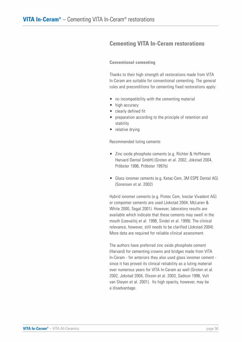
Thanks to their high strength all restorations made from VITAIn-Ceram are suitable for conventional cementing. The generalrules and preconditions for cementing fixed restorations apply:
• no incompatibility with the cementing material• high accuracy• clearly defined fit• preparation according to the principle of retention and
stability• relative drying
Recommended luting cements
• Zinc oxide phosphate cements (e.g. Richter & Hoffmann Harvard Dental GmbH) (Groten et al. 2002, Jokstad 2004, Pröbster 1996, Pröbster 1997b)
• Glass ionomer cements (e.g. Ketac-Cem, 3M ESPE Dental AG)(Sorensen et al. 2002)
Hybrid ionomer cements (e.g. Protec Cem, Ivoclar Vivadent AG)or compomer cements are used (Jokstad 2004, McLaren &White 2000, Segal 2001). However, laboratory results are available which indicate that these cements may swell in themouth (Leevailoj et al. 1998, Sindel et al. 1999). The clinicalrelevance, however, still needs to be clarified (Jokstad 2004).More data are required for reliable clinical assessment.
The authors have preferred zinc oxide phosphate cement(Harvard) for cementing crowns and bridges made from VITA In-Ceram - for anteriors they also used glass ionomer cement -since it has proved its clinical reliability as a luting materialover numerous years for VITA In-Ceram as well (Groten et al.2002, Jokstad 2004, Olsson et al. 2003, Sadoun 1996, Vult van Steyen et al. 2001). Its high opacity, however, may be a disadvantage.
VITA In-Ceram® – VITA All-Ceramics page 57
VITA In-Ceram® – Cementing VITA In-Ceram® restorations
Other cementation materials (Mc Laren & White) should beselected for crowns made from VITA In-Ceram SPINELL since they feature translucent properties or are available as translucentmaterial variations (see table on page 58).
Adhesive luting
The number of supporters of adhesive luting is increasingsteadily (Burke et al. 2002). This must be mainly attributed tothe positive experience gained with luting of ceramic inlaysand veneers but also to reports on reduced failure rates of single tooth crowns made from feldspar ceramics when adhesive cementing with composite material was performedinstead of conventional cementing (Malament & Socransky 2001).
According to the results of laboratory studies the main reasonis the positive bond between the restoration and the preparedtooth which results in considerably enhanced fracture toughnessof the ceramic (Burke et al. 2002, Groten & Pröbster 1997). Theprecondition to achieve this effect in the clinical use is firmadhesion of the luting composite to the ceramic and the tooth(enamel or dentine).
While sufficient bonding to enamel and dentine can obviouslybe ensured with modern adhesive luting systems, bonding to high-strength ceramic materials (oxide ceramics) is more complex and problematic since, unlike feldspar / glass ceramics,they can not simply be etched with hydrofluoric acid (HF-gels).The microretentive surface structure must be achieved using adifferent method (e.g. by sandblasting). But apparently successfulroughening of the surface with or without silanizing depends on the respective structural ceramic material. From the clinicalpoint of view, however, control of subgingival preparation marginsand the presence of different substrate materials (sclerosed orcarious dentine and various build-up materials) is a significantproblem so that some restorations can not be luted. Moreoverdrying and removal of excess material are much more difficult.
The respective suitable luting method can be taken from thetable on the next page:
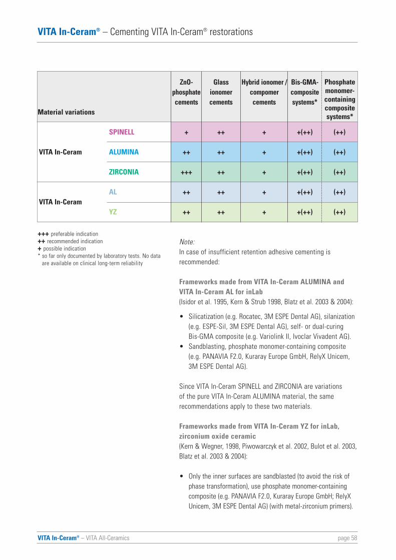
page 58VITA In-Ceram® – VITA All-Ceramics
VITA In-Ceram® – Cementing VITA In-Ceram® restorations
Note:In case of insufficient retention adhesive cementing is recommended:
Frameworks made from VITA In-Ceram ALUMINA andVITA In-Ceram AL for inLab(Isidor et al. 1995, Kern & Strub 1998, Blatz et al. 2003 & 2004):
• Silicatization (e.g. Rocatec, 3M ESPE Dental AG), silanization(e.g. ESPE-Sil, 3M ESPE Dental AG), self- or dual-curing Bis-GMA composite (e.g. Variolink II, Ivoclar Vivadent AG).
• Sandblasting, phosphate monomer-containing composite (e.g. PANAVIA F2.0, Kuraray Europe GmbH, RelyX Unicem, 3M ESPE Dental AG).
Since VITA In-Ceram SPINELL and ZIRCONIA are variations of the pure VITA In-Ceram ALUMINA material, the same recommendations apply to these two materials.
Frameworks made from VITA In-Ceram YZ for inLab, zirconium oxide ceramic (Kern & Wegner, 1998, Piwowarczyk et al. 2002, Bulot et al. 2003,Blatz et al. 2003 & 2004):
• Only the inner surfaces are sandblasted (to avoid the risk of phase transformation), use phosphate monomer-containing composite (e.g. PANAVIA F2.0, Kuraray Europe GmbH; RelyX Unicem, 3M ESPE Dental AG) (with metal-zirconium primers).
ZnO- Glass Hybrid ionomer / Bis-GMA-phosphate ionomer compomer compositecements cements cements systems*
+ ++ + +(++) (++)
++ ++ + +(++) (++)
+++ ++ + +(++) (++)
++ ++ + +(++) (++)
++ ++ + +(++) (++)
SPINELL
VITA In-Ceram ALUMINA
ZIRCONIA
ALVITA In-Ceram
YZ
+++ preferable indication ++ recommended indication + possible indication * so far only documented by laboratory tests. No data
are available on clinical long-term reliability
Material variations
Phosphatemonomer-containingcompositesystems*
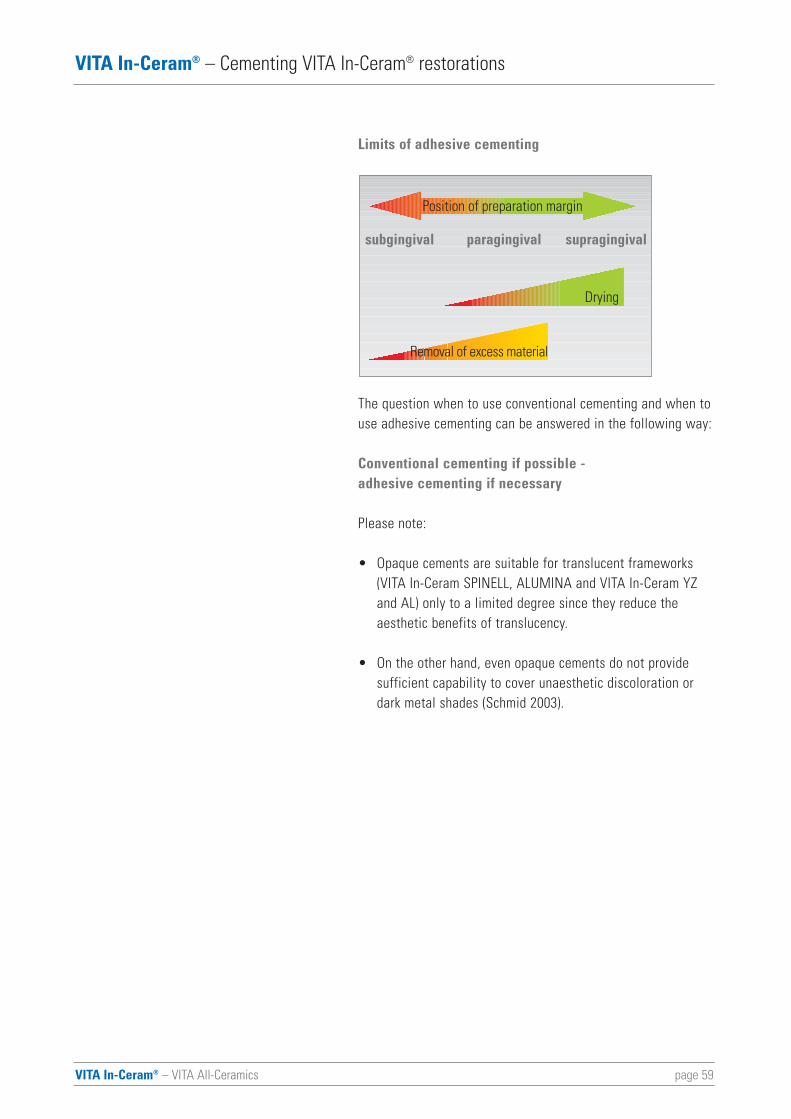
VITA In-Ceram® – VITA All-Ceramics page 59
VITA In-Ceram® – Cementing VITA In-Ceram® restorations
Limits of adhesive cementing
The question when to use conventional cementing and when touse adhesive cementing can be answered in the following way:
Conventional cementing if possible -adhesive cementing if necessary
Please note:
• Opaque cements are suitable for translucent frameworks (VITA In-Ceram SPINELL, ALUMINA and VITA In-Ceram YZ and AL) only to a limited degree since they reduce the aesthetic benefits of translucency.
• On the other hand, even opaque cements do not provide sufficient capability to cover unaesthetic discoloration or dark metal shades (Schmid 2003).
Position of preparation margin
Drying
Removal of excess material
subgingival paragingival supragingival
page 60VITA In-Ceram® – VITA All-Ceramics
VITA In-Ceram® – Cementing VITA In-Ceram® restorations
Clinical procedure
Convenient, conventional cementing is possible for all restorations made from VITA In-Ceram.
Fig. 1 After cleaning and degreasing the inner surfaces of the crown (alcohol), the conventional cement is applied into the crown (in this example: Ketac-Cem, ESPE DENTAL AG). The prepared tooth is also cleaned and disinfected under relativedrying.
Fig. 2 The Ketac Cem glass ionomer cement can be mixed and easily applied thanks to a capsule system (Applicap, 3M ESPE Dental AG).
Fig. 3 The reliable alternative: zinc oxide phosphate cements (e.g. Richter & Hoffmann Harvard Dental GmbH).
Fig. 4 The cement is spread across the entire surface up to the margin of the restoration and a suitable instrument (brush or Heidemann spatula) is used to obtain uniform layer thickness.Excessive application should be avoided.
Fig. 5 Initially, the restoration is positioned slowly by pressing it down with the finger and lowered to the final position in a way to ensure that excess cement can flow off easily. Then the patient is made to bite carefully; afterwards the masticatory pressure is gradually increased to slowly "displace"the remaining excess cement under the increasing masticatorypressure. During this process, the patient may bite on a cottonroll (attention: it must be ensured that the restoration is not moved away from the final position and accurate fit is retained!).
Fig. 6 After hardening completely, all excess cement can be easily removed with a probe or a scaler. This requires thorough probing of the restoration margins and the sulcus to remove the residual cement and takes considerable time - a fact, which is frequently underestimated.
Fig. 7 Complete removal of all residual cement along the sulcus and control of the static and dynamic occlusion result in perfect periodontal and functional integration of the crownmade from VITA In-Ceram.
1
2
3
5
7
4
6
VITA In-Ceram® – VITA All-Ceramics page 61
VITA In-Ceram® – Cementing VITA In-Ceram® restorations
The use of translucent and tooth-colored cements may provideadvantages for anteriors or demanding aesthetic situations -e.g. like in this example of paragingival preparation margins:
Fig. 8 Disinfecting and degreasing the cleaned preparations with alcohol.
Fig. 9 If required, deposits or residual temporary cement can be removed with pumice powder or cleaning paste and a rubber cup in a gentle and reliable manner.
Fig. 10 After this preparatory work the dentine surfaces are dried by gently spraying them with air. Relative drying is considered to be sufficient.
Fig. 11 The hybrid cementation material (in this case the adhesive cement RelyX Unicem, 3M ESPE Dental AG) is filled into the degreased and cleaned crowns and spread whilst avoiding excess application. Additional conditioning of the inner surfaces of the crown is not required unless positive adhesive bonding is to be achieved.
Fig. 12 Crowns are lowered whilst slowly increasing the pressure exerted with a finger to "displace" all excess material until the final position is reached. Due to extraaxial stress which results when the patient bites there is a particularly high risk for anterior crowns to become wedged in an incorrect position.
8
9
10
11
12
page 62VITA In-Ceram® – VITA All-Ceramics
VITA In-Ceram® – Cementing VITA In-Ceram® restorations
Fig. 13 If the restorations are positioned accurately, the cementmargins are polymerized for approx. 3 seconds.
Fig. 14 This level of polymerization allows easy removal of excess material such as residual conventional cement since final hardness and adhesion to the tooth surface have not been achieved yet. The risk of dislocating the crowns no longer exists.
Fig. 15 After removing all excess material polymerization can be completed by curing each side for approx. 20 seconds. The material, however, also hardens automatically (autopoly-merizing) within approx. 3 to 4 minutes and is therefore also suitable for opaque frameworks made from VITA In-Ceram.
Fig. 16 Condition of the crowns 11, 21 made from VITA In-Ceram ALUMINA after definitive cementation with RelyX Unicem.
Fig. 17 Adhesive cementation of a bridge (45 - 47) made from VITA In-Ceram ZIRCONIA for CELAY with PANAVIA F due to limited retention to the distal abutment.
13
14
15
16
17
VITA In-Ceram® – VITA All-Ceramics page 63
VITA In-Ceram® – Cementing VITA In-Ceram® restorations
Fig. 18 The adhesive resin cement PANAVIA F (Kuraray Europe GmbH) is white-opaque. No aesthetic disadvantages need to be expected in the area where the material is applied and for the opaque framework made from VITA In-Ceram ZIRCONIA. Excess material can be easily recognized and should be removed as far as possible before polymerization is completed.
Fig. 19 Chairside adhesive conditioning of the inner surfaces ofthe anchors of the crown can be performed with the CoJet system (3M ESPE Dental AG). Alternatively, sandblasting is also possible if PANAVIA F is used.
Fig. 20 Translucent, tooth colored luting materials/cements provide superior comfort in aesthetically sensitive areas. In these situations of paragingival preparations for a bridge made from VITA In-Ceram YZ for inLab (23 - 25) with a cantilever element (26) reliable retention and coverage of thecement joint in the transitional zone of restoration - tooth neck are required.
Fig. 21 Adhesive cementation under relative drying using the translucent variation of RelyX Unicem. All excess cement hadbeen removed before polymerization was completed.
Fig. 22 Aesthetic integration: paragingival transitional zone of the restoration to the natural tooth is almost invisible thanksto the framework made from VITA In-Ceram YZ for inLaband the veneer. The combination of their optical properties creates a result which is almost identical to natural enamel. This effect, however, must not be impaired by the properties of the cement to achieve a perfect aesthetic result.
18
19
20
21
22
page 64VITA In-Ceram® – VITA All-Ceramics
VITA In-Ceram® – Clinical experience with VITA In-Ceram® restorations
Clinical experience with VITA In-Ceramrestorations
The table (see page 66) provides a survey on the data on the clini-cal behavior of VITA In-Ceram restorations available in literature.The following aspects need to be considered for the interpretationof the data listed in the table:
There is no standardized method based on which clinical long-term studies are generally performed. Additionally, each authorplaces different emphasis on acquisition and evaluation of patientdata. As a result all reports of studies differ from each other andno direct comparisons can be made. Yet data must be handled in away to obtain useful and pragmatic results for the clinical practicesince different (or "better" - whatever that means) data are notavailable.
In our approach the following aspects provided in literature wereconsidered to be factors which contribute essentially to the suc-cess of restorations made from VITA In-Ceram:
• Material variation (VITA In-Ceram SPINELL / ALUMINA / ZIRCONIA) and preparation type (shoulder / chamfer).
• Restoration type (crowns / bridges).
• Restored teeth according to occlusal-functional jaw regions (anteriors / premolars / molars).
Additionally, the sample sizes were determined, which is not sim-ple since the information provided is not always complete andsometimes contradictory.
• Number of patients who received VITA In-Ceram restorations under "observation" (in the broadest sense). The relation of the total number of patients with restorations to patients under "observation" could frequently not be determined.
VITA In-Ceram® – VITA All-Ceramics page 65
VITA In-Ceram® – Clinical experience with VITA In-Ceram® restorations
• Number of restorations under "observation" - definition as indicated for "Number of patients".
In the result data on the failure, i.e. failure of a restoration madefrom VITA In-Ceram, resulting in removal or remake were included:
• Number of restorations / percentage of the total number given.
• Type of failure (fracture / loss of retention / pain after cementation / loss of abutment / secondary caries / necessary endodontic treatment / other reasons)
• Indication of a (mostly cumulative) survival rate in percent which was frequently not included by the authors and had to be estimated as precisely as possible based on failure information published.
Accordingly, the table includes interpretations if the authors didnot provide any specific/direct information. These interpretationsresult in considerable simplification so that a certain degree ofarbitrariness could not be avoided. It was attempted to meet therequirements for the dental practice rather than those of science.
At best, any data which extend beyond a clinical follow-up periodof 5 years have limited significance. Mostly, follow-up periods of3-6 years were found in literature. The studies mainly deal withcrowns and bridges made from VITA In-Ceram ALUMINA in theclassic slip casting technique.
Few data are available on VITA In-Ceram restorations producedfrom industrially prefabricated VITA In-Ceram blocks (Bindl &Mörmann 2002, for CEREC; Groten et al. 2002, for CELAY). Basedon enhanced material properties and characteristic values ofindustrially prefabricated ceramics, however, it must be assumedthat the clinical properties of these restorations can be comparedto those of restorations fabricated using the slip casting technique.
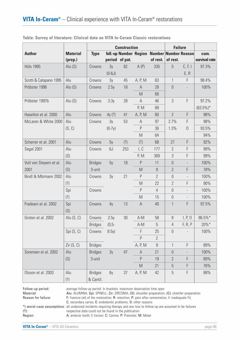
page 66VITA In-Ceram® – VITA All-Ceramics
VITA In-Ceram® – Clinical experience with VITA In-Ceram® restorations
Table: Survey of literature: Clinical data on VITA In-Ceram Classic restorations
Follow-up period: average follow-up period. In brackets: maximum observation time spanMaterial: Alu: ALUMINA; Spi: SPINELL; Zir: ZIRCONIA; (S): shoulder preparation; (C): chamfer preparation
F: fracture (at) of the restoration; R: retention; P: pain after cementation; I: inadequate fit; C: secondary caries; E: endodontic problems; O: other reasons
*) worst-case assumptions: all undesired incidents requiring therapy and any loss to follow-up are assumed to be failures(?): respective data could not be found in the publicationRegion:
Construction FailureAuthor Material Type foll.-up Number Region Number Number Reason cum.
(prep.) period of pat. of rest. of rest. survival rate
Hüls 1995 Alu (S) Crowns 3y 82 A (P) 335 5 C, F, I 97.3%
(0-6J) E, R
Scotti & Catapano 1995 Alu Crowns 3y 45 A, P, M 63 1 F 98.4%
Pröbster 1996 Alu (S) Crowns 2.5y 18 A 28 0 - 100%
M 68
Pröbster 1997b Alu (S) Crowns 3.3y 28 A 46 3 F 97.2%
P, M 89 (63.5%)*
Haselton et al. 2000 Alu Crowns 4y (?) 41 A, P, M 80 2 F 98%
McLaren & White 2000 Alu Crowns 3y 53 A 97 2.7% F 98%
(S, C) (0-7y) P 36 1.3% O 93.5%
M 64 94%
Scherrer et al. 2001 Alu Crowns 5y (?) (?) 68 27 F 92%
Segal 2001 Alu Crowns 6J 253 I, C 177 2 F 99%
(S) P, M 369 3 F 99%
Vult von Steyern et al. Alu Bridges 5y 18 P 11 0 - 100%
2001 (S) 3-unit M 9 2 F 78%
Bindl & Mörmann 2002 Alu Crowns 3y 21 P 2 0 - 100%
(?) M 22 2 F 90%
Spi Crowns P 4 0 - 100%
(?) M 15 0 - 100%
Fradeani et al. 2002 Spi Crowns 4y 13 A 40 1 F 97.5%
(S)
Groten et al. 2002 Alu (S, C) Crowns 2.5y 30 A-M 58 8 I, P, O 86.5%*
Bridges (0,5- A-M 5 4 F, R, P 20%*
Spi (S, C) Crowns 8.5y) F 25 0 - 100%
P 2
Zir (S, C) Bridges A, P, M 9 1 F 89%
Sorensen et al. 2002 Alu Bridges 3y 47 A 21 0 - 100%
(S) 3-unit P 19 2 F 89%
M 21 5 F 76%
Olsson et al. 2003 Alu Bridges 6y 37 A, P, M 42 5 F 88%
(?) & Cantil.
Reason for failure:
A: anterior tooth; I: Incisor; C: Canine; P: Premolar; M: Molar

VITA In-Ceram® – VITA All-Ceramics page 67
VITA In-Ceram® – Clinical experience with VITA In-Ceram® restorations
VITA In-Ceram SPINELL crowns
Crowns made from VITA In-Ceram SPINELL are preferably used in the anterior and premolar area. A uniform high rate of success is published: 97.5 to 100% for a period of 2 to 4 years (Bindl & Mörmann 2002, Fradeani et al. 2002, Groten et al. 2002).
VITA In-Ceram ALUMINA crowns
Crowns made from VITA In-Ceram ALUMINA have proved to be very reliable in the area of anterior teeth. Most authors reportsuccess rates of approx. 97 to 100° over average follow-up periods of 3 to 6 years (Bindl & Mörmann 2002, Groten et al. 2002,Haselton & Diaz-Arnold 2000, Hüls 1995, McLaren & White 2000,Pröbster 1997, Sadoun 1996, Scotti & Catapano 1995, Segal 2001,Scherrer et al. 2001). The failure rate in the posterior area is alsoinsignificant; two reports, however, indicate a rate of approx. 7 to10% (Bindl & Mörmann, McLaren & White 2000).
VITA In-Ceram ALUMINA bridges
Obviously the success of bridges made from VITA In-Ceram ALUMINA mainly depends on the restored jaw region. Average success rates of clearly less than 90% within 3 to 6 years are considerably lower than those of metal-based restorations. Thelosses - almost exclusively due to fracture of the bridges - areclearly higher in the posterior region: 11% of failures in the premolararea within approx. 3 years and 24 % of failures if bridge spans (3-unit) extended up to the molar area. Compared to that, therewere no failures/losses of anterior bridges within the same period(Sorensen et al. 2002). A Swedish project team (Olsson et al. 2003)observed 5 failures (12%) in 42 bridges (more than 50% were cantilever bridges) within periods up to 9 years (5-6 years on average). The posterior area was affected more strongly (fracture of 3 cantilever bridges) than the anterior area. Both failures ofanterior cantilever bridges were caused by traumatic loss of teethand do not depend on the restoration type or material. In their 5-year study of 3-unit bridges made from VITA In-Ceram ALUMINAanother Swedish team (Vult von Steyern et al. 2001) found similarresults as Sorensen and his coauthors: 22% of failures in the posterior area and no failures of anterior bridges. Sadoun, the"inventor" of VITA In-Ceram, also reports a failure rate of approx.10% in posterior bridges within a period up to 8 years and only 2%in the anterior area (Sadoun 1996).
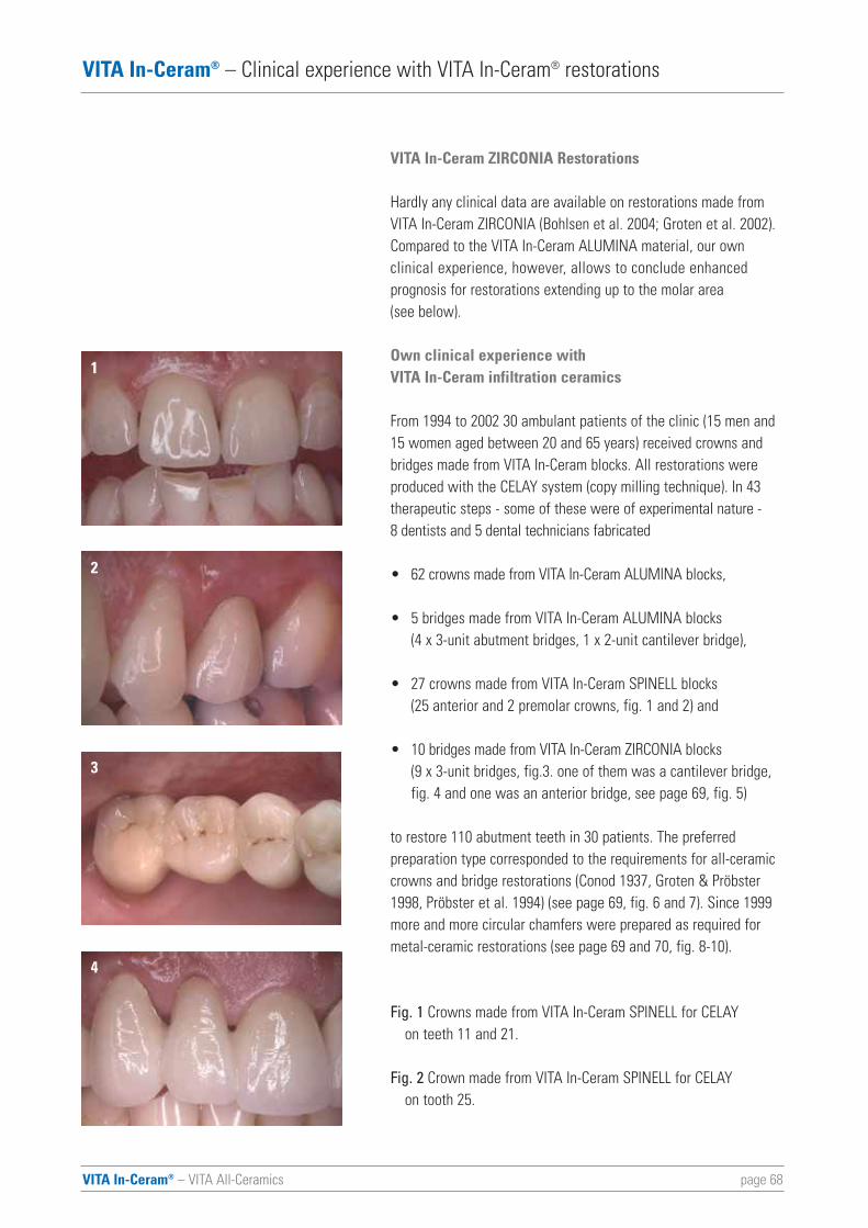
page 68VITA In-Ceram® – VITA All-Ceramics
VITA In-Ceram® – Clinical experience with VITA In-Ceram® restorations
VITA In-Ceram ZIRCONIA Restorations
Hardly any clinical data are available on restorations made fromVITA In-Ceram ZIRCONIA (Bohlsen et al. 2004; Groten et al. 2002).Compared to the VITA In-Ceram ALUMINA material, our own clinical experience, however, allows to conclude enhancedprognosis for restorations extending up to the molar area (see below).
Own clinical experience with VITA In-Ceram infiltration ceramics
From 1994 to 2002 30 ambulant patients of the clinic (15 men and15 women aged between 20 and 65 years) received crowns andbridges made from VITA In-Ceram blocks. All restorations wereproduced with the CELAY system (copy milling technique). In 43therapeutic steps - some of these were of experimental nature - 8 dentists and 5 dental technicians fabricated
• 62 crowns made from VITA In-Ceram ALUMINA blocks,
• 5 bridges made from VITA In-Ceram ALUMINA blocks (4 x 3-unit abutment bridges, 1 x 2-unit cantilever bridge),
• 27 crowns made from VITA In-Ceram SPINELL blocks (25 anterior and 2 premolar crowns, fig. 1 and 2) and
• 10 bridges made from VITA In-Ceram ZIRCONIA blocks(9 x 3-unit bridges, fig.3. one of them was a cantilever bridge, fig. 4 and one was an anterior bridge, see page 69, fig. 5)
to restore 110 abutment teeth in 30 patients. The preferred preparation type corresponded to the requirements for all-ceramiccrowns and bridge restorations (Conod 1937, Groten & Pröbster1998, Pröbster et al. 1994) (see page 69, fig. 6 and 7). Since 1999more and more circular chamfers were prepared as required formetal-ceramic restorations (see page 69 and 70, fig. 8-10).
Fig. 1 Crowns made from VITA In-Ceram SPINELL for CELAY on teeth 11 and 21.
Fig. 2 Crown made from VITA In-Ceram SPINELL for CELAY on tooth 25.
1
2
3
4
VITA In-Ceram® – VITA All-Ceramics page 69
5
VITA In-Ceram® – Clinical experience with VITA In-Ceram® restorations
Fig. 3 Bridge made from VITA In-Ceram ZIRCONIA for CELAY from teeth 15 to 17.
Fig. 4 Bridge made from VITA In-Ceram ZIRCONIA for CELAY from teeth 13 to 11 (same female patient).
Fig. 5 Cantilever bridge made from VITA In-Ceram ZIRCONIA for CELAY with abutments 17, 16 to replace tooth 15 (cantilever element).
Fig. 6 Classic preparation type for all-ceramic crowns: circular 90°shoulder with a preparation depth of 1 mm and rounded inner angle (see fig. 5-6, page 48).
Fig. 7 Classic preparation for crown made from VITA In-Ceram ALUMINA on tooth 12 and a bridge made from VITA In-Ceram ALUMINA from tooth 11 to tooth 22. The example of anterior teeth shows that this invasive preparation may be accompanied by weakening of the abutment tooth and damaging the vitality of the pulp (see fig. 7, page 48).
Fig. 8 Currently, chamfer preparations such as those for PFM restorations are preferred to minimize the risks to the integrity of the abutment tooth. In this case a pronounced circular chamferof teeth 11 and 21 was prepared to receive the crowns made from VITA In-Ceram SPINELL for CELAY (see fig.1).
Fig. 9 Circular chamfer preparations for a bridge made from VITA In-Ceram ZIRCONIA for CELAY from teeth 13 to 11 (see page 68,fig. 4).
Fig. 10 Moderate circular chamfer preparations to restore teeth 11 and 21 with crowns made from VITA In-Ceram ALUMINAproduced using the WOL-CERAM technique (see fig. 3, page 12).
All restorations were cemented conventionally - preferably withphosphate cement (Harvard Cement, fast setting, Richter &Hoffmann Harvard Dental GmbH). Most of the translucentcrowns made from VITA In-Ceram SPINELL were fixed withhybrid ionomer cement (Protec Cem, Ivoclar Vivadent AG). Some crowns were integrated using glass ionomer cement(Ketac Cem, 3M ESPE Dental AG). In some cases, the restora-tions were cemented to be worn temporarily for a limited timeor because of planned orthodontic treatment over an extended
6
7
8
9
page 70VITA In-Ceram® – VITA All-Ceramics
VITA In-Ceram® – Clinical experience with VITA In-Ceram® restorations
period (Temp Bond, Kerr GmbH). 10
k) under follow-up resp. followed until failure occurred*) „follow-up therapy“ describes any undesired impact on a restoration or a restored tooth resulting in clinical intervention
(regardless of type and cause).The sum of the number in the lines and columns of the block "follow-up therapy" do not have to be identical with the total number of follow-up therapies since in some follow-up units repairs were performed and failures resulted.
#) „Repair“ is any clinical intervention performed on a restoration or tooth - after an undesired incidence - as long as the restoration is retained.
+) „Failure“ is any loss of a restoration regardless of the cause. x) „Total loss“ is any loss of a restoration (regardless of the cause) which can not be retreated with VITA In-Ceram Classic for CELAY.†) „worst case“ indicates that all patient contacts/loss to follow-up (l4) must be considered "failures" (like l6).‡) „best case“ " indicates that all patient contacts lost/loss to follow-up (l4) must be considered "in function" (like l7).l) is an abbreviation for "line" in the equations1) borderline / experimental indications (cf. page 72)
Own clinical experience with VITA In-Ceram
Start of therapies: Number Number
January 1994 of pat. Ther. Number restorations Number of teeth
follow-up: Crowns Crowns Bridges Bridges
l April 2002 SPINELL ALUMINA ALUMINA ZIRCONIA SPINELL ALUMINA ZIRCONIA
1 Ther. started 30 43 27 62 5 10 27 63 20
2 Ther. discontinued 1 1 - 3 - - - 3 -
3 Ther. completed 29 42 27 59 5 10 27 60 20
(l3=l1-l2)
4 Contact lost 3 3 4 3 - 2 4 3 4
5 Under follow-upk 26 39 23 56 5 8 23 57 16
(l5=l3-l4)
Follow-up therapy* 9 11 2 7 4 1 2 12 2
(-repair)# (3) (3) (2) (1) - - (2) (1) -
6 (-failure)+ (7) (8) - (6) (4)1 (1) - (11) 2
(--retreated) (5) (5) - (4) (2) - - (8) -
6 (--total loss)x (3) (4) - (2) (3) (1) - (5) (2)
7 function (worst 23 35 23 50 1 7 23 52 14
case = [l5-l6]/z1)† (77%) (81%) (85%) (81%) (20%) (70%) (85%) (83%) (70%)
8 function (best 26 38 27 53 1 9 27 55 18
case = [l7+l4]/z1)‡ (87%) (88%) (100%) (85%) (20%) (90%) (100%) (87%) (90%)
VITA In-Ceram® – VITA All-Ceramics page 71
VITA In-Ceram® – Clinical experience with VITA In-Ceram® restorations
-6-9 -3 63 9 12 1815 21 24 3027 33 36 4239 45 48 5451 57 60 6663 69 72 7875 81 84 9087-12 9693 10299
1 2 3 4 5 6 7 8-1 -0,5 0,5 1,5 2,5 3,5 4,5 5,5 6,5 7,5 8,5
[month]
[year]
C 22C 01
C 27
C 15
C 05
C 07
C 55C 38
C 39C 19
C 50C 49
C 52
C 02
C 31
C 30
C 04
C 16
C 41
C 36
C 20C 09
C 06
C 53
C 33
C 10
C 37
C 03
C 08
C 47
10 month
Follow-up: (dashed line: Loss to follow-up)
ALUMINA (Crowns)
SPINELL (Crowns)
ZIRCONIA (Bridges)
Therapy: (bar width proportional to the number ofabutments restored with VITA In-Ceram CELAY)
Study therapy
Failure => follow-up therapy
Complaints/deficiencies => repair/correction
ALUMINA (Bridges)
C 39
C 47
Modified Lexis diagram for comparative representation of the clinical process of treatment with crowns and bridges made from VITA In-Ceram for CELAY. One or several bars indicate the patients according to the number of therapeutic phases. The width of each bar is proportional to the number of abutments restored in the respective therapeutic phase. Color coding is used to identify the respectiveframework material and the occurrence of incidences such as complaints, failure and follow-up treatment according to the legend.Numerous patients had been treated several times and received complex restorations (restorations made from VITA In-Ceram SPINELL,ALUMINA and ZIRCONIA) and are indicated by several bars and using several colors. The task of such an apparently "confusing" diagram is to illustrate complex processes and clinical situations in a graphical form.
C) refers to the patient ID in the studies
Lexis diagram - Fig. 11
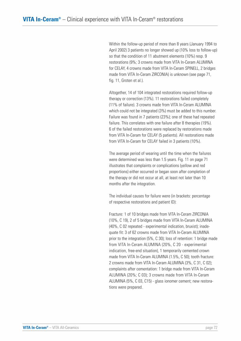
page 72VITA In-Ceram® – VITA All-Ceramics
VITA In-Ceram® – Clinical experience with VITA In-Ceram® restorations
Within the follow-up period of more than 8 years (January 1994 toApril 2002) 3 patients no longer showed up (10% loss to follow-up)so that the condition of 11 abutment elements (10%) resp. 9restorations (9%; 3 crowns made from VITA In-Ceram ALUMINAfor CELAY, 4 crowns made from VITA In-Ceram SPINELL, 2 bridgesmade from VITA In-Ceram ZIRCONIA) is unknown (see page 71,fig. 11, Groten et al.).
Altogether, 14 of 104 integrated restorations required follow-uptherapy or correction (13%). 11 restorations failed completely(11% of failure). 3 crowns made from VITA In-Ceram ALUMINAwhich could not be integrated (3%) must be added to this number.Failure was found in 7 patients (23%); one of these had repeatedfailure. This correlates with one failure after 8 therapies (19%). 6 of the failed restorations were replaced by restorations madefrom VITA In-Ceram for CELAY (5 patients). All restorations madefrom VITA In-Ceram for CELAY failed in 3 patients (10%).
The average period of wearing until the time when the failureswere determined was less than 1.5 years. Fig. 11 on page 71 illustrates that complaints or complications (yellow and red proportions) either occurred or began soon after completion of the therapy or did not occur at all, at least not later than 10months after the integration.
The individual causes for failure were (in brackets: percentage of respective restorations and patient ID):
Fracture: 1 of 10 bridges made from VITA In-Ceram ZIRCONIA(10%, C 19), 2 of 5 bridges made from VITA In-Ceram ALUMINA(40%, C 02 repeated - experimental indication, bruxist); inade-quate fit: 3 of 62 crowns made from VITA In-Ceram ALUMINAprior to the integration (5%, C 30); loss of retention: 1 bridge madefrom VITA In-Ceram ALUMINA (20%, C 20 - experimental indication, free-end situation), 1 temporarily cemented crownmade from VITA In-Ceram ALUMINA (1.5%, C 50); tooth fracture: 2 crowns made from VITA In-Ceram ALUMINA (3%, C 31, C 02);complaints after cementation: 1 bridge made from VITA In-CeramALUMINA (20%; C 03); 3 crowns made from VITA In-Ceram ALUMINA (5%, C 03, C15) - glass ionomer cement; new restora-tions were prepared.
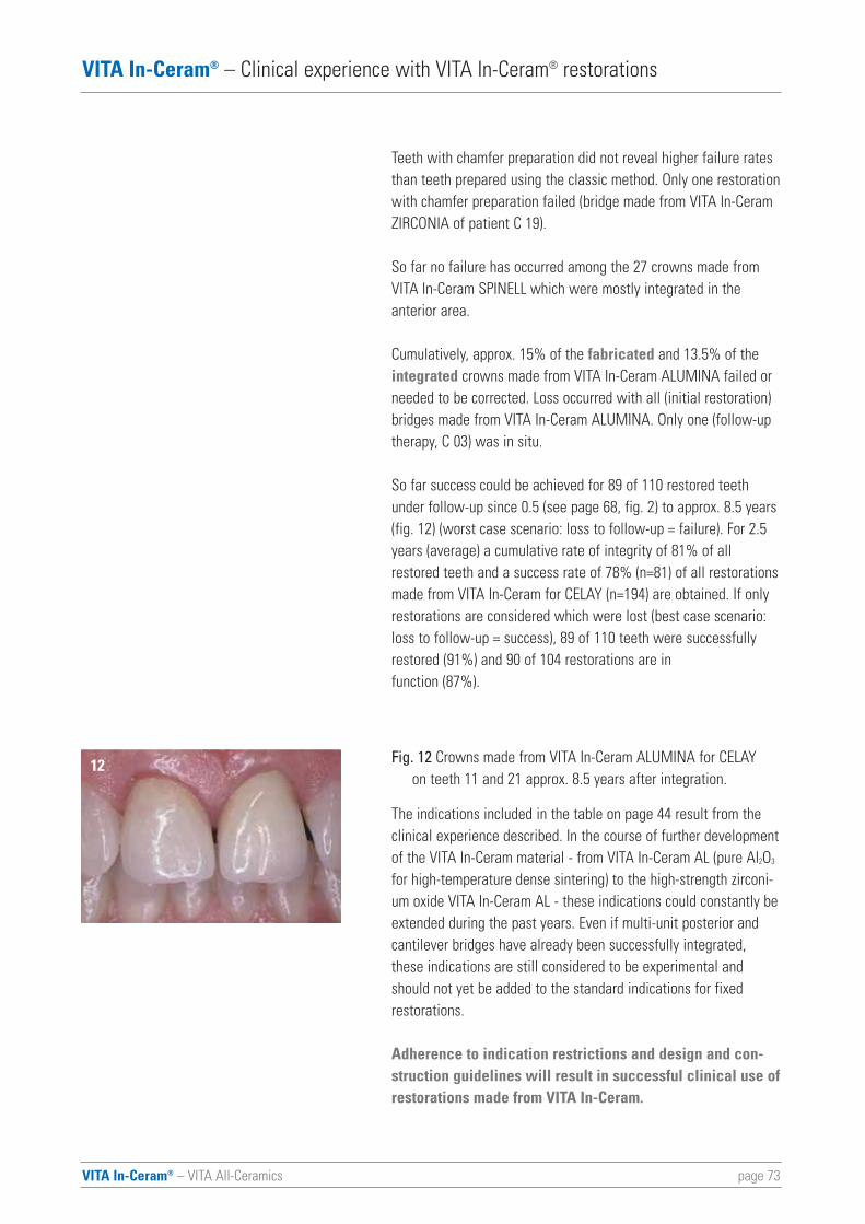
VITA In-Ceram® – VITA All-Ceramics page 73
VITA In-Ceram® – Clinical experience with VITA In-Ceram® restorations
12
Teeth with chamfer preparation did not reveal higher failure ratesthan teeth prepared using the classic method. Only one restorationwith chamfer preparation failed (bridge made from VITA In-CeramZIRCONIA of patient C 19).
So far no failure has occurred among the 27 crowns made fromVITA In-Ceram SPINELL which were mostly integrated in the anterior area.
Cumulatively, approx. 15% of the fabricated and 13.5% of theintegrated crowns made from VITA In-Ceram ALUMINA failed orneeded to be corrected. Loss occurred with all (initial restoration)bridges made from VITA In-Ceram ALUMINA. Only one (follow-uptherapy, C 03) was in situ.
So far success could be achieved for 89 of 110 restored teethunder follow-up since 0.5 (see page 68, fig. 2) to approx. 8.5 years(fig. 12) (worst case scenario: loss to follow-up = failure). For 2.5years (average) a cumulative rate of integrity of 81% of allrestored teeth and a success rate of 78% (n=81) of all restorationsmade from VITA In-Ceram for CELAY (n=194) are obtained. If onlyrestorations are considered which were lost (best case scenario:loss to follow-up = success), 89 of 110 teeth were successfullyrestored (91%) and 90 of 104 restorations are infunction (87%).
Fig. 12 Crowns made from VITA In-Ceram ALUMINA for CELAY on teeth 11 and 21 approx. 8.5 years after integration.
The indications included in the table on page 44 result from theclinical experience described. In the course of further developmentof the VITA In-Ceram material - from VITA In-Ceram AL (pure Al2O3
for high-temperature dense sintering) to the high-strength zirconi-um oxide VITA In-Ceram AL - these indications could constantly beextended during the past years. Even if multi-unit posterior andcantilever bridges have already been successfully integrated,these indications are still considered to be experimental andshould not yet be added to the standard indications for fixedrestorations.
Adherence to indication restrictions and design and con-struction guidelines will result in successful clinical use ofrestorations made from VITA In-Ceram.
page 74VITA In-Ceram® – VITA All-Ceramics
VITA In-Ceram® – Clinical documentations
Clinical documentations
Documentation 1Patient, 33 yearsDiagnosis: insufficient crowns and bridgesTherapy: crowns 11, 21 made from VITA In-Ceram SPINELL for CELAY
and bridge (13-15) from VITA In-Ceram ZIRCONIA for CELAY
1
3 4
5
7
6
8
2
VITA In-Ceram® – VITA All-Ceramics page 75
VITA In-Ceram® – Clinical documentations
11
13 14
15
17
16
18
12
9 10
VITA In-Ceram® – VITA All-Ceramics page 76
Documentation 2
Female patient, 54 yearsDiagnosis: insufficient prosthetic restoration of partially edentulous arch with gaps and
tooth migration with chronic periodontitisTherapy: preprosthetic, systematic periodontitis therapy, all-ceramic restorations for
total rehabilitation to restore the function and for gap closure with bridgesmade from VITA In-Ceram ZIRCONIA for CELAY (teeth 17-16/15)
VITA In-Ceram® – Clinical documentations
10 11
1 32
4 65
7 98
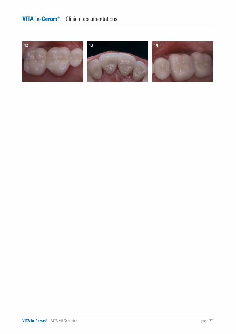
VITA In-Ceram® – VITA All-Ceramics page 77
VITA In-Ceram® – Clinical documentations
12 1413
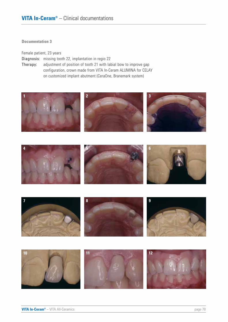
VITA In-Ceram® – VITA All-Ceramics page 78
VITA In-Ceram® – Clinical documentations
Documentation 3
Female patient, 23 yearsDiagnosis: missing tooth 22, implantation in regio 22Therapy: adjustment of position of tooth 21 with labial bow to improve gap
configuration, crown made from VITA In-Ceram ALUMINA for CELAY on customized implant abutment (CeraOne, Branemark system)
1 32
4 65
7 98
10 1211
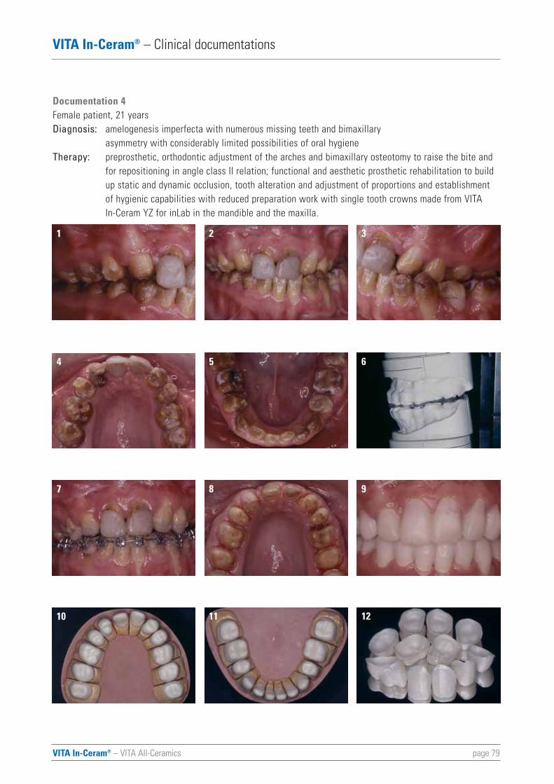
VITA In-Ceram® – VITA All-Ceramics page 79
VITA In-Ceram® – Clinical documentations
Documentation 4Female patient, 21 yearsDiagnosis: amelogenesis imperfecta with numerous missing teeth and bimaxillary
asymmetry with considerably limited possibilities of oral hygieneTherapy: preprosthetic, orthodontic adjustment of the arches and bimaxillary osteotomy to raise the bite and
for repositioning in angle class II relation; functional and aesthetic prosthetic rehabilitation to build up static and dynamic occlusion, tooth alteration and adjustment of proportions and establishment of hygienic capabilities with reduced preparation work with single tooth crowns made from VITA In-Ceram YZ for inLab in the mandible and the maxilla.
1 32
4 65
7 98
10 1211
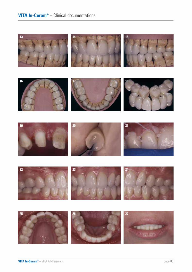
VITA In-Ceram® – VITA All-Ceramics page 80
VITA In-Ceram® – Clinical documentations
13 1514
16 1817
19 2120
22 2423
25 2726
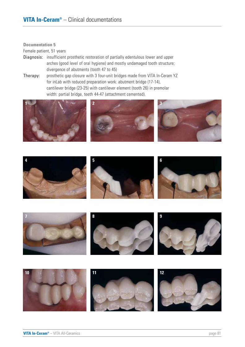
VITA In-Ceram® – VITA All-Ceramics page 81
VITA In-Ceram® – Clinical documentations
Documentation 5Female patient, 51 years Diagnosis: insufficient prosthetic restoration of partially edentulous lower and upper
arches (good level of oral hygiene) and mostly undamaged tooth structure;divergence of abutments (tooth 47 to 45)
Therapy: prosthetic gap closure with 3 four-unit bridges made from VITA In-Ceram YZ for inLab with reduced preparation work: abutment bridge (17-14),cantilever bridge (23-25) with cantilever element (tooth 26) in premolarwidth: partial bridge, teeth 44-47 (attachment cemented).
1 32
4 65
7 98
10 1211
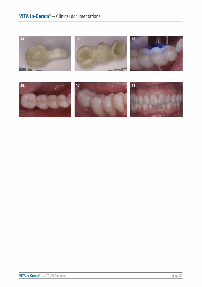
VITA In-Ceram® – VITA All-Ceramics page 82
VITA In-Ceram® – Clinical documentations
13 1514
16 1817
VITA In-Ceram® – VITA All-Ceramics page 83
Bindl A, Mörmann WH. An up to 5-year clinical evaluation of posterior In-Ceram CAD/CAM Core Crowns.International Journal of Prosthodontics 15, 451-456 (2002).
Blatz MB, Sadan A, Kern M. Resin-ceramic bonding: A review of the literature. Journal of Prosthetic Dentistry 89, 268-274 (2003).
Blatz MB, Sadan A, Kern M. Adhäsive Befestigung hochfester Vollkeramikrestaurationen. Quintessenz 55, 33-41 (2004).
Bohlsen, F., Wolfart, S., Wegner, S., Kern, M. Klinische Bewährung von vollkeramischen Brücken aus In-CeramZirconia im pagenzahnbereich. 53. Jahrestagung der DGZPW, 13. - 16.05.04
Bulot D, Sadan A, Burgess JO, Blatz MB. Bond strength of a self-adhesive universal resin cement to Lava Zirconiaafter two surface treatments. Journal of Dental Research 82, (Special Issue A), „Abstract # 578“ (2003).
Burke FJT, Fleming GJP, Nathanson D, Marquis PM. Are adhesive technologies needed to support ceramics? Anassessment of the current evidence. Journal of Adhesive Dentistry 4, 7-22 (2002).
Conod H. Étude sur la statique de la couronne jaquette. Schweizer Monatsschrift für Zahnmedizin 47, 485-529 (1937)
Fradeani M, Aquilano A, Corrado M. Klinische Erfahrungen mit In-Ceram-Spinell-Kronen – Eine Fünfjahres-nachuntersuchung. Internationales Journal für Parodontologie & Restaurative Zahnheilkunde 22, 505-513 (2002).
Groten M, Pröbster L. The influence of different cementation modes on the fracture resistance of feldspathicceramic crowns. International Journal of Prosthodontics 10, 169-177 (1997).
Groten M, Pröbster L. Das Celay-System: Herstellung vollkeramischer Restaurationen im Kopierschleifverfahren.in: Pröbster L (Hrsg). Innovationen für die Zahnheilkunde. Spitta-Verlag, Balingen, Teil 30 (1998)
Groten M, Axmann D, Pröbster L, Weber H. Vollkeramische Kronen und Brücken auf Basis industriell vorgefertigterGerüstkeramiken. Quintessenz 53, 1307-1316 (2002).
Haselton DR, Diaz-Arnold AM, Hillis SL. Clinical assessment of high strength all-ceramic crowns. Journal of Prosthetic Dentistry 83, 396-401 (2000).
Hüls A.: Zum Stand der klinischen Bewährung infiltrationskeramischer Verblendkronen. Deutsch Zahnärztl Z 50, 674-676 (1995) 9
Isidor F, Stokholm R, Ravnholt G. Tensile bond strength of resin luting cements to class infiltrated porous Alumi-nium oxide cores (In-Ceram). European Journal of Prosthodontics and Restorative Dentistry 3, 199-202 (1995).
Jokstad A. A split mouth randomized clinical trial of single crowns retained with resin-modified glass-ionomer andzinc phosphate luting cements. International Journal of Prosthodontics 17, 411-416 (2004).
VITA In-Ceram® – References
VITA In-Ceram® – VITA All-Ceramics page 84
VITA In-Ceram® – References
Kern M, Strub JR. Bonding to alumina ceramic in restorative dentistry: clinical results over up to 5 years. Journal of Dentistry 26, 245-249 (1998).
Kern M, Wegner SM. Bonding to zirconia ceramic: adhesion methods and their durability. Dental Materials 14, 64-71 (1998).
Leevailoj C, Platt JA, Cochran MA, Moore K. In vitro study of fracture incidence and compressive fracture load ofall-ceramic crowns cemented with resin-modified glass ionomer and other luting agents. Journal of Prosthetic Dentistry 80, 699-707 (1998).
Malament KA, Socransky SS. Survival of Dicor glass-ceramic dental restorations over 16 years. Part III: effect of luting agent and tooth-substitute core structure. Journal of Prosthetic Dentistry 86, 511-519 (2001).
McLaren EA, White SM. Survival of In-Ceram crowns in a private practice: A prospective clinical trial. Journal of Prosthetic Dentistry 83, 216-222 (2000).
Olsson K, Fürst B, Andresson B, Carlsson GE. A long-term restrospective and clinical follow-up study of In-Ceram Alumina FPDs. International Journal of Prosthodontics 16, 150-156 (2003).
Piwowarczyk A, Berge HX, Lauer H-Ch, Sorensen. Shearbond strength of cements to Zirconia and Lithium disilicateceramics. Journal of Dental Research 81 (Spec Iss A), 401 (2002).
Pröbster L. Survival Rate of In-Ceram Restorations. International Journal of Prosthodontics 6, 259-263 (1993).
Pröbster L, Groten M, Girthofer S. Kopiergefräste, glasinfiltrierte Aluminiumoxid-Kronen. Phillip Journal 11, 577-584 (1994).
Pröbster L. Four year clinical study of glass-infiltrated sintered alumina crowns. Journal of Oral Rehabilitation 23, 147-151 (1996).
Pröbster L, Girthofer S, Groten M, Rein B. Copy-milled all-ceramic Celay-In-Ceram crowns for modified CeraOneabutments: A technical note. International Journal of Oral & Maxillofacial Implants. 11, 201-204 (1996).
Pröbster L, Groten M. Celay-In-Ceram-Kronen für individualisierte CeraOne-Abutments bei der Einzelzahn-implantatversorgung. Quintessenz Zahntechnik 23, 1105-1111 (1997).
Pröbster L. All-ceramic crowns on modified CeraOne abutments: A case report. Quintessence International 29, 52-65 (1997a).
Pröbster L. Klinische Langzeiterfahrungen mit vollkeramischen Kronen aus In-Ceram. Quintessenz 48, 1639-1646 (1997b).
Sadoun M. In-Ceram: 10 Jahre in der Erprobung. In: Kappert HF. (Hrsg.) Vollkeramik. Werkstoffkunde –Zahntechnik – klinische Erfahrungen. Berlin: Quintessenz Verlags-GmbH, 193-210 (1996).
VITA In-Ceram® – VITA All-Ceramics page 85
VITA In-Ceram® – References
Scherrer SS, De Rijk WG, Wiskott HW, Belser UC. Incidence of fractures and lifetime predictions of all-ceramiccrown systems using censored data. American Jounal of Dentistry 14, 72-80 (2001).
Schmid S. Die Opazität von Kompositmaterialien zur Befestigung von Adhäsivbrücken, Inauguraldissertation,Universität Tübingen in Poliklinik für Zahnärztliche Prothetik. Tübingen, (2003).
Scotti R, Catapano S. A clinical evaluation of In-Ceram crowns. International Journal of Prosthodontics 8, 320-323 (1995)
Segal BS. Retrospective assessment of 546 All-ceramic anterior and posterior crowns in a general practice.Journal of Prosthetic Dentistry 85, 544-550 (2001).
Sindel J, Frankenberger R, Krämer N, Petschelt A. Crack formation of all-ceramic crowns depending on differentcore build-up and luting materials. Journal of Dentistry 27, 175-181 (1999).
Sorensen JA, Kang S-K, Torres TJ, Knode H. In-Ceram fixed partial dentures: Three-year clinical trial results.Journal of the California Dental Association 26, 207-214 (1998).
Stephan, M.; Corten, A.: Aluminiumoxid – der Korund, Mineralogische Betrachtungen des Korund. Quintessenz Zahntech 31, 2, 128-133 (2005).
Vult von Steyern P, Jönsson O, Nilner K. Five-year evaluation of posterior all-ceramic three-unit (In-Ceram) FPDs.International Journal of Prosthodontics 14, 379-384 (2001).


With the unique VITA SYSTEM 3D-MASTERall natural tooth shades are systematically determined and completely reproduced.
Please note: Our products should be used according to the working instructions. We cannot be heldliable for damages resulting from incorrect handling or usage. The user is furthermore obliged to checkthe product before use with regard to its suitability for the intended area of applications. We cannotaccept any liability if the product is used in conjunction with porcelains and equipment from other manufacturers which are not compatible or not authorized for use with our product. Furthermore, ourliability for the correctness of this information is independent of the legal ground and, in as far aslegally permissible, is limited to the invoiced value of the goods supplied excluding turnover tax. In particular, as far as legally permissible, we do not assume any liability for profit loss, for indirect damages, for consequential damages or for claims of third parties against the purchaser. Claims fordamages based on fault liability (culpa in contrahendo, breach of contract, unlawful acts, etc.) can onlybe made in the case of intent or gross negligence. The VITA Modulbox is not necessarily a componentof the product. Date of issue: 02-06
VITA Zahnfabrik H. Rauter GmbH & Co.KGPostfach 1338 · D-79704 Bad Säckingen . GermanyTel. +49/ 7761/562-222 · Fax +49/ 7761/562-446www.vita-zahnfabrik.com · [email protected]
1323
E-03
/06
(2.6
) SI
3D - MasterVita System
Equipment All-Ceramics
Serv
ice
Veneering Materials
Teeth
VITA Zahnfabrik has been certified according to the Medical Device Directive and the following products bear the CE mark :
VITA In-Ceram® SPINELL, VITA In-Ceram® ALUMINA, VITA In-Ceram® ZIRCONIA,VITA In-Ceram® YZ for inLab, VITA In-Ceram® AL for inLab
CEREC® and inLab® are registered trademarks of Sirona Dental Systems GmbH, Bensheim, GermanyCeHa WHITE ECS® is a registered trademark of C. Hafner GmbH & Co. KG, Pforzheim, GermanyCELAY® is a registered trademark of Mikrona Technologie AG, Spreitenbach, SwitzerlandDCS Precident® is a registered trademark of DCS Dental AG, Allschwill, SwitzerlandDigident® is a registered trademark of Digident GmbH, Pforzheim, GermanyHarvard Cement® is a registered trademark of Richter & Hoffmann Harvard Dental GmbH, Berlin, GermanyKetac-Cem® und RelyX® are registered trademarks of 3M ESPE Dental AG, Seefeld, GermanyPANAVIA® is a registered trademark of Kuraray Europe GmbH, Düsseldorf, GermanyProtec Cem® is a registered trademark of Ivoclar Vivadent AG, Schaan, LiechtensteinsynOcta® is a registered trademark of Straumann AG, Waldenbuch, SwitzerlandWOL-CERAM® is a registered trademark of Teamziereis GmbH, Engelsbrand, Germany