13 dilatation of the main pulmonary artery
10
13 Dilatation of the Main Pulmonary Artery
-
Upload
muhammad-bin-zulfiqar -
Category
Education
-
view
40 -
download
3
Transcript of 13 dilatation of the main pulmonary artery

13 Dilatation of the Main Pulmonary Artery

CLINICAL IMAGAGINGAN ATLAS OF DIFFERENTIAL DAIGNOSIS
EISENBERG
DR. Muhammad Bin Zulfiqar PGR-FCPS III SIMS/SHL
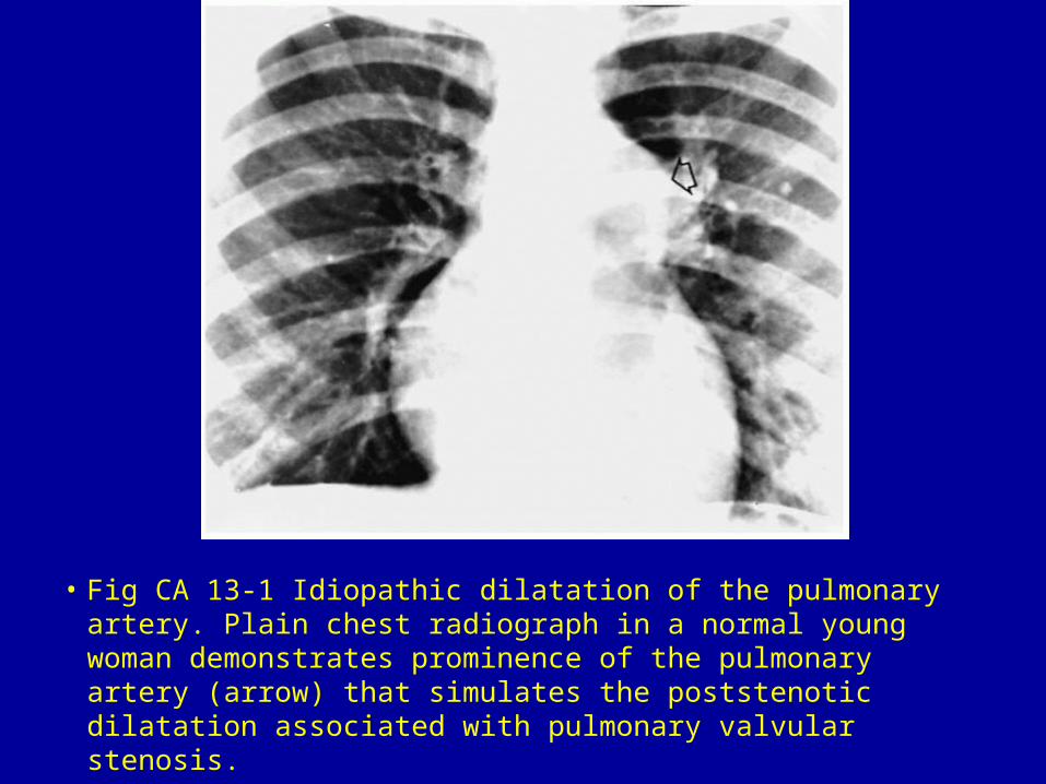
• Fig CA 13-1 Idiopathic dilatation of the pulmonary artery. Plain chest radiograph in a normal young woman demonstrates prominence of the pulmonary artery (arrow) that simulates the poststenotic dilatation associated with pulmonary valvular stenosis.

• Fig CA 13-2 Thyrotoxicosis. Generalized cardiomegaly with increased pulmonary vascularity.

• Fig CA 13-3 Cor pulmonale (primary pulmonary hypertension). (A) Frontal and (B) lateral views of the chest show prominence of the pulmonary outflow tract and markedly dilated central pulmonary vessels. The lateral displacement of the cardiac apex and filling of the retrosternal air space indicate right ventricular enlargement.

• Fig CA 13-4 Eisenmenger syndrome in atrial septal defect. (A) Frontal and (B) lateral films demonstrate slight but definite cardiomegaly and a great increase in the size of the pulmonary trunk. The right and left pulmonary artery branches are huge, but the peripheral pulmonary vasculature is relatively sparse. Long-standing pulmonary hypertension has produced degenerative intimal changes in the pulmonary arteries, which have become densely calcified.1

• Fig CA 13-5 Ventricular septal defect. The pulmonary trunk is very large and overshadows the normal-sized aorta, which seems small by comparison. The pulmonary artery branches in the hilum and in the periphery of the lung are enlarged, and the pulmonary vascular volume is increased. The heart is enlarged and somewhat triangular.1

• Fig CA 13-6 Pulmonary valvular stenosis. Severe poststenotic dilatation of the pulmonary outflow tract (arrow). The heart size and pulmonary vascularity remain within normal limits.

• Fig CA 13-7 Trilogy of Fallot. Marked poststenotic dilatation (arrow) of the pulmonary artery with decrease in overall pulmonary vascularity. There is enormous right atrial and moderate right ventricular enlargement.6