
(12) United States Patent (10) Patent N0.: US 8,478,012 B2 ...
13
US008478012B2 (12) United States Patent (10) Patent N0.: US 8,478,012 B2 Punithakumar et al. (45) Date of Patent: Jul. 2, 2013 (54) METHODS, APPARATUS AND ARTICLES OF 7,327,862 132* 2/2008 Murphy etal. ............. .. 382/128 MANUFACTURE TO PROCESS CARDIAC IMAGES TO DETECT HEART MOTION ABNORMALITIES (75) Inventors: Kumaradevan Punithakumar, London (CA); Shuo Li, London (CA); Ismail Ben Ayed, London (CA); Ian Ross, London (CA); Ali Islam, London (CA); Richard Rankin, London (CA) (73) Assignee: General Electric Company, Schenectady, NY (US) ( * ) Notice: Subject to any disclaimer, the term of this patent is extended or adjusted under 35 U.S.C. 154(b) by 793 days. (21) Appl.No.: 12/633,519 (22) Filed: Dec. 8, 2009 (65) Prior Publication Data US 2011/0064284 A1 Mar. 17, 2011 Related US. Application Data (60) Provisional application No. 61/242,215, ?led on Sep. 14, 2009. (51) Int. Cl. G06K 9/36 (52) US. Cl. USPC ......................................... .. 382/131; 382/132 (58) Field of Classi?cation Search USPC ............ .. 382/128, 13(k132; 378/65; 600/425 See application ?le for complete search history. (2006.01) (56) References Cited U.S. PATENT DOCUMENTS 4,202,340 A 5/1980 Langer et al. 5,797,399 A 8/1998 Morris et al. 5,891,047 A 4/1999 Lander et a1. HEART ABNORMALITY DETECTION COLLECT IMAGES 7,471,760 B2* 12/2008 Boese . . . . . . . . . . . . . . . . . .. 378/8 7,620,447 B2* 11/2009 Harelet al. 600/510 7,653,227 B2* 1/2010 Krishnan et a1. 382/128 7,889,912 B2* 2/2011 Orderud ....... .. 382/154 7,899,222 B2* 3/2011 Rinck et al. 382/128 7,957,572 B2* 6/2011 Von Berg et al. .. 382/128 8,009,887 B2* 8/2011 Ionasec et al. 382/128 8,009,910 B2* 8/2011 Lotjonen ...... .. .. 382/173 8,027,430 B2* 9/2011 Nordet a1. .. 378/65 8,077,953 B2* 12/2011 Qu et al. 382/130 8,094,772 B2 * 1/2012 Grass et al. . ..... .. 378/8 8,144,930 B2 * 3/2012 Li et al. ........ .. 382/103 8,218,845 B2 * 7/2012 Lynch et al. 382/131 8,271,070 B2 * 9/2012 Maier ......................... .. 600/425 2005/0209519 A1 9/2005 Krishnan et a1. OTHER PUBLICATIONS Kumaradevan Punithakumar et al., “Heart Motion Abnormality Detection via an Information Measure and Bayesian Fitering,” MICCAI 2009, Part II, LNCS 5762, pp. 373-380, 2009* US. Appl. No. 12/325,226, entitled “Systems and Methods for Tracking Images,” and ?led on Nov. 30, 2008 (31 pages). Ben Ayed et al., “Left Ventricle Tracking Using Overlap Priors,” Medical Image Computing and Computer-Assisted Interventioni MICCAI 2008, Part I, LNCS 5241, pp. 1025-1033 (9 pages). Ben Ayed et al., “Embedding Overlap Priors in Variational Left Ventricle Tracking,” Jun. 17, 2008 (29 pages). * cited by examiner Primary Examiner * Allen C. Ho (74) Attorney, Agent, or Firm * Hanley, Flight & Zimmerman, LLC (57) ABSTRACT Example methods, apparatus and articles of manufacture to process cardiac images to detect heart motion abnormalities are disclosed. A disclosed example method includes adapting a state of a state-space model based on a plurality of cardiac images to characterize motion of a heart, computing an infor mation-theoretic metric from the state of the state-space model, and comparing the information-theoretic metric to a threshold to determine Whether the motion of the heart is abnormal. 19 Claims, 4 Drawing Sheets SEGMENT IMAGES MODEL MOTION OF LEFT VENTRICULAR CAVITY /_ 515 POINTS I COMPUTE INFORMATION /— 520 THEORETIC METRIC I COMPARE METRIC TO DECISION CRITERIA @
Transcript of (12) United States Patent (10) Patent N0.: US 8,478,012 B2 ...
US008478012B2
(12) United States Patent (10) Patent N0.: US 8,478,012 B2 Punithakumar et al. (45) Date of Patent: Jul. 2, 2013
(54) METHODS, APPARATUS AND ARTICLES OF 7,327,862 132* 2/2008 Murphy etal. ............. .. 382/128 MANUFACTURE TO PROCESS CARDIAC IMAGES TO DETECT HEART MOTION ABNORMALITIES
(75) Inventors: Kumaradevan Punithakumar, London (CA); Shuo Li, London (CA); Ismail Ben Ayed, London (CA); Ian Ross, London (CA); Ali Islam, London (CA); Richard Rankin, London (CA)
(73) Assignee: General Electric Company, Schenectady, NY (US)
( * ) Notice: Subject to any disclaimer, the term of this patent is extended or adjusted under 35 U.S.C. 154(b) by 793 days.
(21) Appl.No.: 12/633,519
(22) Filed: Dec. 8, 2009
(65) Prior Publication Data
US 2011/0064284 A1 Mar. 17, 2011
Related US. Application Data
(60) Provisional application No. 61/242,215, ?led on Sep. 14, 2009.
(51) Int. Cl. G06K 9/36
(52) US. Cl. USPC ......................................... .. 382/131; 382/132
(58) Field of Classi?cation Search USPC ............ .. 382/128, 13(k132; 378/65; 600/425 See application ?le for complete search history.
(2006.01)
(56) References Cited
U.S. PATENT DOCUMENTS
4,202,340 A 5/1980 Langer et al. 5,797,399 A 8/1998 Morris et al. 5,891,047 A 4/1999 Lander et a1.
HEART ABNORMALITY DETECTION
COLLECT IMAGES
7,471,760 B2* 12/2008 Boese . . . . . . . . . . . . . . . . . .. 378/8
7,620,447 B2* 11/2009 Harelet al. 600/510 7,653,227 B2* 1/2010 Krishnan et a1. 382/128 7,889,912 B2* 2/2011 Orderud ....... .. 382/154 7,899,222 B2* 3/2011 Rinck et al. 382/128 7,957,572 B2* 6/2011 Von Berg et al. .. 382/128 8,009,887 B2* 8/2011 Ionasec et al. 382/128 8,009,910 B2* 8/2011 Lotjonen ...... .. .. 382/173
8,027,430 B2* 9/2011 Nordet a1. .. 378/65 8,077,953 B2* 12/2011 Qu et al. 382/130 8,094,772 B2 * 1/2012 Grass et al. . ..... .. 378/8
8,144,930 B2 * 3/2012 Li et al. ........ .. 382/103 8,218,845 B2 * 7/2012 Lynch et al. 382/131 8,271,070 B2 * 9/2012 Maier ......................... .. 600/425
2005/0209519 A1 9/2005 Krishnan et a1.
OTHER PUBLICATIONS
Kumaradevan Punithakumar et al., “Heart Motion Abnormality Detection via an Information Measure and Bayesian Fitering,” MICCAI 2009, Part II, LNCS 5762, pp. 373-380, 2009* US. Appl. No. 12/325,226, entitled “Systems and Methods for Tracking Images,” and ?led on Nov. 30, 2008 (31 pages). Ben Ayed et al., “Left Ventricle Tracking Using Overlap Priors,” Medical Image Computing and Computer-Assisted Interventioni MICCAI 2008, Part I, LNCS 5241, pp. 1025-1033 (9 pages). Ben Ayed et al., “Embedding Overlap Priors in Variational Left Ventricle Tracking,” Jun. 17, 2008 (29 pages).
* cited by examiner
Primary Examiner * Allen C. Ho
(74) Attorney, Agent, or Firm * Hanley, Flight & Zimmerman, LLC
(57) ABSTRACT
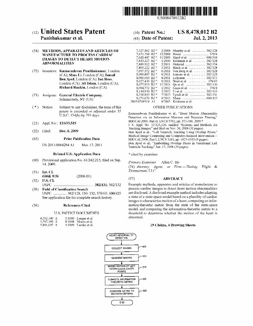
Example methods, apparatus and articles of manufacture to process cardiac images to detect heart motion abnormalities are disclosed. A disclosed example method includes adapting a state of a state-space model based on a plurality of cardiac images to characterize motion of a heart, computing an infor mation-theoretic metric from the state of the state-space model, and comparing the information-theoretic metric to a threshold to determine Whether the motion of the heart is abnormal.
19 Claims, 4 Drawing Sheets
SEGMENT IMAGES
MODEL MOTION OF LEFT VENTRICULAR CAVITY /_ 515
POINTS
I COMPUTE INFORMATION /— 520 THEORETIC METRIC
I COMPARE METRIC TO DECISION CRITERIA
@

US. Patent Jul. 2,2013 Sheet 1 of4 US 8,478,012 B2
IMAGE ACQUISITION SCHEDULER SYSTEM(S) E m
WAGE DIAGNOSTIC
MANAGER <_> 'MAG'NG 120 WORKSTATION — E
IMAGE DATABASE
11_5
\— 100
FIG. 1
ACQUIRE, & —» IIQ’IIIQIT:
MODWY
ARCHIVE & ORGANIzE & RECALL ‘ IvIANAGE
FIG. 2 \T 200

US. Patent Jul. 2, 2013 Sheet 2 of4 US 8,478,012 B2
TO TO IMAGE
ScHEDuLIER 1_§ MANAGER Q9 '- _ — _ — _ _ _ _ _ — _ _ _ _ _ _ _ _ _ — _ — _ — —l
I I l INPUT DEV|CE(S) l
l l I IMAGE PRE- I I I PROCESSING I I MODULE 2Q <—> I I I USER |NTERFACE(S) I I E5 | | IMAGE PROCESSING <—> | I ' MODULE 3_2§ |
I I I I I I D|SPLAY(S) I
I DIAGNOSTIC IMAGING m l l WORKSTATION J \- _. _ _ _ _ _ _ _ _ _ _ _ _ _ _ _ _ _ _ _ _ _ _ _
105 J FIG. 3
[ 325 l _ _ _ _ — _ _ _ _ — _ - _ — _ — _ _ _ _ _ ‘_ _ _ _ — -\
I IMAGE PROCESSING MODULE I
' I l INFORMATION I l MOT'ON METRIC CLASSIFIER
—I-> ESTIMATOR -> PROCESSOR —> % -J->I | “J M |
L I

US. Patent Jul. 2, 2013 Sheet 3 of4 US 8,478,012 B2
HEART ABNORMALITY DETECTION
I COLLECT IMAGES /_ 505
I SEGMENT IMAGES /_ 510
MODEL MOTION OF LEFT 515 VENTRICULAR CAVITY /_
POINTS
I COMPUTE INFORMATION /— 520 THEORETIC METRIC
I COMPARE METRIC TO /— 525 DECISION CRITERIA
FIG. 5

US. Patent US 8,478,012 B2 Jul. 2, 2013 Sheet 4 of4
/— P100
RANDOM ACCESS MEMORY INPUT
m 0 DEV|CE(S) CODED w
INSTRUCTIONS ¢ L10 NTE FACE | R
4" P130
REA/@1132? » 1 P120 OUTPUT — DEV|CE(S)
CODED w INSTRUCTIONS
P112
/- P125
PROCESSOR P105 ' '
FIG. 6
US 8,478,012 B2 1
METHODS, APPARATUS AND ARTICLES OF MANUFACTURE TO PROCESS CARDIAC IMAGES TO DETECT HEART MOTION
ABNORMALITIES
RELATED APPLICATION
This patents claims bene?t from US. Provisional Patent Application Ser. No. 61/242,215, entitled “Methods, Appa ratus and Articles of Manufacture to Detect Heart Motion Abnormalities,” and ?led Sep. 14, 2009, Which is hereby incorporated by reference in its entirety.
FIELD OF THE DISCLOSURE
This disclosure relates generally to cardiac images and, more particularly, to methods, apparatus and articles of manufacture to process cardiac images to detect heart motion abnormalities.
BACKGROUND
A Widely used cardiac diagnostic technique involves the imaging of different portions of a heart during various phases of a heartbeat or cardiac cycle to detect or diagnose cardiac disease, abnormalities and/or damage. Example cardiac imaging tools are a magnetic resonance imaging (MRI) sys tem and a computed topography (CT) imaging system.
BRIEF DESCRIPTION OF THE INVENTION
In vieW of the following descriptions and ?gures, it should be clear that the present disclosure describes methods, appa ratus and articles of manufacture to process cardiac images to detect heart motion abnormalities. Coronary heart disease is the most common type of cardiovascular disease, and early detection of heart motion abnormality(-ies) may be used to diagnose and/or control heart disease. Accordingly, quantita tive scoring of heart Wall motion may be extremely useful in the clinical environment. HoWever, due to the vast amount of information and uncertainty associated With heart motion, early detection of heart motion abnormalities may be dif?cult via visual inspection of cardiac images.
The example methods, apparatus and articles of manufac ture disclo sed herein provide certain advantages over existing heart motion classi?cation methods. For example, the subj ec tive evaluation of heart images by a radiologist can be reduced and/ or eliminated, thereby reducing inter and/ or intra-ob server variability. The examples disclosed herein also enabled automated analysis, Which can reduce the time required to obtain a diagnosis and/or begin treatment. As disclosed herein, left-ventricle heart motion abnormali
ties may be detected by processing a sequence of cardiac images. The cardiac images may be segmented into one or more regions, and then processed or ?ltered With a cyclic model such as a recursive Bayesian ?lter or a Kalman ?lter. An example segmented region corresponds to the left-ven tricular heart cavity. The cyclic model may be adapted and/or adjusted to apply temporal smoothing to the segmented car diac images of the left-ventricular heart cavity. Such smooth ing may be used to reduce the effect of image noise and/or segmentation inaccuracies. States and/or coef?cients of the cyclic model may be used to characterize and/or represent the dynamics and/ or motion of the segment left-ventricular heart cavity. Due to statistical similarity betWeen normal and abnormal
hearts, the classi?cation and/ or discrimination of heart
20
25
30
35
40
45
50
55
60
65
2 motion based on distribution moments such as the mean
systolic velocity may be di?icult and/or inaccurate. Instead, an information-theoretic measure or metric of left-ventricle
Wall motion may be computed from the cyclic model states and/or coe?icients. An example information-theoretic metric comprises the Shannon differential entropy (SDE), Which provides a global theoretically-grounded measure of statisti cal distributions and, thus, may be used to accurately dis criminate betWeen different types of heart motion. Other example information-theoretic metrics include, but are not limited to, the Rényi entropy and Fisher information. The information-theoretic metric may be compared to decision criteria to determine Whether the heart motion depicted in the cardiac images is normal or abnormal.
According to certain aspects of this disclosure, an example method includes adapting a state of a state-space model based on a plurality of cardiac images to characterize motion of a heart, computing an information-theoretic metric from the state of the state-space model, and comparing the informa tion-theoretic metric to a threshold to determine Whether the motion of the heart is abnormal.
According to further aspects of this disclosure, an example apparatus includes a motion estimator to adapt a state of a state-space model based on a plurality of cardiac images to characterize motion of a heart, an information metric proces sor to compute an information-theoretic metric from the state of the state-space model, and a classi?er to compare the information-theoretic metric to a threshold to determine Whether the motion of the heart is abnormal.
BRIEF DESCRIPTION OF THE DRAWINGS
FIG. 1 is a schematic illustration of an example diagnostic imaging system Within Which the example methods, appara tus and articles of manufacture described herein may be implemented.
FIG. 2 illustrates an example image lifecycle management ?oW Within Which the example methods, apparatus and articles of manufacture described herein may be imple mented.
FIG. 3 illustrates an example manner of implementing the example diagnostic Workstation of FIG. 1.
FIG. 4 illustrates an example manner of implementing the example image processing module of FIG. 3.
FIG. 5 is a ?owchart representative of example process that may be carried out to implement the example diagnostic Workstation of FIGS. 1 and 3.
FIG. 6 is a schematic illustration of an example processor platform that may be used and/ or programmed to carry out the example process of FIG. 5 and/or to implement any or all of the example methods, apparatus and articles of manufacture described herein.
DETAILED DESCRIPTION
In the interest of brevity and clarity, throughout the folloW ing disclosure references Will be made to an example diag nostic imaging Workstation 105. HoWever, the methods, apparatus and articles of manufacture described herein to process cardiac left-ventricle images to detect heart motion abnormalities may be implemented by and/or Within any number and/or type(s) of additional and/or alternative diag nostic imaging systems. For example, the methods, apparatus and articles of manufacture described herein could be imple

US 8,478,012 B2 3
mented by or Within a device and/or system that captures diagnostic images (e. g., a computed tomography (CT) imag ing system and/or magnetic resonance imaging (MRI) sys tem), and/or by or Within a system and/or Workstation designed for use in vieWing, analyzing, storing and/or archiving diagnostic images (e. g., the GE® picture archiving and communication system (PACS), and/or the GE advanced Workstation (AW)). Moreover, the example methods, appa ratus and articles of manufacture disclosed herein may be used to process any number and/or type(s) of other images, including other types of cardiac images, to detect motion abnormalities.
FIG. 1 illustrates an example diagnostic imaging system 100 including the example diagnostic imaging Workstation 105 to process cardiac left-ventricle images to detect heart motion abnormalities. The cardiac left-ventricle images may be captured by any number and/ or type(s) of image acquisi tion system(s) 110, and stored in any number and/or type(s) of image database(s) 115 managed by any number and/or type(s) of image manager(s) 120. The processing of cardiac left-ventricle images by the example diagnostic imaging Workstation 105 may be scheduled by any number and/or type(s) of scheduler(s) 125. Example image acquisition sys tems 110 include, but are not limited to, a CT imaging system and/ or an MRI system. Images may be stored and/or archived in the example image database 115 of FIG. 1 using any number and/or type(s) of data structures, and the example image database 115 may be implemented using any number and/or type(s) of memory(-ies), memory device(s) and/or storage device(s) such as a hard disk drive, a compact disc (CD), a digital versatile disc (DVD), a ?oppy drive, etc.
FIG. 2 illustrates an example image lifecycle management How 200 that may be implemented by the example diagnostic imaging system 100 of FIG. 1. Images (e.g., left-ventricle images) are acquired, created and/or modi?ed by the image acquisition system(s) 110. The image manager(s) 120 repli cate, distribute, organiZe and/or otherWise manage the cap tured images. The example diagnostic imaging Workstation 105 of FIG. 1 processes a sequence of replicated, distributed, organiZed and/ or otherWise managed images to, among other things, detect heart motion abnormalities. Information cre ated, computed and/ or otherWise determined during the clas si?cation and/ or detection of heart motion by the diagnostic imaging Workstation 105 can be used to reduce the number of image(s) and/or the amount of data that must be stored, archived and/or otherWise maintained for future recall.
FIG. 3 is a schematic illustration of an example diagnostic imaging Workstation Within Which the example methods, apparatus and articles of manufacture to detect heart motion abnormalities described herein may be implemented. To alloW a user (not shoWn) to interact With the example diag nostic imaging Workstation 105 of FIG. 3, the diagnostic imaging Workstation 105 includes any number and/or type(s) of user interface module(s) 305, any number and/or type(s) of display(s) 310 and any number and/or type(s) of input device(s) 315. The example user interface module(s) 305 of FIG. 3 implements an operating system to present informa tion (e. g., images, WindoWs, screens, interfaces, dialog boxes, etc.) at the display(s) 310, and to alloW a user to control, con?gure and/or operate the example diagnostic imaging Workstation 105. The user provides and/or makes inputs and/ or selections to the user interface module 305 and/or, more generally, to the example diagnostic imaging Workstation 1 05 via the input device(s) 315. Example input devices 315 include, but are not limited to, a keyboard, a touch screen and/or a mouse. In an example, a patient search WindoW is presented at the display 310, and the input device(s) 315 are
20
25
30
35
40
45
50
55
60
65
4 used to enter search criteria to identify a particular patient. When a patient is identi?ed and selected, the example user interface 305 presents a list of available diagnostic images for the patient at the display 310, and the user selects one or more sequences of diagnostic images using the input device(s) 315. The user interface 305 then obtains the selected image sequence(s) from the example image manager 120. An image-processing module 325 processes the selected image sequence(s) to determine Whether any heart motion abnor malities are present, and presents information related to the presence or absence of heart motion abnormalities at the display 310 for vieWing by the user. An example manner of implementing the example image processing module 325 is described beloW in connection With FIG. 4.
In the illustrated example of FIG. 3, selected image sequence(s) are pre-processed by an image pre-processing module 320 before processing by the example image-pro cessing module. Using any number and/or type(s) of method(s) and/or algorithm(s), the example image pre-pro cessing module 320 of FIG. 3 processes the selected images to detect the boundary of the left-ventricle heart cavity in each selected image. In other Words, the example pre-processing module 320 processes the selected images to identify the endocardium in each of the selected images. Example sys tems and methods for pre-processing images are described in Us. patent application Ser. No. 12/325,226, noW U.S. Pat. No. 8,144,930, ?led on Nov. 30, 2008, and entitled “Systems and Methods For Tracking Images,” Which is hereby incor porated by reference in its entirety.
Let I represent a cardiac sequence containing K frames lkzQ C ER ZQiR +, ke[1, . . . , K]. The example pre-processing
module 320 of FIG. 3 preprocesses the set of images I to detect the boundary of the left-ventricle cavity of the heart (the endocardium) in each frame ke[2, . . . , K]. The example pre-processing module 320 determines the boundary of the endocardium by evolving a closed planar parametric curve
?k(s): [0,1]—>Q toWard the endocardium. In some examples,
the parametric curve E1“ is evolved by minimiZing a cost function F“ that includes, among other things, an overlap prior term or constraint that prevents the papillary muscles of the left-ventricle cavity from being included erroneously in the heart myocardium. HoWever, because the papillary muscles and the myocardium are connected and have almost the same intensity pro?le their separation may be dif?cult. Minimization of the cost function Fk results in each frame k being segmented into tWo regions: the left-ventricle cavity Ck
a
corresponding to the interior of the parametric curve F k
CKIRFk' (1) ~ %
Where R? denotes the region enclosed by curve I“, and the background Bk corresponding to the region outside the para
. a)‘: metric curve F
BkIRFkC (2) An example cost functional Fk includes three terms: an over lap-prior term, a mean-matching term and a regulariZation/ gradient term, Which are de?ned as folloWs. An example overlap-prior term is de?ned using the folloW
ing de?nitions. P R J is the nonparametric (kemel-based) esti mate of the intensity distribution Within region R in frame Ie{Ik,k:1, . . . , K}

US 8,478,012 B2 5
NZ — I(x)) dx (3)
V z e 91*, PR,I(Z) = R 61R
Where aR is the area of region R
61,; : fdx (4) R
Example kernels N (') include, but are not limited to, the Dirac function and the Gaussian kernel. B(f/ g) is the Bhatta charyya coef?cient representing the amount of overlap betWeen tWo statistical samples f and g
B(f/g)= 2 Wow (5) zeR+
In the examples described herein, the values of B are selected from [0,1], Where 0 indicates that there is no overlap, and 1 indicates a perfect match.
In some examples, the cavity and myocardium regions in the ?rst frame I1, denoted respectively by C1 and M, are provided by a user of the example diagnostic imaging Work station 105. Based on the example de?nitions of EQNS (3) (5), an example cavity/myocardium overlap measure is expressed mathematically as
(6)
Where Bk represents the amount of overlap betWeen the inten sity distribution Within the heart cavity region Ck in frame Ik and the myocardium model learned from the ?rst frame I1. In most instances, Bk is approximately constant over the cardiac sequence I. Consequently, the value of B1 estimated from the segmentation of the ?rst frame I1 in the sequence I can be used as an overlap-prior to constrain the tracking of the boundary cavity in the subsequent frames I2, . . . , IK. To embed prior information about the overlap betWeen the intensity distribu tion Within the cavity and myocardium, the example image pre-processing module 320 of FIG. 3 minimiZes the folloW ing constraint for each frame ke[2, . . . , K]
Where Ok represents the overlap betWeen the intensity distri butions Within the cavity and prior myocardium ?ts BIZ-n. An example mean-matching term, Which represents con
formity of an intensity mean computed for the left-ventricle cavity in the current frame Ik to a mean computed for the ?rst frame I1, is de?ned by the folloWing mathematical expres s1on:
M”:(u”—ul)2, (8)
Where pk the estimate of intensity mean Within C“ for ke [1, . . . , K], Which is expressed mathematically as
f 1'‘ dx (9) k : ck
Lick I
An example regulariZation/ gradient term, Which may be used to bias the curve toWard a high intensity gradient and/or to enforce curve smoothness, is de?ned mathematically as
20
25
30
35
40
45
50
55
60
65
Where c is a positive constant and gk is an edge indicator function, Which is de?ned as
(11)
Based on the example terms de?ned above in EQNS (3) (11), an example cost function Fk that may be mimiZed to identify, compute and/ or otherWise determine the parametric
curve T31“ is de?ned by
Where the variables or Weights 0t, [3 and 7», are selected to adjust the relative importance or contribution of the three terms described above in connection With EQNS (3)-(11). The example image pre-processing module 320 of FIG. 3
solves for the parametric curve T>k by minimiZing the example cost function Pk of EQN (12) using, for example, Euler-Lagrange descent minimiZation. In some examples, the example image pre-processing module 320 embeds the curve % ~ ~ %
F in a one-parameter family of curves: I“(s,t):[0,1]>< iR +—>Q, and solves the partial differential equation
(13)
denotes the functional derivative of F With respect to The example expression of EQN (13) can be reWritten as:
Q
Where KK is the mean curvature function of F k and kk is the
outWard unit normal to TT“, and assuming that the function R (') used in the kernel density estimation is the Dirac func tion. The example image pre-processing module 320 solves and/or converges the example expression of EQN (14) for each frame Ik, With the left-ventricle cavity boundary for
frame k given by the thus obtained curve Bk.

US 8,478,012 B2 7
While level-set formalisms are used to derive the example curve evolution method described above, any number and/or type(s) of alternative and/or additional method(s), algorithm(s) and/or formalisms may be used to obtain the
a
curve F k for each image lk.
While an example manner of implementing the example diagnostic imaging workstation 105 of FIG. 1 has been illus trated in FIG. 3, one or more of the interfaces, data structures, elements, processes and/or devices illustrated in FIG. 3 may be combined, divided, re-arranged, omitted, eliminated and/ or implemented in any other way. The example user interface(s) 305, the example display(s) 310, the example input device(s) 315, the example image pre-processing mod ule 320, the example image processing module 325 and/or, more generally, the example diagnostic imaging workstation 105 of FIG. 3 may be implemented by hardware, software, ?rmware and/or any combination of hardware, software and/ or ?rmware. Thus, for example, any of the example user interface(s) 305, the example display(s) 310, the example input device(s) 315, the example image pre-processing mod ule 320, the example image processing module 325 and/or, more generally, the example diagnostic imaging workstation 105 may be implemented by one or more circuit(s), program mable processor(s), application speci?c integrated circuit(s) (ASlC(s)), programmable logic device(s) (PLD(s)) and/or ?eld programmable logic device(s) (FPLD(s)), etc. When any apparatus claim of any patent resulting from this provisional application is read to cover a purely software and/ or ?rmware implementation, at least one of the example user interface(s) 305, the example display(s) 310, the example input device(s) 315, the example image pre-processing module 320, the example image processing module 325 and/or, more gener ally, the example diagnostic imaging workstation 105 are hereby expressly de?ned to include a tangible computer readable medium such as a memory, a DVD, a CD, etc. storing the ?rmware and/or software. Further still, the example diagnostic imaging workstation 105 may include interfaces, data structures, elements, processes and/or devices instead of, or in addition to, those illustrated in FIG. 3 and/or may include more than one of any or all of the illustrated
interfaces, data structures, elements, processes and/or devices.
FIG. 4 illustrates an example manner of implementing the example image processing module 325 of FIG. 3. To compute ?lter coef?cients and/or parameters of a state model that predicts the future position and/ or movement of left ventricu lar cavity points, the example image processing module 325 of FIG. 4 includes a motion estimator 405. The example motion estimator 405 of FIG. 4 predicts left-ventricular cav ity points by applying a Bayesian ?lter, such as the Kalman ?lter to a sequence of segmented left-ventricle cardiac images.
Let (x, y) be a Cartesian point on the endocardial boundary
?k(s) identi?ed and/or determined by the example image pre-processing module 320, as described above. Let E be an example state vector E:[x x x] T that represents the dynamics of the point in the x-coordinate direction, where x and x denote, respectively, the velocity and the mean position of the point over a cardiac cycle or heartbeart. Assuming motion of the heart is substantially periodic, an example continuous state-space model that describes the cyclic motion of the point can be expressed as
20
25
30
35
40
45
50
55
60
65
where u) is the angular frequency, and w(t) is white noise that represents the unpredictable modeling errors arising in heart motion detection. The example mathematical model of EQN (15) is linear for a given 00, and is an approximation of a temporal periodic model where higher-order terms of the Fourier expansion are neglected. A substantially equivalent discrete-time version of EQN (l 5) can be expressed as
where the covariance of the process noise given by
Qk:cov(wk) is
Qk:|.qijJ3><3' (17)
and the Qlj’s are de?ned to be
‘111 = qiT (18)
q?wT - 51mm) (19) 21 = i ‘112 =
a)
[113 = [131 = [1%(1 — mum) (10>
3wT — 4sin(wT) + wT — (21) #1 q%[ ] _ l cos(wT)sin(wT) cos(wT)sin(wT) ‘I22 — 5 ' m3
2 l — 2cos(wT) + 2 2 (22) + ‘ T 1 £11 [ COSZWT) ] qzsln (w )
£123 — £132 — 5' m2
q%w2(cos(wT)sin(wT) — wT) — (23)
Letting s represent an example state vector s:[x x x y y y] T representing the dynamics of the left-ventricle cavity in the x-y plane, an example discrete state-space model for the motion of the left-ventricle in the x-y plane is given by
Fcy(k) 03x3 (24)
Using any number and/or type(s) of algorithm(s) and/or method(s), the example motion estimator 405 of FIG. 4 pro cesses the segmented images from the example image pre processing module 320 to obtain an estimate of the state vector s. For example, the example motion estimator 405 applies a Bayesian ?lter such as a Kalman ?lter, which may be used as a state estimator for linear and/or Gaussian systems, to recursively update an estimate the state vector s. Letting Zk:[Zk’x ZkdJT represent an estimate of the point (x, y) on the

US 8,478,012 B2 9
. aA: endocard1al boundary T (s) for frame ke[l, . . . , K], an
example measurement equation is given by
zk:H;,s‘k+vk (25)
Where
0 1 0 0 0 0 (26) Hk =
O O O O l O
and vk is a Zero-mean Gaussian noise sequence With covari ance
(27)
The example motion estimator 405 computes a predicted state using the example model of EQN (24) and by taking an expectation conditioned on Zl:k:{Zl, . . . , Zk}. Letting mkIE [sk] be the mean of the state vector, the motion estimator 405 computes the predicted state using the folloWing mathemati cal expression
mk+ITIFkmk (28)
The corresponding state prediction covariance is given by
The example motion estimator 405 of FIG. 4 computes the measurement residual or innovation by taking the expectation conditioned on Z M
(29)
With the corresponding innovation covariance de?ned by
and the ?lter gain given by
The motion estimator 405 computes the updated state esti mate using the folloWing expression
The motion estimator 405 computes the updated state cova riance using the folloWing mathematical expression
In some examples, the initial state vector sl may not be knoW a priori. In such examples, the example motion estima tor 405 of FIG. 4 implements a tWo-point differencing method to initialiZe position and velocity components of the state vector s. For instance, the motion estimator 405 may compute the initial position and velocity elements in x-coor dinate direction using the folloWing equations
XAIIZIJ, (35)
and
(12,; — 11;) (36)
The mean position x over the cardiac cycle may be computed by the motion estimator 405 by taking an expectation over all corresponding measurements:
20
25
30
35
40
45
50
55
60
65
10
K (37)
The example motion estimator 405 likeWise computes the initial state elements in y-coordinate direction, 91, 91, and 7371 using the measurements {zkiy}. The motion estimator 405 computes the corresponding initial covariance by computing the folloWing mathematical expression
(1)1 (3 3) P1 = [ 03x3
03x3 ] (1)1
Where
(39)
In some instances, segmentation of the left-ventricle cavity may not be consistent over a cardiac cycle. The example motion estimator 405 of FIG. 4 detects such inconsistencies by gating the center of the segmented left-ventricle cavity. For
' ii_i i~i_i i-iT/i example, lett1ng {sk *[xk xk xk yk yk yk] .1*l,...,N}bea sample point on the left-ventricle boundary in frame k, the center of the left-ventricle cavity (cLk cyak) may be computed by the motion estimator 405 using the folloWing:
(40)
If \/(cx,k+l— x,k)2+(cy,k+1—cy,k)2>g (Where g is a prede?ned constant), the motion estimator 405 ignores the segmentation results. In such instances, the motion estimator 405 only predicts the sample points using the model of EQN (24) Without updating the Kalman ?lter.
In order to identify a sequence of corresponding points over time, the example motion estimator 405 determines symmetric nearest-neighbor correspondences by sampling a set of equally-spacedpoints along the left-ventricle boundary. The selected sequence of points may be used to analyZe Wall motion regionally. For example, using spline interpolation, the motion estimator 405 samples NS, points along the left ventricle cavity in each frame, and N points are chosen as inputs for the Kalman ?lter. The motion estimator 405 com putes a kernel density estimation based on the Gaussian ker nel to obtain the probability density. The motion estimator 405 may normalize the radial distance for each dataset With respect to maximum value, to analyZe different long-axis segments, namely, apical, mid and basal, Without additional processing.
To characterize the motion of the heart, the example image processing module 325 includes an information metric pro cessor 410. The example information metric processor 410 of FIG. 4 processes the state model and/or ?lter coef?cients computed by the example motion estimator 405 to compute,

US 8,478,012 B2 11
estimate and/or otherwise determine one or more informa
tion-theoretic measures or metrics representative of heart motion. Consistent With industry usage, the terms “informa tion-theoretic metric” and “information-theoretic measures” used herein refer to any values that are computed based on one or more properties of information theory. As is Well known, the ?eld of information theory is based on probability theory and statistics, and is concerned With the quanti?cation of information and/ or the computation of measures of informa tion. Example information-theoretic metrics are entropy, Which is the information in a random variable, and mutual information, Which represents the amount of information in common betWeen tWo random variables. Additional example information-theoretic metrics include, but are not limited to, the Shannon differential entropy (SDE), Which provides a global theoretical ground measure of distributions, the Rényi entropy, and/ or Fisher information.
The example information metric processor 410 of FIG. 4 computes a normaliZed radial distance rki, Which can be expressed mathematically as
(41)
Where >2; and xyki are the estimates of xki and yki respectively. The values >2; and 9,; are computed using the Kalman ?lter described above. Letting reER be a random variable, the example information metric processor 410 computes a kernel density estimate of the normalized radial distance using the folloWing equation
is the Gaussian kernel.
An example SDE computed by the example information metric processor 410 of FIG. 4 is de?ned by the folloWing mathematical expression
(44)
will NK
Other example information-theoretic metrics that may be computed by the example information metric processor 410 include, but are not limited to, the Rényi entropy
20
25
30
35
40
45
50
55
60
65
12
NK
0 (45)
and Fisher information
4P4 Le,“ lVg(r)l2dr Where
To classify the motion of the heart, the example image pro cessing module 325 includes a classi?er 415. The example classi?er 415 of FIG. 4 compares the information-theoretic metrics computed by the information metric processor 410 to one or more thresholds. Based on the comparison(s), the motion of the heart is classi?ed as normal or abnormal.
While an example manner of implementing the example image pre-processing module 325 of FIG. 3 is illustrated in FIG. 4, one or more of the interfaces, data structures, ele ments, processes and/ or devices illustrated in FIG. 4 may be combined, divided, re-arranged, omitted, eliminated and/or implemented in any other Way. The example motion estimator 405, the example information metric processor 410, the example classi?er 415 and/or, more generally, the example image processing module 325 of FIG. 4 may be implemented by hardWare, softWare, ?rmWare and/or any combination of hardWare, softWare and/ or ?rmWare. Thus, for example, any of the example motion estimator 405, the example informa tion metric processor 410, the example classi?er 415 and/or, more generally, the example image processing module 325 may be implemented by one or more circuit(s), program mable processor(s), ASlC(s), PLD(s) and/or FPLD(s), etc. Any apparatus claim of any patent resulting from this provi sional application is read to cover a purely softWare and/or ?rmWare implementation, at least one of the example motion estimator 405, the example information metric processor 410, the example classi?er 415 and/or, more generally, the example image processing module 325 are hereby expressly de?ned to include a tangible computer-readable medium such as a memory, a DVD, a CD, etc. storing the ?rmWare and/or softWare. Further still, the example image processing module 325 may include interfaces, data structures, elements, pro cesses and/or devices instead of, or in addition to, those illustrated in FIG. 4 and/or may include more than one of any or all of the illustrated interfaces, data structures, elements, processes and/or devices.
FIG. 5 illustrates an example process that may be carried out to implement the example image processing module 325 and/or, more generally, the example diagnostic Workstation 105 of FIGS. 1, 3 and 4. The example process of FIG. 5 may be carried out by a processor, a controller and/or any other suitable processing device. For example, the example process of FIG. 5 may be embodied in coded instructions stored on a tangible computer-readable medium such as a ?ash memory, a CD, a DVD, a ?oppy disk, a read-only memory (ROM), a random-access memory (RAM), a programmable ROM (PROM), an electronically-programmable ROM (EPROM), and/or an electronically-erasable PROM (EEPROM), an optical storage disk, an optical storage device, magnetic stor age disk, a magnetic storage device, and/ or any other medium Which can be used to carry or store program code and/or
(46)
(47)

US 8,478,012 B2 13
instructions in the form of computer-executable instructions or data structures, and which can be accessed by a processor, a general purpose or special purpose computer or other machine with a processor (e.g., the example processor plat form P100 discussed below in connection with FIG. 6). Com binations of the above are also included within the scope of computer-readable media. Computer-executable instructions comprise, for example, instructions and data that cause a processor, a general purpose computer, special purpose com puter, or a special purpose processing machine to perform one or more particular processes. Alternatively, some or all of the example process of FIG. 5 may be implemented using any combination(s) of ASlC(s), PLD(s), FPLD(s), discrete logic, hardware, ?rmware, etc. Also, some or all of the example process of FIG. 5 may be implemented manually or as any combination of any of the foregoing techniques, for example, any combination of ?rmware, software, discrete logic and/or hardware. Further, many other methods of implementing the example operations of FIG. 5 may be employed. For example, the order of execution of the blocks may be changed, and/or one or more of the blocks described may be changed, elimi nated, sub-divided, or combined. Additionally, any or all of the example process of FIG. 5 may be carried out sequentially and/or carried out in parallel by, for example, separate pro cessing threads, processors, devices, discrete logic, circuits, etc.
The example process of FIG. 5 begins with the example diagnostic imaging workstation 105 collecting a sequence of left-ventricle cardiac images from the example image man ager 120 (block 505). The example image pre-processing module 320 segments the images (block 510). The example motion estimator 405 applies a Kalman ?lter to generate a model and/or ?lter representative of the motion of the seg mented imaged left-ventricle images (block 515). Based on the generated model and/or ?lters, the example information metric processor 410 computes one or more information theoretic metrics, such as the SDE, the Rényi entropy, and/or the Fisher information (block 520). Based on the computed information-theoretic metric(s), the example classi?er 415 determines whether the motion of the imaged heart is normal or abnormal (block 525). Control then exits from the example process of FIG. 5.
FIG. 6 is a schematic diagram of an example processor platform P100 that may be used and/or programmed to imple ment any or all of the example diagnostic imaging worksta tion 105, the example image pre-processing module 320, the example image processing module 325, the example motion estimator 405, the example information metric processor 410, and/or the example classi?er 415 of FIGS. 1, 3 and 4. For example, the processor platform P1 00 can be implemented by one or more general-purpose processors, processor cores,
microcontrollers, etc. The processor platform P100 of the example of FIG. 6
includes at least one general-purpose programmable proces sor P105. The processor P105 executes coded instructions P110 and/or P112 present in main memory of the processor P105 (e.g., within a RAM P115 and/or a ROM P120). The processor P105 may be any type of processing unit, such as a processor core, a processor and/or a microcontroller. The processor P105 may execute, among other things, the example process of FIG. 5 to implement the example cardiac left-ventricle image-processing methods and apparatus described herein.
The processor P105 is in communication with the main memory (including a ROM P120 and/or the RAM P115) via a bus P125. The RAM P115 may be implemented by dynamic random access memory (DRAM), synchronous dynamic ran
5
20
25
30
35
40
45
50
55
60
65
14 dom access memory (SDRAM), and/or any other type of RAM device, and ROM may be implemented by ?ash memory and/or any other desired type of memory device. Access to the memory P115 and the memory P120 may be controlled by a memory controller (not shown). The example memory P115 may be used to implement the example image database 115 of FIG. 1. The processor platform P100 also includes an interface
circuit P130. The interface circuit P130 may be implemented by any type of interface standard, such as an external memory interface, serial port, general-purpose input/output, etc. One or more input devices P135 and one or more output devices P140 are connected to the interface circuit P130. The input devices P135 may be used to, for example, implement the example input device(s) 315 of FIG. 3. The example output devices P140 may be used to, for example, implement the example display 310 of FIG. 3.
Generally, computer-executable instructions include rou tines, programs, objects, components, data structures, etc., that perform particular tasks or implement particular abstract data types. Computer-executable instructions, associated data structures, and program modules represent examples of program code for executing the processes to implement the example methods and systems disclosed herein. The particu lar sequence of such executable instructions and/or associ ated data structures represent examples of corresponding acts for implementing the examples described herein. The example methods and apparatus described herein may
be practiced in a networked environment using logical con nections to one or more remote computers having processors. Logical connections may include a local area network (LAN) and a wide area network (WAN) that are presented here by way of example and not limitation. Such networking environ ments are commonplace in of?ce-wide or enterprise-wide computer networks, intranets and the Internet and may use a wide variety of different communication protocols. Such net work computing environments may encompass many types of computer system con?gurations, including personal com puters, hand-held devices, multi-processor systems, micro processor-based or programmable consumer electronics, net work PCs, minicomputers, mainframe computers, and the like. The example methods and apparatus described herein may, additionally or alternatively, be practiced in distributed computing environments where tasks are performed by local and remote processing devices that are linked (either by hard wired links, wireless links, or by a combination of hardwired or wireless links) through a communications network. In a distributed computing environment, program modules may be located in both local and remote memory storage devices.
Although certain example methods, apparatus and articles of manufacture have been described herein, the scope of coverage of this patent is not limited thereto. On the contrary, this patent covers all methods, apparatus and articles of manufacture fairly falling within the scope of the appended claims either literally or under the doctrine of equivalents.
What is claimed is: 1. A method comprising: adapting a state of a state-space model based on a plurality
of cardiac images to characterize motion of a heart; computing, via a processor, an information-theoretic met
ric from the state of the state-space model; and comparing the information-theoretic metric to a threshold
to determine whether the motion of the heart is abnor mal.
2. A method as de?ned in claim 1, wherein the state com prises a plurality of mean positions, a plurality of current

US 8,478,012 B2 15
positions and a plurality of velocities for respective ones of a plurality of points on the heart.
3. A method as de?ned in claim 1, Wherein the state is adapted by applying at least one of a Bayesian ?lter or a Kalman ?lter.
4. A method as de?ned in claim 1, Wherein the information theoretic metric comprises a Shannon differential entropy.
5. A method as de?ned in claim 1, Wherein the information theoretic metric comprises a Rényi entropy.
6. A method as de?ned in claim 1, Wherein the information theoretic metric comprises Fisher information.
7. A method as de?ned in claim 1, Wherein computing the information-theoretic metric comprises:
computing a kernel density estimate of a normalized radial distance based on the state; and
computing the information-theoretic metric from the ker nel density estimate.
8. A method as de?ned in claim 1, further comprising segmenting the plurality of cardiac images to form respective ones of a plurality of segmented images, Wherein the state of the state-space model is adapted based on the plurality of segmented images.
9. An article of manufacture storing machine-readable instructions that, When executed, cause a machine to:
adapt a state of a state-space model based on a plurality of cardiac images to characterize motion of a heart;
compute an information-theoretic metric from the state of the state-space model; and
compare the information-theoretic metric to a threshold to determine Whether the motion of the heart is abnormal.
10.An article of manufacture as de?ned in claim 9, Wherein the machine-readable instructions, When executed, cause the machine to adapt the state by applying at least one of a Bayesian ?lter or a Kalman ?lter.
11 . An article of manufacture as de?ned in claim 9, Wherein the information-theoretic metric comprises a Shannon differ ential entropy.
12.An article of manufacture as de?ned in claim 9, Wherein the machine-readable instructions, When executed, cause the machine to:
segment the plurality of cardiac images to form respective ones of a plurality of segmented images; and
adapt the state based on the plurality of segmented images.
20
25
30
35
16 13 . An article of manufacture as de?ned in claim 9, Wherein
the machine-readable instructions, When executed, cause the machine to compute the information-theoretic metric by:
computing a kernel density estimate of a normalized radial distance based on the state; and
computing the information-theoretic metric from the ker nel density estimate.
14. An apparatus comprising: a motion estimator to adapt a state of a state-space model
based on a plurality of cardiac images to characterize motion of a heart;
an information metric processor to compute an informa tion-theoretic metric from the state of the state-space model; and
a classi?er to compare the information-theoretic metric to a threshold to determine Whether the motion of the heart is abnormal.
15.An apparatus as de?ned in claim 14, further comprising an imaging device to capture the plurality of cardiac images of the heart.
16.An apparatus as de?ned in claim 14, further comprising an image pre-processor to segment the plurality of cardiac images to form respective ones of a plurality of segmented images, Wherein motion estimator is con?gured to adapt the state of the state-space model based on the plurality of seg mented images.
17. An apparatus as de?ned in claim 14, Wherein the motion estimator comprises at least one of a Bayesian ?lter or a Kalman ?lter.
18. An apparatus as de?ned in claim 14, Wherein the infor mation-theoretic metric comprises a Shannon differential entropy.
19. An apparatus as de?ned in claim 14, Wherein the infor mation metric processor is to compute the information-theo retic metric by:
computing a kernel density estimate of a normalized radial distance based on the state; and
computing the information-theoretic metric from the ker nel density estimate.
* * * * *
![(12) United States Patent (10) Patent N0.: US 8,470,342 B2 · US008470342B2 (12) United States Patent (10) Patent N0.: US 8,470,342 B2 Klinman et a]. (45) Date of Patent: Jun. 25,](https://static.fdocuments.net/doc/165x107/60676d57ebc6a70cbe1a6e0a/12-united-states-patent-10-patent-n0-us-8470342-b2-us008470342b2-12-united.jpg)


![(12) (10) Patent N0.: US 7,198,213 B2 United States Patent · 2017. 1. 19. · United States Patent US007198213B2 (12) (10) Patent N0.: US 7,198,213 B2 Kolbet et a]. (45) Date of](https://static.fdocuments.net/doc/165x107/60bfc79629246005d7520b44/12-10-patent-n0-us-7198213-b2-united-states-patent-2017-1-19-united.jpg)

