12 Lead ECGs: Ischemia, Injury & Infarction Part...
32
12 Lead ECGs: Ischemia, Injury & Infarction Part 1 McHenry Western Lake County McHenry Western Lake County EMS EMS
-
Upload
truonghanh -
Category
Documents
-
view
225 -
download
3
Transcript of 12 Lead ECGs: Ischemia, Injury & Infarction Part...

12 Lead ECGs:
Ischemia, Injury & InfarctionPart 1
McHenry Western Lake County McHenry Western Lake County EMS EMS

Ischemia, Injury & Infarction
DefinitionsInjury/Infarct RecognitionLocalization & EvolutionReciprocal ChangesThe High Acuity Patient

The Three I’s
Ischemialack of oxygenationST segment depression or T wave inversion
Injuryprolonged ischemiaST segment elevation
Infarctdeath of tissuemay or may not show a Q wave

Review of Waveform Components

Waveform Components R Wave
First positive deflectionR wave includes the down stroke returning to the baseline

Waveform Components Q Wave
First negative deflection before the R waveQ wave includes the negative down stroke and return to baseline

Waveform Components S Wave
Negative deflection following the R waveS wave includes departure from and return to baseline

Waveform Components QRS
Q wavesCan occur normally in several leads
Normal Q waves called physiologic
Physiologic Q waves< .04 sec (40ms)
Pathologic Q>.04 sec (40ms)

Waveform Components QRS
Q waveMeasure widthPathologic if greater than or equal to 0.04 seconds (1 small box)

Waveform Components QS Complex
Entire complex is negatively deflectedNo R wave is present
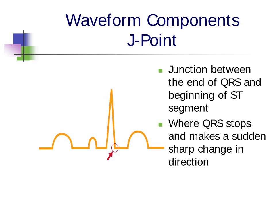
Waveform Components J-Point
Junction between the end of QRS and beginning of ST segmentWhere QRS stops and makes a sudden sharp change in direction

Waveform Components ST Segment
Segment between J-Point and beginning of T wave

Waveform Components ST Segment
Need reference pointCompare to TP segmentDO NOT use PR segment as reference!

Waveform Components Practice
Find the J Point and ST segment

Waveform Components Practice
J POINTS ST SEGMENT

Injury/Infarct Recognition
Epicardial Coronary Artery
Lateral Wall of LV
Positive Electrode
Septum
Interior Wall of LV
Well Perfused Myocardium

Injury/Infarct RecognitionNormal ECG

Injury/Infarct Recognition
Epicardial Coronary Artery
Lateral Wall of LVSeptum
Interior Wall of LV
Ischemia
Positive Electrode
Left Ventricular
Cavity

Injury/Infarct Recognition
Ischemia
Inadequate oxygen to tissue
Represented by ST depression or T inversion
May or may not result in infarct or Q waves

Injury/Infarct RecognitionST Segment Depression

Injury/Infarct Recognition
Thrombus
Ischemia
InjuryInjury
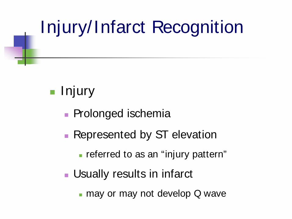
Injury/Infarct Recognition
Injury
Prolonged ischemia
Represented by ST elevation
referred to as an “injury pattern”
Usually results in infarct
may or may not develop Q wave

Injury/Infarct RecognitionST Segment Elevation

Injury/Infarct Recognition
Infarcted AreaElectrically Silent
Depolarization
Infarct

Injury/Infarct Recognition
Infarct
Death of tissue
Represented by Q wave
Not all infarcts develop Q waves

Injury/Infarct RecognitionQ Waves

Injury/Infarct Recognition
Infarcted Area Electrically Silent
Thrombus
Depolarization
Ischemia

Injury/Infarct Recognition
What to Look for:ST segment elevationPresent in two or more anatomically contiguous leads

Injury/Infarct Recognition: Practice
Look at J points and ST segments

Localization
Inferior: II, III, AVFInferior: II, III, AVFSeptal: V1, V2Septal: V1, V2Anterior: V3, V4Anterior: V3, V4Lateral: I, AVL, V5, V6Lateral: I, AVL, V5, V6
I
II
III
aVR
aVL
aVF
V1
V2
V3
V4
V5
V6

Localization
I Lateral
II Inferior
III Inferior
aVR
aVL Lateral
V1 Septal
aVF Inferior
V2 Septal
V3 Anterior
V4 Anterior
V5 Lateral
V6 Lateral
Which coronary arteries are most likely associated with each group of
contiguous leads?
Please continue to part 2 of this presentation
Thanks!