#12 Induction of Pluripotent Stem Cells From Mouse Embryonic and Adult Fibroblast Cultures by...
14
Induction of Pluripotent Stem Cells from Mouse Embryonic and Adult Fibroblast Cultures by De fi ned Factors Kazutoshi Takahashi 1 and Shinya Yamanaka 1,2, * 1 Department of Stem Cell Biology, Institute for Frontier Medical Sciences, Kyoto University, Kyoto 606-8507, Japan 2 CREST, Japan Science and Technology Agency, Kawaguchi 332-0012, Japan *Contact: [email protected] DOI 10.1016/j.cell.2006.07.024 SUMMARY Differentiated cells can be reprogrammed to an embryonic-like state by transfer of nuclear con- tents into oocytes or by fusion with embryonic stem (ES) cells. Little is known about factors that induce this reprogramming. Here, we dem- onstrate induct ion of plur ipot ent stem cell s from mouse embryonic or adult fibroblasts by introducing four factors, Oct3/4, Sox2, c-Myc, and Klf 4, under ES cel l culture conditions. Unexpectedly, Nanog was dispensable. These cells, which we designated iPS (induced plurip- otent stem) cells, exhibit the morphology and growth properties of ES cells and express ES cell marker genes. Subcutaneous transplanta- ti on of iPS cells into nude mi ce resulte d in tumors containing a variety of tissues from all three germ layers. Following injection into blas- tocysts, iPS cells contributed to mouse embry- oni c development. These data demonstrate that pluripotent stem cells can be directly gen- erated from fibroblast cultures by the addition of only a few defined factors. INTRODUCTION Embryonic stem (ES) cells, which are derived from the in- ner cell mass of mammalian blastocysts, have the ability to grow indefinite ly while maintaini ng plurip oten cy and the ability to differentiate into cells of all three germ layers ( Eva ns and Kau fma n, 1981; Marti n, 1981 ) . Human ES cell s might be used to treat a host of diseases, such as Parkin- son’s disease, spinal cord injury, and diabetes ( Thomson et al., 1998 ). However, there are ethical difficulties regard- ing the use of human embryos, as well as the problem of tissue rejection following transplantation in patients. One way to circumvent these issues is the generation of plu- ripotent cells directly from the patients’ own cells. Somatic cells can be repro grammed by transferr ing their nuclear contents into oocytes ( Wilmut et al., 1997 ) or by fusion wi th ES cells ( Cowan et al., 2005; Tada et al., 2001 ), indicating that unfertilized eggs and ES cells contain fact ors that can confe r toti pote ncy or plurip oten cy to somatic cells. We hypothesized that the factors that play important roles in the maintenance of ES cell identity also play pivotal roles in the induction of pluripotency in somatic cells. Several transcription factors, including Oct3/4 ( Nichols et al., 1998; Niwa et al., 2000 ), Sox2 ( Avilion et al., 2003 ), and Nanog ( Chambers et al., 2003; Mitsui et al., 2003 ), function in the maintenance of pluripotency in both early embryos and ES cells. Several genes that are frequently upreg ulated in tumor s, such as Stat3 ( Mats uda et al., 1999; Niwa et al., 1998 ), E-Ras ( Takahashi et al., 2003 ), c- myc ( Cartwright et al., 2005 ), Klf4 ( Li et al., 2005 ), and b-catenin ( Kielman et al., 2002; Sato et al., 2004 ), have been shown to contribute to the long-term maintenance of the ES cell phenotype and the rapid proliferation of ES cells in culture. In addition, we have identified several other gene s that are spec ifical ly expre ssed in ES cells ( Maruyama et al., 2005; Mitsui et al., 2003 ). In this study, we examined whether these factors could induce pluripotency in somatic cells. By combining four selec ted factors, we were able to generate pluripot ent cells, which we call induced pluripotent stem (iPS) cells, direc tly from mouse embryo nic or adult fibrobl ast cul- tures. RESULTS We sel ect ed 24 gen es as candidat es for fac tor s that induce pl ur ipotency in somati c cells, based on our hypothesis that such factors also play pivotal roles in the mai ntenance of ES cel l identi ty (se e Tab le S1 in the Supplemental Data available with this article online). For b-ca ten in, c-Myc, and St at3 , we use d act ive for ms, S33Y-b-catenin ( Sadot et al., 2002 ), T58A-c-Myc ( Chang et al., 2000 ), and Stat3-C ( Bromberg et al., 1999 ), respec- tively. Because of the reported negative effect of Grb2 on pluripotency ( Burdon et al., 1999; Cheng et al., 1998 ), we inclu ded its dominant-ne gati ve muta nt Grb2 DSH2 ( Miyamoto et al., 2004 ) as 1 of the 24 candidates. Cell 126, 663–676, August 25, 2006 ª2006 Elsevier Inc. 663
Transcript of #12 Induction of Pluripotent Stem Cells From Mouse Embryonic and Adult Fibroblast Cultures by...
8/14/2019 #12 Induction of Pluripotent Stem Cells From Mouse Embryonic and Adult Fibroblast Cultures by Defined Factors
http://slidepdf.com/reader/full/12-induction-of-pluripotent-stem-cells-from-mouse-embryonic-and-adult-fibroblast 1/14
Induction of Pluripotent Stem Cells
from Mouse Embryonic and AdultFibroblast Cultures by Defined FactorsKazutoshi Takahashi1 and Shinya Yamanaka1,2,*1Department of Stem Cell Biology, Institute for Frontier Medical Sciences, Kyoto University, Kyoto 606-8507, Japan2CREST, Japan Science and Technology Agency, Kawaguchi 332-0012, Japan
*Contact: [email protected]
DOI 10.1016/j.cell.2006.07.024
SUMMARY
Differentiated cells can be reprogrammed to anembryonic-like state by transfer of nuclear con-
tents into oocytes or by fusion with embryonic
stem (ES) cells. Little is known about factors
that induce this reprogramming. Here, we dem-
onstrate induction of pluripotent stem cells
from mouse embryonic or adult fibroblasts by
introducing four factors, Oct3/4, Sox2, c-Myc,
and Klf4, under ES cell culture conditions.
Unexpectedly, Nanog was dispensable. These
cells, which we designated iPS (induced plurip-
otent stem) cells, exhibit the morphology and
growth properties of ES cells and express EScell marker genes. Subcutaneous transplanta-
tion of iPS cells into nude mice resulted in
tumors containing a variety of tissues from all
three germ layers. Following injection into blas-
tocysts, iPS cells contributed to mouse embry-
onic development. These data demonstrate
that pluripotent stem cells can be directly gen-
erated from fibroblast cultures by the addition
of only a few defined factors.
INTRODUCTION
Embryonic stem (ES) cells, which are derived from the in-
ner cell mass of mammalian blastocysts, have the ability
to grow indefinitely while maintaining pluripotency and
the ability to differentiate into cells of all three germ layers
( Evans and Kaufman, 1981; Martin, 1981 ). Human ES cells
might be used to treat a host of diseases, such as Parkin-
son’s disease, spinal cord injury, and diabetes ( Thomson
et al., 1998 ). However, there are ethical difficulties regard-
ing the use of human embryos, as well as the problem of
tissue rejection following transplantation in patients. One
way to circumvent these issues is the generation of plu-
ripotent cells directly from the patients’ own cells.
Somatic cells can be reprogrammed by transferring
their nuclear contents into oocytes ( Wilmut et al., 1997 )
or by fusion with ES cells ( Cowan et al., 2005; Tada
et al., 2001 ), indicating that unfertilized eggs and ES cells
contain factors that can confer totipotency or pluripotencyto somatic cells. We hypothesized that the factors that
play important roles in the maintenance of ES cell identity
also play pivotal roles in the induction of pluripotency in
somatic cells.
Several transcription factors, including Oct3/4 ( Nichols
et al., 1998; Niwa et al., 2000 ), Sox2 ( Avilion et al., 2003 ),
and Nanog ( Chambers et al., 2003; Mitsui et al., 2003 ),
function in the maintenance of pluripotency in both early
embryos and ES cells. Several genes that are frequently
upregulated in tumors, such as Stat3 ( Matsuda et al.,
1999; Niwa et al., 1998 ), E-Ras ( Takahashi et al., 2003 ),
c- myc ( Cartwright et al., 2005 ), Klf4 ( Li et al., 2005 ), and
b-catenin ( Kielman et al., 2002; Sato et al., 2004 ), havebeen shown to contribute to the long-term maintenance
of the ES cell phenotype and the rapid proliferation of
ES cells in culture. In addition, we have identified several
other genes that are specifically expressed in ES cells
( Maruyama et al., 2005; Mitsui et al., 2003 ).
In this study, we examined whether these factors could
induce pluripotency in somatic cells. By combining four
selected factors, we were able to generate pluripotent
cells, which we call induced pluripotent stem (iPS) cells,
directly from mouse embryonic or adult fibroblast cul-
tures.
RESULTS
We selected 24 genes as candidates for factors that
induce pluripotency in somatic cells, based on our
hypothesis that such factors also play pivotal roles in the
maintenance of ES cell identity (see Table S1 in the
Supplemental Data available with this article online). For
b-catenin, c-Myc, and Stat3, we used active forms,
S33Y-b-catenin ( Sadot et al., 2002 ), T58A-c-Myc ( Chang
et al., 2000 ), and Stat3-C ( Bromberg et al., 1999 ), respec-
tively. Because of the reported negative effect of Grb2
on pluripotency ( Burdon et al., 1999; Cheng et al., 1998 ),
we included its dominant-negative mutant Grb2DSH2
( Miyamoto et al., 2004 ) as 1 of the 24 candidates.
Cell 126, 663–676, August 25, 2006 ª2006 Elsevier Inc. 663
8/14/2019 #12 Induction of Pluripotent Stem Cells From Mouse Embryonic and Adult Fibroblast Cultures by Defined Factors
http://slidepdf.com/reader/full/12-induction-of-pluripotent-stem-cells-from-mouse-embryonic-and-adult-fibroblast 2/14
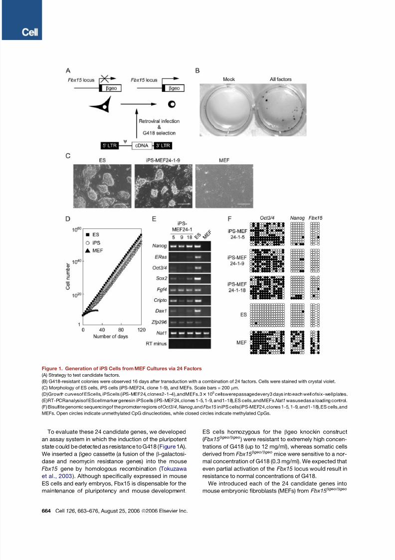
To evaluate these 24 candidate genes, we developed
an assay system in which the induction of the pluripotent
state could be detected as resistance to G418 ( Figure 1 A).
We inserted a b geo cassette (a fusion of the b-galactosi-
dase and neomycin resistance genes) into the mouse
Fbx15 gene by homologous recombination ( Tokuzawa
et al., 2003 ). Although specifically expressed in mouse
ES cells and early embryos, Fbx15 is dispensable for the
maintenance of pluripotency and mouse development.
ES cells homozygous for the bgeo knockin construct
( Fbx15b geo/ b geo ) were resistant to extremely high concen-
trations of G418 (up to 12 mg/ml), whereas somatic cells
derived from Fbx15b geo/ b geo mice were sensitive to a nor-
mal concentration of G418 (0.3 mg/ml). We expected that
even partial activation of the Fbx15 locus would result in
resistance to normal concentrations of G418.
We introduced each of the 24 candidate genes into
mouse embryonic fibroblasts (MEFs) from Fbx15b geo/ b geo
Figure 1. Generation of iPS Cells from MEF Cultures via 24 Factors
(A) Strategy to test candidate factors.
(B) G418-resistant colonies were observed 16 days after transduction with a combination of 24 factors. Cells were stained with crystal violet.
(C) Morphology of ES cells, iPS cells (iPS-MEF24, clone 1-9), and MEFs. Scale bars = 200 mm.
(D)Growth curvesof EScells, iPScells (iPS-MEF24, clones2-1–4),andMEFs.33105cellswerepassagedevery3 days into each well ofsix-wellplates.
(E)RT-PCRanalysisof EScellmarker genesin iPScells (iPS-MEF24,clones 1-5, 1-9, and1-18),ES cells,andMEFs.Nat1wasusedas a loading control.
(F) Bisulfite genomic sequencingof the promoter regions of Oct3/4,Nanog,andFbx15 iniPS cells(iPS-MEF24,clones 1-5, 1-9, and1-18),ES cells,and
MEFs. Open circles indicate unmethylated CpG dinucleotides, while closed circles indicate methylated CpGs.
664 Cell 126, 663–676, August 25, 2006 ª2006 Elsevier Inc.
8/14/2019 #12 Induction of Pluripotent Stem Cells From Mouse Embryonic and Adult Fibroblast Cultures by Defined Factors
http://slidepdf.com/reader/full/12-induction-of-pluripotent-stem-cells-from-mouse-embryonic-and-adult-fibroblast 3/14
embryos by retroviral transduction ( Morita et al., 2000 ).
Transduced cells were then cultured on STO feeder cells
in ES cell medium containing G418 (0.3 mg/ml). We did
not, however, obtain drug-resistant colonies with any sin-
gle factor, indicating that no single candidate gene was
sufficient to activate the Fbx15 locus ( Figure 1B; see
also Table S2, which summarizes all of the transduction
experiments in this study).
In contrast, transduction of all 24 candidates together
generated 22 G418-resistant colonies ( Figure 1B). Of the
12 clones for which we continued cultivating under selec-
tion, 5 clones exhibited morphology similar to ES cells,
including a round shape, large nucleoli, and scant cyto-
plasm ( Figure 1C). We repeated the experiments and ob-
served 29 G418-resistant colonies, from which we picked
6 colonies. Four of these clones possessed ES cell-like
morphology and proliferation properties ( Figure 1D). The
doubling time of these cells (19.4, 17.5, 18.7, and 18.6
hr) was equivalent to that of ES cells (17.0 hr). We desig-
nated thesecells iPS-MEF24 for ‘‘pluripotent stem cells in-
duced from MEFs by 24 factors.’’ Reverse transcription
PCR (RT-PCR) analysis revealed that the iPS-MEF24
clones expressed ES cell markers, including Oct3/4,
Nanog, E-Ras, Cripto, Dax1, and Zfp296 ( Mitsui et al.,
2003 ) and Fgf4 ( Yuan et al., 1995 ) ( Figure 1E). Bisulfite
genomic sequencing demonstrated that the promoters
of Fbx15 and Nanog were demethylated in iPS cells
( Figure 1F). By contrast, the Oct3/4 promoter remained
methylated in these cells. These data indicate that some
combination of these 24 candidate factors induced the
expression of ES cell marker genes in MEF culture.
Next, to determine which of the 24 candidates were crit-
ical, we examined the effect of withdrawal of individual
factors from the pool of transduced candidate genes on
the formation of G418-resistant colonies ( Figure 2 A). We
identified 10 factors (3, 4, 5, 11, 14, 15, 18, 20, 21, and
22) whose individual withdrawal from the bulk transduc-
tion pool resulted in no colony formation 10 days after
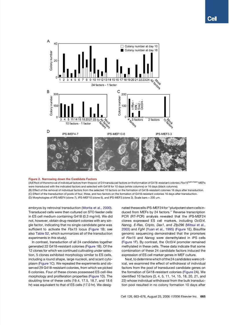
Figure 2. Narrowing down the Candidate Factors
(A)Effect of theremoval of individual factors from thepool of 24 transduced factors on theformation of G418-resistant colonies.Fbx15b geo/ b geo MEFs
were transduced with the indicated factors and selected with G418 for 10 days (white columns) or 16 days (black columns).
(B) Effect of the removal of individual factors from the selected 10 factors on the formation of G418-resistant colonies 16 days after transduction.
(C) Effect of the transduction of pools of four, three, and two factors on the formation of G418-resistant colonies 16 days after transduction.
(D) Morphologies of iPS-MEF4 (clone 7), iPS-MEF10 (clone 6), and iPS-MEF3 (clone 3). Scale bars = 200 mm.
Cell 126, 663–676, August 25, 2006 ª2006 Elsevier Inc. 665
8/14/2019 #12 Induction of Pluripotent Stem Cells From Mouse Embryonic and Adult Fibroblast Cultures by Defined Factors
http://slidepdf.com/reader/full/12-induction-of-pluripotent-stem-cells-from-mouse-embryonic-and-adult-fibroblast 4/14
transduction and fewer colonies 16 days after transduc-
tion. Combination of these 10 genes alone produced
more ES cell-like colonies than transduction of all 24
genes did ( Figure 2B).
We next examined the formation of colonies after with-
drawal of individual factors from the 10-factor pool trans-
duced into MEFs ( Figure 2B). G418-resistant colonies did
not form when either Oct3/4 (factor 14) or Klf4 (factor 20)
was removed. Removal of Sox2 (factor 15) resulted in
only a few G418-resistant colonies. When we removed
c-Myc (factor 22), G418-resistant colonies did emerge,
but these had a flatter, non-ES-cell-like morphology. Re-
moval of the remaining factors did not significantly affect
colony numbers. These results indicate that Oct3/4, Klf4,
Sox2, and c-Myc play important roles in the generation
of iPS cells from MEFs.
Combination of the four genes produced a number of
G418-resistant colonies similar to that observed with thepool of 10 genes ( Figure 2C). We continued cultivation of
12 clones for each transduction and were able to establish
4 iPS-MEF4 and 5 iPS-MEF10 clones. In addition, we
could generate iPS cells (iPS-MEF4wt) with wild-type
c-Myc instead of the T58A mutant ( Table S2 ). These data
demonstrate that iPS cells can be induced from MEF
culture by the introduction of four transcription factors,
Oct3/4, Sox2, c-Myc, and Klf4.
No combination of two factors could induce the forma-
tion of G418-resistant colonies ( Figure 2C). Two combina-
tions of three factors—Oct3/4, Sox2, and c-Myc (minus
Klf4) or Klf4, Sox2, and c-Myc (minus Oct3/4)—generated
a single, small colony in each case, but these could not bemaintained in culture. With the combination of Oct3/4,
Klf4, and Sox2 (minus c-Myc), we observed the formation
of 36 G418-resistant colonies, which, however, exhibited
a flat, non-ES-cell-like morphology. With the combination
of Oct3/4, Klf4, and c-Myc (minus Sox2), we observed
the formation of 54 G418-resistant colonies, of which we
picked 6. Although all 6 clones could be maintained over
several passages, the morphology of these cells (iPS-
MEF3) differed from that of iPS-MEF4 and iPS-MEF10
cells, with iPS-MEF3 colonies exhibiting rough surfaces
( Figure 2D). These data indicate that the combination of
Oct3/4, c-Myc, and Klf4 can activate the Fbx15 locus,
but the change induced by these three factors alone is dif-
ferent from that seen in iPS-MEF4 or iPS-MEF10 cells.
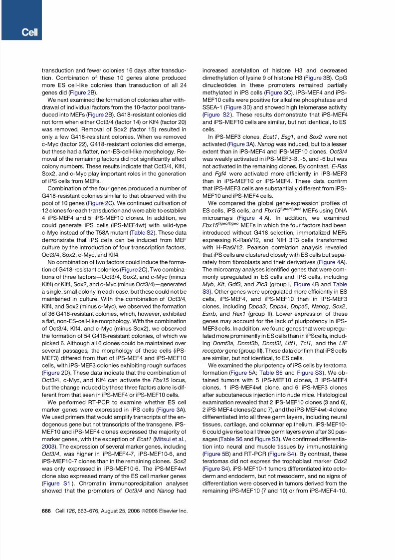
We performed RT-PCR to examine whether ES cell
marker genes were expressed in iPS cells ( Figure 3 A).
We used primers that would amplify transcripts of the en-
dogenous gene but not transcripts of the transgene. iPS-
MEF10 and iPS-MEF4 clones expressed the majority of
marker genes, with the exception of Ecat1 ( Mitsui et al.,
2003 ). The expression of several marker genes, including
Oct3/4, was higher in iPS-MEF4-7, iPS-MEF10-6, and
iPS-MEF10-7 clones than in the remaining clones. Sox2
was only expressed in iPS-MEF10-6. The iPS-MEF4wt
clone also expressed many of the ES cell marker genes
( Figure S1 ). Chromatin immunoprecipitation analyses
showed that the promoters of Oct3/4 and Nanog had
increased acetylation of histone H3 and decreased
dimethylation of lysine 9 of histone H3 ( Figure 3B). CpG
dinucleotides in these promoters remained partially
methylated in iPS cells ( Figure 3C). iPS-MEF4 and iPS-
MEF10 cells were positive for alkaline phosphatase and
SSEA-1 ( Figure 3D) and showed high telomerase activity
( Figure S2 ). These results demonstrate that iPS-MEF4
and iPS-MEF10 cells are similar, but not identical, to ES
cells.
In iPS-MEF3 clones, Ecat1, Esg1, and Sox2 were not
activated ( Figure 3 A). Nanog was induced, but to a lesser
extent than in iPS-MEF4 and iPS-MEF10 clones. Oct3/4
was weakly activated in iPS-MEF3-3, -5, and -6 but was
not activated in the remaining clones. By contrast, E-Ras
and Fgf4 were activated more efficiently in iPS-MEF3
than in iPS-MEF10 or iPS-MEF4. These data confirm
that iPS-MEF3 cells are substantially different from iPS-
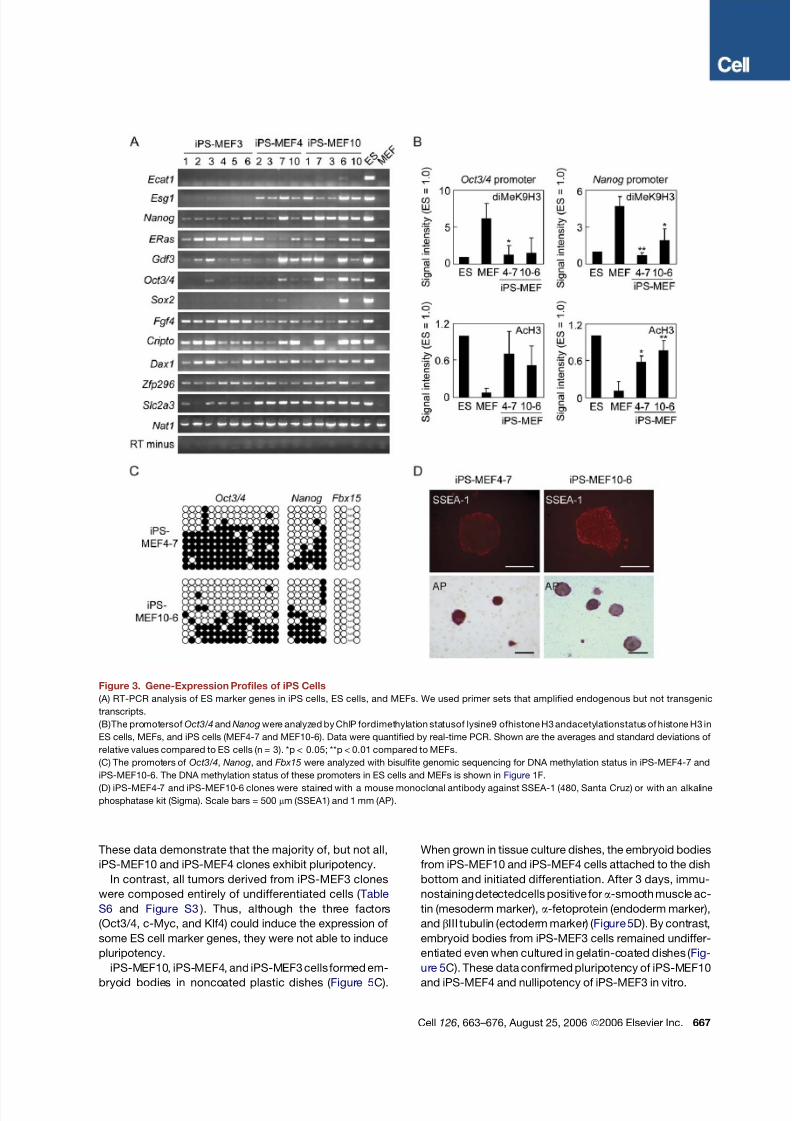
MEF10 and iPS-MEF4 cells.We compared the global gene-expression profiles of
ES cells, iPS cells, and Fbx15b geo/ b geo MEFs using DNA
microarrays ( Figure 4 A). In addition, we examined
Fbx15b geo/ b geo MEFs in which the four factors had been
introduced without G418 selection, immortalized MEFs
expressing K-RasV12, and NIH 3T3 cells transformed
with H-RasV12. Pearson correlation analysis revealed
that iPS cells are clustered closely with ES cells but sepa-
rately from fibroblasts and their derivatives ( Figure 4 A).
The microarray analyses identified genes that were com-
monly upregulated in ES cells and iPS cells, including
Myb, Kit , Gdf3, and Zic3 (group I, Figure 4B and Table
S3 ). Other genes were upregulated more efficiently in EScells, iPS-MEF4, and iPS-MEF10 than in iPS-MEF3
clones, including Dppa3, Dppa4, Dppa5, Nanog, Sox2,
Esrrb, and Rex1 (group II). Lower expression of these
genes may account for the lack of pluripotency in iPS-
MEF3 cells. In addition, we found genes that were upregu-
lated more prominently in ES cells than in iPScells, includ-
ing Dnmt3a, Dnmt3b, Dnmt3l , Utf1, Tcl1, and the LIF
receptor gene (group III). These data confirm that iPS cells
are similar, but not identical, to ES cells.
We examined the pluripotency of iPS cells by teratoma
formation ( Figure 5 A; Table S6 and Figure S3 ). We ob-
tained tumors with 5 iPS-MEF10 clones, 3 iPS-MEF4
clones, 1 iPS-MEF4wt clone, and 6 iPS-MEF3 clones
after subcutaneous injection into nude mice. Histological
examination revealed that 2 iPS-MEF10 clones (3 and 6),
2 iPS-MEF4 clones (2 and 7), and the iPS-MEF4wt-4 clone
differentiated into all three germ layers, including neural
tissues, cartilage, and columnar epithelium. iPS-MEF10-
6 could give rise to all three germ layers even after 30 pas-
sages ( Table S6 and Figure S3 ). We confirmed differentia-
tion into neural and muscle tissues by immunostaining
( Figure 5B) and RT-PCR ( Figure S4 ). By contrast, these
teratomas did not express the trophoblast marker Cdx2
( Figure S4 ). iPS-MEF10-1 tumors differentiated into ecto-
derm and endoderm, but not mesoderm, and no signs of
differentiation were observed in tumors derived from the
remaining iPS-MEF10 (7 and 10) or from iPS-MEF4-10.
666 Cell 126, 663–676, August 25, 2006 ª2006 Elsevier Inc.
8/14/2019 #12 Induction of Pluripotent Stem Cells From Mouse Embryonic and Adult Fibroblast Cultures by Defined Factors
http://slidepdf.com/reader/full/12-induction-of-pluripotent-stem-cells-from-mouse-embryonic-and-adult-fibroblast 5/14
8/14/2019 #12 Induction of Pluripotent Stem Cells From Mouse Embryonic and Adult Fibroblast Cultures by Defined Factors
http://slidepdf.com/reader/full/12-induction-of-pluripotent-stem-cells-from-mouse-embryonic-and-adult-fibroblast 6/14
Figure 4. Global Gene-Expression
Analyses by DNA Microarrays
(A) Pearson correlation analysis of 10,517
probes was performed to cluster ES cells,
iPS cells (MEF4- 7, MEF10-6, MEF3-2, and
MEF3-3), MEFs, MEFs expressing the four
factors, immortalized MEFs expressing
K-RasV12, and NIH 3T3 cells transformed
by H-RasV12. Red indicates increased
expression compared to median levels of
the eight samples, whereas green means
decreased expression.
(B) Genes upregulated in ES and/or iPS cells.
Genes in group I are genes upregulated in ES
cells and iPS cells. Genes in group II are
upregulated more in ES cells, iPS-MEF4-7,
and iPS-MEF10-6 than in iPS-MEF3 cells.
Genes in group III are upregulated more in
ES cells than in iPS cells. Lists of genes are
shown in Tables S3–S5.
668 Cell 126, 663–676, August 25, 2006 ª2006 Elsevier Inc.
8/14/2019 #12 Induction of Pluripotent Stem Cells From Mouse Embryonic and Adult Fibroblast Cultures by Defined Factors
http://slidepdf.com/reader/full/12-induction-of-pluripotent-stem-cells-from-mouse-embryonic-and-adult-fibroblast 7/14
We next introduced the four selected factors into tail-tip
fibroblasts (TTFs) of four 7-week-old male Fbx15b geo/ b geo
mice on a C57/BL6-129 hybrid background. We obtained
3 G418-resistant colonies, from each of which we could
establish iPS cells (iPS-TTF4). We also introduced the
four factors into TTFs from a 12-week-old female
Fbx15b geo/ b geo mouse, which also constitutively ex-
pressed green fluorescent protein (GFP) from the CAG
promoter and had a C57/BL6-129-ICR hybrid back-
ground. Of the 13 G418-resistant colonies obtained, we
isolated 6 clones from which we could establish iPS cells
(iPS-TTFgfp4, clones 1–6). In addition, we established
another iPS-TTFgfp4 (clone 7), in which the cDNA for
each of the four factors was flanked with two loxP sites
in the transgene. These cells were morphologically indis-
tinguishable from ES cells ( Figure 6 A). RT-PCR showed
that clones 3 and 7 of iPS-TTFgfp4 expressed the majority
of ES cell marker genes at high levels and the others at
lower levels ( Figure6B).In another attempt, we used either
the T58A mutant or the wild-type c-Myc for transduction
and established 5 iPS-TTFgfp4 clones (clones 8–12) and
3 iPS-TTFgfp4wt clones (clones 1–3) ( Figure S5 ). RT-PCR
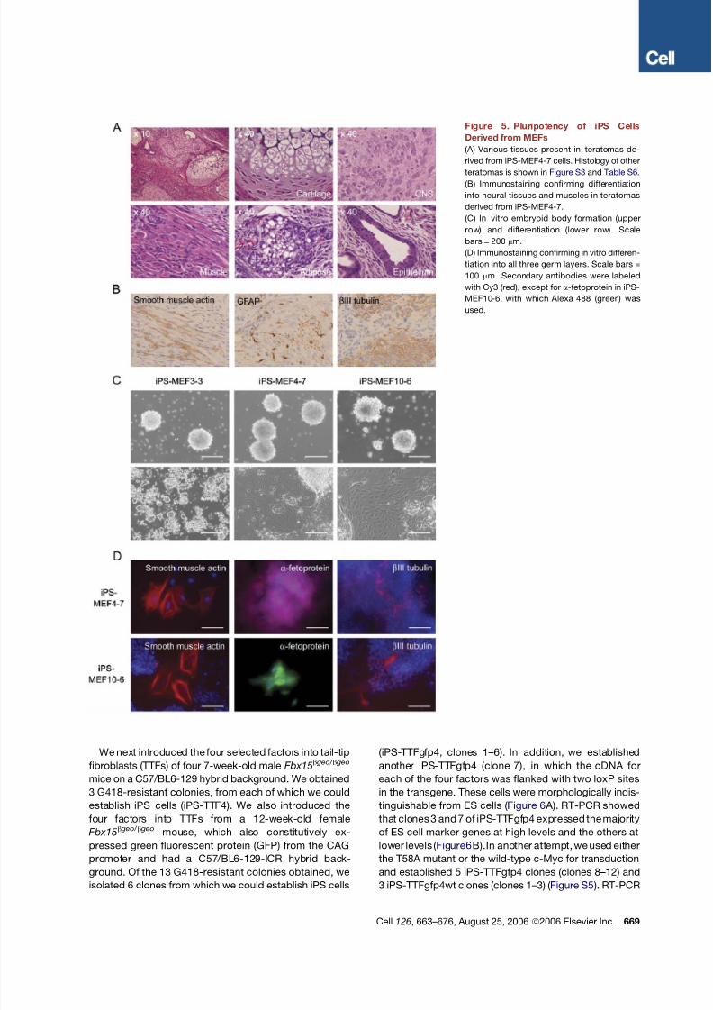
Figure 5. Pluripotency of iPS Cells
Derived from MEFs
(A) Various tissues present in teratomas de-
rived from iPS-MEF4-7 cells. Histology of other
teratomas is shown in Figure S3 and Table S6.
(B) Immunostaining confirming differentiation
into neural tissues and muscles in teratomas
derived from iPS-MEF4-7.
(C) In vitro embryoid body formation (upper
row) and differentiation (lower row). Scale
bars = 200 mm.
(D) Immunostaining confirming in vitro differen-
tiation into all three germ layers. Scale bars =
100 mm. Secondary antibodies were labeled
with Cy3 (red), except for a-fetoprotein in iPS-
MEF10-6, with which Alexa 488 (green) was
used.
Cell 126, 663–676, August 25, 2006 ª2006 Elsevier Inc. 669
8/14/2019 #12 Induction of Pluripotent Stem Cells From Mouse Embryonic and Adult Fibroblast Cultures by Defined Factors
http://slidepdf.com/reader/full/12-induction-of-pluripotent-stem-cells-from-mouse-embryonic-and-adult-fibroblast 8/14
showed that iPS-TTFgfp4wt cells also expressed most
of the ES cell marker genes ( Figure S6 ).
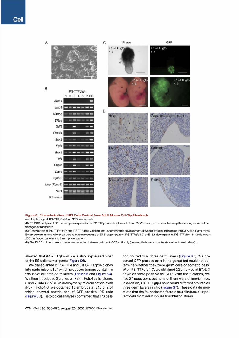
We transplanted 2 iPS-TTF4 and 6 iPS-TTFgfp4 clones
into nude mice, all of which produced tumors containing
tissues of all three germ layers ( Table S6 and Figure S3 ).
We then introduced 2 clones of iPS-TTFgfp4 cells (clones
3 and 7) into C57/BL6 blastocysts by microinjection. With
iPS-TTFgfp4-3, we obtained 18 embryos at E13.5, 2 of
which showed contribution of GFP-positive iPS cells
( Figure 6C). Histological analyses confirmed that iPS cells
contributed to all three germ layers ( Figure 6D). We ob-
served GFP-positive cells in the gonad but could not de-
termine whether they were germ cells or somatic cells.
With iPS-TTFgfp4-7, we obtained 22 embryos at E7.5, 3
of which were positive for GFP. With the 2 clones, we
had 27 pups born, but none of them were chimeric mice.
In addition, iPS-TTFgfp4 cells could differentiate into all
three germ layers in vitro ( Figure S7 ). These data demon-
strate that the four selected factors could induce pluripo-
tent cells from adult mouse fibroblast cultures.
Figure 6. Characterization of iPS Cells Derived from Adult Mouse Tail-Tip Fibroblasts
(A) Morphology of iPS-TTFgfp4-3 on STO feeder cells.
(B) RT-PCR analysis of ES marker gene expression in iPS-TTFgfp4 cells (clones 1–5 and 7). We used primer sets that amplified endogenous but not
transgenic transcripts.
(C) Contribution of iPS-TTFgfp4-7 and iPS-TTFgfp4-3 cellsto mouseembryonic development. iPScells were microinjected intoC57/BL6 blastocysts.
Embryos were analyzed with a fluorescence microscope at E7.5 (upper panels, iPS-TTFgfp4-7) or E13.5 (lower panels, iPS-TTFgfp4-3). Scale bars =
200 mm (upper panels) and 2 mm (lower panels).
(D) The E13.5 chimeric embryo was sectioned and stained with anti-GFP antibody (brown). Cells were counterstained with eosin (blue).
670 Cell 126, 663–676, August 25, 2006 ª2006 Elsevier Inc.
8/14/2019 #12 Induction of Pluripotent Stem Cells From Mouse Embryonic and Adult Fibroblast Cultures by Defined Factors
http://slidepdf.com/reader/full/12-induction-of-pluripotent-stem-cells-from-mouse-embryonic-and-adult-fibroblast 9/14
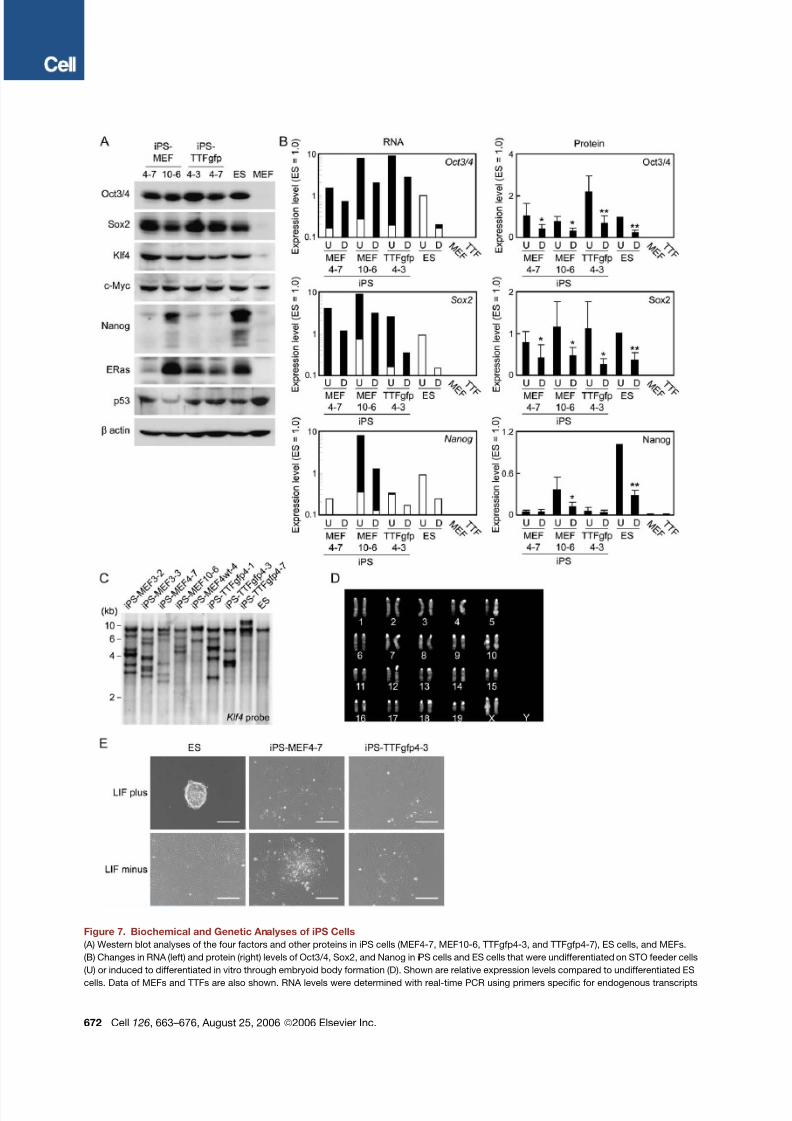
We further characterized the expression of the four fac-
tors and others in iPS cells. Real-time PCR confirmed that
endogenous expression of Oct3/4 and Sox2 was lower in
iPS cells than in ES cells ( Figure S8 ). However, the total
amount of the four factors from the endogenous genes
and the transgenes exceeded the normal expression
levels in ES cells. In contrast, Western blot analyses
showed that the total protein amounts of the four factors
in iPS cells were comparable to those in ES cells ( Fig-
ure 7 A; Figure S8 ).We could detectNanog andE-Ras pro-
teins in iPS cells, but at lower levels than those in ES cells
( Figures 7 A and 7B; Figure S8 ). The p53 levels in iPS cells
were lower than those in MEFs and equivalent to those in
ES cells ( Figure 7 A; Figure S9 ). The p21 levels in iPS cells
varied in each clone and were between those in ES cells
and MEFs ( Figure S9 ). Upon differentiation in vitro, the to-
talmRNA expression levelsof Oct3/4andSox2decreased
but remained much higher than in ES cells. In contrast,their protein levels decreased to comparable levels in
iPS cells and ES cells ( Figure 7B).
Southern blot analyses showed that each iPS clone has
a unique transgene integration pattern ( Figure 7C). Karyo-
typing analyses of the iPS-TTFgfp4 (clones 1, 2, 3, 7, and
11) and iPS-TTFgfp4wt (clones 1–3) demonstrated that 2
iPS-TTFgfp4 clones and all of the iPS-TTFgfp4wt clones
showed a normal karyotype of 40XX ( Figure 7D), while
the other 3 iPS-TTFgfp4 clones were 39XO, 40XO +10,
and 40Xi(X). Analyses of PCR-based simple sequence
length polymorphisms (SSLPs) demonstrated that iPS-
MEF clones have a mixed background of C57/BL6 and
129 ( Table S7 ), whereas iPS-TTFgfp clones have a mixedbackground of ICR, C57/BL6, and 129 ( Table S8 ). Finally,
we found that iPS cells could not remain undifferentiated
when cultured in the absence of feeder cells, even with
the presence of LIF ( Figure 7E). These results, together
with the different gene-expression patterns, exclude the
possibility that iPS cells are merely contamination of pre-
existing ES cells. Finally, subclones of iPS cells were pos-
itive for alkaline phosphatase and could differentiate into
all three germ layers in vitro ( Figure S10 ), confirming their
clonal nature.
DISCUSSION
Oct3/4, Sox2, and Nanog have been shown to function
as core transcription factors in maintaining pluripotency
( Boyer et al., 2005; Loh et al., 2006 ). Among the three,
we found that Oct3/4 and Sox2 are essential for the gen-
eration of iPS cells. Surprisingly, Nanog is dispensable. In
addition,we identifiedc-Mycand Klf4 as essential factors.
These two tumor-related factors could not be replaced by
other oncogenes including E-Ras, Tcl1, b-catenin, and
Stat3 ( Figures 2 A and 2B).
The c-Myc protein has many downstream targets that
enhance proliferation and transformation ( Adhikary and
Eilers, 2005 ), many of which may have roles in the gener-
ation of iPS cells. Of note, c-Myc associates with histone
acetyltransferase (HAT) complexes, including TRRAP,
which is a core subunit of the TIP60 and GCN5 HAT com-
plexes ( McMahon et al., 1998 ), CREB binding protein
(CBP), and p300 ( Vervoorts et al., 2003 ). Within the mam-
malian genome, there may be up to 25,000 c-Myc binding
sites ( Cawley et al., 2004 ), many more than the predicted
number of Oct3/4 and Sox2 binding sites ( Boyer et al.,
2005; Loh et al., 2006 ). c-Myc protein may induce global
histone acetylation ( Fernandez et al., 2003 ), thus allowing
Oct3/4 and Sox2 to bind to their specific target loci.
Klf4 has been shown to repress p53 directly ( Rowland
et al., 2005 ), and p53 protein has been shown to suppress
Nanog during ES cell differentiation ( Lin et al., 2004 ). We
found that iPS cells showed levels of p53 protein lower
than those in MEFs ( Figure 7 A). Thus, Klf4 might contrib-
uteto activation of Nanogand other ES cell-specific genes
through p53 repression. Alternatively, Klf4 might function
as an inhibitor of Myc-induced apoptosis through the re-pression of p53 in our system ( Zindy et al., 1998 ). On the
other hand, Klf4 activates p21CIP1, thereby suppressing
cell proliferation ( Zhang et al., 2000 ). This antiproliferation
function of Klf4 might be inhibited by c-Myc, which sup-
presses the expression of p21CIP1 ( Seoane et al., 2002 ).
The balance between c-Myc and Klf4 may be important
for the generation of iPS cells.
One question that remains concerns the origin of our
iPS cells. With our retroviral expression system, we esti-
mated that only a small portion of cells expressing the
four factors became iPS cells ( Figure S11 ). The low fre-
quency suggests that rare tissue stem/progenitor cells
that coexisted in the fibroblast cultures might have givenrise to the iPS cells. Indeed, multipotent stem cells have
been isolated from skin ( Dyce et al., 2004; Toma et al.,
2001, 2005 ). These studies showed that 0.067% of
mouse skin cells are stem cells. One explanation for the
low frequency of iPS cell derivation is that the four factors
transform tissue stem cells. However, we found that the
four factors induced iPS cells with comparably low effi-
ciency even from bone marrow stroma, which should be
more enriched in mesenchymal stem cells andothermulti-
potent cells ( Tables S2 and S6 ). Furthermore, cells in-
duced by the three factors were nullipotent ( Table S6
and Figure S3 ). DNA microarray analyses suggested that
iPS-MEF4 cells and iPS-MEF3 cells have the same origin
( Figure 4 ). These results do not favor multipotent tissue
stem cells as the origin of iPS cells.
There are several other possibilities for the low fre-
quency of iPS cell derivation. First, the levels of the four
factors required for generation of pluripotent cells may
have narrow ranges, and only a small portion of cells ex-
pressing allfour of thefactorsat therightlevels canacquire
ES cell-like properties. Consistent with this idea, a mere
50% increase or decrease in Oct3/4 proteins induces
differentiation of ES cells ( Niwa et al., 2000 ). iPS clones
overexpressed the four factors when RNA levels were an-
alyzed, buttheirprotein levelswere comparable to those in
EScells( Figures 7 Aand7B; Figure S8 ), suggestingthat the
iPS clones possess a mechanism (or mechanisms) that
Cell 126, 663–676, August 25, 2006 ª2006 Elsevier Inc. 671
8/14/2019 #12 Induction of Pluripotent Stem Cells From Mouse Embryonic and Adult Fibroblast Cultures by Defined Factors
http://slidepdf.com/reader/full/12-induction-of-pluripotent-stem-cells-from-mouse-embryonic-and-adult-fibroblast 10/14
Figure 7. Biochemical and Genetic Analyses of iPS Cells
(A) Western blot analyses of the four factors and other proteins in iPS cells (MEF4-7, MEF10-6, TTFgfp4-3, and TTFgfp4-7), ES cells, and MEFs.
(B) Changes in RNA (left) and protein (right) levels of Oct3/4, Sox2, and Nanog in iPS cells and ES cells that were undifferentiated on STO feeder cells
(U) or induced to differentiated in vitro through embryoid body formation (D). Shown are relative expression levels compared to undifferentiated ES
cells. Data of MEFs and TTFs are also shown. RNA levels were determined with real-time PCR using primers specific for endogenous transcripts
672 Cell 126, 663–676, August 25, 2006 ª2006 Elsevier Inc.
8/14/2019 #12 Induction of Pluripotent Stem Cells From Mouse Embryonic and Adult Fibroblast Cultures by Defined Factors
http://slidepdf.com/reader/full/12-induction-of-pluripotent-stem-cells-from-mouse-embryonic-and-adult-fibroblast 11/14
tightly regulates the protein levels of the four factors. We
speculate that high amounts of the four factors are re-
quired in the initial stage of iPS cell generation, but, once
they acquire ES cell-like status, too much of the factors
are detrimental for self-renewal. Only a small portion of
transduced cells show such appropriate transgene ex-
pression. Second, generation of pluripotent cells may
require additional chromosomal alterations, which take
place spontaneously during culture or are induced by
some of the four factors. Although theiPS-TTFgfp4 clones
had largely normal karyotypes ( Figure 7D), we cannot rule
out the existence of minor chromosomal alterations. Site-
specific retroviral insertion may also play a role. Southern
blot analyses showed that each iPSclone has20 retroviral
integrations ( Figure 7C). Some of these may have caused
silencing or fusionwith endogenous genes. Further studies
will be required to determine the origin of iPS cells.
Another unsolved question is whether the four factorswe identified play roles in reprogramming induced by fu-
sion with ES cells or nuclear transfer into oocytes. Since
the four factors are expressed in ES cells at high levels,
it is reasonable to speculate that they are involved in the
reprogramming machinery that exists in ES cells. Our re-
sult is also consistent with the finding that the reprogram-
ming activity resides in the nucleus, but not in the cyto-
plasm, of ES cells ( Do and Scholer, 2004 ). However, iPS
cells were not identical to ES cells, as shown by the global
gene-expression patterns and DNA methylation status. It
is possible that we have missed additional important fac-
tors. One such candidate is ECAT1, although its forced
expression in iPS cells did not consistently upregulateES cell marker genes ( Figure S12 ).
More obscure arethe roles of thefour factors, especially
Klf4 and c-Myc, in the reprogramming observed in oo-
cytes.Both Klf4 and c-Myc aredispensable forpreimplan-
tation mouse development ( Baudino et al., 2002; Katz
et al., 2002 ). Furthermore, c- myc is not detected in oo-
cytes ( Domashenko et al., 1997 ). In contrast, L- myc is ex-
pressed maternally in oocytes.Klf17 andKlf7 , but notKlf4,
are found in expressed sequence-tag libraries derived
from unfertilized mouse eggs. Klf4 and c-Myc might be
compensated by these related proteins. It is highly likely
that other factors are also required to induce complete
reprogramming and totipotency in oocytes.
It is likely that the four factors from the transgenes are
required for maintaining the iPS cells since the expression
of Oct3/4 and Sox2 from theendogenous genes remained
low ( Figure 7B; Figure S8 ). We intended to prove this by
using transgenes flanked by two loxP sites and obtained
an iPS clone (TTF4gfp4-7). However, we noticed that
these cells contain multiple loxP sites on multiple chromo-
somes, and, thus, the Cre-mediated recombination would
cause not only deletion of the transgenes but also inter-
and intrachromosomal rearrangements. Studies with
conditional expression systems, such as the tetracycline-
mediated system, are required to answer this question.
We showed that the iPS cells can differentiate in vitro
and in vivo even with the presence of the retroviral vectors
containing the four factors. We found that Oct3/4 and
Sox2 proteins decreased significantly during in vitro differ-
entiation ( Figure 7B). Retroviral expression has been
shown to be suppressed in ES cells and further silenced
upon differentiation by epigenetic modifications, such as
DNA methylation ( Yao etal.,2004 ). Thesame mechanisms
are likely to play roles in transgene repression in iPS cells
since they express Dnmt3a, 3b, and 3l , albeit at lower
levels than ES cells do ( Table S5 ). In addition, we found
that iPS cells possess a mechanism (or mechanisms)that lowers protein levels of the transgenes and Nanog
( Figure 7B; Figure S8 ). The same mechanism may be
enhanced during differentiation. However, silencing of
Oct3/4 in iPS-TTFgfp4-3 cells was not complete, which
may explain our inability to obtain live chimeric mice after
blastocyst microinjection of iPS cells.
An unexpected finding in this study was the efficient ac-
tivation of Fgf4 and Fbx15 by the combination of the three
factors devoid of Sox2 since these two genes have been
shown to be regulated synergistically by Oct3/4 and
Sox2 ( Tokuzawa et al., 2003; Yuan et al., 1995 ). It is also
surprising that Nanog is dispensable for induction and
maintenance of iPS cells. More detailed analyses of iPScells will enhance our understanding of transcriptional
regulation in pluripotent stem cells.
Our findings may have wider applications, as we have
found that transgene reporters with other ES cell marker
genes,such asNanog, canreplace theFbx15knockin dur-
ing selection (K. Okita and S.Y., unpublished data). How-
ever, we still do notknow whether thefour factors cangen-
erate pluripotent cells from human somatic cells. Use of
c-Myc may not be suitable for clinical applications, and
the process may require specific culture environments.
Nevertheless, thefinding is an importantstep in controlling
pluripotency, which may eventually allow the creation of
pluripotent cells directly from somatic cells of patients.
EXPERIMENTAL PROCEDURES
Mice
Fbx15b geo/ b geo mice were generated with 129SvJae-derived RF8 ES
cells as described previously ( Tokuzawa et al., 2003 ) and were
(white columns) or those common for both endogenous and transgenic transcripts (white and black columns). RNA expression levels are shown on
logarithmic axes. Protein levels were determined by Western blot normalized with b-actin. Protein levels are shown as the averages and standard
deviations on linear axes (n = 4). *p < 0.05 compared to undifferentiated cells.
(C) Southern blot analyses showing the integration of transgenes. Genomic DNA isolated from iPS cells and ES cells was digested with EcoRI and
BamHI, separated on agarose gel, transferred to a nylon membrane, and hybridized with a Klf4 cDNA probe.
(D) Normal karyotype of iPS-TTFgfp4-2 clone.
(E) Morphology of ES cells and iPS cells cultured without feeder cells. One thousand cells were cultured on gelatin-coated six-well plates for 5 days,
with or without LIF. Scale bars = 200 mm.
Cell 126, 663–676, August 25, 2006 ª2006 Elsevier Inc. 673
8/14/2019 #12 Induction of Pluripotent Stem Cells From Mouse Embryonic and Adult Fibroblast Cultures by Defined Factors
http://slidepdf.com/reader/full/12-induction-of-pluripotent-stem-cells-from-mouse-embryonic-and-adult-fibroblast 12/14
backcrossed to the C57/BL6 strain for at least five generations. These
mice were used forprimary mouse embryonicfibroblast (MEF) andtail-
tip fibroblast (TTF) preparations. To generate Fbx15b geo/ b geo mice with
constitutive expression of GFP, an Fbx15b geo/ b geo mouse (C57/BL6-
129 background) was mated with an ICR mouse with the GFP trans-
gene driven by the constitutive CAG promoter ( Niwa et al., 1991 ).
The resulting Fbx15b geo/+,GFP/+ mice were intercrossed to generate
Fbx15b geo/ b geo,GFP/GFP mice. Nude mice (BALB/Jcl-nu) were pur-
chased from CLEA.
Cell Culture
RF8ES cells andiPS cells were maintained on feederlayers of mitomy-
cin C-treated STO cells as previously described ( Meiner et al., 1996 ).
As a source of leukemia inhibitory factor (LIF), we used conditioned
medium (1:10,000 dilution) from Plat-E cell cultures that had been
transduced with a LIF-encoding vector. ES and iPS cells were pas-
saged every 3 days. Plat-E packaging cells ( Morita et al., 2000 ), which
were also used to produce retroviruses, were maintained in DMEM
containing 10% FBS, 50 units/50 mg/ml penicillin/streptomycin,
1 mg/ml puromycin(Sigma), and100 mg/ml of blasticidin S (Funakoshi).
For MEF isolation, uteri isolated from 13.5-day-pregnant mice were
washed with phosphate-buffered saline (PBS). The head and visceral
tissues were removed from isolated embryos. The remaining bodies
were washedin fresh PBS, minced using a pair of scissors,transferred
into a 0.1 mM trypsin/1 mM EDTA solution (3 ml per embryo), and
incubated at 37C for 20 min. After incubation, an additional 3 ml per
embryo of 0.1 mM trypsin/1 mM EDTA solution was added, and the
mixture wasincubated at 37C for20 min. After trypsinization,an equal
amount of medium (6 ml per embryo DMEM containing 10% FBS) was
added and pipetted up and down a few times to help with tissue dis-
sociation. After incubation of the tissue/medium mixture for 5 min at
room temperature, the supernatant was transferred into a new tube.
Cells were collected by centrifugation (200 3 g for 5 min at 4C) and
resuspended in fresh medium. 13 106 cells (passage 1) were cultured
on 100 mm dishes at 37C with 5% CO2. In this study, we used MEFs
within three passages to avoid replicative senescence.To establishTTFs,the tails from adult mice were peeled, mincedinto
1 cm pieces, placed on culture dishes, and incubated in MF-start me-
dium (Toyobo) for 5 days. Cells that migrated out of the graft pieces
were transferred to new plates (passage 2) and maintained in DMEM
containing 10%FBS.We used TTFs atpassage 3 foriPS cell induction.
Retroviral Infection
The day before transduction, Plat-E cells ( Morita et al., 2000 ) were
seeded at83106 cellsper 100mm dish.On thenextday,pMXs-based
retroviral vectors were introduced into Plat-E cells using Fugene 6
transfection reagent (Roche) according to the manufacturer’s recom-
mendations. Twenty-seven microliters of Fugene 6 transfection re-
agentwas diluted in300ml DMEMandincubated for5 minat room tem-
perature. Nine micrograms of plasmid DNA was added to the mixture,
which was incubated for another 15 min at room temperature. After in-
cubation, the DNA/Fugene 6 mixture was added drop by drop onto
Plat-Ecells. Cells were then incubatedovernight at 37Cwith5%CO2.
Twenty-four hours after transduction, the medium was replaced.
MEFs or TTFs were seeded at 8 3 105 cells per 100 mm dish on mito-
mycin C-treated STO feeders. After 24 hr, virus-containing superna-
tants derived from these Plat-E cultures were filtered through a
0.45 mm cellulose acetate filter (Schleicher & Schuell) and supple-
mented with 4 mg/ml polybrene (Nacalai Tesque). Target cells were in-
cubated in the virus/polybrene-containing supernatants for 4 hr to
overnight. After infection, the cells were replated in 10 ml fresh
medium. Three days after infection, we added G418 at a final concen-
tration of 0.3 mg/ml. Clones were selected for 2 to 3 weeks.
Plasmid Construction
To generate pMXs-gw, we introduced a Gateway cassette rfA (Invitro-
gen) into the EcoRI/XhoI site of the pMXs plasmid. Primers used are
listed in Table S9. Mutations inb-catenin, c- myc, and Stat3 were intro-
duced by PCR-based site-directed mutagenesis. For forced expres-
sion, we amplified the coding regions of candidate genes by RT-PCR,
cloned these sequences into pDONR201 or pENTR-D-TOPO (Invitro-
gen), and recombined the resulting plasmids with pMXs-gw by LR
reaction (Invitrogen).
Teratoma Formation and Histological Analysis
EScells oriPS cells weresuspended at1 3 107 cells/ml in DMEM con-
taining 10% FBS. Nude mice were anesthetized with diethyl ether. We
injected 100 ml of the cell suspension (1 3 106 cells) subcutaneously
into the dorsal flank. Four weeks after the injection, tumors were surgi-
cally dissected from the mice. Samples were weighed, fixed in PBS
containing 4% formaldehyde, and embedded in paraffin. Sections
were stained with hematoxylin and eosin.
Bisulfite Genomic Sequencing
Bisulfite treatment was performed using the CpGenome modification
kit (Chemicon) according to the manufacturer’s recommendations.
PCR primers are listed in Table S9. Amplified products were cloned
intopCR2.1-TOPO (Invitrogen). Tenrandomlyselectedclones werese-
quenced with theM13 forward andM13 reverse primers foreachgene.
Determination of Karyotypes and SSLP by PCR
Karyotypes were determined with quinacrine-Hoechst staining at the
International Council for Laboratory Animal Science (ICLAS) Monitor-
ing Center (Japan). We obtained PCR primer sequences for SSLP
from the Mouse Genome Informatics website (The Jackson Labora-
tory, http://www.informatics.jax.org ). Allele sizes were approximated
on the basis of the known allele sizes in various inbred strains.
Western Blot Analyses
Western blot was performed as previously described ( Takahashiet al.,
2003 ). The primary antibodies used were anti-Oct3/4 monoclonal
antibody (C-10, Santa Cruz), anti-Sox2 antiserum ( Maruyama et al.,
2005 ), anti-Klf4 polyclonal antibody (H-180, Santa Cruz), anti-c-Mycpolyclonal antibody (A-14, Santa Cruz), anti-Nanog antiserum ( Mitsui
et al., 2003 ), anti-E-Ras antiserum ( Takahashi et al., 2003 ), anti-p53
polyclonal antibody(FL-393, SantaCruz), and anti-b-actinmonoclonal
antibody (A5441, Sigma).
RT-PCR for Marker Genes
We performed reverse transcription reactions using ReverTra Ace -a-
(Toyobo) and the oligo dT20 primer. PCR was done with ExTaq
(Takara). Real-time PCR was performed with Platinum SYBR Green
qPCR SuperMix-UDG with ROX (Invitrogen) according to manufac-
turer’s instructions. Signals were detected with an ABI7300 Real-
Time PCR System (Applied Biosystems). Primer sequences are listed
in Table S9.
DNA Microarray
Total RNA from ES cells, iPS cells, or MEFs were labeled with Cy3.
Samples were hybridized to a Mouse Oligo Microarray (G4121B, Agi-
lent) according to the manufacturer’s protocol. Arrays were scanned
with a G2565BA Microarray Scanner System (Agilent). Data were ana-
lyzed using GeneSpring GX software (Agilent).
In Vitro Differentiation of iPS Cells
Cells were harvested by trypsinization and transferred to bacterial cul-
ture dishes in the ES medium without G418 or LIF. After 3 days, aggre-
gated cells were plated onto gelatin-coated tissue culture dishes and
incubated for another 3 days. The cells were stained with anti-
a-smooth muscle actin monoclonal antibody (N1584, Dako), anti-a-
fetoprotein polyclonal antibody (N1501, Dako) or anti-bIII tubulin
monoclonal antibody (CBL412, Abcam) along with 40-6-diamidino-
2-phenylindole (Sigma). Total RNA derived from plated embryoid
bodies on day 6 was used for RT-PCR analysis.
674 Cell 126, 663–676, August 25, 2006 ª2006 Elsevier Inc.
8/14/2019 #12 Induction of Pluripotent Stem Cells From Mouse Embryonic and Adult Fibroblast Cultures by Defined Factors
http://slidepdf.com/reader/full/12-induction-of-pluripotent-stem-cells-from-mouse-embryonic-and-adult-fibroblast 13/14
Chromatin Immunoprecipitation Assay
We performed chromatin immunoprecipitation (ChIP) as previously
described ( Maruyama et al., 2005 ). Antibodies used in this experiment
were anti-dimethyl K9 H3 rabbit polyclonal antibody (ab7312-100,
Abcam) and anti-acetyl H3 rabbit polyclonal antibody (06-599,
Upstate). PCR primers are listed in Table S9.
Statistical Analyses
Data are shown as averages and standard deviations. We used
Student’s t test for protein-level analyses and one-factor ANOVA
with Scheffe’s post hoc test for ChIP analyses. All statistical analyses
were done with Excel 2003 (Microsoft) with theStatcel2add-on (OMS).
Supplemental Data
Supplemental Data include 12 figures and 9 tables and can be found
with this article online at http://www.cell.com/cgi/content/full/126/4/
663/DC1/ .
ACKNOWLEDGMENTS
We are grateful to Tomoko Ichisaka for preparation of mice and Mit-
suyo Maeda and Yoshinobu Toda for histological analyses. We thank
Megumi Kumazaki, Mirei Murakami, Masayoshi Maruyama, and Nor-
iko Tsubooka for technical assistance; Masato Nakagawa, Keisuke
Okita, and Koji Shimozaki for scientific comments; and Yumi Ohuchi
for administrative assistance. We also thank Dr. Robert Farese, Jr.
for RF8 ES cells and Dr. Toshio Kitamura for the Plat-E cells and
pMX retroviral vectors. This work was supported in part by research
grants from the Ministry of Education, Culture, Sports, Science and
Technology of Japan to S.Y. This work is also supported in part by
theTakedaScience Foundation, theOsaka Cancer Research Founda-
tion, the Inamori Foundation, the Mitsubishi Pharma Research Foun-
dation, and the Sankyo Foundation of Life Science and by a Grant-
in-Aid from the Japan Medical Association to S.Y. K.T. was supportedby a fellowship from the Japan Society for the Promotion of Science.
Received: April 24, 2006
Revised: June 18, 2006
Accepted: July 20, 2006
Published online: August 10, 2006
REFERENCES
Adhikary, S., and Eilers, M. (2005). Transcriptional regulation and
transformation by Myc proteins. Nat. Rev. Mol. Cell Biol. 6, 635–645.
Avilion,A.A.,Nicolis,S.K.,Pevny, L.H., Perez,L., Vivian, N.,and Lovell-
Badge, R. (2003). Multipotent cell lineages in early mouse develop-
ment depend on SOX2 function. Genes Dev. 17 , 126–140.
Baudino, T.A., McKay, C., Pendeville-Samain, H., Nilsson, J.A.,
Maclean, K.H., White, E.L., Davis, A.C., Ihle, J.N., and Cleveland,
J.L. (2002). c-Myc is essential for vasculogenesis and angiogenesis
during development and tumor progression. Genes Dev. 16, 2530–
2543.
Boyer,L.A.,Lee, T.I., Cole, M.F., Johnstone, S.E., Levine, S.S., Zucker,
J.P., Guenther, M.G., Kumar, R.M., Murray, H.L., Jenner, R.G., et al.
(2005). Core transcriptional regulatory circuitry in human embryonic
stem cells. Cell 122, 947–956.
Bromberg, J.F., Wrzeszczynska, M.H., Devgan, G., Zhao, Y., Pestell,
R.G., Albanese, C., and Darnell, J.E., Jr. (1999). Stat3 as an oncogene.
Cell 98, 295–303.
Burdon, T.,Stracey, C.,Chambers, I., Nichols, J.,and Smith,A. (1999).
Suppression of SHP-2 and ERK signalling promotes self-renewal of
mouse embryonic stem cells. Dev. Biol. 210, 30–43.
Cartwright, P., McLean, C., Sheppard, A., Rivett, D., Jones, K., and
Dalton, S. (2005). LIF/STAT3 controls ES cell self-renewal and pluripo-
tency by a Myc-dependent mechanism. Development 132, 885–896.
Cawley, S., Bekiranov, S., Ng, H.H., Kapranov, P., Sekinger, E.A.,
Kampa, D., Piccolboni, A., Sementchenko, V., Cheng, J., Williams, A.J., et al. (2004). Unbiased mapping of transcription factor binding
sites along human chromosomes 21 and 22 points to widespread reg-
ulation of noncoding RNAs. Cell 116, 499–509.
Chambers, I., Colby, D., Robertson, M., Nichols, J., Lee, S., Tweedie,
S.,and Smith,A. (2003). Functional expressioncloning of nanog,a plu-
ripotency sustaining factor in embryonic stem cells. Cell113, 643–655.
Chang, D.W., Claassen, G.F., Hann, S.R., and Cole, M.D. (2000). The
c-Myctransactivation domain is a direct modulator of apoptotic versus
proliferative signals. Mol. Cell. Biol. 20, 4309–4319.
Cheng,A.M.,Saxton, T.M., Sakai,R., Kulkarni,S., Mbamalu, G.,Vogel,
W., Tortorice, C.G., Cardiff, R.D., Cross, J.C., Muller, W.J., and Paw-
son, T. (1998). Mammalian Grb2 regulates multiple steps in embryonic
development and malignant transformation. Cell 95, 793–803.
Cowan, C.A., Atienza, J., Melton, D.A., and Eggan, K. (2005). Nuclear
reprogramming of somatic cells after fusion with human embryonic
stem cells. Science 309, 1369–1373.
Do, J.T., and Scholer, H.R. (2004). Nuclei of embryonic stem cells
reprogram somatic cells. Stem Cells 22, 941–949.
Domashenko, A.D., Latham, K.E., and Hatton, K.S. (1997). Expression
of myc-family, myc-interacting, and myc-target genes during preim-
plantation mouse development. Mol. Reprod. Dev. 47 , 57–65.
Dyce, P.W., Zhu, H., Craig, J., and Li, J. (2004). Stem cells with multi-
lineage potential derived from porcine skin. Biochem. Biophys. Res.
Commun. 316, 651–658.
Evans, M.J., and Kaufman, M.H. (1981). Establishment in culture of
pluripotential cells from mouse embryos. Nature 292, 154–156.
Fernandez, P.C., Frank,S.R.,Wang,L., Schroeder, M.,Liu, S.,Greene,
J., Cocito, A., and Amati, B. (2003). Genomic targets of the human
c-Myc protein. Genes Dev. 17 , 1115–1129.
Katz, J.P., Perreault, N., Goldstein, B.G., Lee, C.S., Labosky, P.A.,
Yang, V.W., and Kaestner, K.H. (2002). The zinc-finger transcription
factor Klf4 is required for terminal differentiation of goblet cells in the
colon. Development 129, 2619–2628.
Kielman, M.F., Rindapaa, M., Gaspar, C., van Poppel, N., Breukel, C.,
van Leeuwen, S., Taketo, M.M., Roberts, S., Smits, R., and Fodde, R.
(2002). Apc modulates embryonic stem-cell differentiation by control-
ling the dosage of beta-catenin signaling. Nat. Genet. 32, 594–605.
Li, Y., McClintick, J., Zhong, L., Edenberg, H.J., Yoder, M.C., and
Chan, R.J. (2005). Murine embryonic stem cell differentiation is pro-
moted by SOCS-3 and inhibited by the zinc finger transcription factor
Klf4. Blood 105, 635–637.
Lin, T., Chao, C., Saito, S., Mazur, S.J., Murphy, M.E., Appella, E., and
Xu, Y. (2004). p53 induces differentiation of mouse embryonic stem
cells by suppressing Nanog expression. Nat. Cell Biol. 7 , 165–171.
Published online December 26, 2004. 10.1038/ncb1211.
Loh, Y.H., Wu, Q., Chew, J.L., Vega, V.B., Zhang, W., Chen, X., Bour-
que, G., George, J., Leong, B., Liu, J., et al. (2006). The Oct4 and
Nanog transcription network regulates pluripotency in mouse embry-
onic stem cells. Nat. Genet. 38, 431–440.
Martin, G.R. (1981). Isolation of a pluripotent cell line from early mouse
embryos cultured in medium conditioned by teratocarcinoma stem
cells. Proc. Natl. Acad. Sci. USA 78, 7634–7638.
Maruyama, M., Ichisaka, T., Nakagawa, M., and Yamanaka, S. (2005).
Differential roles for sox15 and sox2 in transcriptional control in mouse
embryonic stem cells. J. Biol. Chem. 280, 24371–24379.
Matsuda, T., Nakamura, T., Nakao, K., Arai, T., Katsuki, M., Heike, T.,
and Yokota, T. (1999). STAT3 activation is sufficient to maintain an
Cell 126, 663–676, August 25, 2006 ª2006 Elsevier Inc. 675
8/14/2019 #12 Induction of Pluripotent Stem Cells From Mouse Embryonic and Adult Fibroblast Cultures by Defined Factors
http://slidepdf.com/reader/full/12-induction-of-pluripotent-stem-cells-from-mouse-embryonic-and-adult-fibroblast 14/14
undifferentiated state of mouse embryonic stem cells. EMBO J. 18,
4261–4269.
McMahon, S.B., Van Buskirk, H.A., Dugan, K.A., Copeland, T.D., and
Cole, M.D. (1998). The novel ATM-related protein TRRAP is an essen-
tial cofactor for the c-Myc and E2F oncoproteins. Cell 94, 363–374.Meiner, V.L., Cases, S., Myers, H.M., Sande, E.R., Bellosta, S.,
Schambelan, M., Pitas, R.E., McGuire, J., Herz, J., and Farese, R.V.,
Jr. (1996). Disruption of the acyl-CoA:cholesterol acyltransferase
gene in mice: evidence suggesting multiple cholesterol esterification
enzymes in mammals. Proc. Natl. Acad. Sci. USA 93, 14041–14046.
Mitsui, K., Tokuzawa, Y., Itoh, H., Segawa, K., Murakami, M., Takaha-
shi, K., Maruyama, M., Maeda, M., and Yamanaka, S. (2003). The ho-
meoprotein Nanog is required for maintenance of pluripotency in
mouse epiblast and ES cells. Cell 113, 631–642.
Miyamoto,Y., Yamauchi,J., Mizuno, N.,and Itoh, H. (2004). Theadap-
tor protein Nck1 mediates endothelin A receptor-regulated cell migra-
tion through the Cdc42-dependent c-Jun N-terminal kinase pathway.
J. Biol. Chem. 279, 34336–34342.
Morita, S., Kojima, T., and Kitamura, T. (2000). Plat-E: an efficient and
stable system for transient packaging of retroviruses. Gene Ther. 7 ,
1063–1066.
Nichols, J., Zevnik, B., Anastassiadis, K., Niwa, H., Klewe-Nebenius,
D.,Chambers, I.,Scholer,H., andSmith, A. (1998). Formationof plurip-
otent stem cells in the mammalian embryo depends on the POU tran-
scription factor Oct4. Cell 95, 379–391.
Niwa, H., Yamamura, K., and Miyazaki, J. (1991). Efficient selection for
high-expression transfectants with a novel eukaryotic vector. Gene
108, 193–199.
Niwa, H., Burdon, T., Chambers, I., and Smith, A. (1998). Self-renewal
of pluripotent embryonic stem cells is mediated via activation of
STAT3. Genes Dev. 12, 2048–2060.
Niwa, H., Miyazaki, J., and Smith, A.G. (2000). Quantitative expression
of Oct-3/4 defines differentiation, dedifferentiation or self-renewal of
ES cells. Nat. Genet. 24, 372–376.Rowland, B.D., Bernards, R., and Peeper, D.S. (2005). The KLF4 tu-
mour suppressor is a transcriptional repressor of p53 that acts as
a context-dependent oncogene. Nat. Cell Biol. 7 , 1074–1082.
Sadot, E., Conacci-Sorrell, M., Zhurinsky, J., Shnizer, D., Lando, Z.,
Zharhary,D., Kam,Z., Ben-Ze’ev, A.,and Geiger, B. (2002). Regulation
of S33/S37 phosphorylated beta-catenin in normal and transformed
cells. J. Cell Sci. 115, 2771–2780.
Sato, N., Meijer, L., Skaltsounis, L., Greengard, P., and Brivanlou, A.H.
(2004). Maintenance of pluripotency in human and mouse embryonic
stem cells through activation of Wnt signaling by a pharmacological
GSK-3-specific inhibitor. Nat. Med. 10, 55–63.
Seoane, J., Le, H.V., and Massague, J. (2002). Myc suppression of the
p21(Cip1) Cdk inhibitor influences the outcome of the p53 response to
DNA damage. Nature 419, 729–734.
Tada, M., Takahama, Y., Abe, K., Nakatsuji, N., and Tada, T. (2001).
Nuclear reprogramming of somatic cells by in vitro hybridization with
ES cells. Curr. Biol. 11, 1553–1558.
Takahashi, K., Mitsui, K., and Yamanaka, S. (2003). Role of ERas in
promoting tumour-like properties in mouse embryonic stem cells.Nature 423, 541–545.
Thomson, J.A., Itskovitz-Eldor,J., Shapiro, S.S., Waknitz, M.A., Swier-
giel, J.J., Marshall, V.S., and Jones, J.M. (1998). Embryonic stem cell
lines derived from human blastocysts. Science 282, 1145–1147.
Tokuzawa, Y., Kaiho, E., Maruyama, M., Takahashi, K., Mitsui, K.,
Maeda, M.,Niwa, H., andYamanaka, S. (2003). Fbx15 is a novel target
of Oct3/4 but is dispensable for embryonic stem cell self-renewal and
mouse development. Mol. Cell. Biol. 23, 2699–2708.
Toma, J.G., Akhavan, M., Fernandes, K.J., Barnabe-Heider, F., Sadi-
kot, A., Kaplan, D.R., and Miller, F.D. (2001). Isolation of multipotent
adult stem cells from the dermis of mammalian skin. Nat. Cell Biol. 3,
778–784.
Toma, J.G., McKenzie, I.A., Bagli, D., and Miller, F.D. (2005). Isolation
and characterization of multipotent skin-derived precursors from
human skin. Stem Cells 23, 727–737.
Vervoorts, J., Luscher-Firzlaff, J.M., Rottmann, S., Lilischkis, R., Wal-
semann, G., Dohmann, K., Austen, M., and Luscher, B. (2003). Stimu-
lation of c-MYC transcriptional activity and acetylation by recruitment
of the cofactor CBP. EMBO Rep. 4, 484–490.
Wilmut, I., Schnieke, A.E., McWhir, J., Kind, A.J., and Campbell, K.H.
(1997). Viable offspring derived from fetal and adult mammalian cells.
Nature 385, 810–813.
Yao, S., Sukonnik, T., Kean, T., Bharadwaj, R.R., Pasceri, P., and Ellis,
J. (2004). Retrovirus silencing, variegation, extinction, and memory are
controlled by a dynamic interplay of multiple epigenetic modifications.
Mol. Ther. 10, 27–36.
Yuan, H., Corbi, N., Basilico, C., and Dailey, L. (1995). Developmental-
specific activity of the FGF-4 enhancer requires the synergistic action
of Sox2 and Oct-3. Genes Dev. 9, 2635–2645.Zhang, W., Geiman, D.E., Shields, J.M., Dang, D.T., Mahatan, C.S.,
Kaestner, K.H., Biggs, J.R., Kraft, A.S., and Yang, V.W. (2000). The
gut-enriched Kruppel-like factor (Kruppel-like factor 4) mediates the
transactivating effect of p53 on the p21WAF1/Cip1 promoter. J. Biol.
Chem. 275, 18391–18398.
Zindy, F., Eischen, C.M., Randle, D.H., Kamijo, T., Cleveland, J.L.,
Sherr, C.J., and Roussel, M.F. (1998). Myc signaling via the ARF tumor
suppressor regulates p53-dependent apoptosis and immortalization.
Genes Dev. 12, 2424–2433.
Accession Numbers
Microarray data are available in GEO (Gene Expression Omnibus,
http://www.ncbi.nlm.nih.gov/projects/geo/index.cgi ) with the acces-
sion number GSE5259.