10.1021@ac3033245
5
In Vivo Solid-Phase Microextraction with in Vitro Calibration: Determination of Off-Flavor Components in Live Fish Ziwei Bai, † Alexandre Pilote, ‡ Pallab Kumer Sarker, ‡ Grant Vandenberg, ‡ and Janusz Pawliszyn* ,† † Department of Chemistry, University of Waterloo, Waterloo, Ontario N2L 3G1, Canada ‡ Department des Sciences Animales, Universite ́ Laval, Pavillon Paul-Comtois, Qué bec, Qué bec G1K 0A6, Canada ABSTRACT: The presence of off-flavor compounds in fish represents a significant economic problem encountered in aquaculture production. The off-flavor compounds are due to the absorption of substances produced by a range of microorganisms. Currently, a number of strategies have been used to prevent or limit the growth of these microorganisms. Therefore, it is important to evaluate the effectiveness of strategies via monitoring the concentrations of off-flavor compounds in the recirculating aquaculture system. In vivo solid-phase microextraction (SPME), a rapid and simple sample preparation method, will allow monitoring the concentration of off-flavor compounds in live fish. In this research, geosmin and 2-methylisoborneol (2-MIB) produced by cyanobacteria and actinomycetes, which are the major sources for “earthy” and “muddy” flavors in fish, were selected as representatives. In order to accurately quantify these compounds in fish muscle, two kinetic calibration methods, on-fiber standardization and measurement using predetermined sampling rate, were used as quantification methods, which were both validated by traditional methods. The detection limit of in vivo SPME in fish muscle was 0.12 ng/g for geosmin and 0.21 ng/g for 2-MIB, which are both below the human sensory thresholds. R ecirculating aquaculture systems (RAS) are fish culture systems in which water is largely reused after undergoing treatment. 1 Currently, commercial RAS production systems typically recirculate over 99% of their water usage, thus significantly reducing water consumption. 2 In addition, RAS improves opportunities for waste management and nutrient recycling, 3 thus providing better hygiene and disease manage- ment, 4 as well as biological pollution control. 5 However, one major disadvantage of producing fish in RAS is the presence of off-flavor compounds. Among those flavors, the “earthy” and “muddy” odors constitute more than 80% of the off-flavor problems found in farm-raised fish. 6 Such flavors come from the absorption by fish of substances including geosmin and 2- methylisoborneol (2-MIB), which are produced by a broad group of microorganisms in water. 7-9 The presence of undesirable off-flavors in products raised in RAS may cause a major reduction in the consumption of the products, or make them unsuitable for sale. The presence of off-flavors therefore represents a major hurdle for the wide-scale adoption of RAS technologies as a production technique. Currently in RAS research, there is focus on developing strategies to prevent or limit the development of micro- organisms that produce the off-flavor compounds found in fish. Therefore, in order to evaluate the effectiveness of these strategies on microorganisms, a method needs to be implemented that can monitor the level of target compounds by repeatedly sampling the individual living fish in RAS over time. Recently, solid-phase microextraction (SPME) has been widely used as a rapid, inexpensive, and solvent-free sample preparation method, which combines sampling, analyte isolation, and enrichment into one step. 10 Successful use of in vivo SPME depends on the selection of an appropriate calibration method. The on-fiber standardization method is one of the most widely used calibration methods for SPME in vivo sampling, 11-15 which uses the symmetry between the desorption of deuterated standards from the extraction phase to the sample and the absorption of analytes from sample to the extraction phase. 16 Recently, another calibration method using predetermined sampling rates of the analytes has been reported by Ouyang et al. 17 It assumes that the rate of mass transfer or sampling rate remains constant throughout the duration of sampling within the linear range. The relationship between the concentration of target analytes in the sample matrixes (C 0 ) and the extracted amount of analytes at time t(n) can be expressed with eq 1: = C nRt / 0 s (1) where R s is the sampling rate for the target analyte and t is the sampling time. Use of eq 1 assumes that the intersample matrix differences in semisolid tissues (such as fish muscle) are slight between individuals of the same species. Consequently, the Received: November 15, 2012 Accepted: January 18, 2013 Published: January 18, 2013 Article pubs.acs.org/ac © 2013 American Chemical Society 2328 dx.doi.org/10.1021/ac3033245 | Anal. Chem. 2013, 85, 2328-2332
-
Upload
audry-arias -
Category
Environment
-
view
92 -
download
0
Transcript of 10.1021@ac3033245

In Vivo Solid-Phase Microextraction with in Vitro Calibration:Determination of Off-Flavor Components in Live FishZiwei Bai,† Alexandre Pilote,‡ Pallab Kumer Sarker,‡ Grant Vandenberg,‡ and Janusz Pawliszyn*,†
†Department of Chemistry, University of Waterloo, Waterloo, Ontario N2L 3G1, Canada‡Department des Sciences Animales, Universite ́ Laval, Pavillon Paul-Comtois, Queb́ec, Queb́ec G1K 0A6, Canada
ABSTRACT: The presence of off-flavor compounds in fishrepresents a significant economic problem encountered inaquaculture production. The off-flavor compounds are due tothe absorption of substances produced by a range ofmicroorganisms. Currently, a number of strategies have beenused to prevent or limit the growth of these microorganisms.Therefore, it is important to evaluate the effectiveness ofstrategies via monitoring the concentrations of off-flavorcompounds in the recirculating aquaculture system. In vivosolid-phase microextraction (SPME), a rapid and simplesample preparation method, will allow monitoring theconcentration of off-flavor compounds in live fish. In thisresearch, geosmin and 2-methylisoborneol (2-MIB) producedby cyanobacteria and actinomycetes, which are the major sources for “earthy” and “muddy” flavors in fish, were selected asrepresentatives. In order to accurately quantify these compounds in fish muscle, two kinetic calibration methods, on-fiberstandardization and measurement using predetermined sampling rate, were used as quantification methods, which were bothvalidated by traditional methods. The detection limit of in vivo SPME in fish muscle was 0.12 ng/g for geosmin and 0.21 ng/g for2-MIB, which are both below the human sensory thresholds.
Recirculating aquaculture systems (RAS) are fish culturesystems in which water is largely reused after undergoing
treatment.1 Currently, commercial RAS production systemstypically recirculate over 99% of their water usage, thussignificantly reducing water consumption.2 In addition, RASimproves opportunities for waste management and nutrientrecycling,3 thus providing better hygiene and disease manage-ment,4 as well as biological pollution control.5 However, onemajor disadvantage of producing fish in RAS is the presence ofoff-flavor compounds. Among those flavors, the “earthy” and“muddy” odors constitute more than 80% of the off-flavorproblems found in farm-raised fish.6 Such flavors come fromthe absorption by fish of substances including geosmin and 2-methylisoborneol (2-MIB), which are produced by a broadgroup of microorganisms in water.7−9 The presence ofundesirable off-flavors in products raised in RAS may cause amajor reduction in the consumption of the products, or makethem unsuitable for sale. The presence of off-flavors thereforerepresents a major hurdle for the wide-scale adoption of RAStechnologies as a production technique.Currently in RAS research, there is focus on developing
strategies to prevent or limit the development of micro-organisms that produce the off-flavor compounds found in fish.Therefore, in order to evaluate the effectiveness of thesestrategies on microorganisms, a method needs to beimplemented that can monitor the level of target compoundsby repeatedly sampling the individual living fish in RAS overtime. Recently, solid-phase microextraction (SPME) has been
widely used as a rapid, inexpensive, and solvent-free samplepreparation method, which combines sampling, analyteisolation, and enrichment into one step.10
Successful use of in vivo SPME depends on the selection ofan appropriate calibration method. The on-fiber standardizationmethod is one of the most widely used calibration methods forSPME in vivo sampling,11−15 which uses the symmetry betweenthe desorption of deuterated standards from the extractionphase to the sample and the absorption of analytes from sampleto the extraction phase.16 Recently, another calibration methodusing predetermined sampling rates of the analytes has beenreported by Ouyang et al.17 It assumes that the rate of masstransfer or sampling rate remains constant throughout theduration of sampling within the linear range. The relationshipbetween the concentration of target analytes in the samplematrixes (C0) and the extracted amount of analytes at time t(n)can be expressed with eq 1:
=C n R t/0 s (1)
where Rs is the sampling rate for the target analyte and t is thesampling time. Use of eq 1 assumes that the intersample matrixdifferences in semisolid tissues (such as fish muscle) are slightbetween individuals of the same species. Consequently, the
Received: November 15, 2012Accepted: January 18, 2013Published: January 18, 2013
Article
pubs.acs.org/ac
© 2013 American Chemical Society 2328 dx.doi.org/10.1021/ac3033245 | Anal. Chem. 2013, 85, 2328−2332

sampling rate of the SPME fiber can be predetermined underlaboratory conditions and directly used for real sampleanalysis.18 With this method, no distribution coefficient (K)determination and deuterated standard uploading prior tosampling are needed any longer.The objective of this study was to develop an effective and
simple method to determine geosmin and 2-MIB in fish usingin vivo sampling technique. In order to accomplish this goal,two kinetic calibration methods were investigated and verified:(1) on-fiber standardization and (2) measurement usingpredetermined sampling rates of analytes. Finally, thedeveloped methods were applied for on-site determination ofoff-flavors. These results were compared to those obtained fromthe traditional methods of analysis using lethal sampling andmicrowave distillation prior to SPME.
■ EXPERIMENTAL SECTIONChemicals and Materials. Geosmin and 2-MIB were
purchased from Wako Chemicals U.S.A., Inc. Deuterated MIB(MIB-d3) was obtained from CDN Isotopes Inc. (Pointe-Claire,Quebec, Canada), and deuterated geosmin (geosmin-d3) waspurchased from Sigma-Aldrich (Oakville, Ontario, Canada),with the purity of 99%. All chemicals purchased were of thehighest possible purity and were used without furtherpurification. Chloroform for fat content measurement waspurchased from EMD Chemicals Inc. (Damstadt, Germany).HPLC (high-performance liquid chromatography) grademethanol was purchased from Fisher Scientific (Unionville,Ontario, Canada). HPLC grade acetone was obtained fromCaledon Laboratories Ltd. (Georgetown, Ontario, Canada).Pump oil was purchased from Varian Vacuum Technologies(Lexington, MA). Nanopure water was obtained using theBarnstead Nanopure water system. The 1 cm metal corecommercial PDMS fibers were obtained from Supelco(Bellefonte, PA). Taint-free rainbow trout fillets werepurchased from Sobeys Inc. (Ontario, Canada). Rainbowtrout fillets contaminated with geosmin and 2-MIB wereobtained from Alma Aquaculture Research Station (Universityof Guelph, Ontario, Canada). All fish tissue experiments wereperformed in accordance with protocols approved by ourinstitutional Animal Care Committee (AUP no. A-12-01) in theChemistry Lab Facility at the University of Waterloo. All in vivofish experimental procedures were approved by the AnimalCare Committee at University of Guelph (AUP no. 12R066).Symmetry of Geosmin and 2-MIB on SPME under in
Vitro Condition for the On-Fiber StandardizationCalibration Method. Desorption time profiles of deuteratedstandards and absorption time profiles of analytes wereperformed simultaneously to verify the symmetry. Deuteratedstandards preloaded onto the fiber prior to the extraction wereprepared by using headspace sampling from deuteratedstandards pump oil solution under 25 °C. Both homogenizedand nonhomogenized fish tissue samples were utilized.Contaminated homogenized fish muscle was obtained byspiking specific amounts of geosmin and 2-MIB methanolsolution into taint-free homogenized fish tissue. Nonhomogen-ized fish tissue with off-flavors was prepared by cutting eachsample in 4 g size. Sampling was conducted by directly insertingthe SPME fibers in the homogenized and nonhomogenizedfish. All the experiments were performed under 8.5 °C, thesame temperature as in vivo sampling. The absorption timeprofile was obtained by extracting from geosmin and 2-MIBcontaminated samples for different sampling times. Meanwhile,
the desorption time profile was drawn by preloading sameamounts of standards and desorbing in the samples for differenttimes.
In Vivo Sampling System Setup. For this study, in vivosampling experiments were performed at Alma AquacultureResearch Station, University of Guelph. The source of water inthe station was from the underground wells nearby and wasproved to contain no detectable 2-MIB or geosmin by using aheadspace SPME analysis technique.19 The water temperaturewas kept constant at 8.5 °C. Rainbow trout (Oncorhynchusmykiss) were of marketable table size at 900 ± 154 g (n = 48).Before experiments, fish were acclimated with the diet in a flow-through system 1 week prior to experimentation. The systemwas located indoors and consisted of 16 (729 L) tanks. Eachtank held four fish, three for in vivo sampling and one for lethalsampling. Before exposure to tainted water, the fish weredeprived of food for 48 h. This was in order to minimizevariations in metabolic rates and ventilation rates betweenindividual fish with different feeding histories.20 During theexposure time, the fish were deprived of food as well. Theuptake of off-flavor compounds from contaminated water byfish as well as the in vivo sampling experiments were bothperformed in a static, albeit oxygen offered system (no waterflow rate). The water inside each static system (round tank)was transferred from flow-through system, and the volume was90 L.The procedure for operating in vivo sampling was as follows:
First, fibers were prepared by uploading deuterated geosminstandard from the headspace of pump oil solution. Before eachSPME sampling, fish were anesthetized in a 80 μg/mL MS-222water solution for the duration of approximately 1 min. Afterthat, an 18 gauge needle was used to pierce the fish skin, whereit is below the anterior point of the dorsal fin (as shown inFigure 1), and a 21 gauge long stainless steel wire was
introduced into the same perforation and penetrated to thesame length as the SPME fiber, because the needle was not aslong as the SPME fiber. Then the SPME fiber was inserted, andthe hub was pushed to the end. Following fiber placement, fishwere placed back in the static tank. After 30 min of SPMEextraction, the fish was anesthetized again. At last, the fiber wasremoved, gently washed with Nanopure water, wiped with aKimwipe, sealed with a Teflon cap, wrapped with aluminumfoil, and stored in the dry ice until gas chromatography massspectrometry (GC/MS) analysis.
Figure 1. SPME fiber inserting position for in vivo sampling in fishmuscle.
Analytical Chemistry Article
dx.doi.org/10.1021/ac3033245 | Anal. Chem. 2013, 85, 2328−23322329

Instrumentation. A Varian 3800 GC coupled with a Varian4000 electron ionization ion-trap mass spectrometer (GC/EI-IT-MS) (Varian, Inc. Walnut Creek, CA, U.S.A.) was used foranalysis. The GC was equipped with a capillary column (RTX-5, 30 m × 0.25 mm i.d., 0.25 μm film thickness) (RestekCorporation, Bellefonte, PA, U.S.A.). The oven temperatureprogram was 60 °C (0.5 min) increasing at 40 °C/min to 110°C, increasing at 1 °C/min to 117 °C, increasing at 5 °C/minto 156 °C, increasing at 40 °C/min to 250 °C (10 min). Thesplit/splitless injector of the GC was equipped with a Merlinmicroseal injector adapter (Merlin Instrument Company, HalfMoon Bay, CA, U.S.A.) for metal fiber desorption. For bothliquid injection and SPME, splitless mode was used, and splitwas opened after 5 min of desorption. The temperature of thetransfer line was 280 and 240 °C for the ion trap. The columnflow rate of the helium carrier gas was 1 mL/min. The 1079injector was equipped with a programmed temperaturevaporizer. The temperature of the injector was 250 °C. Whenusing selected ion storage (SIS) scan mode, m/z 112, m/z 150,m/z 115, and m/z 138 were selected for quantification ofgeosmin, 2-MIB, geosmin-d3, and MIB-d3, respectively. Asqualifier ions, m/z 126 and 182 were for geosmin, m/z 168 wasto represent 2-MIB, m/z 129 and 185 were for geosmin-d3, andm/z 171 were selected for MIB-d3. When performing the invivo sampling analysis, 2-MIB required using MS/MS mode,and geosmin kept using SIS mode. Under MS/MS mode, theparent ion of 2-MIB m/z 95 yielded daughter ions m/z 67 andm/z 91. The excitation amplitude was set as 1.0 V applied withresonant waveform. The emission current and maximumionization time were set as 80 μA and 65 000 μs, respectively.
■ RESULTS AND DISCUSSION
Preliminary Experiments. Selected ion storage mode wasutilized for symmetry determination. To determine geosmin,geosmin-d3 was used as an internal standard. In this case, the EIspectrum of the labeled compound produced a base peak at m/z 115, which contained three deuterium atoms, in theagreement with the fragmentation proposed by Lloyd andGrimm.21 The quantification of geosmin and geosmin-d3 wasachieved using the ion pair at m/z 112 and 115, andqualification was established by using the ions m/z 126 and182 for geosmin and m/z 129 and 185 for geosmin-d3.However, for 2-MIB and MIB-d3, the base peak ion at m/z
95 for both was not a deuterated fragment. Finally, only m/z150 and 138 could be decided to represent 2-MIB and MIB-d3.However, in that case, the limit of detection for 2-MIB was ashigh as 10 ng/g in fish tissue, which was more than 10 timeshigher than the human sensory threshold.22 Consequently, theon-fiber standardization calibration method is not suitable fordetermination of low contamination of 2-MIB in fish.Therefore, measurement using the predetermined samplingrates method, which does not require the use of the internalstandard, was placed.
Fouling issues due to the adsorption of macromolecularcompounds on the SPME coating surface, such as lipids andprotein,23 was found after the fiber was desorbed in the GC inthe form of 2-MIB and geosmin peak intensity enhancement.To address this, following the desorption, the fiber was gentlywiped with an acetone-soaked Kimwipes and rinsed with largeamounts of water, followed by conditioning in the GC injectorfor 10 min at 250 °C. As a result, the repeatability of the fiberwas kept acceptable.
Symmetry of Desorption and Absorption Verificationunder in Vivo and in Vitro Conditions. The kinetics ofabsorption and desorption was performed in homogenized fishtissue, nonhomogenized fish tissue, and live fish. Time profileswere used for this verification. Because this calibration methodcould not be used to determine a low concentration of 2-MIBin fish tissue, the concentrations of 2-MIB in all these threesystems were high. The details about the concentration for bothanalytes were as follows: homogenized fish tissue 0.5 μg/g for2-MIB and 0.025 μg/g for geosmin; nonhomogenized fishtissue and live fish were around 0.3 μg/g for both analytes inthese two systems. The time constants of absorption anddesorption were very similar for both 2-MIB and geosmin in allthree systems, which means the symmetry was kept all throughthe desorption and absorption processes (as shown in Table 1).
Different Concentrations of Analytes in Homogen-ized Fish Tissue. A various range of concentrations (0.5, 2.5,5.0, 12.5, 25.0 ng/g) of 2-MIB and geosmin in homogenizedfish tissue were prepared and followed by a 30 min extraction.Equation 2 describes the entire absorption kinetics of theanalyte from the sample matrixes to SPME liquid, in which n isthe amount of extracted analyte at time t, ne is the amount ofanalyte extracted at equilibrium, and a is a absorption timeconstant.24 According to eq 2, the extraction amount should beproportional to the initial concentration, if all the parameterskeep constant. The linearity displayed in Figure 2 indicates thatthe extraction time constant for both analytes remainedconsistent in this concentration range. In addition, the valueof the extraction time constant was obtained in Table 1. Thisdiscovery was important for both calibration methods. For theon-fiber standardization calibration method, it needs the
Table 1. Time Constant Results and Conclusions for 2-MIB and Geosmin under Both in Vitro and in Vivo Conditionsa
2-MIB geosmin
desorption a (min−1) (R2) absorption a (min−1) (R2) desorption a (min−1) (R2) absorption a (min−1) (R2)
homogenized fish tissue 0.012 ± 0.002 (0.946) 0.011 ± 0.001 (0.984) 0.008 ± 0.001 (0.987) 0.008 ± 0.001 (0.984)nonhomogenized fish tissue 0.012 ± 0.003 (0.844) 0.012 ± 0.006 (0.780) 0.007 ± 0.001 (0.884) 0.010 ± 0.004 (0.837)live fish 0.010 ± 0.003 (0.852) 0.013 ± 0.006 (0.790) 0.012 ± 0.003 (0.747) 0.011 ± 0.007 (0.839)
aCorrelation coefficients are given in parentheses.
Figure 2. Different concentrations of analytes in homogenized fishtissue.
Analytical Chemistry Article
dx.doi.org/10.1021/ac3033245 | Anal. Chem. 2013, 85, 2328−23322330
symmetry of desorption and absorption during the samplingtime. For the calibration method using a predeterminedsampling rate, it requires the constant sampling rate.
= − − = − −+
n at n atK V V
K VC[1 exp( )] [1 exp( )]e
fs f s
fs s0
(2)
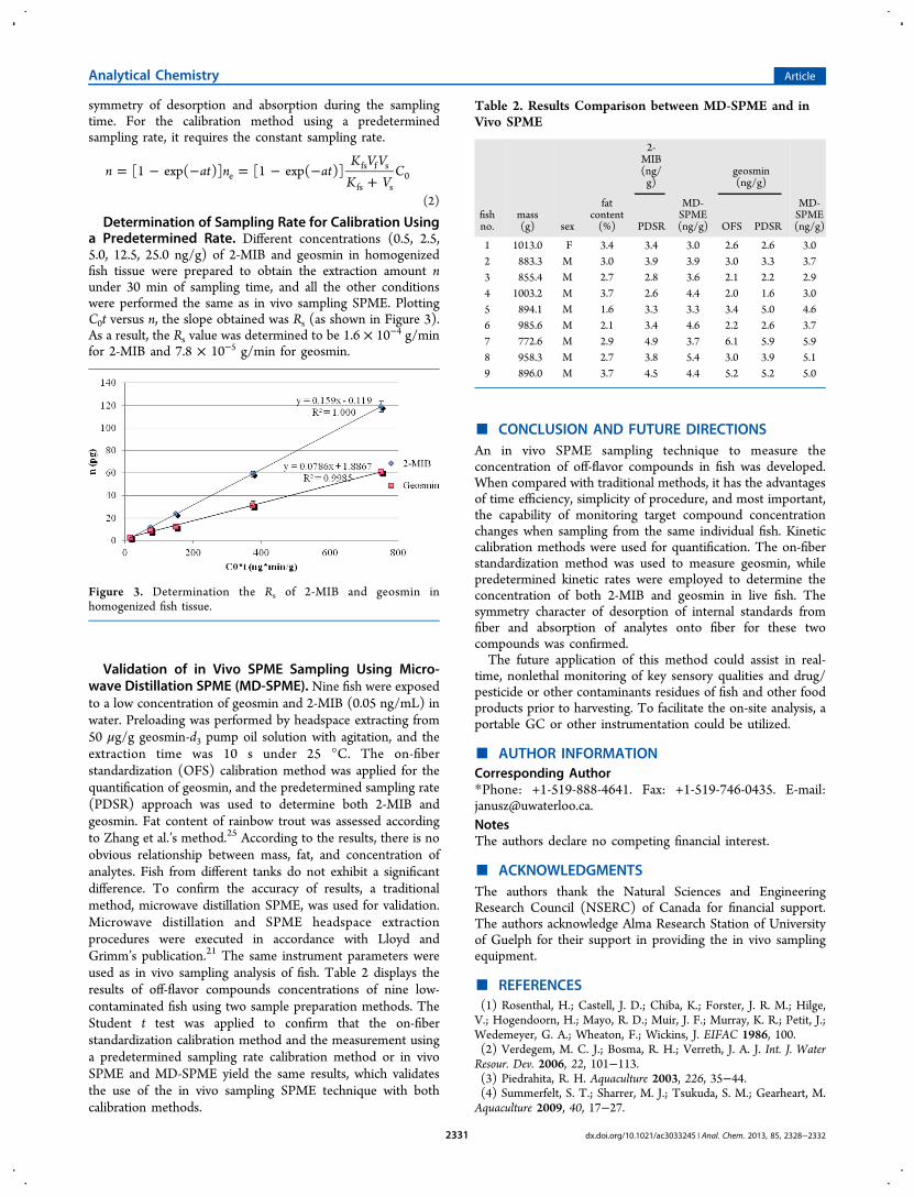
Determination of Sampling Rate for Calibration Usinga Predetermined Rate. Different concentrations (0.5, 2.5,5.0, 12.5, 25.0 ng/g) of 2-MIB and geosmin in homogenizedfish tissue were prepared to obtain the extraction amount nunder 30 min of sampling time, and all the other conditionswere performed the same as in vivo sampling SPME. PlottingC0t versus n, the slope obtained was Rs (as shown in Figure 3).As a result, the Rs value was determined to be 1.6 × 10⁻⁴ g/minfor 2-MIB and 7.8 × 10⁻⁵ g/min for geosmin.
Validation of in Vivo SPME Sampling Using Micro-wave Distillation SPME (MD-SPME). Nine fish were exposedto a low concentration of geosmin and 2-MIB (0.05 ng/mL) inwater. Preloading was performed by headspace extracting from50 μg/g geosmin-d3 pump oil solution with agitation, and theextraction time was 10 s under 25 °C. The on-fiberstandardization (OFS) calibration method was applied for thequantification of geosmin, and the predetermined sampling rate(PDSR) approach was used to determine both 2-MIB andgeosmin. Fat content of rainbow trout was assessed accordingto Zhang et al.’s method.25 According to the results, there is noobvious relationship between mass, fat, and concentration ofanalytes. Fish from different tanks do not exhibit a significantdifference. To confirm the accuracy of results, a traditionalmethod, microwave distillation SPME, was used for validation.Microwave distillation and SPME headspace extractionprocedures were executed in accordance with Lloyd andGrimm’s publication.21 The same instrument parameters wereused as in vivo sampling analysis of fish. Table 2 displays theresults of off-flavor compounds concentrations of nine low-contaminated fish using two sample preparation methods. TheStudent t test was applied to confirm that the on-fiberstandardization calibration method and the measurement usinga predetermined sampling rate calibration method or in vivoSPME and MD-SPME yield the same results, which validatesthe use of the in vivo sampling SPME technique with bothcalibration methods.
■ CONCLUSION AND FUTURE DIRECTIONSAn in vivo SPME sampling technique to measure theconcentration of off-flavor compounds in fish was developed.When compared with traditional methods, it has the advantagesof time efficiency, simplicity of procedure, and most important,the capability of monitoring target compound concentrationchanges when sampling from the same individual fish. Kineticcalibration methods were used for quantification. The on-fiberstandardization method was used to measure geosmin, whilepredetermined kinetic rates were employed to determine theconcentration of both 2-MIB and geosmin in live fish. Thesymmetry character of desorption of internal standards fromfiber and absorption of analytes onto fiber for these twocompounds was confirmed.The future application of this method could assist in real-
time, nonlethal monitoring of key sensory qualities and drug/pesticide or other contaminants residues of fish and other foodproducts prior to harvesting. To facilitate the on-site analysis, aportable GC or other instrumentation could be utilized.
■ AUTHOR INFORMATIONCorresponding Author*Phone: +1-519-888-4641. Fax: +1-519-746-0435. E-mail:[email protected] authors declare no competing financial interest.
■ ACKNOWLEDGMENTSThe authors thank the Natural Sciences and EngineeringResearch Council (NSERC) of Canada for financial support.The authors acknowledge Alma Research Station of Universityof Guelph for their support in providing the in vivo samplingequipment.
■ REFERENCES(1) Rosenthal, H.; Castell, J. D.; Chiba, K.; Forster, J. R. M.; Hilge,V.; Hogendoorn, H.; Mayo, R. D.; Muir, J. F.; Murray, K. R.; Petit, J.;Wedemeyer, G. A.; Wheaton, F.; Wickins, J. EIFAC 1986, 100.(2) Verdegem, M. C. J.; Bosma, R. H.; Verreth, J. A. J. Int. J. WaterResour. Dev. 2006, 22, 101−113.(3) Piedrahita, R. H. Aquaculture 2003, 226, 35−44.(4) Summerfelt, S. T.; Sharrer, M. J.; Tsukuda, S. M.; Gearheart, M.Aquaculture 2009, 40, 17−27.
Figure 3. Determination the Rs of 2-MIB and geosmin inhomogenized fish tissue.
Table 2. Results Comparison between MD-SPME and inVivo SPME
2-MIB(ng/g)
geosmin(ng/g)
fishno.
mass(g) sex
fatcontent(%) PDSR
MD-SPME(ng/g) OFS PDSR
MD-SPME(ng/g)
1 1013.0 F 3.4 3.4 3.0 2.6 2.6 3.02 883.3 M 3.0 3.9 3.9 3.0 3.3 3.73 855.4 M 2.7 2.8 3.6 2.1 2.2 2.94 1003.2 M 3.7 2.6 4.4 2.0 1.6 3.05 894.1 M 1.6 3.3 3.3 3.4 5.0 4.66 985.6 M 2.1 3.4 4.6 2.2 2.6 3.77 772.6 M 2.9 4.9 3.7 6.1 5.9 5.98 958.3 M 2.7 3.8 5.4 3.0 3.9 5.19 896.0 M 3.7 4.5 4.4 5.2 5.2 5.0
Analytical Chemistry Article
dx.doi.org/10.1021/ac3033245 | Anal. Chem. 2013, 85, 2328−23322331

(5) Zohar, Y.; Tal, Y.; Schreier, H. J.; Steven, C.; Stubblefield, J.;Place, A. Urban Aquacult. 2005, 159−171.(6) Grimm, C. C.; Lloyd, S. W.; Batista, R.; Zimba, P. V. J.Chromatogr. Sci. 2000, 38, 289−296.(7) Gerbek, N. N. J. Antibiot. 1969, 22, 508−509.(8) Rosen, A. A.; Mashni, C. I.; Safferman, R. S. Water Treat. Exam.1970, 19, 106−119.(9) Yurkowski, M.; Tabachek, J. L. J. Fish. Res. Board Can. 1976, 33,25−35.(10) Arthur, C. L.; Pawliszyn, J. Anal. Chem. 1990, 62, 2145−2148.(11) Zhou, S.; Zhao, W.; Pawliszyn, J. Anal. Chem. 2008, 80, 481−490.(12) Togunde, O. P.; Oakes, K.; Servos, M.; Pawliszyn, J. Anal. Chim.Acta 2012, 742, 2−9.(13) Zhang, X.; Oakes, K. D.; Hoque, M. E.; Luong, D.; Metcalfe, C.D.; Pawliszyn, J.; Servos, M. R. Anal. Chem. 2011, 83, 3365−3370.(14) Zhou, S.; Zhang, X.; Ouyang, G.; Es-haghi, A.; Pawliszyn, J.Anal. Chem. 2007, 79, 1221−1230.(15) Zhou, S.; Oakes, K. D.; Servos, M. R.; Pawliszyn, J. Environ. Sci.Technol. 2008, 42, 6073−6079.(16) Chen, Y.; Pawliszyn, J. Anal. Chem. 2004, 76, 5807−5815.(17) Ouyang, G.; Vuckovic, D.; Pawliszyn, J. Chem. Rev. 2011, 111,2784−2814.(18) Ouyang, G.; Oakes, K. D.; Bragg, L.; Wang, S.; Cui, S.; Servos,M. R.; Dixon, D. G.; Pawliszyn, J. Environ. Sci. Technol. 2010, 45,7792−7798.(19) Lloyd, S. W.; Lea, J. M.; Zimba, P. V.; Grimm, C. C. Water Res.1998, 32, 2140−2146.(20) Robertsona, R. F.; Jaunceyb, K.; Beveridgec, M. C. M.; Lawton,L. A. Aquaculture 2005, 245, 89−99.(21) Lloyd, S.W.; Grimm, C. C. J. Agric. Food Chem. 1999, 47, 164−169.(22) Robertson, R. F.; Jauncey, K.; Beveridge, M. C. M.; Lawton, L.A. Aquaculture 2005, 245, 89−99.(23) Pawliszyn, J. Anal. Chem. 2003, 75, 2543−2558.(24) Pawliszyn, J. Handbook of Solid Phase Microextraction; ChemicalIndustry Press: Beijing, China, 2009.(25) Zhang, X.; Oakes, K. D.; Cui, S.; Bragg, L.; Servos, M. R.;Pawliszyn, J. Environ. Sci. Technol. 2010, 44, 3417−3422.
Analytical Chemistry Article
dx.doi.org/10.1021/ac3033245 | Anal. Chem. 2013, 85, 2328−23322332