10 Relationship between Cell Biology and...
27
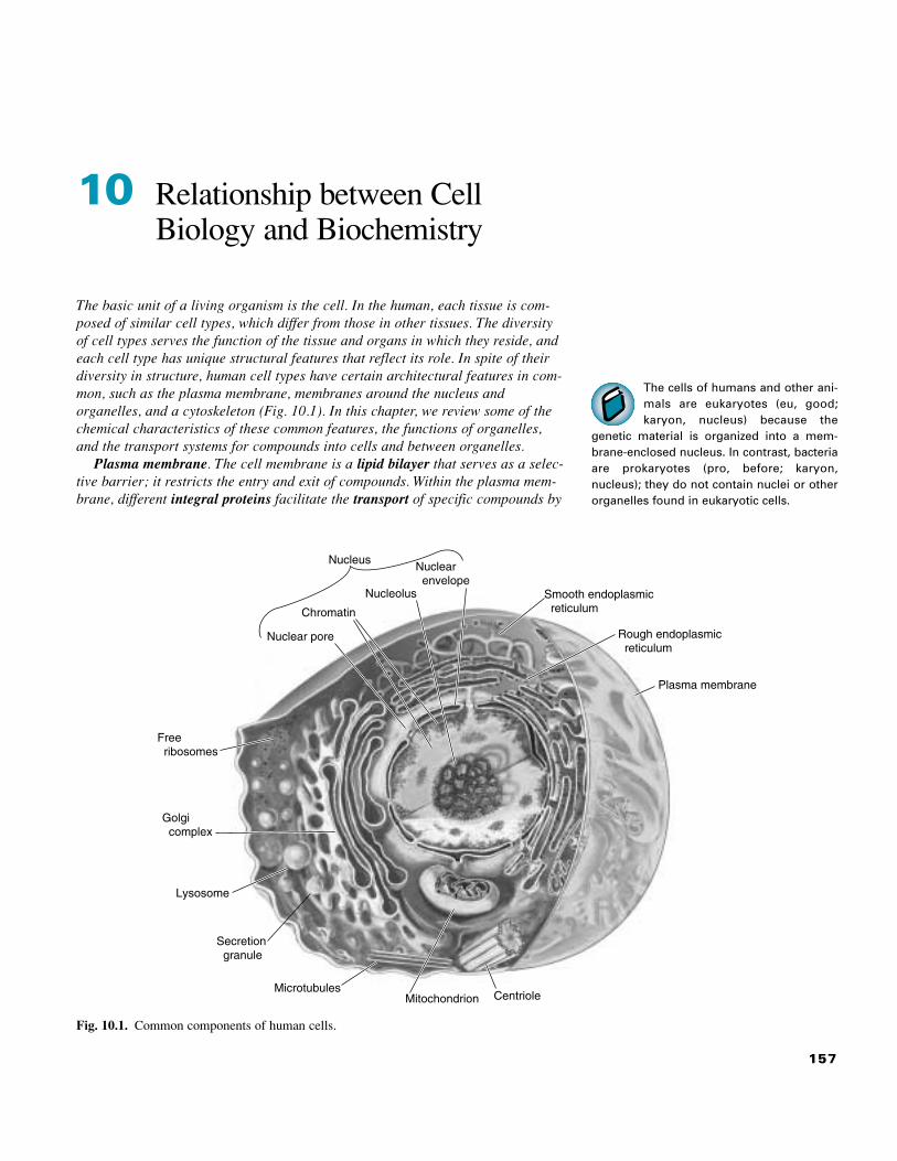
157 The cells of humans and other ani- mals are eukaryotes (eu, good; karyon, nucleus) because the genetic material is organized into a mem- brane-enclosed nucleus. In contrast, bacteria are prokaryotes (pro, before; karyon, nucleus); they do not contain nuclei or other organelles found in eukaryotic cells. 10 Relationship between Cell Biology and Biochemistry The basic unit of a living organism is the cell. In the human, each tissue is com- posed of similar cell types, which differ from those in other tissues. The diversity of cell types serves the function of the tissue and organs in which they reside, and each cell type has unique structural features that reflect its role. In spite of their diversity in structure, human cell types have certain architectural features in com- mon, such as the plasma membrane, membranes around the nucleus and organelles, and a cytoskeleton (Fig. 10.1). In this chapter, we review some of the chemical characteristics of these common features, the functions of organelles, and the transport systems for compounds into cells and between organelles. Plasma membrane. The cell membrane is a lipid bilayer that serves as a selec- tive barrier; it restricts the entry and exit of compounds. Within the plasma mem- brane, different integral proteins facilitate the transport of specific compounds by Smooth endoplasmic reticulum Rough endoplasmic reticulum Plasma membrane Nuclear pore Chromatin Free ribosomes Nucleolus Golgi complex Lysosome Secretion granule Nuclear envelope Nucleus Microtubules Mitochondrion Centriole Fig. 10.1. Common components of human cells.
Transcript of 10 Relationship between Cell Biology and...
157
The cells of humans and other ani-mals are eukaryotes (eu, good;karyon, nucleus) because the
genetic material is organized into a mem-brane-enclosed nucleus. In contrast, bacteriaare prokaryotes (pro, before; karyon,nucleus); they do not contain nuclei or otherorganelles found in eukaryotic cells.
10 Relationship between CellBiology and Biochemistry
The basic unit of a living organism is the cell. In the human, each tissue is com-posed of similar cell types, which differ from those in other tissues. The diversityof cell types serves the function of the tissue and organs in which they reside, andeach cell type has unique structural features that reflect its role. In spite of theirdiversity in structure, human cell types have certain architectural features in com-mon, such as the plasma membrane, membranes around the nucleus andorganelles, and a cytoskeleton (Fig. 10.1). In this chapter, we review some of thechemical characteristics of these common features, the functions of organelles,and the transport systems for compounds into cells and between organelles.
Plasma membrane. The cell membrane is a lipid bilayer that serves as a selec-tive barrier; it restricts the entry and exit of compounds. Within the plasma mem-brane, different integral proteins facilitate the transport of specific compounds by
Smooth endoplasmicreticulum
Rough endoplasmicreticulum
Plasma membrane
Nuclear pore
Chromatin
Freeribosomes
Nucleolus
Golgicomplex
Lysosome
Secretiongranule
Nuclearenvelope
Nucleus
MicrotubulesMitochondrion Centriole
Fig. 10.1. Common components of human cells.

158 SECTION TWO / CHEMICAL AND BIOLOGICAL FOUNDATIONS OF BIOCHEMISTRY
T H E W A I T I N G R O O M
Al Martini had been drinking heavily when he drove his car off the road andwas taken to the hospital emergency room (see Chapters 8 and 9). Althoughhe suffered only minor injuries, his driving license was suspended.
Two years after Dennis “the Menace” Veere successfully recovered fromhis malathion poisoning, he visited his grandfather, Percy Veere. Mr. Veeretook Dennis with him to a picnic at the shore, where they ate steamed
crabs. Later that night, Dennis experienced episodes of vomiting and watery diar-rhea, and Mr. Veere rushed him to the hospital emergency room. Dennis’s hands andfeet were cold, he appeared severely dehydrated, and he was approaching hypov-olemic shock (a severe drop in blood pressure). He was diagnosed with cholera,caused by the bacteria Vibrio cholerae.
Before Lotta Topaigne was treated with allopurinol (see Chapter 8), herphysician administered colchicine (acetyltrimethylcolchicinic acid) for theacute attack of gout affecting her great toe. After taking a high dose of
colchicine divided over several-hour intervals, the throbbing pain in her toe hadabated significantly. The redness and swelling also seemed to have lessened slightly.
I. COMPARTMENTATION IN CELLS
Membranes are lipid structures that separate the contents of the compartment theysurround from its environment. An outer plasma membrane separates the cell from the
V. cholerae epidemics are associ-ated with unsanitary conditionsaffecting the drinking water supply
and are rare in the United States. However,these bacteria grow well under the alkalineconditions found in seawater and attach tochitin in shellfish. Thus, sporadic casesoccur in the southeast United States associ-ated with the ingestion of contaminatedshellfish.
Cell lysis, the breaking of the cellmembrane and release of cellcontents, occurs when the conti-
nuity of the cell membrane is disrupted.
The cytoplasm of the cell is theportion of the cell between the cellmembrane and the nucleus. Mito-
chondria, lysososmes and peroxisomes arereferred to as cytoplasmic organelles. TheGolgi and the endoplasmic reticulum arereferred to as cytoplasmic membrane sys-tems. The plasma membrane can be gentlydisrupted by detergents or shear stress with-out damage to the other membrane sys-tems. When a suspension that has beentreated this way is centrifuged for a longperiod of time (100,000g for 1 hour), theorganelles and membrane systems will col-lect at the bottom of the tube. The remainingclear liquid of soluble enzymes, cofactors,and metabolites is the cytosol.
energy-requiring active transport, facilitated diffusion, or by forming pores orgated-channels. The plasma membrane is supported by a membrane skeletoncomposed of proteins.
Organelles and cytoplasmic membrane systems. Most organelles within thecell are compartments surrounded by a membrane system that restricts exchangeof compounds and information with other compartments (see Fig. 10.1). In gen-eral, each organelle has unique functions that are served by the enzymes andother compounds it contains, or the environment it maintains. Lysosomes containhydrolytic enzymes that degrade proteins and other large molecules. The nucleuscontains the genetic material and carries out gene replication and transcriptionof DNA, the first step of protein synthesis. The last phase of protein synthesisoccurs on ribosomes. For certain proteins, the ribosomes become attached to thecomplex membrane system called the endoplasmic reticulum; for other proteins,synthesis is completed on ribosomes that remain in the cytoplasm. The endoplas-mic reticulum is also involved in lipid synthesis and transport of molecules to theGolgi. The Golgi forms vesicles for transport of molecules to the plasma mem-brane and other membrane systems, and for secretion. Mitochondria areorganelles of fuel oxidation and ATP generation. Peroxisomes contain manyenzymes that use or produce hydrogen peroxide. The cytosol is the intracellularcompartment free of organelles and membrane systems.
Cytoskeleton. The cytoskeleton is a flexible fibrous protein support system thatmaintains the geometry of the cell, fixes the position of organelles, and movescompounds within the cell or the cell itself. It is composed principally of actinmicrofilaments, intermediate filaments, tubulin microtubules, and their attachedproteins.

159CHAPTER 10 / RELATIONSHIP BETWEEN CELL BIOLOGY AND BIOCHEMISTRY
The variable carbohydrate compo-nents of the glycolipids on the cellsurface function as cell recognition
markers. For example, the A, B, or O bloodgroups are determined by the carbohydratecomposition of the glycolipids. Cell surfaceglycolipids may also serve as binding sitesfor viruses and bacterial toxins before pene-trating the cell. For example, the cholera ABtoxin binds to GM1-gangliosides on the sur-face of the intestinal epithelial cells. Thetoxin is then endocytosed in caveolae(invaginations or “caves” that can form inspecific regions of the membrane).
external aqueous environment. Organelles (such as the nucleus, mitochondria, lyso-somes, and peroxisosmes) are also surrounded by a membrane system that separatesthe internal compartment of the organelle from the cytosol. The function of thesemembranes is to collect or concentrate enzymes and other molecules serving a com-mon function into a compartment with a localized environment. The transporters andreceptors in each membrane system control this localized environment and commu-nication of the cell or organelle with the surrounding milieu.
The following sections describe various organelles and membrane systemsfound in most human cells and outline the relationship between their propertiesand function. Each organelle has different enzymes and carries out different gen-eral functions. For example, the nucleus contains the enzymes for DNA andRNA synthesis.
Not all cells in the human are alike. Different cell types differ quantitativelyin their organelle content, or their organelles may contain vastly differentamounts of a particular enzyme, consistent with the function of the cell. Forexample, liver mitochondria contain a key enzyme for synthesizing ketone bod-ies, but they lack a key enzyme for their use. The reverse is true in muscle mito-chondria. Thus, the enzymic content of the organelles varies somewhat from celltype to cell type.
II. PLASMA MEMBRANE
A. Structure of the Plasma Membrane
All mammalian cells are enclosed by a plasma membrane composed of a lipidbilayer (two layers) containing embedded proteins (Fig. 10.2). The membranesare continuous and sealed so that the hydrophobic lipid bilayer selectivelyrestricts the exchange of polar compounds between the external fluid and theintracellular compartment. The membrane is referred to as a fluid mosaicbecause it consists of a mosaic of proteins and lipid molecules that can, for themost part, move laterally in the plane of the membrane. The proteins are classi-fied as integral proteins, which span the cell membrane, or peripheral proteins,which are attached to the membrane surface through electrostatic bonds to lipidsor integral proteins. Many of the proteins and lipids on the external leaflet con-tain covalently bound carbohydrate chains and therefore are glycoproteins andglycolipids. This layer of carbohydrate on the outer surface of the cell is calledthe glycocalyx.
1. LIPIDS IN THE PLASMA MEMBRANE
Each layer of the plasma membrane lipid bilayer is formed primarily by phos-pholipids, which are arranged with their hydrophilic head groups facing the aque-ous medium and their fatty acyl tails forming a hydrophobic membrane core (seeFig. 10.2). The principle phospholipids in the membrane are the glycerol lipidsphosphatidylcholine, phosphatidylethanolamine, and phosphatidylserine and thesphingolipid sphingomyelin (Fig. 10.3). The lipid composition varies among dif-ferent cell types, with phosphatidylcholine being the major plasma membrane lipidin most cell types and sphingolipids the most variable.
The lipid composition of the bilayer is asymmetric, with a higher content of phos-phatidylcholine and sphingomyelin in the outer leaflet and a higher content of phos-phatidylserine and phosphatidylethanolamine in the inner leaflet. Phosphatidylserinecontains a net negative charge that contributes to the membrane potential and might beimportant for binding positively charged molecules within the cell. Phosphatidylinosi-tol, which is found only in the inner membrane, functions in the transfer of informa-tion from hormones and neurotransmitters across the cell membrane into the cell(Fig. 10.4).
Bacteria are single cells sur-rounded by a cell membrane and acell wall exterior to the membrane.
They are prokaryotes, which do not containnuclei or other organelles (i.e. membrane-surrounded subcellular structures) found ineukaryotic cells. Nonetheless, bacteria carryout many similar metabolic pathways, withthe enzymes located in either the intracellu-lar compartment or the cell membrane.
The Vibrio cholerae responsible for Dennis
Veere’s cholera are gram-negative bacteria.Their plasma membrane is surrounded by athin cell wall composed of a protein–polysac-charide structure called peptidoglycan and anouter membrane. In contrast, gram-positivebacteria have a plasma membrane and a thickpeptidoglycan cell wall that retains the Gramstain. Vibrio grow best under aerobic condi-tions, but also can grow under low oxygenconditions. They possess enzymes similar tothose in human cells for glycolysis, the TCAcycle, and oxidative phosphorylation. Theyhave a low tolerance for acid, which partiallyaccounts for their presence in slightly basicseawater and shellfish.
One of the bacterial toxins secretedby Clostridium perefringens, thebacteria that cause gas gangrene,
is a lipase that hydrolyzes phosphocholinefrom phosphatidylcholine and from sphin-gomyelin. The resulting lysis of the cellmembrane releases intracellular contentsthat provide the bacteria with nutrients forrapid growth. These bacteria are strict anaer-obes and grow only in the absence of oxy-gen. As their toxins lyse membranes in theendothelial cells of blood vessels, the capil-laries are destroyed, and the bacteria areprotected from oxygen transported by thered blood cells. They are also protected fromantibiotics and components of the immunesystem carried in the blood.

160 SECTION TWO / CHEMICAL AND BIOLOGICAL FOUNDATIONS OF BIOCHEMISTRY
Phospholipid
Peripheralprotein
Hydrophilicregion
Hydrophobicregion
Hydrophilicregion
Glycocalyx
Carbohydrate
GlycoproteinGlycoprotein
Cholesterol GlycolipidCholesterol
Exterior
Interior
Glycolipid
Integral protein
Fig. 10.2. Basic structure of an animal cell membrane.
Fig. 10.3. Common phospholipids in the mammalian cell membrane. The polar head groupsshown for ethanolamine and serine replace the choline in phosphatidylcholine to form phos-phatidylethanolamine and phosphatidylserine, respectively. Phosphatidylcholine, phos-phatidylethanolamine, and phosphatidylserine are phosphoacylglycerols. In contrast, sphin-gomyelin does not contain the glycerol backbone but has a sphingosine backbone and is asphingolipid.
CH2
P O—O
O
NH
O
CH2
N
CH2
CH2
CH
CH2
CH2
CH2
CH2
CH2
CH2
CH2
CH2
CH2
CH2
CH2
CH3
CH2
CH2
CH2
CH2
CH
CH
CH2
CH2
CH2
CH2
CH2
CH2
CH2
CH3
HC
HOCH
CH2
CH2
CH2
C O
HC
CH3CH3
O
CH3
Sphingomyelin
+
P O—O
O
O
CH2
NH3
CH2
+
+
O
P O—O
CH
O
CH2
C
N
CH2
CH2
CH2
CH2
CH2
CH2
CH2
CH2
CH2
CH2
CH2
CH2
CH2
CH2
CH2
CH2
CH2
CH2
CH2
CH
CH
CH2
CH2
CH2
CH2
CH2
CH2
CH2
CH3
CH2
CH3
CH2
C
CH2
CH2
CH2
C O
H2C CH2
CH3CH3
CH3
Phosphatidyl-choline
Ethanolamine
P O—
O—
O
O
HH3N
O
CH2
C
CO
Serine
+Polar
head
Hydrophobic
tails

161CHAPTER 10 / RELATIONSHIP BETWEEN CELL BIOLOGY AND BIOCHEMISTRY
Fig. 10.4. Phosphatidylinositol bisphosphate (PIP2). R1 and R2 are fatty acyl chains. Theportion of PIP2 that becomes inositol triphosphate, the polar head group extending into thecytosol, is shown in blue.
Fig. 10.5. Cholesterol in the plasma membrane. The polar hydroxyl group of cholesterol isoriented toward the surface. The hydrocarbon tail and the steroid nucleus (blue) lie in thehydrophobic core. A cis double bond in the fatty acyl chain of a phospholipid bends the chainto create a hydrophobic binding site for cholesterol.
Cholesterol, which is interspersed between the phospholipids, maintainsmembrane fluidity. In the phosphoacylglycerols, unsaturated fatty acid chains bentinto the cis conformation form a pocket for cholesterol, which binds with itshydroxyl group in the external hydrophilic region of the membrane and itshydrophobic steroid nucleus in the hydrophobic membrane core (Fig. 10.5). Thepresence of cholesterol and the cis unsaturated fatty acids in the membrane preventthe hydrophobic chains from packing too closely together. As a consequence, lipidand protein molecules that are not bound to external or internal structural proteinscan rotate and move laterally in the plane of the leaflet. This movement enables theplasma membrane to partition between daughter cells during cell division, to
Al Martini is suffering from bothshort-term and long-term effects ofethanol on his central nervous sys-
tem. Data support the theory that the short-term effects of ethanol on the brain partiallyarise from an increase in membrane fluiditycaused when ethanol intercalates betweenthe membrane lipids. The changes in mem-brane fluidity may affect proteins that spanthe membrane (integral proteins), such asion channels and receptors for neurotrans-mitters involved in conducting the nerveimpulse.
R2 C O C H
H2C
Phosphatidylinositol4,5–bisphosphate
(PIP2)
Inositoltriphosphate
(IP3)
OO P
O–
O
O–O P
O–
O
O–O P
O–
O
H2C R1O C
O
HO
H HOH
H
H
HO
H
1 6
5
43
2
O
Phospholipid
Polar OH group
Cholesterol

162 SECTION TWO / CHEMICAL AND BIOLOGICAL FOUNDATIONS OF BIOCHEMISTRY
Two of the prominent integral pro-teins in the red blood cell mem-brane are glycophorin, which pro-
vides an external negative charge that repelsother cells, and band 3, which is a channelfor bicarbonate and chloride exchange. Thetransport of bicarbonate into the red bloodcell in exchange for chloride helps to carrythe bicarbonate to the lungs, where it isexpired as CO2.
All cells contain an inner mem-brane skeleton of spectrin-like pro-teins. Red blood cell spectrin was
the first member of the spectrin familydescribed. The protein dystrophin present inskeletal muscle cells is a member of thespectrin family. Genetic defects in the dys-trophin gene are responsible for Duchenne’sand Becker’s muscular dystrophies.
deform as cells pass through capillaries, and to form and fuse with vesicle mem-branes. The fluidity of the membrane is partially determined by the unsaturatedfatty acid content of the diet.
The composition of the membrane is dynamic. Sections of membrane form budsthat pinch off into vesicles and membrane vesicles formed in the Golgi and else-where bring new and recycled components back to the membrane. Individual fattyacyl chains turn over as they are hydrolyzed from the lipids and replaced, andenzymes called flipases transfer lipids between leaflets.
2. PROTEINS IN THE PLASMA MEMBRANE
The integral proteins contain transmembrane domains with hydrophobic amino acidside chains that interact with the hydrophobic portions of the lipids to seal the mem-brane (see Fig. 10.2). Hydrophilic regions of the proteins protrude into the aqueousmedium on both sides of the membrane. Many of these proteins function as eitherchannels or transporters for the movement of compounds across the membrane, asreceptors for the binding of hormones and neurotransmitters, or as structural pro-teins (Fig. 10.6).
Peripheral membrane proteins, which were originally defined as those proteinsthat can be released from the membrane by ionic solvents, are bound through weakelectrostatic interactions with the polar head groups of lipids or with integral pro-teins. One of the best-characterized classes of peripheral proteins is the spectrinfamily of proteins, which are bound to the intracellular membrane surface and pro-vide mechanical support for the membrane. Spectrin is bound to actin, whichtogether form a structure that is called the inner membrane skeleton or the corticalskeleton (see Fig. 10.6).
A third classification of membrane proteins consists of lipid-anchored proteinsbound to the inner or outer surface of the membrane. The glycophosphatidylinosi-tolglycan (GPI) anchor is a covalently attached lipid that anchors proteins to the
GlycophorinBand 3
ActinAnkyrin Protein 4.1
Spectrin
Fig. 10.6. Proteins in the red blood cell membrane. The proteins named Band 3 (the bicar-bonate-chloride exchange transporter) and glycophorin contain nonpolar �-helical segmentsspanning the lipid bilayer. These proteins contain a large number of polar and chargedhydrophilic amino acids in the intracellular and extracellular domains. On the inside of thecell, they are attached to peripheral proteins constituting the inner membrane skeleton. Band3 is connected to spectrin filaments via the protein ankyrin. Glycophorin is connected to shortactin filaments and spectrin via protein 4.1.

163CHAPTER 10 / RELATIONSHIP BETWEEN CELL BIOLOGY AND BIOCHEMISTRY
The prion protein, present in neu-ronal membranes, provides anexample of a protein attached to
the membrane through a GPI anchor. This isthe protein that develops an altered patho-genic conformation in both mad cow dis-ease and Creutzfeldt-Jakob disease (seeChapter 7, Biochemical Comments).
external surface of the membrane (Fig.10.7). A number of proteins involved in hor-monal regulation are anchored to the internal surface of the membrane throughpalmityl (C16) or myristyl (C14) fatty acyl groups or through geranylgeranyl(C20) or farnesyl (C15) isoprenyl groups (see Ras, Chapter 9, Fig. 9.14, orChapter 6, Fig. 6.14). However, many integral proteins also contain attached lipidgroups to increase their stability in the membrane.
3. THE GLYCOCALYX OF THE PLASMA MEMBRANE
Some of the proteins and lipids on the external surface of the membrane containshort chains of carbohydrates (oligosaccharides) that extend into the aqueousmedium. Carbohydrates therefore constitute 2 to10% of the weight of plasma mem-branes. This hydrophilic carbohydrate layer, called the glycocalyx, protects the cellagainst digestion and restricts the uptake of hydrophobic compounds.
The glycoproteins generally contain branched oligosaccharide chains ofapproximately 15 sugar residues that are attached through N-glycosidic bonds tothe amide nitrogen of an asparagine side chain (N-glycosidic linkage), or througha glycosidic bond to the oxygen of serine (O-glycoproteins). The membrane gly-colipids are usually galactosides or cerebrosides. Specific carbohydrate chains onthe glycolipids serve as cell recognition molecules (see Chapter 5 for structures ofclasses of compounds).
B. Transport of Molecules across the Plasma Membrane
Membranes form hydrophobic barriers around cells to control the internal environ-ment by restricting the entry and exit of molecules. As a consequence, cells requiretransport systems to permit entry of small polar compounds that they need (e.g.,glucose) to concentrate compounds inside the cell (e.g., K�) and to expel other
Fig. 10.7. The glycosylphosphatidylinositol glycan anchor (GPI). The carboxy terminus ofthe protein is attached to phosphoethanolamine, which is bound to a branched oligosaccha-ride that is attached to the inositol portion of phosphatidylinositol. The hydrophobic fattyacyl chains of the phosphatidylinositol portion are bound in the hydrophobic core of themembrane.
CH2
O
CH2CH2NH3 Inositol
Glucosamine
N-acetylgalactosamine
Ethanolamine
C terminus
Protein
Mannose
NH
OC
CH2
P
P+
164 SECTION TWO / CHEMICAL AND BIOLOGICAL FOUNDATIONS OF BIOCHEMISTRY
Dennis Veere has become dehy-drated because he has lost somuch water through vomiting and
diarrhea (see Chapter 4). Cholera toxinincreases the efflux of sodium and chlorideions from his intestinal mucosal cells intothe intestinal lumen. The increase of water inhis stools results from the passive transfer ofwater from inside the cell and body fluids,where it is in high concentration (i.e., intra-cellular Na� and Cl� concentrations are low),to the intestinal lumen and bowel, wherewater is in lower concentration (relative tohigh Na� and Cl�). The watery diarrhea isalso high in K� ions and bicarbonate. All ofthe signs and symptoms of cholera gener-ally derive from this fluid loss.
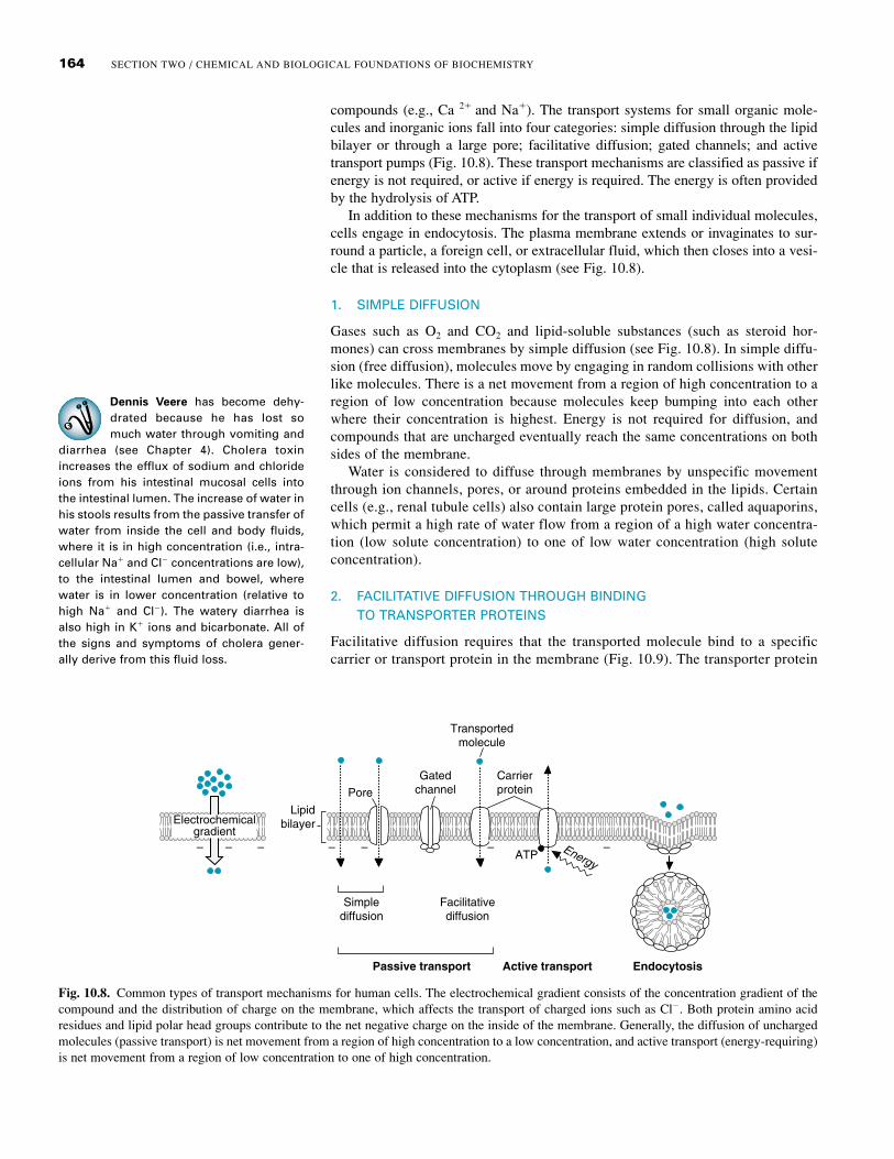
compounds (e.g., Ca 2� and Na�). The transport systems for small organic mole-cules and inorganic ions fall into four categories: simple diffusion through the lipidbilayer or through a large pore; facilitative diffusion; gated channels; and activetransport pumps (Fig. 10.8). These transport mechanisms are classified as passive ifenergy is not required, or active if energy is required. The energy is often providedby the hydrolysis of ATP.
In addition to these mechanisms for the transport of small individual molecules,cells engage in endocytosis. The plasma membrane extends or invaginates to sur-round a particle, a foreign cell, or extracellular fluid, which then closes into a vesi-cle that is released into the cytoplasm (see Fig. 10.8).
1. SIMPLE DIFFUSION
Gases such as O2 and CO2 and lipid-soluble substances (such as steroid hor-mones) can cross membranes by simple diffusion (see Fig. 10.8). In simple diffu-sion (free diffusion), molecules move by engaging in random collisions with otherlike molecules. There is a net movement from a region of high concentration to aregion of low concentration because molecules keep bumping into each otherwhere their concentration is highest. Energy is not required for diffusion, andcompounds that are uncharged eventually reach the same concentrations on bothsides of the membrane.
Water is considered to diffuse through membranes by unspecific movementthrough ion channels, pores, or around proteins embedded in the lipids. Certaincells (e.g., renal tubule cells) also contain large protein pores, called aquaporins,which permit a high rate of water flow from a region of a high water concentra-tion (low solute concentration) to one of low water concentration (high soluteconcentration).
2. FACILITATIVE DIFFUSION THROUGH BINDING TO TRANSPORTER PROTEINS
Facilitative diffusion requires that the transported molecule bind to a specificcarrier or transport protein in the membrane (Fig. 10.9). The transporter protein
Carrierprotein
Transportedmolecule
Simplediffusion
Facilitativediffusion
ATP
Passive transport Active transport Endocytosis
GatedchannelPore
Energy
Lipidbilayer
– – – – – ––
Electrochemicalgradient
Fig. 10.8. Common types of transport mechanisms for human cells. The electrochemical gradient consists of the concentration gradient of thecompound and the distribution of charge on the membrane, which affects the transport of charged ions such as Cl�. Both protein amino acidresidues and lipid polar head groups contribute to the net negative charge on the inside of the membrane. Generally, the diffusion of unchargedmolecules (passive transport) is net movement from a region of high concentration to a low concentration, and active transport (energy-requiring)is net movement from a region of low concentration to one of high concentration.

165CHAPTER 10 / RELATIONSHIP BETWEEN CELL BIOLOGY AND BIOCHEMISTRY
All of the cells in the body havefacilitative glucose transportersthat transport glucose across the
plasma membrane down an electrochemical(concentration) gradient as it is rapidlymetabolized in the cell. In muscle and adi-pose tissue, insulin increases the content offacilitative glucose transporters in the cellmembrane, thus increasing the ability ofthese tissues to take up glucose. Patientswith type 1 diabetes mellitus, who do notproduce insulin (e.g., Di Abietes, see Chapter7), have a decreased ability to transport glu-cose into these tissues, thereby contributingto hyperglycemia (high blood glucose).
then undergoes a conformational change that allows the transported molecule tobe released on the other side of the membrane. Although the transported mole-cules are bound to proteins, the transport process is still classified as diffusionbecause energy is not required, and the compound equilibrates (achieves a bal-ance of concentration and charge) on both sides of the membrane.
Transporter proteins, like enzymes, exhibit saturation kinetics; when all thebinding sites on all of the transporter proteins in the membrane are occupied,the system is saturated and the rate of transport reaches a plateau (the maxi-mum velocity). By analogy to enzymes, the concentration of a transportedcompound required to reach 1⁄2 the maximum velocity is often called the Km
(Fig. 10.10). Facilitative transporters are similar to enzymes with respect totwo additional features: they are relatively specific for the compounds theybind and they can be inhibited by compounds that block their binding sites orchange their conformation.
Highconcentration
Lowconcentration
1 2 3 4
Fig. 10.9. Facilitative transport. Although the molecule being transported must bind to theprotein transporter, the mechanism is passive diffusion, and the molecule moves from aregion of high concentration to one of low concentration. “Passive” refers to the lack of anenergy requirement for the transport.
Fig. 10.10. Saturation kinetics of transporter proteins. When a compound must bind to aprotein to be transported across a membrane, the velocity of transport depends on theamount of compound bound. It reaches a maximum rate when the compound’s concentra-tion is raised so high that all of the transporter binding sites are occupied. The curve is a rec-tangular hyperbola that approaches Vmax at infinite substrate concentration, identical to thatof Michaelis-Menten enzymes. The Km of transport is the concentration of compoundrequired for 1⁄2 Vmax. In contrast, simple diffusion of a compound does not require its bind-ing to a protein, and the rate of transport increases linearly with increasing concentration ofthe compound.
Carrier-mediateddiffusion
Simplediffusion
Concentration of transported molecule
Rat
e of
tran
spor
t
Vmax
Km
Vmax1
2

166 SECTION TWO / CHEMICAL AND BIOLOGICAL FOUNDATIONS OF BIOCHEMISTRY
The cystic fibrosis transmembraneconductance regulator (CFTR) wasnamed for its role in cystic fibrosis.
A mutation in the gene encoding its trans-membrane subunits results in dried mucusaccumulation in the airways and pancreaticducts.
The CFTR is also involved in the dehydra-tion experienced by cholera patients such asDennis Veere. In intestinal mucosal cells,cholera A toxin indirectly activates phospho-rylation of the regulatory domain of CFTR byprotein kinase A. Thus, the channel stays openand Cl� and H2O flow from the cell into theintestinal lumen, resulting in dehydration.
Protein-mediated transport sys-tems, whether facilitative or active,are classified as antiports if they
specifically exchange compounds of similarcharge across a membrane; they are calledsymports or cotransporters if they simultane-ously transport two molecules across themembrane in the same direction. Band 3 inthe red blood cell membrane, whichexchanges chloride ion for bicarbonate, pro-vides an example of an antiport.
3. GATED CHANNELS IN PLASMA MEMBRANES
In gated channels, transmembrane proteins form a pore for ions that is either openedor closed in response to a stimulus: voltage changes across the membrane (voltage-gated channels), the binding of a compound (ligand-gated channels), or a regulatorychange in the intracellular domain (phosphorylation-gated and pressure-gated chan-nels). For example, the conduction of a nerve impulse along the axon depends onthe passive flux of Na� ions through a voltage-gated channel that is opened bydepolarization of the membrane. CFTR (cystic fibrosis transmembrane conduc-tance regulator) is a Cl� channel that provides an example of a ligand-gated chan-nel regulated through phosphorylation (phosphorylation-gated) (Fig. 10.11). CFTRis a member of the ABC (adenine nucleotide binding cassette, or ATP binding cas-sette) superfamily of transport proteins. It has two transmembrane domains thatform a closed channel, each connected to an ATP binding site, and a regulatorydomain that sits in front of the channel. When the regulatory domain is phosphory-lated by a kinase, its conformation changes and it moves away from the ATP bind-ing domains. As ATP binds and is hydrolyzed, the transmembrane domains changeconformation and open the channel, and chloride ions diffuse through. As the con-formation reverts back to its original form, the channel closes.
Transport through a ligand-gated channel is considered diffusion, although ATPis involved, because only a few ATP molecules are being used to open and close thechannel through which many, many chloride ions diffuse. However, the distinctionbetween ligand-gated channels and facilitative transporters is not always as clear.Many gated channels show saturation kinetics at very high concentrations of thecompounds being transported.
4. ACTIVE TRANSPORT REQUIRES ENERGY AND TRANSPORTER PROTEINS
Both active transport and facilitative transport are mediated by protein transporters(carriers) in the membrane. However, in facilitative transport, the compound istransported down an electrochemical gradient (the balance of concentration andcharge across a membrane), usually from a high concentration to a low concentra-tion, to equilibrate between the two sides of the membrane. In active transport,energy is used to concentrate the compound on one side of the membrane. If energyis directly applied to the transporter (e.g., ATP hydrolysis by Na�,K�-ATPase), thetransport is called primary active transport; if energy is used to establish an ion gra-dient (e.g., the Na� gradient), and the gradient is used to concentrate another com-pound, the transport is called secondary active transport.
The Na�,K�-ATPase spans the plasma membrane, much like a gated pore, witha binding site for 3 Na� ions open to the intracellular side (Fig. 10.12). Energy from
Membrane
Out
In
PKA
2 ADPCl–
+ 2 Pi
R ABDABD ATPATP
PPP
R
P
R
PPPP
1 2
Fig. 10.11. CFTR, a ligand-gated channel controlled by phosphorylation. Two intracellular binding domains control opening ofthe channel, an adenine nucleotide binding domain (ABD) and a regulatory domain (R). 1 Phosphorylation of the regulatorysubunit by protein kinase A causes a conformational change that allows ATP to bind to the adenine nucleotide binding domain(ABD). 2 Hydrolysis of bound ATP opens the channel so that chloride ions can diffuse through.

167CHAPTER 10 / RELATIONSHIP BETWEEN CELL BIOLOGY AND BIOCHEMISTRY
The Ca2�-ATPase, a calcium pump,uses a mechanism similar to thatof Na�,K�-ATPase to maintainintracellular Ca2� concentration
below 10�7 M in spite of the high extracellu-lar concentration of 10-3 M. This transporteris inhibited by binding of the regulatory pro-tein calmodulin. When the intracellular Ca2�
concentration increases, Ca2� binds tocalmodulin, which dissociates from thetransporter, thereby activating it to pumpCa2� out of the cell (see Chapter 9 for thestructure of calmodulin). High levels of intra-cellular Ca2� are associated with irreversibleprogression from cell injury to cell death.
ATP hydrolysis is used to phosphorylate an internal domain and change the trans-porters’ conformation so that bound Na� ions are released to the outside, and twoexternal K� ions bind. K� binding triggers hydrolysis of the bound phosphate groupand a return to the original conformation, accompanied by release of K� ions insidethe cell. As a consequence, cells are able to maintain a much lower intracellular Na�
concentration and much higher intracellular K� ion concentration than present inthe external fluid.
The Na� gradient, which is maintained by primary active transport, is used topower the transport of glucose, amino acids, and many other compounds into the cellthrough secondary active transport. An example is provided by the transport of glu-cose into cells of the intestinal epithelium in conjunction with Na� ions (Fig. 10.13).
Fig. 10.12. Active transport by Na�,K�-ATPase. Three sodium ions bind to the transporter protein on the cytoplasmic side of the membrane.When ATP is hydrolyzed to ADP, the carrier protein is phosphorylated and undergoes a change in conformation that causes the sodium ions tobe released into the extracellular fluid. Two potassium ions then bind on the extracellular side. Dephosphorylation of the carrier protein producesanother conformational change, and the potassium ions are released on the inside of the cell membrane. The transporter protein then resumes itsoriginal conformation, ready to bind more sodium ions.
Extracellularfluid
Cytoplasm3 Na+ 2 K+
3 Na+ 2 K+
ATP ADP Pi
P P P
Na+ Na+
Na+
K+
ATP
ADP
K+
Glucose GlucoseGlucose
Lumen Extracellular fluid
Transport protein
Fig. 10.13. Secondary active transport of glucose by the Na�-glucose cotransporter. Onesodium ion binds to the carrier protein in the luminal membrane, stimulating the binding ofglucose. After a conformational change, the protein releases Na� and glucose into the celland returns to its original conformation. Na�,K�-ATPase in the basolateral membrane pumpsNa� against its concentration gradient into the extracellular fluid. Thus, the Na� concentra-tion in the cell is low, and Na� moves from the lumen down its concentration gradient intothe cell and is pumped against its gradient into the extracellular fluid. Glucose, consequently,moves against its concentration gradient from the lumen into the cell by traveling on the samecarrier as Na�. Glucose then passes down its concentration gradient into the extracellularfluid on a passive transporter protein.

168 SECTION TWO / CHEMICAL AND BIOLOGICAL FOUNDATIONS OF BIOCHEMISTRY
The dehydration of cholera is oftentreated with an oral rehydrationsolution containing Na�, K�, and
glucose or a diet of rice (which contains glu-cose and amino acids). Glucose is absorbedfrom the intestinal lumen via the Na�-dependent glucose cotransporters, whichcotransport Na� into the cells together withglucose. Many amino acids are alsoabsorbed by Na�-dependent cotransport.With the return of Na� to the cytoplasm,water efflux from the cell into the intestinallumen decreases.
The vitamin folate provides anexample of a compound trans-ported into cells by caveolae,
which form around the occupied folatereceptor. In contrast, endocytosis of manycompounds such as membrane hormonereceptors occurs through clathrin-coatedpits. The receptors are targeted for these pitsby adaptor proteins that bind to a specificamino acid sequence in the receptor.
These cells create a gradient in Na� and then use this gradient to drive the transportof glucose from the intestinal lumen into the cell against its concentration gradient.
D. Vesicular Transport across the Plasma Membrane
Vesicular transport occurs when a membrane completely surrounds a compound,particle, or cell and encloses it into a vesicle. When the vesicle fuses with anothermembrane system, the entrapped compounds are released. Endocytosis refers tovesicular transport into the cell, and exocytosis to transport out of the cell. Endocy-tosis is further classified as phagocytosis if the vesicle forms around particulatematter (such as whole bacterial cells or metals and dyes from a tattoo), and pinocy-tosis if the vesicle forms around fluid containing dispersed molecules. Receptor-mediated endocytosis is the name given to the formation of clathrin-coated vesiclesthat mediate the internalization of membrane-bound receptors in vesicles coated onthe intracellular side with subunits of the protein clathrin (Fig. 10.14). Potocytosisis the name given to endocytosis that occurs via caveolae (small invaginations or“caves”), which are regions of the cell membrane with a unique lipid and proteincomposition (including the protein caveolin-1).
III. LYSOSOMES
Lysosomes are the intracellular organelles of digestion enclosed by a singlemembrane that prevents the release of its digestive enzymes into the cytosol.They are central to a wide variety of body functions that involve elimination ofunwanted material and recycling their components, including destruction of
Ligand
PitClathrin
GTPDynamin
Adaptors
Receptors
GTPhydrolysis
Clathrincoatedvesicle
Fig. 10.14. Formation of a clathrin-coated vesicle. Ligands entering the cell through recep-tor-mediated endocytosis bind to receptors that cluster in an area of the membrane. Adaptorproteins bind to the receptor tails and to the clathrin molecules to enclose the budding mem-brane in a cage-like clathrin coat. Molecules of a monomeric G protein called dynamin (from the Rab family) constrict theneck of the vesicle and pinch it off from the membrane as GTP is hydrolyzed.
169CHAPTER 10 / RELATIONSHIP BETWEEN CELL BIOLOGY AND BIOCHEMISTRY
Lysosomal storage diseases.Genetic defects in lysosomalenzymes, or in proteins such as the
mannose 6-phosphate receptors required fortargeting the enzymes to the lysosome, leadto an abnormal accumulation of undigestedmaterial in lysosomes that may be con-verted to residual bodies. The accumulationmay be so extensive that normal cellularfunction is compromised, particularly inneuronal cells. Genetic diseases such as theTay-Sachs disease (an accumulation of par-tially digested gangliosides in lysosomes),and Pompe’s disease (an accumulation ofglycogen particles in lysosomes) are causedby the absence or deficiency of specific lyso-somal enzymes.
infectious bacteria and yeast, recovery from injury, tissue remodeling, involution oftissues during development, and normal turnover of cells and organelles.
A. Lysosomal Hydrolases
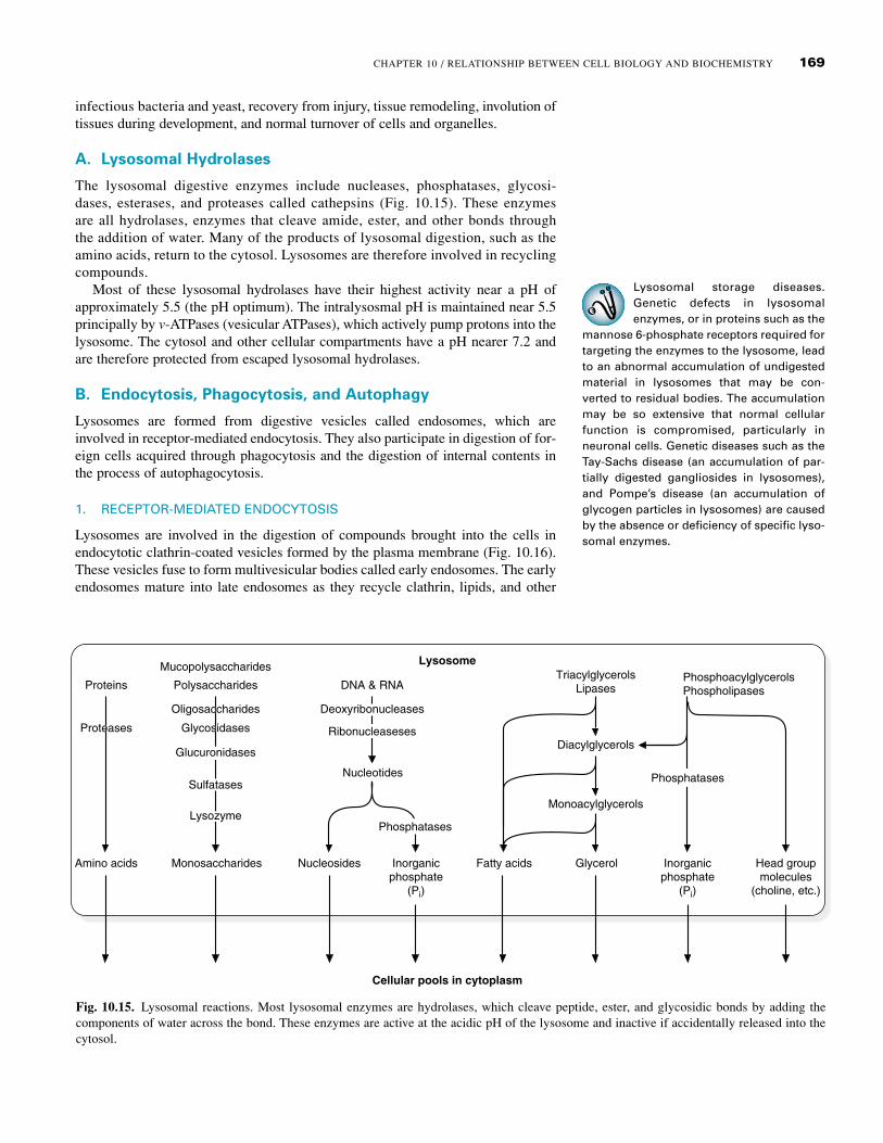
The lysosomal digestive enzymes include nucleases, phosphatases, glycosi-dases, esterases, and proteases called cathepsins (Fig. 10.15). These enzymesare all hydrolases, enzymes that cleave amide, ester, and other bonds throughthe addition of water. Many of the products of lysosomal digestion, such as theamino acids, return to the cytosol. Lysosomes are therefore involved in recyclingcompounds.
Most of these lysosomal hydrolases have their highest activity near a pH ofapproximately 5.5 (the pH optimum). The intralysosmal pH is maintained near 5.5principally by v-ATPases (vesicular ATPases), which actively pump protons into thelysosome. The cytosol and other cellular compartments have a pH nearer 7.2 andare therefore protected from escaped lysosomal hydrolases.
B. Endocytosis, Phagocytosis, and Autophagy
Lysosomes are formed from digestive vesicles called endosomes, which areinvolved in receptor-mediated endocytosis. They also participate in digestion of for-eign cells acquired through phagocytosis and the digestion of internal contents inthe process of autophagocytosis.
1. RECEPTOR-MEDIATED ENDOCYTOSIS
Lysosomes are involved in the digestion of compounds brought into the cells inendocytotic clathrin-coated vesicles formed by the plasma membrane (Fig. 10.16).These vesicles fuse to form multivesicular bodies called early endosomes. The earlyendosomes mature into late endosomes as they recycle clathrin, lipids, and other
Fig. 10.15. Lysosomal reactions. Most lysosomal enzymes are hydrolases, which cleave peptide, ester, and glycosidic bonds by adding thecomponents of water across the bond. These enzymes are active at the acidic pH of the lysosome and inactive if accidentally released into thecytosol.
Proteins Polysaccharides
Mucopolysaccharides
DNA & RNA
Lysosome
Cellular pools in cytoplasm
TriacylglycerolsLipases
Diacylglycerols
Monoacylglycerols
PhosphoacylglycerolsPhospholipases
Amino acids Monosaccharides Nucleosides
Nucleotides
Inorganicphosphate
(Pi)
Fatty acids Glycerol Inorganicphosphate
(Pi)
Head groupmolecules
(choline, etc.)
OligosaccharidesOligosaccharides
GlycosidasesGlycosidases
GlucuronidasesGlucuronidases
SulfatasesSulfatases
LysozymeLysozyme
DeoxyribonucleasesDeoxyribonucleases
RibonucleasesesRibonucleaseses
PhosphatasesPhosphatases
PhosphatasesPhosphatases
ProteasesProteases

170 SECTION TWO / CHEMICAL AND BIOLOGICAL FOUNDATIONS OF BIOCHEMISTRY
The elevated level of uric acid inLotta Topaigne’s blood led to thedeposition of monosodium urate
crystals in the joint space (synovial fluid) ofher right great toe, resulting in podagra(painful great toe). Neutrophils, the media-tors of the acute inflammation that followed,attempted to phagocytose the urate crystals.The engulfed urate crystals were depositedin the late endosomes and lysosomes of theneutrophil. Because urate crystals are parti-cles that cannot be degraded by any of thelysosomal acid hydolases, their accumula-tion caused lysis of the lysosomal mem-branes, followed by cell lysis and release oflysosomal enzymes into the joint space. Theurate crystals also resulted in release ofchemical mediators of inflammation thatrecruited other cells into the area. This fur-ther amplified the acute inflammatory reac-tion in the tissues of the joint caspsule (syn-ovitis), leading to the extremely painfulswelling of acute gouty arthritis.
Phagocytosis and autophagy arepart of the normal turnover of bodycomponents, such as degradation
of cells that have a shorter lifespan than thewhole organism and remodeling of tissuesduring pregnancy. For example, phagocytes,located mainly in the spleen and liver,remove approximately 3 � 1011 red blood cellsfrom the circulation each day. During preg-nancy, breast tissue is remodeled to developthe capacity for lactation; after weaning of aninfant, the lactating breast returns to its orig-inal state (involution).
membrane components back to the plasma membrane in vesicles called recyclingendosomes. The late endosomes mature into lysosomes as they progressively accu-mulate newly synthesized acid hydrolases and vesicular proton pumps brought tothem in clathrin-coated vesicles from the Golgi. Thus, lysosomes do not acquiretheir full digestive power until after sorting of membrane lipids and proteins forrecycling.
Within the Golgi, enzymes are targeted for endosomes (and eventually lyso-somes) by addition of mannose 6-phosphate residues that bind to mannose 6-phosphate receptor proteins in the Golgi membrane. The mannose 6-phosphatereceptors together with their bound acid hydrolases are incorporated into theclathrin-coated Golgi transport vesicles and released. The transport vesicles losetheir clathrin coat and then fuse with the late endosomal membrane. The acidity ofthe endosome releases the acid hydrolases from the receptors into the vesicle lumen.The receptors are eventually recycled back to the Golgi.
2. PHAGOCYTOSIS AND AUTOPHAGY
One of the major roles of lysosomes is phagocytosis (Fig. 10.17). Neutrophils andmacrophages, the major phagocytic cells, devour pathogenic microorganisms andclean up wound debris and dead cells, thus aiding in repair. As bacteria or other par-ticles are enclosed into clathrin-coated pits in the plasma membrane, these vesiclesbud off to form intracellular phagosomes. The phagosomes fuse with lysosomes,where the acidity and digestive enzymes destroy the contents. Pinocytotic vesciclesalso may fuse with lysosomes.
In autophagy (self-eating), intracellular components such as organelles or glyco-gen particles are surrounded by a membrane derived from ER vesicles, forming anautophagosome. The autophagosome fuses with a lysosome, and the contents of thephagolysosome are digested by lysosomal enzymes. Organelles usually turn overmuch more rapidly than the cells in which they reside (e.g., approximately fourmitochondria in each liver cell are degraded per hour). Cells that are damaged butstill viable recover, in part, by using autophagy to eliminate damaged components.
Golgi
Endocytoticvesicle
Early endosome
Late endosome
Recycling endosome
Lysosome
Receptor
pH<5.5
1
2
3 4
Fig. 10.16. Lysosomes in receptor-mediated endocytosis via clathrin-coated pits. 1 Endo-cytotic vesicles fuse to form early endosomes. 2 Vesicle contents are sorted, and receptors,clathrin, and lipids are sent back to the plasma membrane. 3 Transport vesicles from thetrans-Golgi carry lysosomal hydrolases to the late endosome. 4 Lysosomes containing con-centrated hydrolases digest proteins and other components acquired from endocytoticvesicles.

171CHAPTER 10 / RELATIONSHIP BETWEEN CELL BIOLOGY AND BIOCHEMISTRY
Fig. 10.18. Mitochondrion. Electron micro-graph (top); three-dimensional drawing (bot-tom).
Mitochondial diseases. Mitochon-dria contain DNA and can repro-duce by replicating their DNA and
then dividing in half. Although nuclear DNAencodes most of the enzymes found in mito-chondria, mitochondrial DNA encodes someof the subunits of the electron transportchain proteins and ATP synthase. Mutationsin mitochondrial DNA result in a number ofgenetic diseases that affect skeletal muscle,neuronal, and renal tissues. They are impli-cated in aging.
If a significant amount of undigestible material remains within the lysosomeafter the digestion process is completed, the lysosome is called a residual body.Depending on the cell type, residual bodies may be expelled (exocytosis) or remainindefinitely in the cell as lipofuscin granules that accumulate with age.
IV. MITOCHONDRIA
Mitochondria contain most of the enzymes for the pathways of fuel oxidationand oxidative phosphorylation and thus generate most of the ATP required bymammalian cells. Each mitochondrion is surrounded by two membranes, anouter membrane and an inner membrane, separating the mitochondrial matrixfrom the cytosol (Fig. 10.18). The inner membrane forms invaginations knownas cristae containing the electron transport chain and ATP synthase. Most of theenzymes for the TCA cycle and other pathways for oxidation are located in themitochondrial matrix, the compartment enclosed by the inner mitochondrialmembrane. (The TCA cycle and electron transport chain are described in moredetail in Chapters 20 and 21.)
The inner mitochondrial membrane is highly impermeable, and the proton gra-dient that is built up across this membrane during oxidative phosphorylation isessential for ATP generation from ADP and phosphate. The transport of ions occursprincipally through facilitative transporters in a type of secondary active transportpowered by the proton gradient established by the electron transport chain. Theouter membrane contains pores made from proteins called porins and is permeableto molecules with a molecular weight up to about 1000 g/mole.
Mitochondria can replicate by division; however, most of their proteins mustbe imported from the cytosol. Mitochondria contain a small amount of DNA,which encodes for only 13 different subunits of proteins involved in oxidativephosphorylation. Most of the enzymes and proteins in mitochondria are encodedby nuclear DNA and synthesized on cytoplasmic ribosomes. They are imported
Fig. 10.17. Phagocytosis and autophagy. Cells and large particles are phagocytosed. Thephagosomes fuse with lysosomes to form phagolysosomes. Recyclable amino acids (AA),fatty acids (FA), and carbohydrates (CHO) are released into the cytosol. Autophagosomes areformed in the ER as the cell digests mitochondria and its own large particles. These alsomerge with lysosomes. Undigested material may remain in the lysosomes to form residualbodies, which are either extruded or remain in the cell as lipofuscin granules.
Residual body
PHAGOCYTOSIS
AUTOPHAGOCYTOSIS
EXOCYTOSIS
Phagososome
Phagolysosome
Plasma membrane
Lysosomes
AA, FA,CHO
AA, FA,CHO
Lipofuscingranule
Autophagosome Autophagolysosme
Mitochondrion
RER
Inner membranefolded into cristae
Inner membranefolded into cristae
1 µm
Outermembrane
Outermembrane
Matrix
Matrix

172 SECTION TWO / CHEMICAL AND BIOLOGICAL FOUNDATIONS OF BIOCHEMISTRY
Peroxisomal Diseases. Peroxisomaldiseases are caused by mutationsaffecting either the synthesis of
functional peroxisomal enzymes or theirincorporation into peroxisomes. For example,adrenoleukodystrophy probably involves amutation that decreases the content of atransporter in the peroxisomal membrane.Zellweger’s syndrome is caused by the failureto complete the synthesis of peroxisomes.
Fig. 10.20. Nucleus. Electron micrograph (top); three-dimensional drawing (bottom).
Fig. 10.19. Types of reactions in peroxisomes.
through membrane pores by a receptor-mediated process involving members ofthe heat shock family of proteins.
V. PEROXISOMES
Peroxisomes are cytoplasmic organelles, similar in size to lysosomes, that areinvolved in oxidative reactions using molecular oxygen (Fig. 10.19). These reac-tions produce the toxic chemical hydrogen peroxide (H2O2), which is subsequentlyused or degraded within the peroxisome by catalase and other enzymes. Peroxi-somes function in the oxidation of very long chain fatty acids (containing 20 ormore carbons) to shorter chain fatty acids, the conversion of cholesterol to bileacids, and the synthesis of ether lipids called plasmalogens. They are bounded by asingle membrane.
Like mitochondria, peroxisomes can replicate by division. However, they aredependent on the import of proteins to function. They contain no DNA.
VI. NUCLEUS
The largest of the subcellular organelles of animal cells is the nucleus (Fig. 10.20).Most of the genetic material of the cell is located in the chromosomes of thenucleus, which are composed of DNA, an equal weight of small, positively chargedproteins called histones, and a variable amount of other proteins. This nucleoprotein
O2
catalase
H2O2
Compound
Oxidizedcompound
Other oxidativereactions
O2 H2O
Euchromatin
Chromatin(DNA + histonesand other proteins)
Outermembrane(continuouswith RER)
3–10 µm
Nucleolus
Nuclearenvelope
Heterochromatin
Pores
Nucleolus
Nuclearenvelope
Pores

173CHAPTER 10 / RELATIONSHIP BETWEEN CELL BIOLOGY AND BIOCHEMISTRY
The Ras family of monomeric G
proteins. Ras and Ran belong to asuperfamily of proteins called
small G proteins (also called small GTP-binding proteins, small GTPases, ormonomeric G proteins: see Chapter 9, sec-tion III.C.2). These proteins function as tim-ing regulators for a variety of cell functions.They are referred to as “small” because theyare composed of a single subunit with aweight of 20 to 40 kDa, and they are calledGTPases because they slowly hydrolyzebound GTP. When small G proteins containbound GTP, they bind to and activate theirtarget proteins. As their bound GTP ishydrolyzed to GDP and Pi, their conforma-tion changes dramatically, and they dissoci-ate from the target protein. They thus serveas “automatic clocks” that shut themselvesoff. Many of the monomeric GTP bindingproteins are regulated by GAPs (GTPaseactivating proteins), GEFs (GTP exchangeproteins which stimulate GDP dissociationand GTP binding), or GDIs (GDP-dissociationinhibitory proteins). (See Chapter 9 for amore complete discussion of these regula-tors). The function of each of the five majorclasses of Ras monomeric G proteins in cellbiology is summarized in Table 10.1.
complex is called chromatin. The nucleolus, a substructure of the nucleus, is the siteof rRNA transcription and processing, and of ribosome assembly. Replication, tran-scription, translation, and the regulation of these processes are the major focus ofthe molecular biology section of this text (see Section Three).
The nucleus is separated from the rest of the cell (the cytoplasm) by the nuclearenvelope, which consists of two membranes joined at nuclear pores. The outernuclear membrane is continuous with the rough endoplasmic reticulum. To convertthe genetic code of the DNA into the primary sequence of a protein, DNA is tran-scribed into RNA, which is modified and edited into mRNA. The mRNA travelsthrough the nuclear pores into the cytoplasm, where it is translated into the primarysequence of a protein on ribosomes (Fig. 10.21). Ribosomes, which are generatedin the nucleolus, also must travel through nuclear pores to the cytoplasm. Conversely,proteins required for replication, transcription, and other processes pass into thenucleus through these pores. Thus, transport through the pore is specific for themolecule and the direction of transport.
Specificity and direction of travel through the nuclear pore (import vs. export) isdictated by binding proteins (importins vs. exportins), by a small GTP proteincalled Ran, and by the location of the regulatory protein, RanGAP (GTPase acti-vating protein) only on the cytoplasmic side (Fig. 10.22). Proteins transported intothe nucleus have a nuclear localization signal that causes them to bind to one of thesubunits of cytosolic proteins called importins. The other subunit of the importinmolecule binds to cytoplasmic filaments attached to the outer ring of the nuclearpore. As the importin-protein complex enters the nucleus, the small GTP-bindingprotein Ran binds to an importin subunit, causing release of the transported proteininto the nucleus. The Ran-importin complex is returned to the cytosol, where Ran-GAP (GTPase activating protein) activates hydrolysis of bound GTP to GDP andphosphate. The energy released by GTP hydrolysis changes the conformation ofRan and the complex dissociates. The free importin can then bind another protein.
Table 10.1 Monomeric G Proteins in the Ras Superfamily
G-Protein Some Family Location and MembraneFamily Function Members Attachment Site
Ras Regulator of gene H-Ras, K-Ras, N-Ras, Anchored to plasma expression and cell Ral A, Rad, Rap, Rit, membrane by farnesyl, growth, found in palmitoyl, or other lipid mutated oncogenic groups forms in many human tumors
Rho Controls organization of Rho (A-E), Cdc42, Anchored to plasma actin cytoskeleton and Rac (1-3) membrane by lipids, and gene expression (F-actin translocates to cytosolbundling, myosin filament assembly)
Arf/Sar Assembly of coatomer- Arf (1-6), Sar 1a,1b; Arf is anchored to coated vesicles (COPI Arl (1-7) vesicular membranes byand COPII) for vesicular myristyl groups, but Sar istrafficking pathways anchored by the proteinoriginating in the Golgi itself.
Rab Targeting of vesicles Dynamin, Rab Anchored to lipid involved in secretory (11-33) membranes with and endocytotic geranylgeranyl (C20 pathways and formation isoprenoid) groups and of v-SNARE–t-SNARE other lipidscomplexes
Ran Transport through nuclear Ran Not anchored to lipid pore complexes. membrane.
Found in cytosol andnucleus

174 SECTION TWO / CHEMICAL AND BIOLOGICAL FOUNDATIONS OF BIOCHEMISTRY
Protein
Nuclear pore
Nucleus
Cytoplasm
Importin
Importin
Pi
Ran
Ran GAP
Ran
GTP
Ran
GTP
GDP
Ran
GDP
Fig. 10.22. Nuclear import. Proteins with the nuclear localization signal bind to importins,which carry them through the nuclear pore into the nucleus. The monomeric G protein Rancontaining bound GTP binds to one of the subunits of importin. This causes dissociation ofthe importin subunits and release of the imported protein in the nucleus. The Ran-importincomplex exits a nuclear pore. On the cytoplasmic side, a RanGAP (GTPase activating pro-tein) activates the hydrolysis of GTP to GDP, which causes dissociation of the complex.RanGDP is subsequently returned to the nucleus, where an accessory protein activates dis-sociation of GDP and association of GTP.
Fig. 10.21. The nuclear pore complex. The approximately 100 different polypeptide chainsof the nuclear pore complex form an assembly of 8 spokes attached to two ring structures(a cytoplasmic ring in the outer nuclear membrane and a nuclear ring through the innermembrane) with a transporter “plug” in the center. Small molecules, ions, and proteins withless than a 50-kDa mass passively diffuse through the pore in either direction. However,RNAs and most proteins are too large to diffuse through, and are actively transported in aprocess that requires energy, is selective for the molecule transported, is unidirectional, andcan be regulated.
Transporter
Spokes
Cytoplasmicring
Cytoplasmicparticles
Nuclearring
Nuclearlamina
Cytoplasmicfilaments

175CHAPTER 10 / RELATIONSHIP BETWEEN CELL BIOLOGY AND BIOCHEMISTRY
Chronic ingestion of ethanol hasincreased the content of MEOS, themicrosomal ethanol oxidizing sys-
tem, in Al Martini’s liver. MEOS is acytochrome P450 enzyme that catalyzes theconversion of ethanol, NADPH and O2 toacetaldehyde, NADP�, and 2 H2O (see Chap-ter 9). The adjective microsomal is a termderived from experimental cell biology thatis sometimes used for processes occurringin the ER. When cells are lysed in the labora-tory, the ER is fragmented into vesiclescalled microsomes, which can be isolated bycentrifugation. Microsomes, as such, are notactually present in cells.
RNAs are transported from the nucleus to the cytoplasm as ribonucleoproteins,which are targeted for export by a specific amino acid sequence called the nuclearexport signal. The nucleoprotein forms a complex with additional proteins calledexportins and with Ran. This complex is transported through the pore to the cyto-plasm, where RanGAP activates hydrolysis of the bound GTP. In the absence ofGTP, the complex dissociates with the release of RNA into the cytoplasm, and theexportins and Ran are transported back to the nucleus.
VII.ENDOPLASMIC RETICULUM
The endoplasmic reticulum (ER) is a network of membranous tubules within the cellconsisting of smooth endoplasmic reticulum (SER), which lacks ribosomes, andrough endoplasmic reticulum (RER), which is studded with ribosomes (Fig. 10.23).The SER has a number of functions. It contains enzymes for the synthesis of manylipids, such as triacylglycerols and phospholipids. It also contains the cytochromeP450 oxidative enzymes involved in metabolism of drugs and toxic chemicals suchas ethanol and the synthesis of hydrophobic molecules such as steroid hormones.Glycogen is stored in regions of liver cells that are rich in SER.
The RER is involved in the synthesis of certain proteins. Ribosomes attached tothe membranes of the RER give them their “rough” appearance. Proteins producedon these ribosomes enter the lumen of the RER, travel to the Golgi complex in vesi-cles, and are subsequently either secreted from the cell, sequestered withinmembrane-enclosed organelles such as lysosomes, or embedded in the plasmamembrane. Posttranslational modifications of these proteins, such as the initiationof N-linked glycosylation and the addition of GPI anchors, occur in the RER. Incontrast, proteins encoded by the nucleus and found in the cytosol, peroxisomes, ormitochondria are synthesized on free ribosomes in the cytosol and are seldom mod-ified by the attachment of oligosaccharides.
Fig. 10.23 A. Smooth endoplasmic reticulum. B. Rough endoplasmic reticulum. A and B areelectron micrographs. A three-dimensional drawing is in the middle.
Smooth endoplasmicreticulum (SER)
Rough endoplasmicreticulum (RER)
A
B
Ribosomes

176 SECTION TWO / CHEMICAL AND BIOLOGICAL FOUNDATIONS OF BIOCHEMISTRY
VIII. GOLGI COMPLEX
The Golgi complex is involved in modifying proteins produced in the RER and insorting and distributing these proteins to the lysosomes, secretory vesicles, or theplasma membrane. It consists of a curved stack of flattened vesicles in the cyto-plasm that is generally divided into three compartments: the cis-Golgi network,which is often convex and faces the nucleus; the medial Golgi stacks; and the transGolgi network, which often faces the plasma membrane (Fig. 10.24).
Proteins are transported to and from the Golgi in at least three kinds of vesicles:coatomer-coated COP I vesicles, coatomer-coated COP II vesicles, and clathrin-coated vesicles (see Fig. 10.24). Proteins produced on the RER travel in COP IIvesicles to an endoplasmic reticulum-Golgi intermediate compartment (ERGIC),and then to the cis-Golgi network, where they enter the lumen. Here N-linkedoligosaccharide chains that were added to proteins in the RER are modified, andO-linked oligosaccharides are added. COP I vesicles recycle material from theGolgi back to the ER and possibly transfer material from the Golgi to other sites.
Trans-Golgi
Medial-Golgi
Cis-Golgi
Clathrin
Clathrin
LysosomePlasmamembrane
Microtubules
Late endosome
COPI
COPI
COPIIFusionof COPIIvesicles
ER-golgiintermediatecompartment
Rough ER
?
?
Fig. 10.24. Vesicular transport to and from the Golgi complex. COP II vesicles (coatomer-coated) form in the rough ER and move to the Golgi. COP I vesicles generally go from thetrans to the cis Golgi to the ER. Vesicles that go to late endosomes (eventually lysosomes)from the Golgi or the plasma membrane are clathrin-coated. Less is known about exocytoticvesicles. Vesicle transport, as well as transport of organelles and secretory proteins, occursalong microtubules (structures formed from the protein tubulin).

177CHAPTER 10 / RELATIONSHIP BETWEEN CELL BIOLOGY AND BIOCHEMISTRY
The monomeric G protein Arf wasnamed for its contribution to thepathogenesis of cholera and not
for its normal function in the assembly ofCOP I vesicles. However, it is also requiredfor the transport of V. cholerae A-toxin. Thecholera toxin is endocytosed in caveolaevesicles that subsequently merge with lyso-somes (or are transformed into lysosomes),where the acidic pH contributes to activationof the toxin. As the toxin is transportedthrough the Golgi and ER, it is furtherprocessed and activated. Arf forms a com-plex with the A-toxin that promotes its travelbetween compartments. The A-toxin is actu-ally an ADP-ribosylase (an enzyme thatcleaves NAD and attaches the ADP portionto a protein) (see Chapter 6, Fig. 6.14), andhence, Arf became known as the ADP-ribo-sylating factor. The ADP-ribosylation of pro-teins regulating the CFTR chloride channelleads to Dennis Veere’s dehydration anddiarrhea.
Vesicles released from the trans face of the Golgi complex travel to endosomes asclathrin-coated vesicles.
COP vesicles are coated with a complex composed of coatomer proteins(COP), an Arf family monomeric G protein that mediates vesicle assembly, andother proteins (Fig. 10.25). COP I vesicles contain the monomeric G protein Arf(ADP-ribosylating factor), and COP II vesicles contain the monomeric G proteinSar (another member of the Arf family). In both types of vesicles, hydrolysis ofGTP causes dissociation of the G-protein and disassembly of the vesicle coat. Thevesicle components are then recycled. Glycoproteins or glycolipids once anchoredin the membrane of the vesicle remain in the plasma membrane when the vesicularand plasma membranes fuse.
Vesicles that have lost their coats are ready to fuse with the target membrane.The vesicle membranes contain proteins called v-SNARES (vesicle-SNARES)(see Fig. 10.25). Each type of v-SNARE is able to recognize and bind to its com-plementary t-SNARE (target SNARE) on the target membrane, thus ensuring that
Fig. 10.25. Transport in COP-coated vesicles. A. Assembly and release. Arf with bound GTPassembles a region of the trans-Golgi membrane containing receptors for the protein cargoand coatomers. As GTP is hydrolyzed to GDP, the coat is released. B. Docking. The small Gprotein Rab assists in docking. v-Snares in the vesicle membrane recognize complementaryt-Snares in the target membrane. C. Fusion. NSF and SNAP are fusion proteins.
Target
Coatomer
Coatomer
ARF
ARF
Pi
GDP
GTP
A. Assembly and release
B. DockingC. Fusion
Rab Rab
Vesicle
v-Snare
t-Snare
Rab
NSFSNAPs
Rab
178 SECTION TWO / CHEMICAL AND BIOLOGICAL FOUNDATIONS OF BIOCHEMISTRY
Secretory vesicles. The hormoneinsulin is synthesized as a prohor-mone, proinsulin, which is incorpo-
rated into secretory vesicles. These vesiclescontain a protease that is activated by theacidic pH of the secretory vesicle. It cleavesproinsulin into the A, B, and C chains (seeFig. 6.13).
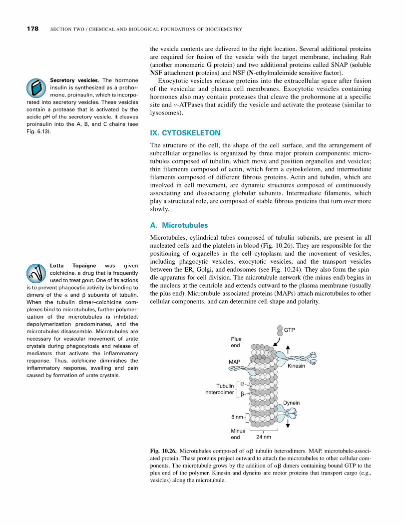
Fig. 10.26. Microtubules composed of �� tubulin heterodimers. MAP, microtubule-associ-ated protein. These proteins project outward to attach the microtubules to other cellular com-ponents. The microtubule grows by the addition of �� dimers containing bound GTP to theplus end of the polymer. Kinesin and dyneins are motor proteins that transport cargo (e.g.,vesicles) along the microtubule.
Lotta Topaigne was givencolchicine, a drug that is frequentlyused to treat gout. One of its actions
is to prevent phagocytic activity by binding todimers of the � and � subunits of tubulin.When the tubulin dimer–colchicine com-plexes bind to microtubules, further polymer-ization of the microtubules is inhibited,depolymerization predominates, and themicrotubules disassemble. Microtubules arenecessary for vesicular movement of uratecrystals during phagocytosis and release ofmediators that activate the inflammatoryresponse. Thus, colchicine diminishes theinflammatory response, swelling and paincaused by formation of urate crystals.
the vesicle contents are delivered to the right location. Several additional proteinsare required for fusion of the vesicle with the target membrane, including Rab(another monomeric G protein) and two additional proteins called SNAP (solubleNSF attachment proteins) and NSF (N-ethylmaleimide sensitive factor).
Exocytotic vesicles release proteins into the extracellular space after fusionof the vesicular and plasma cell membranes. Exocytotic vesicles containinghormones also may contain proteases that cleave the prohormone at a specificsite and v-ATPases that acidify the vesicle and activate the protease (similar tolysosomes).
IX. CYTOSKELETON
The structure of the cell, the shape of the cell surface, and the arrangement ofsubcellular organelles is organized by three major protein components: micro-tubules composed of tubulin, which move and position organelles and vesicles;thin filaments composed of actin, which form a cytoskeleton, and intermediatefilaments composed of different fibrous proteins. Actin and tubulin, which areinvolved in cell movement, are dynamic structures composed of continuouslyassociating and dissociating globular subunits. Intermediate filaments, whichplay a structural role, are composed of stable fibrous proteins that turn over moreslowly.
A. Microtubules
Microtubules, cylindrical tubes composed of tubulin subunits, are present in allnucleated cells and the platelets in blood (Fig. 10.26). They are responsible for thepositioning of organelles in the cell cytoplasm and the movement of vesicles,including phagocytic vesicles, exocytotic vesicles, and the transport vesiclesbetween the ER, Golgi, and endosomes (see Fig. 10.24). They also form the spin-dle apparatus for cell division. The microtubule network (the minus end) begins inthe nucleus at the centriole and extends outward to the plasma membrane (usuallythe plus end). Microtubule-associated proteins (MAPs) attach microtubules to othercellular components, and can determine cell shape and polarity.
Tubulinheterodimer
α
β
8 nm
24 nm
Plusend
Minusend
Kinesin
GTP
MAP
Dynein

179CHAPTER 10 / RELATIONSHIP BETWEEN CELL BIOLOGY AND BIOCHEMISTRY
Colchicine has a narrow therapeu-tic index (i.e., the amount of drugthat produces the desirable thera-
peutic effect is not much lower than theamount that produces an adverse effect). Itstherapeutic effect depends on inhibitingtubulin synthesis in neutrophils, but it canalso prevent tubulin synthesis (and, thus,cell division and other cellular processes) inother cells. Fortunately, neutrophils concen-trate colchicine, so they are affected at lowerintakes than other cell types. Neutrophilslack the transport protein P-glycoprotein, amember of the ABC cassette family (whichincludes the CFTR channel). In most othercell types, P-glycoprotein exports chemicalssuch as colchicine, thus preventing theiraccumulation.
A variety of human cells have ciliaand flagella, hairlike projectionsfrom the surface that have a stroke-
like motion. These projections contain a flex-ible organized array of microtubules. Fluidor mucus is propelled over the surface of cil-iated epithelial cells by the coordinated beat-ing of cilia. A sperm cell swims by means ofa flagellum.
Motor proteins called kinesins and cytoplasmic dyneins use ATP energy to movecargo along the microtubules. Kinesins moves molecules, vesicles, and organellestoward the plus end of microtubules, usually toward the plasma membrane. Cyto-plasmic dyneins are huge proteins that move vesicles and organelles to the minusend, generally toward the nucleus. They are also involved in the positioning of theGolgi complex and the movement of chromosomes during mitosis.
Microtubules consist of polymerized arrays of � and � tubulin dimers that form13 protofilaments organized around a hollow core (see Fig. 10.26). Three differenttubulin polypeptides (�, �, and �) of similar amino acid composition are encodedby related genes; � and � dimers polymerize to form most microtubules, and �-tubuluin is found only in the centrosome. Tubulin dimers composed of one � andone � subunit bind GTP, which creates a conformational change in the dimer thatfavors addition of dimers to the tubulin polymer. The dimers can add to and disso-ciate from both ends of the tubulin, but the end to which they add more rapidly (theplus end) has a net rate of growth, and the end to which they add more slowly (theminus end) has a net rate of loss. As GTP is hydrolyzed to GDP, the binding of tubu-lin subunits is weakened, resulting in their dissociation (dynamic instability). Thus,the net rate and direction of growth is dictated by the fastest growing end of themicrotubule.
B. Actin Filaments
Actin filaments form a network controlling the shape of the cell and movementof the cell surface, thereby allowing cells to move, divide, engulf particles, andcontract. Actin is present in all living cells. The actin polymer, called F-actin, iscomposed of a helical arrangement of globular G-actin subunits (Fig. 10.27).Within the polymer, each G-actin subunit contains a bound ATP or ADP that
Fig. 10.27. Actin filaments. The polymer F-actin is assembled from G-actin subunits con-taining bound ATP. While bound, the ATP is slowly hydrolyzed to ADP. The conformationalchange shifts the equilibrium so that dissociation of the G-actin subunits is favorable at theminus end of the polymer. Once dissociated, the actin subunits exchange ADP for ATP, whichmay again associate with the actin polymer. At the plus end of the molecule, association isfavored over dissociation.
F-actin
Minus end
G-actin subunit
Plus end
Pi
A
A
A
AA
A
A
A
AA
A
A
A
A
A
A
A
A
A

180 SECTION TWO / CHEMICAL AND BIOLOGICAL FOUNDATIONS OF BIOCHEMISTRY
holds the actin fold into a closed conformation (see Chapter 7). The actin poly-mer is dynamic. New subunits of G-actin containing ATP continuously combinewith the assembled F-actin polymer at the plus end. As F-actin elongates, boundATP is hydrolyzed to ADP, so that most of the polymer contains G-actin-ADPsubunits. The conformation of ADP-actin favors dissociation from the minus endof the polymer; thus, the polymer is capable of lengthening from the plus end.This directional growth can account for certain types of cell movement and shapechanges: the formation of pseudopodia that surround other cells during phago-cytosis, the migration of cells in the developing embryo, or the movement ofwhite blood cells through tissues.
Actin polymers form the thin filaments (also called microfilaments) in the cellthat are organized into compact ordered bundles or loose network arrays by cross-linking proteins. Short actin filaments bind to the cross-linking protein spectrin toform the cortical actin skeleton network (see Fig. 10.6). In muscle cells, long actinfilaments combine with thick filaments, composed of the protein myosin, to pro-duce muscle contraction. The assembly of G-actin subunits into polymers, bundlingof fibers, and attachments of actin to spectrin and to the plasma membrane proteinsand organelles, are mediated by a number of actin-binding proteins and G-proteinsfrom the Rho family.
C. Intermediate Filaments
Intermediate filaments (IF) are composed of fibrous protein polymers that providestructural support to membranes of the cells and scaffolding for attachment of othercellular components. Each IF subunit is composed of a long rod-like �-helical corecontaining globular spacing domains, and globular N- and C-terminal domains. The�-helical segments of two subunits coil around each other to form a coiled coil, andthen combine with another dimer coil to form a tetramer. Depending on the type offilament, the dimers may be either hetero- or homo-dimers. The tetramers join end-to end to form protofilaments and approximately eight proto filaments combine toform filaments (Fig. 10.28). Filament assembly is partially controlled through phos-phorylation.
In contrast to actin thin filaments, the 50 or so different types of intermediate fil-aments are each composed of a different protein having the same general structuredescribed above (Table 10.2). Some of the intermediate filaments, such as thenuclear lamins, are common to all cell types. These filaments provide a lattice-likesupport network attached to the inner nuclear membrane. Other intermediate fila-ments are specific for types of cells (e.g., epithelial cells have cytokeratins, and neu-rons have neurofilaments). These provide an internal network that helps to supportthe shape and resilience of the cell.
Polypeptide
Protofilament
Dimer
Filament
Tetramer
N C N
N
C N
C
C
NC
Head
Central rod domain Coiled-coil
Tail
Fig. 10.28. Formation of a cytokeratin filament. The central rod of the keratin monomer is principally �-helical structure. A specific acidic keratin monomer combines with a specific basic keratin monomer to form a heterodimer coil (a coiled coil structure). Two dimerscombine in antiparallel fashion to form a tetramer, and the tetramers combine head-to-tail to form protofilaments. Approximately eight protofil-aments combine to form a filament. The filament is thicker than actin filaments (called thin filaments or microfilaments) and thinner than micro-tubules (thick tubes) and is therefore called an intermediate filament.

181CHAPTER 10 / RELATIONSHIP BETWEEN CELL BIOLOGY AND BIOCHEMISTRY
CLINICAL COMMENTS
Al Martini. Al Martini has been drinking for 5 years and has begun toexhibit mental and systemic effects of chronic alcohol consumption. In hisbrain, ethanol has altered the fluidity of neuronal lipids, causing changes
in their response to neurotransmitters released from exocytotic vesicles. In his liver,increased levels of MEOS (CYP2E1) located in the smooth ER increased his rate ofethanol oxidation to acetaldehyde, a compound that is toxic to the cell. His liver alsocontinues to oxidize ethanol to acetaldehyde through a cytosolic enzyme, liver alco-hol dehydrogenase.
One of the toxic effects of acetaldehyde is inhibition of tubulin polymerization.Tubulin is used in the liver for secretion of very low-density lipoprotein (VLDL)particles containing newly synthesized triacylglycerols. As a result, these triacyl-glycerols accumulate in the liver, and he has begun to develop a fatty liver.Acetaldehyde may also damage protein components of the inner mitochondrialmembrane and affect its ability to pump protons to the cytosol.
Lotta Topaigne had a rapid and gratifying clinical response to thehourly administration of colchicine. This drug diminishes phagocytosisand the subsequent release of the lysosomal enzymes that initiate the
inflammatory response in synovial tissue.The inflammatory response that causes the symptoms of an acute gout attack
begins when neutrophils and macrophages ingest urate crystals. In neutrophils,urate activates the conversion of the polyunsaturated fatty acid arachidonic acid(present in membrane phospholipids) to leukotriene B4. The release of thisprostaglandin contributes to the pain. Colchicine, through its effect on tubulin,inhibits phagocytosis, leukotriene B4 release, and recruitment and cell division ofadditional cells involved in inflammation. Colchicine also inhibits the tubulin-dependent release of histamine from mast cells. As a result, there was a rapidimprovement in the pain and swelling in Lotta’s great toe.
After the gout attack subsided, Ms. Topaigne was placed on allopurinol, a drugthat inhibits urate production (see Chapter 8). During the next 6 months of allop-urinol therapy, Ms. Topaigne’s blood urate levels decreased. She did not haveanother gout attack during this time.
Dennis Veere. Dennis Veere was diagnosed with cholera. He wasplaced on intravenous rehydration therapy, followed by oral rehydrationtherapy with high glucose and Na�-containing fluids (to be continued in
Chapter 11).Vibrio cholerae secrete an A toxin that is processed and transported in the cell in
conjunction with the monomeric G protein Arf (ADP-ribosylation factor). The A
Table 10.2 Intermediate Filaments
Type Protein Location
Type I Acidic keratins Different epithelial cells express different keratins.Type II Basic keratins Epithelial cells. Each cell type has a specific acidic
and a specific basic keratin. Type III Vimentin-like Cells of mesenchymal origin
Vimentin Desmin MuscleGlial fibrillary acid Glial cells in the central nervous systemprotein
Type IV Neurofilaments Neurons, particularly long thin motor axons(NF-L, NF-M, NF-H)
Type V Nuclear lamins Forms a mesh-like network in the inner A, B, and C lining of cellular nuclear envelopes.
Type VI Nestin Stem cells of central nervous system
In epidermolysis bullosa simplex,the skin blisters in response to avery slight mechanical stress. The
familial form of the disease is generallycaused by mutations in either of the twoforms of keratin that constitute the keratinheterodimers of the basal layer of the epider-mis. The weakened keratin cytoskeletonresults in cytolysis when stress is applied.The disease also can be caused by mutationsin plektin, the protein that attaches keratin tothe membrane protein integrin inhemidesmosomes (a structure involved inattaching cells to the extracellular matrix).
Extracellularmatrix
Cytoplasm
β γIntegrinIntegrin
Plaquecontainingplectin
Keratinintermediatefilaments

182 SECTION TWO / CHEMICAL AND BIOLOGICAL FOUNDATIONS OF BIOCHEMISTRY
The mitochondria of eukaryoticcells have many features in com-mon with prokaryotic cells and, in
fact, may have originated when primordialanaerobic eukaryotes engulfed ancient aero-bic prokaryotes, establishing a symbioticrelationship. These prokaryotes providedeukaryotes with a more efficient mechanismfor oxidizing fuels to obtain energy.
toxin ADP-ribosylates the G� subunit of a heterotrimeric G protein (a processdiscussed in Chapter 11.) The net result is activation of protein kinase A, which thenphosphorylates the CFTR (cystic fibrosis transmembrane conductance regulator)chloride channel so that it remains permanently open. The subsequent efflux ofchloride, sodium, and water into the bowel lumen is responsible for Dennis Veere’sdiarrhea and subsequent dehydration.
BIOCHEMICAL COMMENTS
Differences between bacteria and human cells. Differentspecies of bacteria have some common structural features that distinguishthem from animal cells. They are single-cell organisms that are prokary-
otes (“before nucleus”). Their genetic material (DNA) is concentrated in the centralregion of the cell called a nucleoid, rather than a nucleus, because it is not separatedfrom the rest of the cellular contents by a membrane. Likewise, bacteria contain nocytoplasmic organelles defined by membranes. They do have a plasma membranethat encloses the cytoplasm. External to this membrane is a peptidoglycan cell wallcomposed of extensively cross-linked polysaccharides that form a protective shieldon the surface of the cell.
Bacterial cells obtain nutrients from the medium on which they grow. Manyof their metabolic pathways for fuel oxidation are similar to those in eukaryotesand generate NADH and ATP; however, individual steps in these pathways mayuse different coenzymes or very different enzymes for catalysis than do humancells. Like human cells, bacteria use intermediates of glycolysis and other basicdegradative pathways to serve as precurors for biosynthetic pathways, andenergy acquired from catabolic pathways is used in anabolic pathways. Aerobicbacteria, such as Escherichia coli, contain enzymes of the tricarboxylic acid(TCA) cycle and the components of the electron transport chain, which arelocated in the cell membrane. However, many bacteria are anaerobes and canfunction in the absence of oxygen.
Many of the metabolic differences between human cells and bacteria are relatedto their interactions with their environment. Some bacteria, such as E. coli, canadapt to adverse or changing conditions (high versus low O2 tension or a single sup-ply of nutrients from which to synthesize everything) by dramatic shifts in the genesthat are transcribed. Other bacteria find a unique environmental niche where they donot have to compete with other bacteria for nutrients (e.g., Lactobaccili in yogurtare adapted to an acidic pH). In contrast, the human cells are adapted to interactingwith blood and interstitial fluid, which provides a well-controlled pH, a constantnutrient supply, and a medium for communication between very distant cells. As aconsequence of their constant environment, adult human cells seldom need to adapt(or can adapt) to widely fluctuating conditions through large variations in the genestranscribed. As a consequence of being organized into a multicellular organism,human cell types have been able to specialize in function, structure, and enzymecontent.
Suggested References
Emmerson BT. The management of gout. N Engl J Med. 1996;334:445–451.Takai Y, Sasaki T, Matozaki T. Small GTP-binding proteins. Physiol Rev 2001;81:153–208. Although this is
a lengthy review for medical students, it is very useful in identifying the roles of different G proteins.
A number of extremely good cell and molecular biology textbooks have recently been published. Someof the most recent textbooks that we found helpful include:
Karp G. Cell and Molecular Biology: Concepts and Experiments. 3rd Ed. New York: John Wiley andSons, 2002.

183CHAPTER 10 / RELATIONSHIP BETWEEN CELL BIOLOGY AND BIOCHEMISTRY
1. Which of the following is a characteristic of the plasma membrane?
(A) It is composed principally of triacylglycerols and cholesterol.(B) It contains principally nonpolar lipids.(C) It contains phospholipids with their acyl groups extending into the cytosol. (D) It contains more phosphatidylserine in the inner than the outer leaflet.(E) It contains oligosaccharides sandwiched between the inner and outer leaflets.
2. Transmembrane proteins
(A) can usually be dissociated from membranes without disrupting the lipid bilayer.(B) are classified as peripheral membrane proteins.(C) contain hydrophobic amino acid residues at their carboxy terminus.(D) contain hydrophilic amino acid residues extending into the lipid bilayer.(E) contain membrane-spanning regions that are �-helices.
Use the following case history for questions 3 and 4.A patient had a sudden heart attack caused by inadequate blood flow through the vessels of the heart. As a consequence,there was an inadequate supply of oxygen to generate ATP in his cardiomyocytes.
3. The compartment of the cardiomyocyte most directly involved in ATP generation is the
(A) mitochondrion.(B) peroxisome.(C) lyosome.(D) nucleus.(E) Golgi.
4. The transport process most directly affected by his decreased rate of ATP generation is
(A) active transport of Na� out of the cell.(B) facilitative transport of glucose into the cell.(C) passive diffusion of H2O into the cell.(D) ion flux through ligand-gated channels.(E) receptor-mediated endocytosis.
Use the following case history for questions 5 and 6. A patient in a nursing home who developed severe diarrhea was diagnosed with a Clostridium difficile infection. The severediarrhea associated with C. difficile is caused principally by two toxins that are UDP-glucosyltransferases. These toxinsmodify a monomeric G protein, thereby disrupting cellular attachments associated with the actin skeleton.
5. On the basis of its effect, which of the following G protein families is the most likely target of this modification?
(A) Ras(B) Ran(C) Rho(D) Rab(E) Arf
REVIEW QUESTIONS—CHAPTER 10
Cooper GM. The Cell:A Molecular Approach. 2nd Ed. Washington, DC:ASM Press, Sinauer Assoc., 2000.Lodish H, Berk A, Zipursky SL, Matsudaira P, Baltimore D, Darnell J. Molecular Cell Biology. 4thEd. New York: W.H. Freeman, 2000. The section on cell structure is particularly good.Alberts B, Johnson A, Lewis J, Raff M, Roberts K, Walter P. Molecular Biology of the Cell. 4th Ed.New York: Garland Science, 2002.