
1 s2.0-s0092867412007623-main
16
Leading Edge Review Cancer Epigenetics: From Mechanism to Therapy Mark A. Dawson 1,2 and Tony Kouzarides 1, * 1 Gurdon Institute and Department of Pathology, University of Cambridge, Tennis Court Road, Cambridge CB2 1QN, UK 2 Department of Haematology, Cambridge Institute for Medical Research and Addenbrooke’s Hospital, University of Cambridge, Hills Road, Cambridge CB2 0XY, UK *Correspondence: [email protected] http://dx.doi.org/10.1016/j.cell.2012.06.013 The epigenetic regulation of DNA-templated processes has been intensely studied over the last 15 years. DNA methylation, histone modification, nucleosome remodeling, and RNA-mediated target- ing regulate many biological processes that are fundamental to the genesis of cancer. Here, we present the basic principles behind these epigenetic pathways and highlight the evidence suggest- ing that their misregulation can culminate in cancer. This information, along with the promising clin- ical and preclinical results seen with epigenetic drugs against chromatin regulators, signifies that it is time to embrace the central role of epigenetics in cancer. Chromatin is the macromolecular complex of DNA and histone proteins, which provides the scaffold for the packaging of our entire genome. It contains the heritable material of eukaryotic cells. The basic functional unit of chromatin is the nucleosome. It contains 147 base pairs of DNA, which is wrapped around a histone octamer, with two each of histones H2A, H2B, H3, and H4. In general and simple terms, chromatin can be subdi- vided into two major regions: (1) heterochromatin, which is highly condensed, late to replicate, and primarily contains inac- tive genes; and (2) euchromatin, which is relatively open and contains most of the active genes. Efforts to study the coordi- nated regulation of the nucleosome have demonstrated that all of its components are subject to covalent modification, which fundamentally alters the organization and function of these basic tenants of chromatin (Allis et al., 2007). The term ‘‘epigenetics’’ was originally coined by Conrad Wad- dington to describe heritable changes in a cellular phenotype that were independent of alterations in the DNA sequence. Despite decades of debate and research, a consensus definition of epigenetics remains both contentious and ambiguous (Berger et al., 2009). Epigenetics is most commonly used to describe chromatin-based events that regulate DNA-templated pro- cesses, and this will be the definition we use in this review. Modifications to DNA and histones are dynamically laid down and removed by chromatin-modifying enzymes in a highly regulated manner. There are now at least four different DNA modifications (Baylin and Jones, 2011; Wu and Zhang, 2011) and 16 classes of histone modifications (Kouzarides, 2007; Tan et al., 2011). These are described in Table 1. These modifications can alter chromatin structure by altering noncovalent interac- tions within and between nucleosomes. They also serve as docking sites for specialized proteins with unique domains that specifically recognize these modifications. These chromatin readers recruit additional chromatin modifiers and remodeling enzymes, which serve as the effectors of the modification. The information conveyed by epigenetic modifications plays a critical role in the regulation of all DNA-based processes, such as transcription, DNA repair, and replication. Conse- quently, abnormal expression patterns or genomic alterations in chromatin regulators can have profound results and can lead to the induction and maintenance of various cancers. In this Review, we highlight recent advances in our understanding of these epigenetic pathways and discuss their role in oncogen- esis. We provide a comprehensive list of all the recurrent cancer mutations described thus far in epigenetic pathways regulating modifications of DNA (Figure 2), histones (Figures 3, 4, and 5), and chromatin remodeling (Figure 6). Where relevant, we will also emphasize existing and emerging drug therapies aimed at targeting epigenetic regulators (Figure 1). Characterizing the Epigenome Our appreciation of epigenetic complexity and plasticity has dramatically increased over the last few years following the development of several global proteomic and genomic technol- ogies. The coupling of next-generation sequencing (NGS) plat- forms with established chromatin techniques such as chromatin immunoprecipitation (ChIP-Seq) has presented us with a previ- ously unparalleled view of the epigenome (Park, 2009). These technologies have provided comprehensive maps of nucleo- some positioning (Segal and Widom, 2009), chromatin confor- mation (de Wit and de Laat, 2012), transcription factor binding sites (Farnham, 2009), and the localization of histone (Rando and Chang, 2009) and DNA (Laird, 2010) modifications. In addi- tion, NGS has revealed surprising facts about the mammalian transcriptome. We now have a greater appreciation of the fact that most of our genome is transcribed and that noncoding RNA may play a fundamental role in epigenetic regulation (Ama- ral et al., 2008). Most of the complexity surrounding the epigenome comes from the modification pathways that have been identified. 12 Cell 150, July 6, 2012 ª2012 Elsevier Inc.
-
Upload
jimotiva -
Category
Health & Medicine
-
view
70 -
download
1
Transcript of 1 s2.0-s0092867412007623-main
Leading Edge
Review
Cancer Epigenetics:From Mechanism to Therapy
Mark A. Dawson1,2 and Tony Kouzarides1,*1Gurdon Institute and Department of Pathology, University of Cambridge, Tennis Court Road, Cambridge CB2 1QN, UK2Department of Haematology, Cambridge Institute for Medical Research and Addenbrooke’s Hospital, University of Cambridge, Hills Road,Cambridge CB2 0XY, UK*Correspondence: [email protected]
http://dx.doi.org/10.1016/j.cell.2012.06.013
The epigenetic regulation of DNA-templated processes has been intensely studied over the last 15years. DNA methylation, histone modification, nucleosome remodeling, and RNA-mediated target-ing regulate many biological processes that are fundamental to the genesis of cancer. Here, wepresent the basic principles behind these epigenetic pathways and highlight the evidence suggest-ing that their misregulation can culminate in cancer. This information, along with the promising clin-ical and preclinical results seen with epigenetic drugs against chromatin regulators, signifies that itis time to embrace the central role of epigenetics in cancer.
Chromatin is the macromolecular complex of DNA and histone
proteins, which provides the scaffold for the packaging of our
entire genome. It contains the heritable material of eukaryotic
cells. The basic functional unit of chromatin is the nucleosome.
It contains 147 base pairs of DNA, which is wrapped around
a histone octamer, with two each of histones H2A, H2B, H3,
and H4. In general and simple terms, chromatin can be subdi-
vided into two major regions: (1) heterochromatin, which is
highly condensed, late to replicate, and primarily contains inac-
tive genes; and (2) euchromatin, which is relatively open and
contains most of the active genes. Efforts to study the coordi-
nated regulation of the nucleosome have demonstrated that all
of its components are subject to covalent modification, which
fundamentally alters the organization and function of these basic
tenants of chromatin (Allis et al., 2007).
The term ‘‘epigenetics’’ was originally coined by ConradWad-
dington to describe heritable changes in a cellular phenotype
that were independent of alterations in the DNA sequence.
Despite decades of debate and research, a consensus definition
of epigenetics remains both contentious and ambiguous (Berger
et al., 2009). Epigenetics is most commonly used to describe
chromatin-based events that regulate DNA-templated pro-
cesses, and this will be the definition we use in this review.
Modifications to DNA and histones are dynamically laid
down and removed by chromatin-modifying enzymes in a highly
regulated manner. There are now at least four different DNA
modifications (Baylin and Jones, 2011; Wu and Zhang, 2011)
and 16 classes of histone modifications (Kouzarides, 2007; Tan
et al., 2011). These are described in Table 1. Thesemodifications
can alter chromatin structure by altering noncovalent interac-
tions within and between nucleosomes. They also serve as
docking sites for specialized proteins with unique domains that
specifically recognize these modifications. These chromatin
readers recruit additional chromatin modifiers and remodeling
enzymes, which serve as the effectors of the modification.
12 Cell 150, July 6, 2012 ª2012 Elsevier Inc.
The information conveyed by epigenetic modifications plays
a critical role in the regulation of all DNA-based processes,
such as transcription, DNA repair, and replication. Conse-
quently, abnormal expression patterns or genomic alterations
in chromatin regulators can have profound results and can
lead to the induction and maintenance of various cancers. In
this Review, we highlight recent advances in our understanding
of these epigenetic pathways and discuss their role in oncogen-
esis. We provide a comprehensive list of all the recurrent cancer
mutations described thus far in epigenetic pathways regulating
modifications of DNA (Figure 2), histones (Figures 3, 4, and 5),
and chromatin remodeling (Figure 6). Where relevant, we will
also emphasize existing and emerging drug therapies aimed at
targeting epigenetic regulators (Figure 1).
Characterizing the EpigenomeOur appreciation of epigenetic complexity and plasticity has
dramatically increased over the last few years following the
development of several global proteomic and genomic technol-
ogies. The coupling of next-generation sequencing (NGS) plat-
forms with established chromatin techniques such as chromatin
immunoprecipitation (ChIP-Seq) has presented us with a previ-
ously unparalleled view of the epigenome (Park, 2009). These
technologies have provided comprehensive maps of nucleo-
some positioning (Segal and Widom, 2009), chromatin confor-
mation (de Wit and de Laat, 2012), transcription factor binding
sites (Farnham, 2009), and the localization of histone (Rando
and Chang, 2009) and DNA (Laird, 2010) modifications. In addi-
tion, NGS has revealed surprising facts about the mammalian
transcriptome. We now have a greater appreciation of the fact
that most of our genome is transcribed and that noncoding
RNA may play a fundamental role in epigenetic regulation (Ama-
ral et al., 2008).
Most of the complexity surrounding the epigenome comes
from the modification pathways that have been identified.
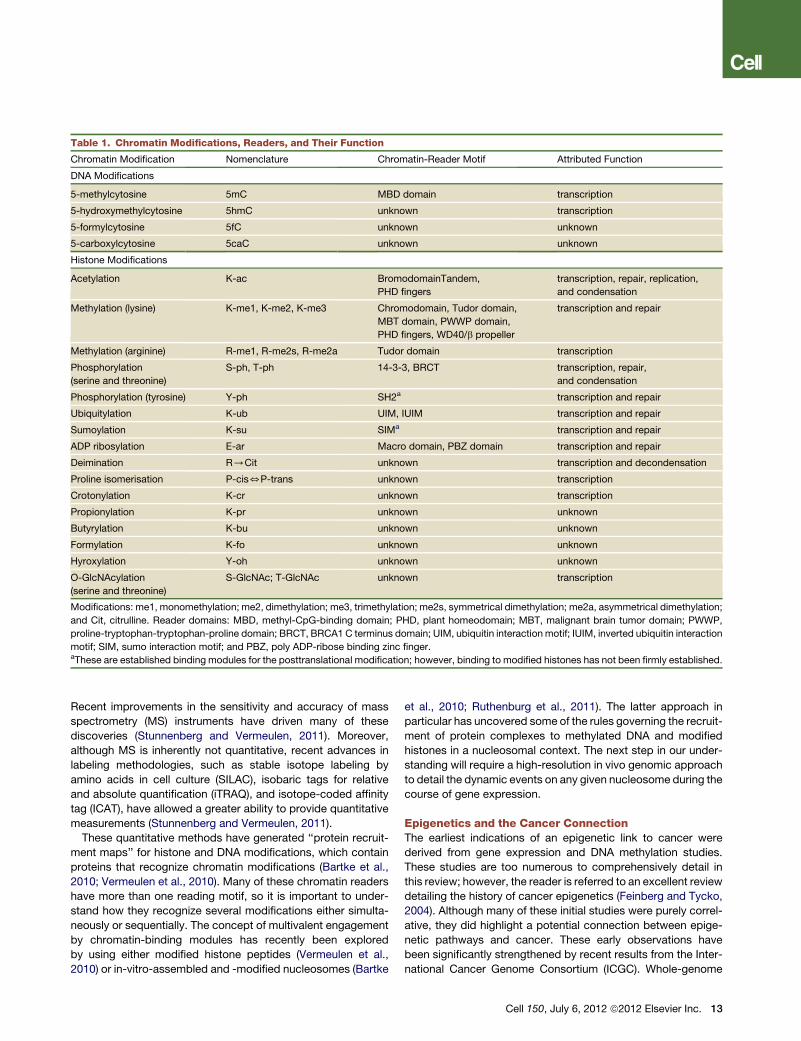
Table 1. Chromatin Modifications, Readers, and Their Function
Chromatin Modification Nomenclature Chromatin-Reader Motif Attributed Function
DNA Modifications
5-methylcytosine 5mC MBD domain transcription
5-hydroxymethylcytosine 5hmC unknown transcription
5-formylcytosine 5fC unknown unknown
5-carboxylcytosine 5caC unknown unknown
Histone Modifications
Acetylation K-ac BromodomainTandem,
PHD fingers
transcription, repair, replication,
and condensation
Methylation (lysine) K-me1, K-me2, K-me3 Chromodomain, Tudor domain,
MBT domain, PWWP domain,
PHD fingers, WD40/b propeller
transcription and repair
Methylation (arginine) R-me1, R-me2s, R-me2a Tudor domain transcription
Phosphorylation
(serine and threonine)
S-ph, T-ph 14-3-3, BRCT transcription, repair,
and condensation
Phosphorylation (tyrosine) Y-ph SH2a transcription and repair
Ubiquitylation K-ub UIM, IUIM transcription and repair
Sumoylation K-su SIMa transcription and repair
ADP ribosylation E-ar Macro domain, PBZ domain transcription and repair
Deimination R/Cit unknown transcription and decondensation
Proline isomerisation P-cis5P-trans unknown transcription
Crotonylation K-cr unknown transcription
Propionylation K-pr unknown unknown
Butyrylation K-bu unknown unknown
Formylation K-fo unknown unknown
Hyroxylation Y-oh unknown unknown
O-GlcNAcylation
(serine and threonine)
S-GlcNAc; T-GlcNAc unknown transcription
Modifications: me1, monomethylation; me2, dimethylation; me3, trimethylation; me2s, symmetrical dimethylation; me2a, asymmetrical dimethylation;
and Cit, citrulline. Reader domains: MBD, methyl-CpG-binding domain; PHD, plant homeodomain; MBT, malignant brain tumor domain; PWWP,
proline-tryptophan-tryptophan-proline domain; BRCT, BRCA1 C terminus domain; UIM, ubiquitin interaction motif; IUIM, inverted ubiquitin interaction
motif; SIM, sumo interaction motif; and PBZ, poly ADP-ribose binding zinc finger.aThese are established binding modules for the posttranslational modification; however, binding to modified histones has not been firmly established.
Recent improvements in the sensitivity and accuracy of mass
spectrometry (MS) instruments have driven many of these
discoveries (Stunnenberg and Vermeulen, 2011). Moreover,
although MS is inherently not quantitative, recent advances in
labeling methodologies, such as stable isotope labeling by
amino acids in cell culture (SILAC), isobaric tags for relative
and absolute quantification (iTRAQ), and isotope-coded affinity
tag (ICAT), have allowed a greater ability to provide quantitative
measurements (Stunnenberg and Vermeulen, 2011).
These quantitative methods have generated ‘‘protein recruit-
ment maps’’ for histone and DNA modifications, which contain
proteins that recognize chromatin modifications (Bartke et al.,
2010; Vermeulen et al., 2010). Many of these chromatin readers
have more than one reading motif, so it is important to under-
stand how they recognize several modifications either simulta-
neously or sequentially. The concept of multivalent engagement
by chromatin-binding modules has recently been explored
by using either modified histone peptides (Vermeulen et al.,
2010) or in-vitro-assembled and -modified nucleosomes (Bartke
et al., 2010; Ruthenburg et al., 2011). The latter approach in
particular has uncovered some of the rules governing the recruit-
ment of protein complexes to methylated DNA and modified
histones in a nucleosomal context. The next step in our under-
standing will require a high-resolution in vivo genomic approach
to detail the dynamic events on any given nucleosome during the
course of gene expression.
Epigenetics and the Cancer ConnectionThe earliest indications of an epigenetic link to cancer were
derived from gene expression and DNA methylation studies.
These studies are too numerous to comprehensively detail in
this review; however, the reader is referred to an excellent review
detailing the history of cancer epigenetics (Feinberg and Tycko,
2004). Although many of these initial studies were purely correl-
ative, they did highlight a potential connection between epige-
netic pathways and cancer. These early observations have
been significantly strengthened by recent results from the Inter-
national Cancer Genome Consortium (ICGC). Whole-genome
Cell 150, July 6, 2012 ª2012 Elsevier Inc. 13
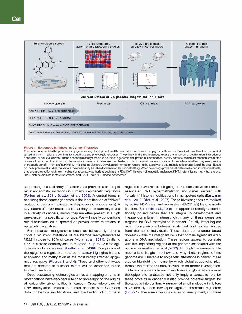
Figure 1. Epigenetic Inhibitors as Cancer TherapiesThis schematic depicts the process for epigenetic drug development and the current status of various epigenetic therapies. Candidate small molecules are firsttested in vitro in malignant cell lines for specificity and phenotypic response. These may, in the first instance, assess the inhibition of proliferation, induction ofapoptosis, or cell-cycle arrest. These phenotypic assays are often coupled to genomic and proteomicmethods to identify potential molecular mechanisms for theobserved response. Inhibitors that demonstrate potential in vitro are then tested in vivo in animal models of cancer to ascertain whether they may providetherapeutic benefit in terms of survival. Animal studies also provide valuable information regarding the toxicity and pharmacokinetic properties of the drug. Basedon these preclinical studies, candidate molecules may be taken forward into the clinical setting. When new drugs prove beneficial in well-conducted clinical trials,they are approved for routine clinical use by regulatory authorities such as the FDA. KAT, histone lysine acetyltransferase; KMT, histone lysine methyltransferase;RMT, histone arginine methyltransferase; and PARP, poly ADP ribose polymerase.
sequencing in a vast array of cancers has provided a catalog of
recurrent somatic mutations in numerous epigenetic regulators
(Forbes et al., 2011; Stratton et al., 2009). A central tenet in
analyzing these cancer genomes is the identification of ‘‘driver’’
mutations (causally implicated in the process of oncogenesis). A
key feature of driver mutations is that they are recurrently found
in a variety of cancers, and/or they are often present at a high
prevalence in a specific tumor type. We will mostly concentrate
our discussions on suspected or proven driver mutations in
epigenetic regulators.
For instance, malignancies such as follicular lymphoma
contain recurrent mutations of the histone methyltransferase
MLL2 in close to 90% of cases (Morin et al., 2011). Similarly,
UTX, a histone demethylase, is mutated in up to 12 histologi-
cally distinct cancers (van Haaften et al., 2009). Compilation of
the epigenetic regulators mutated in cancer highlights histone
acetylation and methylation as the most widely affected epige-
netic pathways (Figures 3 and 4). These and other pathways
that are affected to a lesser extent will be described in the
following sections.
Deep sequencing technologies aimed at mapping chromatin
modifications have also begun to shed some light on the origins
of epigenetic abnormalities in cancer. Cross-referencing of
DNA methylation profiles in human cancers with ChIP-Seq
data for histone modifications and the binding of chromatin
14 Cell 150, July 6, 2012 ª2012 Elsevier Inc.
regulators have raised intriguing correlations between cancer-
associated DNA hypermethylation and genes marked with
‘‘bivalent’’ histone modifications in multipotent cells (Easwaran
et al., 2012; Ohm et al., 2007). These bivalent genes are marked
by active (H3K4me3) and repressive (H3K27me3) histone modi-
fications (Bernstein et al., 2006) and appear to identify transcrip-
tionally poised genes that are integral to development and
lineage commitment. Interestingly, many of these genes are
targeted for DNA methylation in cancer. Equally intriguing are
recent comparisons between malignant and normal tissues
from the same individuals. These data demonstrate broad
domains within the malignant cells that contain significant alter-
ations in DNA methylation. These regions appear to correlate
with late-replicating regions of the genome associated with the
nuclear lamina (Berman et al., 2012). Although there remains little
mechanistic insight into how and why these regions of the
genome are vulnerable to epigenetic alterations in cancer, these
studies highlight the means by which global sequencing plat-
forms have started to uncover avenues for further investigation.
Genetic lesions in chromatinmodifiers and global alterations in
the epigenetic landscape not only imply a causative role for
these proteins in cancer but also provide potential targets for
therapeutic intervention. A number of small-molecule inhibitors
have already been developed against chromatin regulators
(Figure 1). These are at various stages of development, and three
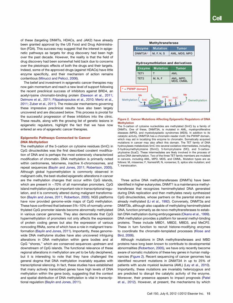
Figure 2. Cancer Mutations Affecting Epigenetic Regulators of DNA
MethylationThe 5-carbon of cytosine nucleotides are methylated (5mC) by a family ofDNMTs. One of these, DNMT3A, is mutated in AML, myeloproliferativediseases (MPD), and myelodysplastic syndromes (MDS). In addition to itscatalytic activity, DNMT3A has a chromatin-reader motif, the PWWP domain,which may aid in localizing this enzyme to chromatin. Somatically acquiredmutations in cancer may also affect this domain. The TET family of DNAhydroxylases metabolizes 5mC into several oxidative intermediates, including5-hydroxymethylcytosine (5hmC), 5-formylcytosine (5fC), and 5-carbox-ylcytosine (5caC). These intermediates are likely involved in the process ofactive DNA demethylation. Two of the three TET family members are mutatedin cancers, including AML, MPD, MDS, and CMML. Mutation types are asfollows: M, missense; F, frameshift; N, nonsense; S, splice site mutation; andT, translocation.
of these (targeting DNMTs, HDACs, and JAK2) have already
been granted approval by the US Food and Drug Administra-
tion (FDA). This success may suggest that the interest in epige-
netic pathways as targets for drug discovery had been high
over the past decade. However, the reality is that the field of
drug discovery had been somewhat held back due to concerns
over the pleiotropic effects of both the drugs and their targets.
Indeed, some of the approved drugs (against HDACs) have little
enzyme specificity, and their mechanism of action remains
contentious (Minucci and Pelicci, 2006).
The belief and investment in epigenetic cancer therapies may
now gain momentum and reach a new level of support following
the recent preclinical success of inhibitors against BRD4, an
acetyl-lysine chromatin-binding protein (Dawson et al., 2011;
Delmore et al., 2011; Filippakopoulos et al., 2010; Mertz et al.,
2011; Zuber et al., 2011). The molecular mechanisms governing
these impressive preclinical results have also been largely
uncovered and are discussed below. This process is pivotal for
the successful progression of these inhibitors into the clinic.
These results, along with the growing list of genetic lesions in
epigenetic regulators, highlight the fact that we have now
entered an era of epigenetic cancer therapies.
Epigenetic Pathways Connected to CancerDNA Methylation
The methylation of the 5-carbon on cytosine residues (5mC) in
CpG dinucleotides was the first described covalent modifica-
tion of DNA and is perhaps the most extensively characterized
modification of chromatin. DNA methylation is primarily noted
within centromeres, telomeres, inactive X-chromosomes, and
repeat sequences (Baylin and Jones, 2011; Robertson, 2005).
Although global hypomethylation is commonly observed in
malignant cells, the best-studied epigenetic alterations in cancer
are the methylation changes that occur within CpG islands,
which are present in �70% of all mammalian promoters. CpG
islandmethylation plays an important role in transcriptional regu-
lation, and it is commonly altered during malignant transforma-
tion (Baylin and Jones, 2011; Robertson, 2005). NGS platforms
have now provided genome-wide maps of CpG methylation.
These have confirmed that between 5%–10%of normally unme-
thylated CpG promoter islands become abnormally methylated
in various cancer genomes. They also demonstrate that CpG
hypermethylation of promoters not only affects the expression
of protein coding genes but also the expression of various
noncoding RNAs, some of which have a role in malignant trans-
formation (Baylin and Jones, 2011). Importantly, these genome-
wide DNA methylome studies have also uncovered intriguing
alterations in DNA methylation within gene bodies and at
CpG ‘‘shores,’’ which are conserved sequences upstream and
downstream of CpG islands. The functional relevance of these
regional alterations in methylation are yet to be fully deciphered,
but it is interesting to note that they have challenged the
general dogma that DNA methylation invariably equates with
transcriptional silencing. In fact, these studies have established
that many actively transcribed genes have high levels of DNA
methylation within the gene body, suggesting that the context
and spatial distribution of DNA methylation is vital in transcrip-
tional regulation (Baylin and Jones, 2011).
Three active DNA methyltransferases (DNMTs) have been
identified in higher eukaryotes. DNMT1 is amaintenancemethyl-
transferase that recognizes hemimethylated DNA generated
during DNA replication and then methylates newly synthesized
CpG dinucleotides, whose partners on the parental strand are
already methylated (Li et al., 1992). Conversely, DNMT3a and
DNMT3b, although also capable of methylating hemimethylated
DNA, function primarily as de novo methyltransferases to estab-
lish DNAmethylation during embryogenesis (Okano et al., 1999).
DNA methylation provides a platform for several methyl-binding
proteins. These include MBD1, MBD2, MBD3, and MeCP2.
These in turn function to recruit histone-modifying enzymes
to coordinate the chromatin-templated processes (Klose and
Bird, 2006).
Although mutations in DNA methyltransferases and MBD
proteins have long been known to contribute to developmental
abnormalities (Robertson, 2005), we have only recently become
aware of somatic mutations of these key genes in human malig-
nancies (Figure 2). Recent sequencing of cancer genomes has
identified recurrent mutations in DNMT3A in up to 25% of
patients with acute myeloid leukemia (AML) (Ley et al., 2010).
Importantly, these mutations are invariably heterozygous and
are predicted to disrupt the catalytic activity of the enzyme.
Moreover, their presence appears to impact prognosis (Patel
et al., 2012). However, at present, the mechanisms by which
Cell 150, July 6, 2012 ª2012 Elsevier Inc. 15
these mutations contribute to the development and/or mainte-
nance of AML remains elusive.
Understanding the cellular consequences of normal and aber-
rant DNA methylation remains a key area of interest, especially
because hypomethylating agents are one of the few epigenetic
therapies that have gained FDA approval for routine clinical
use (Figure 1). Although hypomethylating agents such as azaci-
tidine and decitabine have shown mixed results in various solid
malignancies, they have found a therapeutic niche in the myelo-
dysplastic syndromes (MDS). Until recently, this group of disor-
ders was largely refractory to therapeutic intervention, and MDS
was primarily managed with supportive care. However, several
large studies have now shown that treatment with azacitidine,
even in poor prognosis patients, improves their quality of life
and extends survival time. Indeed, azacitidine is the first therapy
to have demonstrated a survival benefit for patients with MDS
(Fenaux et al., 2009). The molecular mechanisms governing the
impressive responses seen in MDS are largely unknown.
However, recent evidence would suggest that low doses of
these agents hold the key to therapeutic benefit (Tsai et al.,
2012). It is also emerging that the combinatorial use of DNMT
and HDAC inhibitors may offer superior therapeutic outcomes
(Gore, 2011).
DNA Hydroxy-Methylation and Its Oxidation Derivatives
Historically, DNA methylation was generally considered to
be a relatively stable chromatin modification. However, early
studies assessing the global distribution of this modification
during embryogenesis had clearly identified an active global
loss of DNA methylation in the early zygote, especially in the
male pronucleus. More recently, high-resolution genome-wide
mapping of this modification in pluripotent and differentiated
cells has also confirmed the dynamic nature of DNAmethylation,
evidently signifying the existence of an enzymatic activity within
mammalian cells that either erases or alters this chromatin
modification (Baylin and Jones, 2011). In 2009, two seminal
manuscripts describing the presence of 5-hydroxymethylcyto-
sine (5hmC) offered the first insights into the metabolism of
5mC (Kriaucionis and Heintz, 2009; Tahiliani et al., 2009).
The ten-eleven translocation (TET 1–3) family of proteins have
now been demonstrated to be the mammalian DNA hydroxy-
lases responsible for catalytically converting 5mC to 5hmC.
Indeed, iterative oxidation of 5hmC by the TET family results in
further oxidation derivatives, including 5-formylcytosine (5fC)
and 5-carboxylcytosine (5caC). Although the biological signifi-
cance of the 5mC oxidation derivatives is yet to be established,
several lines of evidence highlight their importance in transcrip-
tional regulation: (1) they are likely to be an essential intermediate
in the process of both active and passive DNA demethylation, (2)
they preclude or enhance the binding of several MBD proteins
and, as such, will have local and global effects by altering
the recruitment of chromatin regulators, and (3) genome-wide
mapping of 5hmC has identified a distinctive distribution of this
modification at both active and repressed genes, including its
presence within gene bodies and at the promoters of bivalently
marked, transcriptionally poised genes (Wu and Zhang, 2011).
Notably, 5hmC was also mapped to several intergenic cis-regu-
latory elements that are either functional enhancers or insulator
elements. Consistent with the notion that 5hmC is likely to
16 Cell 150, July 6, 2012 ª2012 Elsevier Inc.
have a role in both transcriptional activation and silencing,
the TET proteins have also been shown to have activating
and repressive functions (Wu and Zhang, 2011). Genome-wide
mapping of TET1 has demonstrated it to have a strong prefer-
ence for CpG-rich DNA and, consistent with its catalytic function,
it also been localized to regions enriched for 5mC and 5hmC.
The TET family of proteins derive their name from the
initial description of a recurrent chromosomal translocation,
t(10;11)(q22;q23), which juxtaposes the MLL gene with TET1 in
a subset of patients with AML (Lorsbach et al., 2003). Notably,
concurrent to the initial description of the catalytic activity for
the TET family of DNA hydroxylases, several reports emerged
describing recurrent mutations in TET2 in numerous hematolog-
ical malignancies (Cimmino et al., 2011; Delhommeau et al.,
2009; Langemeijer et al., 2009) (Figure 2). Interestingly, TET2-
deficient mice develop a chronic myelomonocytic leukemia
(CMML) phenotype, which is in keeping with the high prevalence
of TET2 mutations in patients with this disease (Moran-Crusio
et al., 2011; Quivoron et al., 2011). The clinical implications of
TET2 mutations have largely been inconclusive; however, in
some subsets of AML patients, TET2mutations appear to confer
a poor prognosis (Patel et al., 2012). Early insights into the
process of TET2-mediated oncogenesis have revealed that the
patient-associated mutations are largely loss-of-function muta-
tions that consequently result in decreased 5hmC levels and
a reciprocal increase in 5mC levels within the malignant cells
that harbor them. Moreover, mutations in TET2 also appear to
confer enhanced self-renewal properties to themalignant clones
(Cimmino et al., 2011).
Histone Modifications
In 1964, Vincent Allfrey prophetically surmised that histone
modifications might have a functional influence on the regulation
of transcription (Allfrey et al., 1964). Nearly half a century later,
the field is still grappling with the task of unraveling the mecha-
nisms underlying his enlightened statement. In this time, we
have learned that these modifications have a major influence,
not just on transcription, but in all DNA-templated processes
(Kouzarides, 2007). The major cellular processes attributed to
each of these modifications are summarized in Table 1.
The great diversity in histone modifications introduces a
remarkable complexity that is slowly beginning to be eluci-
dated. Using transcription as an example, we have learned
that multiple coexisting histone modifications are associated
with activation, and some are associated with repression.
However, these modification patterns are not static entities but
a dynamically changing and complex landscape that evolves in
a cell context-dependent fashion. Moreover, active and repres-
sive modifications are not always mutually exclusive, as evi-
denced by ‘‘bivalent domains.’’ The combinatorial influence
that one or more histone modifications have on the deposition,
interpretation, or erasure of other histone modifications has
been broadly termed ‘‘histone crosstalk,’’ and recent evidence
would suggest that crosstalk is widespread and is of great bio-
logical significance (Lee et al., 2010).
It should be noted that the cellular enzymes that modify
histones may also have nonhistone targets and, as such, it has
been difficult to divorce the cellular consequences of individual
histone modifications from the broader targets of many of these
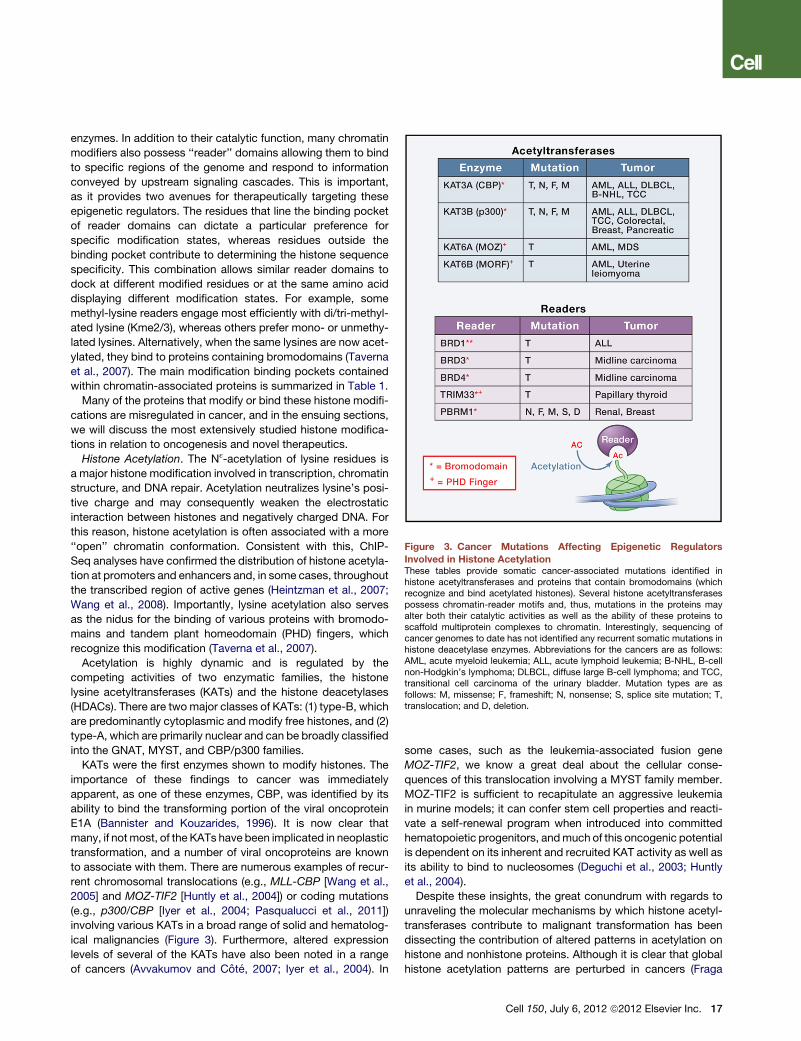
Figure 3. Cancer Mutations Affecting Epigenetic Regulators
Involved in Histone AcetylationThese tables provide somatic cancer-associated mutations identified inhistone acetyltransferases and proteins that contain bromodomains (whichrecognize and bind acetylated histones). Several histone acetyltransferasespossess chromatin-reader motifs and, thus, mutations in the proteins mayalter both their catalytic activities as well as the ability of these proteins toscaffold multiprotein complexes to chromatin. Interestingly, sequencing ofcancer genomes to date has not identified any recurrent somatic mutations inhistone deacetylase enzymes. Abbreviations for the cancers are as follows:AML, acute myeloid leukemia; ALL, acute lymphoid leukemia; B-NHL, B-cellnon-Hodgkin’s lymphoma; DLBCL, diffuse large B-cell lymphoma; and TCC,transitional cell carcinoma of the urinary bladder. Mutation types are asfollows: M, missense; F, frameshift; N, nonsense; S, splice site mutation; T,translocation; and D, deletion.
enzymes. In addition to their catalytic function, many chromatin
modifiers also possess ‘‘reader’’ domains allowing them to bind
to specific regions of the genome and respond to information
conveyed by upstream signaling cascades. This is important,
as it provides two avenues for therapeutically targeting these
epigenetic regulators. The residues that line the binding pocket
of reader domains can dictate a particular preference for
specific modification states, whereas residues outside the
binding pocket contribute to determining the histone sequence
specificity. This combination allows similar reader domains to
dock at different modified residues or at the same amino acid
displaying different modification states. For example, some
methyl-lysine readers engage most efficiently with di/tri-methyl-
ated lysine (Kme2/3), whereas others prefer mono- or unmethy-
lated lysines. Alternatively, when the same lysines are now acet-
ylated, they bind to proteins containing bromodomains (Taverna
et al., 2007). The main modification binding pockets contained
within chromatin-associated proteins is summarized in Table 1.
Many of the proteins that modify or bind these histone modifi-
cations are misregulated in cancer, and in the ensuing sections,
we will discuss the most extensively studied histone modifica-
tions in relation to oncogenesis and novel therapeutics.
Histone Acetylation. The Nε-acetylation of lysine residues is
a major histone modification involved in transcription, chromatin
structure, and DNA repair. Acetylation neutralizes lysine’s posi-
tive charge and may consequently weaken the electrostatic
interaction between histones and negatively charged DNA. For
this reason, histone acetylation is often associated with a more
‘‘open’’ chromatin conformation. Consistent with this, ChIP-
Seq analyses have confirmed the distribution of histone acetyla-
tion at promoters and enhancers and, in some cases, throughout
the transcribed region of active genes (Heintzman et al., 2007;
Wang et al., 2008). Importantly, lysine acetylation also serves
as the nidus for the binding of various proteins with bromodo-
mains and tandem plant homeodomain (PHD) fingers, which
recognize this modification (Taverna et al., 2007).
Acetylation is highly dynamic and is regulated by the
competing activities of two enzymatic families, the histone
lysine acetyltransferases (KATs) and the histone deacetylases
(HDACs). There are two major classes of KATs: (1) type-B, which
are predominantly cytoplasmic and modify free histones, and (2)
type-A, which are primarily nuclear and can be broadly classified
into the GNAT, MYST, and CBP/p300 families.
KATs were the first enzymes shown to modify histones. The
importance of these findings to cancer was immediately
apparent, as one of these enzymes, CBP, was identified by its
ability to bind the transforming portion of the viral oncoprotein
E1A (Bannister and Kouzarides, 1996). It is now clear that
many, if notmost, of the KATs have been implicated in neoplastic
transformation, and a number of viral oncoproteins are known
to associate with them. There are numerous examples of recur-
rent chromosomal translocations (e.g., MLL-CBP [Wang et al.,
2005] and MOZ-TIF2 [Huntly et al., 2004]) or coding mutations
(e.g., p300/CBP [Iyer et al., 2004; Pasqualucci et al., 2011])
involving various KATs in a broad range of solid and hematolog-
ical malignancies (Figure 3). Furthermore, altered expression
levels of several of the KATs have also been noted in a range
of cancers (Avvakumov and Cote, 2007; Iyer et al., 2004). In
some cases, such as the leukemia-associated fusion gene
MOZ-TIF2, we know a great deal about the cellular conse-
quences of this translocation involving a MYST family member.
MOZ-TIF2 is sufficient to recapitulate an aggressive leukemia
in murine models; it can confer stem cell properties and reacti-
vate a self-renewal program when introduced into committed
hematopoietic progenitors, andmuch of this oncogenic potential
is dependent on its inherent and recruited KAT activity as well as
its ability to bind to nucleosomes (Deguchi et al., 2003; Huntly
et al., 2004).
Despite these insights, the great conundrum with regards to
unraveling the molecular mechanisms by which histone acetyl-
transferases contribute to malignant transformation has been
dissecting the contribution of altered patterns in acetylation on
histone and nonhistone proteins. Although it is clear that global
histone acetylation patterns are perturbed in cancers (Fraga
Cell 150, July 6, 2012 ª2012 Elsevier Inc. 17
et al., 2005; Seligson et al., 2005), it is also well established that
several nonhistone proteins, including many important onco-
genes and tumor suppressors such as MYC, p53, and PTEN,
are also dynamically acetylated (Choudhary et al., 2009). A prag-
matic view on this issue is that both histone and nonhistone
acetylation are likely to be important and, in most part, the
abundance of substrates has not deterred the enthusiasm for
the development of histone acetyltransferase inhibitors (KAT-I).
Although there is only modest structural homology between
the different families of KATs, developing specific inhibitors
has proven to be fraught with frustration (Cole, 2008). However,
recent progress with derivatives of the naturally occurring KAT-I,
such as curcumin, anacardic acid, and garcinol, as well as the
synthesis of novel chemical probes, suggest that therapeutically
targeting the various KATs with some specificity is likely to be
achieved in the near future (Cole, 2008).
Histone Deacetylation. HDACs are enzymes that reverse lysine
acetylation and restore the positive charge on the side chain.
There are 18 such enzymes identified, and these are subdivided
into four major classes, depending on sequence homology.
Class I (HDAC 1-3 and HDAC8) and class II (HDAC 4-7 and
HDAC 9-10) represent the HDACs most closely related to yeast
scRpd3 and scHda1, respectively, whereas class IV comprises
only one enzyme, HDAC11. Class I, II, and IV HDACs share a
related catalytic mechanism that requires a zinc metal ion but
does not involve the use of a cofactor. In contrast, class III
HDACs (sirtuin 1–7) are homologous to yeast scSir2 and employ
a distinct catalytic mechanism that is NAD+-dependent. Analo-
gous to the KATs, HDACs target both histone and nonhistone
proteins. Substrate specificity for these enzymes is largely medi-
ated by components of multisubunit complexes in which HDACs
are found, such as Mi2/NuRD, Sin3A, and Co-REST (Bantscheff
et al., 2011; Xhemalce et al., 2011).
In the context of malignancy, chimeric fusion proteins that are
seen in leukemia, such as PML-RARa, PLZF-RARa, and AML1-
ETO, have been shown to recruit HDACs to mediate aberrant
gene silencing, which contributes to leukemogenesis (John-
stone and Licht, 2003). HDACs can also interact with nonchi-
meric oncogenes such as BCL6, whose repressive activity is
controlled by dynamic acetylation (Bereshchenko et al., 2002).
Importantly, inhibitors of histone deactylases (HDAC-I) are able
to reverse some of the aberrant gene repression seen in
these malignancies and induce growth arrest, differentiation,
and apoptosis in the malignant cells (Federico and Bagella,
2011; Johnstone and Licht, 2003). Based on impressive preclin-
ical and clinical data, two pan-HDAC inhibitors, Vorinostat and
Romidepsin, have recently been granted FDA approval (Olsen
et al., 2007; Piekarz et al., 2009) for clinical use in patients with
cutaneous T cell lymphoma (Figure 1). Although somatic muta-
tions in HDACs do not appear to be prominent in cancer
(Figure 3), the expression levels of various HDACs appear to
be altered in numerous malignancies. Consequently, several
novel HDAC inhibitors are currently under investigation for clin-
ical use in a broad range of cancers (Federico and Bagella, 2011;
Johnstone and Licht, 2003). However, the pleiotropic effects of
HDACs continue to pose significant challenges in dissecting the
specific effects on histone and nonhistone proteins (Bantscheff
et al., 2011).
18 Cell 150, July 6, 2012 ª2012 Elsevier Inc.
Histone Acetylation Readers. The primary readers of Nε-acet-
ylation of lysine residues are families of proteins that contain an
evolutionarily conserved binding motif termed a bromodomain.
There are over 40 described human proteins with bromodomains
(Chung and Witherington, 2011). These comprise a diverse
group of proteins that function as chromatin remodelers, histone
acetyltransferases, histone methyltransferases, and transcrip-
tional coactivators. Many of these proteins also contain several
separate evolutionarily conserved ‘‘chromatin-reading’’ motifs
such as PHD fingers, which recognize distinct histone posttrans-
lational modifications (Table 1).
Until recently, it had not been feasible to therapeutically target
protein-protein interactions with small molecules. However,
several recent studies have shown that it is possible to develop
highly specific and chemically distinct small molecules against
the BET family (BRD2, BRD3, BRD4, and BRDt) of bromodo-
main proteins (Dawson et al., 2011; Filippakopoulos et al.,
2010; Nicodeme et al., 2010). The BET family shares a common
structural composition featuring tandem amino-terminal bromo-
domains that exhibit high levels of sequence conservation. BET
proteins play a fundamental role in transcriptional elongation
and cell-cycle progression. Moreover, recurrent translocations
involving BRD3/4 are associated with the aggressive and invari-
ably fatal NUT-midline carcinoma (Filippakopoulos et al., 2010)
(Figure 3).
Targeting the BET bromodomains is a promising therapeutic
avenue in cancer. The BET inhibitors have recently been shown
to have excellent efficacy in NUT-midline carcinoma (Filippako-
poulos et al., 2010) and in a range of hematological malignancies
(Dawson et al., 2011; Delmore et al., 2011; Mertz et al., 2011;
Zuber et al., 2011). A central theme reported in all of the studies
thus far is the downregulation of MYC transcription following
BET inhibition. MYC is a master regulator of cell proliferation
and survival; it is also one of the most common genes dysregu-
lated in cancer (Meyer and Penn, 2008). Following BET inhibition
with either RNAi or specific BET inhibitors, the expression of
MYC was noted to be substantially decreased in a variety of
malignant hematopoietic cell lines, including MLL-translocated
acute myeloid leukemia (Dawson et al., 2011; Zuber et al.,
2011), multiple myeloma (Delmore et al., 2011), and Burkitt’s
lymphoma (Mertz et al., 2011). Furthermore, murine models of
these diseases confirmed the excellent therapeutic efficacy of
BET inhibition in vivo.
Although MYC has a prominent role in these diseases, it is
unlikely that the profound effects observed by BET inhibition
are solelymediated byMYC inhibition. There aremanymalignant
cell lines that overexpress MYC yet fail to respond to BET inhibi-
tion (Mertz et al., 2011); MYC expression is not always affected
by BET inhibition (Mertz et al., 2011); MYC is often equally
downregulated in responsive and nonresponsive malignant cell
lines (Dawson et al., 2011; Zuber et al., 2011); and, importantly,
MYC overexpression fails to rescue the apoptosis induced by
BET inhibition (Zuber et al., 2011). The molecular mechanisms
governing the efficacy of BET inhibition are slowly being deci-
phered. What seems to be clear from the current analyses is
that BET inhibitors specifically regulate a small number of genes,
and inhibition of transcriptional elongation may be a primary
mode of action.
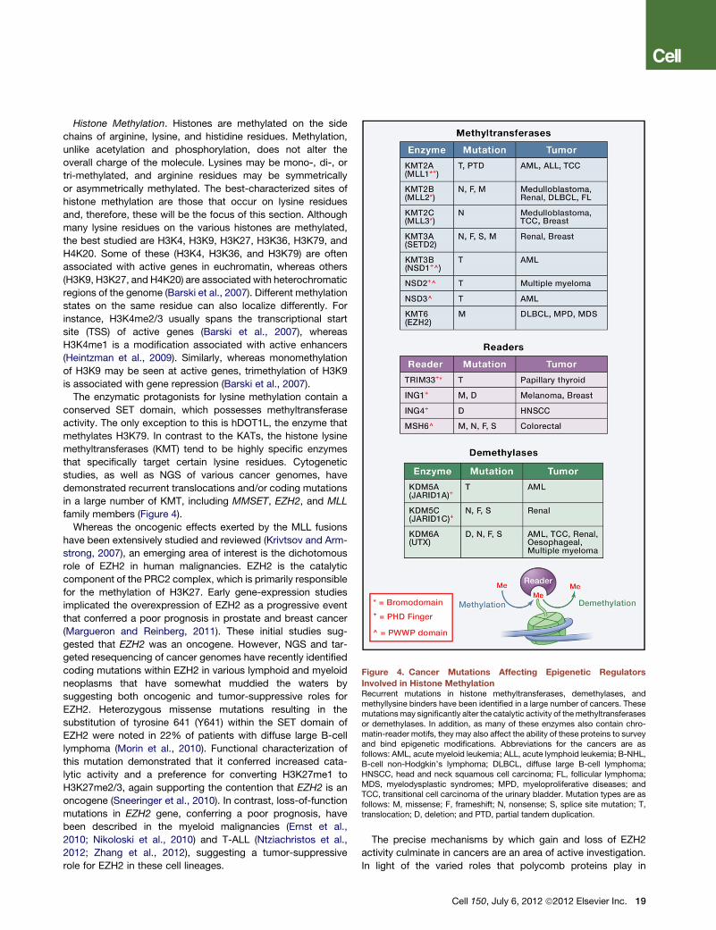
Figure 4. Cancer Mutations Affecting Epigenetic Regulators
Involved in Histone MethylationRecurrent mutations in histone methyltransferases, demethylases, andmethyllysine binders have been identified in a large number of cancers. Thesemutationsmay significantly alter the catalytic activity of themethyltransferasesor demethylases. In addition, as many of these enzymes also contain chro-matin-reader motifs, they may also affect the ability of these proteins to surveyand bind epigenetic modifications. Abbreviations for the cancers are asfollows: AML, acute myeloid leukemia; ALL, acute lymphoid leukemia; B-NHL,B-cell non-Hodgkin’s lymphoma; DLBCL, diffuse large B-cell lymphoma;HNSCC, head and neck squamous cell carcinoma; FL, follicular lymphoma;MDS, myelodysplastic syndromes; MPD, myeloproliferative diseases; andTCC, transitional cell carcinoma of the urinary bladder. Mutation types are asfollows: M, missense; F, frameshift; N, nonsense; S, splice site mutation; T,translocation; D, deletion; and PTD, partial tandem duplication.
Histone Methylation. Histones are methylated on the side
chains of arginine, lysine, and histidine residues. Methylation,
unlike acetylation and phosphorylation, does not alter the
overall charge of the molecule. Lysines may be mono-, di-, or
tri-methylated, and arginine residues may be symmetrically
or asymmetrically methylated. The best-characterized sites of
histone methylation are those that occur on lysine residues
and, therefore, these will be the focus of this section. Although
many lysine residues on the various histones are methylated,
the best studied are H3K4, H3K9, H3K27, H3K36, H3K79, and
H4K20. Some of these (H3K4, H3K36, and H3K79) are often
associated with active genes in euchromatin, whereas others
(H3K9, H3K27, and H4K20) are associated with heterochromatic
regions of the genome (Barski et al., 2007). Different methylation
states on the same residue can also localize differently. For
instance, H3K4me2/3 usually spans the transcriptional start
site (TSS) of active genes (Barski et al., 2007), whereas
H3K4me1 is a modification associated with active enhancers
(Heintzman et al., 2009). Similarly, whereas monomethylation
of H3K9 may be seen at active genes, trimethylation of H3K9
is associated with gene repression (Barski et al., 2007).
The enzymatic protagonists for lysine methylation contain a
conserved SET domain, which possesses methyltransferase
activity. The only exception to this is hDOT1L, the enzyme that
methylates H3K79. In contrast to the KATs, the histone lysine
methyltransferases (KMT) tend to be highly specific enzymes
that specifically target certain lysine residues. Cytogenetic
studies, as well as NGS of various cancer genomes, have
demonstrated recurrent translocations and/or coding mutations
in a large number of KMT, including MMSET, EZH2, and MLL
family members (Figure 4).
Whereas the oncogenic effects exerted by the MLL fusions
have been extensively studied and reviewed (Krivtsov and Arm-
strong, 2007), an emerging area of interest is the dichotomous
role of EZH2 in human malignancies. EZH2 is the catalytic
component of the PRC2 complex, which is primarily responsible
for the methylation of H3K27. Early gene-expression studies
implicated the overexpression of EZH2 as a progressive event
that conferred a poor prognosis in prostate and breast cancer
(Margueron and Reinberg, 2011). These initial studies sug-
gested that EZH2 was an oncogene. However, NGS and tar-
geted resequencing of cancer genomes have recently identified
coding mutations within EZH2 in various lymphoid and myeloid
neoplasms that have somewhat muddied the waters by
suggesting both oncogenic and tumor-suppressive roles for
EZH2. Heterozygous missense mutations resulting in the
substitution of tyrosine 641 (Y641) within the SET domain of
EZH2 were noted in 22% of patients with diffuse large B-cell
lymphoma (Morin et al., 2010). Functional characterization of
this mutation demonstrated that it conferred increased cata-
lytic activity and a preference for converting H3K27me1 to
H3K27me2/3, again supporting the contention that EZH2 is an
oncogene (Sneeringer et al., 2010). In contrast, loss-of-function
mutations in EZH2 gene, conferring a poor prognosis, have
been described in the myeloid malignancies (Ernst et al.,
2010; Nikoloski et al., 2010) and T-ALL (Ntziachristos et al.,
2012; Zhang et al., 2012), suggesting a tumor-suppressive
role for EZH2 in these cell lineages.
The precise mechanisms by which gain and loss of EZH2
activity culminate in cancers are an area of active investigation.
In light of the varied roles that polycomb proteins play in
Cell 150, July 6, 2012 ª2012 Elsevier Inc. 19
self-renewal and differentiation (Margueron andReinberg, 2011),
solution of this problem will necessitate vigilance and apprecia-
tion of the cellular context within which the mutations arise. The
increased awareness of the involvement of KMTs in cancer has
heightened efforts to identify specific inhibitors. These efforts
will only be encouraged by the recent demonstration that
small-molecule inhibition of DOT1L shows preclinical promise
as a targeted therapy in MLL leukemia (Daigle et al., 2011),
a disease in which aberrant DOT1L activity is ill defined but
clearly involved (Krivtsov and Armstrong, 2007).
Histone Demethylation. The initial notion that histone lysine
methylation was a highly stable, nondynamic modification has
now been irrefutably overturned by the identification of two
classes of lysine demethylases (Mosammaparast and Shi,
2010). The prima facie example, LSD1 (KDM1A), belongs to
the first class of demethylases that demethylates lysines via an
amine oxidation reaction with flavin adenine dinucleotide (FAD)
as a cofactor. As this family of enzymes requires a protonated
nitrogen to initiate demethylation, they are limited to demethylat-
ing mono- and dimethyllysine. The second and more expansive
class of enzymes is broadly referred to as the Jumonji demethy-
lases. They have a conserved JmjC domain, which functions via
an oxidative mechanism and radical attack (involving Fe(II) and
a-ketoglutarate). The Jumonji family does not require a free
electron pair on the nitrogen atom to initiate catalysis and,
therefore, unlike LSD1, they can demethylate all three methyl
lysine states. Unsurprisingly, the multisubunit complexes within
which these enzymes reside confer much of their target speci-
ficity. As an example, LSD1 can function as a transcriptional
repressor by demethylating H3K4me1/2 as part of the core-
pressor for RE1-silencing transcription factor (Co-REST) com-
plex, but its activity is linked to gene activation when it associ-
ates with the androgen receptor to demethylate H3K9me2
(Mosammaparast and Shi, 2010). Thus far, recurrent coding
mutations have been noted in KDM5A (JARID1A), KDM5C
(JARID1C), and KDM6A (UTX) (Figure 4). Mutations in UTX, in
particular, are prevalent in a large number of solid and hemato-
logical cancers. Small-molecule inhibitors of the two families of
histone demethylases are at various stages of development,
and this interest will be spurred on by emerging preclinical
data showing the therapeutic potential of compounds that inhibit
LSD1/KDM1A in AML (Barretina et al., 2012; Schenk et al., 2012).
Interestingly, recent findings related to recurrent mutations in
the genes encoding the metabolic enzymes isocitrate dehydro-
genase-1 (IDH1) and IDH2 have broad implications for the
Jumonji class of demethylases, which use a-ketoglutarate
(a-KG). IDH1/2 are nicotinamide adenine dinucleotide phos-
phate (NADP)-dependent enzymes that normally catalyze the
oxidative decarboxylation of isocitrate to a-KG, which is associ-
ated with the production of NADPH. Mutations in IDH1 and IDH2
are seen in up to 70% of patients with secondary glioblastoma
mutiforme and are also noted as recurrent mutations in a range
of myeloid malignancies, most notably AML (Cimmino et al.,
2011). These mutations manifest in a neomorphic enzymatic
activity that results in the NADPH-dependent reduction of
a-KG to 2-hydroxyglutarate (2-HG). Consequently, malignant
cells with IDH1/2 mutations may harbor 2-HG levels that are
up to 100-fold higher than normal (Cimmino et al., 2011). 2-HG
20 Cell 150, July 6, 2012 ª2012 Elsevier Inc.
is a competitive inhibitor of the a-KG-dependent dioxygenases;
in fact, 2-HG has been shown to adopt a near-identical orienta-
tion within the catalytic core of the JmjC domain (Xu et al., 2011).
As 2-HG levels accumulate within the malignant cells, there is
a purported blanket inhibition of the Jumonji class of histone
demethylases. Accordingly, there is a discernable increase in
histone methylation levels (Xu et al., 2011). These remarkable
findings are yet to be fully investigated, and it will be important
to determine whether all the Jumonji family members are equally
susceptible to 2-HG inhibition. A similar question can be posed
for the TET family of enzymes (see above), which also use a-ke-
toglutarate.
Histone Methylation Readers. The various states of lysine
methylation result in considerable physicochemical diversity of
lysine; these modification states are read and interpreted by
proteins containing different specialized recognition motifs.
Broadly speaking, the aromatic cages that engage methyllysine
can be divided into two major families, the Royal Family (Tudor
domains, Chromo domains, and malignant brain tumor [MBT]
domains) and PHD fingers. The structural composition of these
domains that allows for this diversity has recently been expertly
reviewed (Taverna et al., 2007).
Analogous to the situation with bromodomain proteins,
several methyllysine readers have also been implicated in cancer
(Figure 4). For instance, all three isoforms of the chromodomain
protein HP1 have altered expression in numerous cancers (Dia-
lynas et al., 2008). However, thus far, no cancer-specific somatic
mutations have been identified in HP1. In contrast, ING family
members have had coding mutations identified in malignancies
such as melanoma and breast cancer, including those that
specifically target the PHD finger, which recognizes H3K4me3
(Coles and Jones, 2009). Despite these findings, neither of the
aforementioned examples establishes a causal relationship
between cancer and the abrogation of methyllysine binding at
chromatin. The best example of this, and indeed a proof of
principle for therapeutically targeting methyllysine binders, has
recently been shown in a specific form of AML (Wang et al.,
2009). Leukemia, induced by the fusion of NUP98 with the
PHD finger containing part of JARID1A or PHF23, can be abro-
gated by mutations that negate the ability of the PHD finger to
bind H3K4me3. Functional compensation of this effect can be
provided by other PHD fingers that recognize this modification,
but not those that do not bind H3K4me3. Moreover, mechanistic
insights were provided, demonstrating that chromatin binding of
the fusion protein inhibits the deposition of H3K27me3, which
leads to the continued expression of critical hematopoietic
oncogenes such as HoxA9, Meis1, and Pbx1 (Wang et al.,
2009). In light of these findings, and as result of the structural
diversity present in methyllysine-binding modules, it is likely
that small molecules that disrupt this important protein-protein
interaction may be effective anticancer agents.
Histone Phosphorylation. The phosphorylation of serine, thre-
onine, and tyrosine residues has been documented on all core
and most variant histones. Phosphorylation alters the charge
of the protein, affecting its ionic properties and influencing the
overall structure and function of the local chromatin environ-
ment. The phosphorylation of histones is integral to essential
cellular processes such as mitosis, apoptosis, DNA repair,
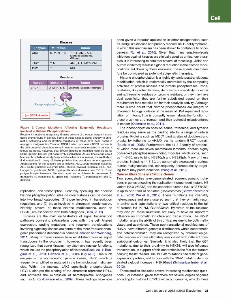
Figure 5. Cancer Mutations Affecting Epigenetic Regulators
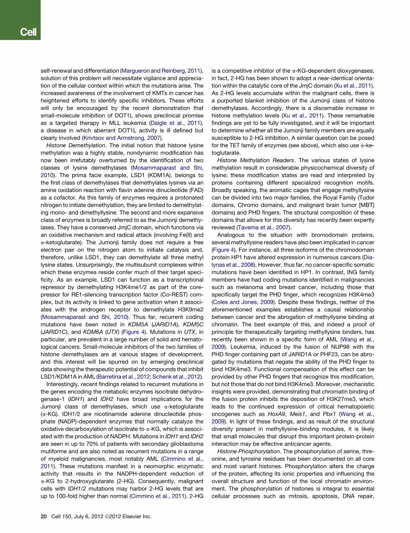
Involved in Histone PhosphorylationRecurrent mutations in signaling kinases are one of the most frequent onco-genic events found in cancer. Some of these kinases signal directly to chro-matin. Activating and inactivating mutations of these have been noted ina range of malignancies. Thus far, BRCA1, which contains a BRCT domain, isthe only potential phosphochromatin reader recurrently mutated in cancer. Itshould be noted, however, that BRCA1 binding to modified histones via itsBRCT domain has not yet been firmly established. As our knowledge abouthistone phosphatases and phosphohistone binders increases, we are likely tofind mutations in many of these proteins that contribute to oncogenesis.Abbreviations for the cancers are as follows: AML, acute myeloid leukemia;ALL, acute lymphoid leukemia; CML, chronic myeloid leukemia; NHL, non-Hodgkin’s lymphoma; MPD, myeloproliferative diseases; and T-PLL, T cellprolymphocytic leukemia. Mutation types are as follows: M, missense; F,frameshift; N, nonsense; S, splice site mutation; T, translocation; and D,deletion.
replication, and transcription. Generally speaking, the specific
histone phosphorylation sites on core histones can be divided
into two broad categories: (1) those involved in transcription
regulation, and (2) those involved in chromatin condensation.
Notably, several of these histone modifications, such as
H3S10, are associated with both categories (Baek, 2011).
Kinases are the main orchestrators of signal transduction
pathways conveying extracellular cues within the cell. Altered
expression, coding mutations, and recurrent translocations
involving signaling kinases are some of the most frequent onco-
genic phenomena described in cancer (Hanahan and Weinberg,
2011). Many of these kinases have established roles as signal
transducers in the cytoplasm; however, it has recently been
recognized that some kinases may also have nuclear functions,
which include the phosphorylation of histones (Baek, 2011; Bun-
gard et al., 2010; Dawson et al., 2009) (Figure 5). One such
enzyme is the nonreceptor tyrosine kinase, JAK2, which is
frequently amplified or mutated in the hematological malignan-
cies. Within the nucleus, JAK2 specifically phosphorylates
H3Y41, disrupts the binding of the chromatin repressor HP1a,
and activates the expression of hematopoietic oncogenes
such as Lmo2 (Dawson et al., 2009). These findings have now
been given a broader application in other malignancies, such
as Hodgkin’s disease and primary mediastinal B-cell lymphoma,
in which this mechanism has been shown to contribute to onco-
genesis (Rui et al., 2010). Given that many small-molecule
inhibitors against kinases are clinically used as anticancer thera-
pies, it is interesting to note that several of these (e.g., JAK2 and
Aurora inhibitors) result in a global reduction in the histone modi-
fications laid down by these enzymes. These agents can there-
fore be considered as potential epigenetic therapies.
Histone phosphorylation is a highly dynamic posttranslational
modification, which is reciprocally controlled by the competing
activities of protein kinases and protein phosphatases. Phos-
phatases, like protein kinases, demonstrate specificity for either
serine/threonine residues or tyrosine residues, or they may have
dual specificity; they are further subdivided based on their
requirement for a metallic ion for their catalytic activity. Although
there is little doubt that histone phosphatases are integral to
chromatin biology, outside of the realm of DNA repair and regu-
lation of mitosis, little is currently known about the function of
these enzymes at chromatin and their potential misadventures
in cancer (Xhemalce et al., 2011).
The phosphorylation sites on serine, threonine, and tyrosine
residues may serve as the binding site for a range of cellular
proteins. Proteins such as MDC1 bind at sites of double-strand
breaks by tethering to gH2AX via its tandem BRCT domain
(Stucki et al., 2005). Furthermore, the 14-3-3 family of proteins,
of which there are seven mammalian isoforms, contain highly
conserved phosphoserine-binding modules which some, such
as 14-3-3z, use to bind H3S10ph and H3S28ph. Many of these
proteins, including 14-3-3z, are abnormally expressed in various
human malignancies and, consequently, therapeutically target-
ing them may prove beneficial (Yang et al., 2012).
Cancer Mutations in Histone Genes
Two recent studies have demonstrated recurrent somatic muta-
tions in genes encoding the replication-independent histone H3
variant H3.3 (H3F3A) and the canonical histone H3.1 (HIST1H3B)
in up to one-third of pediatric glioblastomas (Schwartzentruber
et al., 2012; Wu et al., 2012). These mutations are invariably
heterozygous and are clustered such that they primarily result
in amino acid substitutions at two critical residues in the tail
of histone H3 (K27M, G34R/G34V). By virtue of the residues
they disrupt, these mutations are likely to have an important
influence on chromatin structure and transcription. The K27M
mutation alters the ability of this critical residue to be both meth-
ylated and acetylated. These posttranslational modifications of
H3K27 have different genomic distributions within euchromatin
and heterochromatin; they are recognized by different epige-
netic readers and are ultimately associated with different tran-
scriptional outcomes. Similarly, it is also likely that the G34
mutations, due to their proximity to H3K36, will also influence
transcription. In support of this contention is the fact that tumors
carrying the K27M andG34R/G34Vmutations had distinct gene-
expression profiles, and tumors with the G34V mutation demon-
strated a global increase in H3K36me3 (Schwartzentruber et al.,
2012).
These studies also raise several interesting mechanistic ques-
tions. For instance, given that there are several copies of genes
encoding for histone H3.1/3.3 within our genome, why do these
Cell 150, July 6, 2012 ª2012 Elsevier Inc. 21
Figure 6. Cancer Mutations Affecting Members of the SWI/SNF
Chromatin-Remodeling ComplexSWI/SNF is a multisubunit complex that binds chromatin and disrupts histone-DNA contacts. The SWI/SNF complex alters nucleosome positioning andstructure by sliding and evicting nucleosomes to make the DNA moreaccessible to transcription factors and other chromatin regulators. Recurrentmutations in several members of the SWI/SNF complex have been identified ina large number of cancers. Abbreviations for the cancers are as follows:B-NHL, B-cell non-Hodgkin’s lymphoma; HNSCC, head and neck squamouscell carcinoma; OCC, ovarian clear cell carcinoma; and TCC, transitional cellcarcinoma of the urinary bladder. Mutation types are as follows: M, missense;F, frameshift; N, nonsense; S, splice site mutation; T, translocation; and D,deletion.
mutated histone proteins get incorporated into nucleosomes?
How do these mutated proteins influence the function of histone
chaperones, nucleosome assembly, stability, and mobility? One
possibility uncovered from these studies suggests that telomere
maintenance and heterochromatin stability may be compro-
mised as a consequence of the H3.3 mutations. Several of
these pediatric glioblastoma multiforme (GBMs) also harbored
mutations in the ATRX/DAXX chromatin-remodeling complex,
which is responsible for the deposition of H3.3. These tumors
with mutations in H3F3A/ATRX/DAXX were associated with
increased alternative lengthening of telomeres and genomic
instability (Schwartzentruber et al., 2012). The ATRX/DAXX
mutations described here are also a seminal feature of pancre-
atic neuroendocrine tumors (Jiao et al., 2011) and highlight
emerging evidence suggesting that mutations in members of
chromatin-remodeling complexes are a common feature in
human malignancy.
Chromatin Remodelers
The myriad of covalent modifications on the nucleosome often
provides the scaffold and context for dynamic ATP-dependent
chromatin remodeling. Based on their biochemical activity and
subunit composition, the mammalian chromatin-remodeling
complexes can be broadly split into four major families: the
switching defective/sucrose nonfermenting (SWI/SNF) family,
the imitation SWI (ISWI) family, the nucleosome remodeling
and deacetylation (NuRD)/Mi-2/chromodomain helicase DNA-
binding (CHD) family, and the inositol requiring 80 (INO80) family.
These enzymes are evolutionarily conserved and use ATP as
an energy source to mobilize, evict, and exchange histones.
Each of these families has distinct domain structures and is
populated by members that contain various chromatin reader
motifs (SANT domains, bromodomains, and chromodomains)
that confer some regional and context specificity to their chro-
matin-remodeling activities (Wang et al., 2007).
Several members from the various chromatin-remodeling
families, such as SNF5 (Versteege et al., 1998), BRG1 (Wilson
and Roberts, 2011), and MTA1 (Li et al., 2012), were known to
be mutated in malignancies, raising the possibility that they
may be bone fide tumor suppressors (Figure 6). Strong evidence
in support of this contention has now emerged from the
sequencing of cancer genomes. These efforts have highlighted
high-frequency mutations in several SWI/SNF complex mem-
bers in a range of hematological (Chapman et al., 2011; Morin
et al., 2011) and solid malignancies (Gui et al., 2011; Jones
et al., 2010; Tan et al., 2011; Varela et al., 2011; Wang et al.,
2011). The prevalence of these mutations would suggest that
many of the members of these complexes are involved in the
development and maintenance of cancer; however, functional
insights into the mechanisms of oncogenesis are only just
beginning to emerge. It is clear that the SWI/SNF complexes
have several lineage-specific subunits and interact with tissue-
specific transcription factors to regulate differentiation. They
also have a reciprocal and antagonistic relationship with the pol-
ycomb complexes. One possibility, which remains to be formally
established, is that mutations in SWI/SNF members potentiate
malignancy by skewing the balance between self-renewal and
differentiation. Recent data would also suggest a role for the
SWI/SNF complexes in regulating cell-cycle progression, cell
22 Cell 150, July 6, 2012 ª2012 Elsevier Inc.
motility, and nuclear hormone signaling (Wilson and Roberts,
2011).
Genetic evidence from mouse models has confirmed that
altered expression of these purported tumor suppressors can
increase the propensity to develop cancer. In the case of
BRG1, even haploinsufficiency results in increased tumors (Wil-
son and Roberts, 2011). However, despite the wealth of informa-
tion implicating the SWI/SNF complexes in cancer (Figure 6),
there is no mechanistic evidence to demonstrate that altered
chromatin remodeling due to aberrant chromatin binding or
loss of ATPase activity is involved.
Noncoding RNAs
The high-throughput genomic platforms have established that
virtually the entire genome is transcribed; however, only �2%
of this is subsequently translated (Amaral et al., 2008). The re-
maining ‘‘noncoding’’ RNAs (ncRNAs) can be roughly catego-
rized into small (under 200 nucleotides) and large ncRNAs. These
RNAs are increasingly recognized to be vital for normal develop-
ment and may be compromised in diseases such as cancer. The
small ncRNAs include small nucleolar RNAs (snoRNAs), PIWI-
interacting RNAs (piRNAs), small interfering RNAs (siRNAs),
and microRNAs (miRNAs). Many of these families show a high
degree of sequence conservation across species and are
involved in transcriptional and posttranscriptional gene silencing
through specific base pairing with their targets. In contrast,
the long ncRNAs (lncRNAs) demonstrate poor cross-species
sequence conservation, and their mechanism of action in tran-
scriptional regulation is more varied. Notably, these lncRNAs
appear to have a critical function at chromatin, where they may
act as molecular chaperones or scaffolds for various chromatin
regulators, and their function may be subverted in cancer
(Wang and Chang, 2011).
One of the best-studied lncRNAs that emerges from the
mammalian HOXC cluster but invariably acts in trans is
HOTAIR. HOTAIR provides a concurrent molecular scaffold for
the targeting and coordinated action of both the PRC2 complex
and the LSD1-containing CoREST/REST complex (Wang and
Chang, 2011). HOTAIR is aberrantly overexpressed in advanced
breast and colorectal cancer (Kogo et al., 2011; Wang and
Chang, 2011), and manipulation of HOTAIR levels within malig-
nant cells can functionally alter the invasive potential of these
cancers by changing PRC2 occupancy (Wang and Chang,
2011). An equally intriguing example that has broad implications
for both normal development and aberrant targeting of chro-
matin complexes in cancer is the lncRNA HOTTIP. In contrast
to HOTAIR, HOTTIP is expressed from the mammalian HOXA
cluster and acts in cis to aid in the transcriptional activation of
the 50 HOXA genes (Wang and Chang, 2011). HOTTIP, by means
of chromatin looping, is brought into close proximity of the
50 HOXA genes and recruits MLL1 complexes to lay down
H3K4me3 and potentiate transcription. Given that the 50 HOXA
cluster plays a seminal role in development and maintenance
of a large number of leukemias, these findings raise the possi-
bility that abnormal expression and/or function of HOTTIP may
be a feature of these diseases.
Discerning the molecular mechanisms and nuances of RNA-
protein interactions is a pivotal area of chromatin research, as
the stereochemical nature of these interactions may in the future
lend itself to specific targeting by innovative small molecules as
cancer therapies.
Perspective and ConclusionsInformation from global proteomic and genomic techniques has
confirmed many of the hypotheses regarding the molecular
causes of cancer, but it has challenged others. The principal
tenet in oncology—that cancer is a disease initiated and driven
by genetic anomalies—remains uncontested, but it is now clear
that epigenetic pathways also play a significant role in oncogen-
esis. One concern had been that the endpoint of these pathways
may not necessarily be epigenetic. However, these concerns are
ameliorated by the multiplicity of mutations in epigenetic regu-
lators, including chromatin-remodeling complexes, and the
observation that histones themselves are mutated at sites of
key modifications in cancer. In fact, it is now irrefutable that
many of the hallmarks of cancer, such asmalignant self-renewal,
differentiation blockade, evasion of cell death, and tissue inva-
siveness are profoundly influenced by changes in the epige-
nome.
Despite these assertions, there are still many questions to be
answered before we can use our current basic knowledge in
the clinical arena. The first important issue is that of selectivity.
How can ubiquitously expressed epigenetic regulators serve
as selective targets? The answer may lie in the fact that epige-
netic components control a small number of genes instead of
having global effects on gene expression. For example, the
BET protein inhibitors alter only a few hundred genes, and these
genes differ depending on cell type (Dawson et al., 2011; Nico-
deme et al., 2010). Thus, these drugs can disrupt a selective
set of genes. What remains uncertain and imperative to now
learn is how these epigenetic regulators are targeted to these
‘‘essential’’ genes and what makes these genes solely reliant
on certain epigenetic regulators.
Related to this issue is the observation that epigenetic inhibi-
tors lead to dramatic effects in malignant cells, though their
normal counterparts remain largely unaltered. This suggests
that, during normal homeostasis, epigenetic regulators function
in a multitiered and semiredundant manner, but in cancer, they
may be required to maintain the expression of a few key target
genes. A slight tip in the balance of this regulation is sufficient
to result in a cell catastrophe. This ‘‘epigenetic vulnerability’’ of
certain cancer cells in many ways mirrors the age old axiom of
‘‘oncogene addiction’’ (Weinstein, 2002). Some cancer cells
are reliant on specific epigenetic pathways, whereas normal
cells have alternative compensating pathways to rely on.
Finally, it is now also evident from both clinical and preclinical
studies that hematopoietic malignancies are clearly more vulner-
able to epigenetic interventions than solid malignancies. Thus,
not all cancers are equally susceptible to epigenetic therapies.
The biology underpinning this observation urgently warrants
our attention if epigenetic therapies are to be more widely appli-
cable. Broadly speaking, even aggressive hematopoietic malig-
nancies, such as AML, appear to harbor as few as ten coding
mutations; in contrast, the cancer genomes of solid malignan-
cies appear to be vastly more complex. Furthermore, the in vivo
niche occupied by hematopoietic cells offers a very different
environment for drug exposure, and hematopoietic cells may
metabolize these drugs differently than other tissues. Could
these intrinsic cellular differences account for the varied efficacy
of these agents? Are these therapies being used appropriately in
the solid malignancies?
This latter question raises the more fundamental issue of
rationally designed combination epigenetic therapies. It is likely
that many of these new epigenetic drugs offer synergistic bene-
fits, and these new therapies may also synergize with conven-
tional chemotherapies. This strategy of combination therapy
may not only increase therapeutic efficacy but also reduce the
likelihood of drug resistance.
The plethora of genetic lesions in epigenetic regulators offers
many possible targets for drug discovery and will no doubt
attract the attention of the pharmaceutical industry. However,
given the expense of the drug discovery process, what should
guide the choice of target? The ‘‘drugability’’ of enzymes has
traditionally biased this choice, but the current success of target-
ing acetyl-readers may propel other modification readers (e.g.,
methyl-readers) as the candidates of choice. In addition, one
should not rely solely on the existence of genetic lesions to guide
the target for drug discovery. There are no genetic lesions re-
ported in histone deacetylases, yet clinically safe and effective
Cell 150, July 6, 2012 ª2012 Elsevier Inc. 23
drugs have been developed against these enzymes. A potential
way forward is to use high-throughput genotype/phenotype drug
discovery programs in cancer cells, as has been recently re-
ported (Barretina et al., 2012; Garnett et al., 2012).
Although the biography of cancer will continue to evolve and
surprise us, the prevailing mood within the field of cancer epige-
netics is one of optimism. Clearly, the roads leading to effective
cancer therapies are long and treacherous, and we do not have
a map to lead us to success. However, what wemay now have is
a promising path to follow.
ACKNOWLEDGMENTS
The scope of this review and its space limitations have unfortunately meant
that we have not been able to separately cite many of the original publications
that have contributed substantially to the field. We sincerely apologize to the
authors of these publications. We would like to thank Drs. Andy Bannister
and Brian Huntly for valued input and critical appraisal of the manuscript.
We would also like to thank Dr. Peter Campbell for sharing data from the Inter-
national Cancer Genome Consortium and Prof. Gerald Crabtree for helpful
discussions. Mark Dawson is supported by a Wellcome-Beit Intermediate
Clinical Fellowship, and the Kouzarides lab is funded by a program grant
from Cancer Research UK (CRUK).
REFERENCES
Allfrey, V.G., Faulkner, R., andMirsky, A.E. (1964). Acetylation andmethylation
of histones and their possible role in the regulation of RNA synthesis. Proc.
Natl. Acad. Sci. USA 51, 786–794.
Allis, C.D., Jenuwein, T., and Reinberg, D. (2007). Epigenetics (Cold Spring
Harbor, NY: Cold Spring Harbor Laboratory Press).
Amaral, P.P., Dinger, M.E., Mercer, T.R., and Mattick, J.S. (2008). The eukary-
otic genome as an RNA machine. Science 319, 1787–1789.
Avvakumov, N., and Cote, J. (2007). The MYST family of histone acetyltrans-
ferases and their intimate links to cancer. Oncogene 26, 5395–5407.
Baek, S.H. (2011). When signaling kinases meet histones and histone modi-
fiers in the nucleus. Mol. Cell 42, 274–284.
Bannister, A.J., and Kouzarides, T. (1996). The CBP co-activator is a histone
acetyltransferase. Nature 384, 641–643.
Bantscheff, M., Hopf, C., Savitski, M.M., Dittmann, A., Grandi, P., Michon, A.-
M., Schlegl, J., Abraham, Y., Becher, I., Bergamini, G., et al. (2011). Chemo-
proteomics profiling of HDAC inhibitors reveals selective targeting of HDAC
complexes. Nat. Biotechnol. 29, 255–265.
Barretina, J., Caponigro, G., Stransky, N., Venkatesan, K., Margolin, A.A., Kim,
S., Wilson, C.J., Lehar, J., Kryukov, G.V., Sonkin, D., et al. (2012). The Cancer
Cell Line Encyclopedia enables predictive modelling of anticancer drug sensi-
tivity. Nature 483, 603–607.
Barski, A., Cuddapah, S., Cui, K., Roh, T.-Y., Schones, D.E., Wang, Z.,Wei, G.,
Chepelev, I., and Zhao, K. (2007). High-resolution profiling of histone methyl-
ations in the human genome. Cell 129, 823–837.
Bartke, T., Vermeulen, M., Xhemalce, B., Robson, S.C., Mann,M., and Kouzar-
ides, T. (2010). Nucleosome-interacting proteins regulated by DNA and
histone methylation. Cell 143, 470–484.
Baylin, S.B., and Jones, P.A. (2011). A decade of exploring the cancer
epigenome - biological and translational implications. Nat. Rev. Cancer 11,
726–734.
Bereshchenko, O.R., Gu, W., and Dalla-Favera, R. (2002). Acetylation inacti-
vates the transcriptional repressor BCL6. Nat. Genet. 32, 606–613.
Berger, S.L., Kouzarides, T., Shiekhattar, R., and Shilatifard, A. (2009). An
operational definition of epigenetics. Genes Dev. 23, 781–783.
Berman, B.P., Weisenberger, D.J., Aman, J.F., Hinoue, T., Ramjan, Z., Liu, Y.,
Noushmehr, H., Lange, C.P., van Dijk, C.M., Tollenaar, R.A., et al. (2012).
24 Cell 150, July 6, 2012 ª2012 Elsevier Inc.
Regions of focal DNA hypermethylation and long-range hypomethylation in
colorectal cancer coincide with nuclear lamina-associated domains. Nat.
Genet. 44, 40–46.
Bernstein, B.E., Mikkelsen, T.S., Xie, X., Kamal, M., Huebert, D.J., Cuff, J., Fry,
B., Meissner, A., Wernig, M., Plath, K., et al. (2006). A bivalent chromatin
structure marks key developmental genes in embryonic stem cells. Cell 125,
315–326.
Bungard, D., Fuerth, B.J., Zeng, P.Y., Faubert, B., Maas, N.L., Viollet, B., Carl-
ing, D., Thompson, C.B., Jones, R.G., and Berger, S.L. (2010). Signaling kinase
AMPK activates stress-promoted transcription via histone H2B phosphoryla-
tion. Science 329, 1201–1205.
Chapman, M.A., Lawrence, M.S., Keats, J.J., Cibulskis, K., Sougnez, C.,
Schinzel, A.C., Harview, C.L., Brunet, J.P., Ahmann, G.J., Adli, M., et al.
(2011). Initial genome sequencing and analysis of multiple myeloma. Nature
471, 467–472.
Choudhary, C., Kumar, C., Gnad, F., Nielsen, M.L., Rehman, M., Walther, T.C.,
Olsen, J.V., andMann, M. (2009). Lysine acetylation targets protein complexes
and co-regulates major cellular functions. Science 325, 834–840.
Chung, C.W., and Witherington, J. (2011). Progress in the discovery of small-
molecule inhibitors of bromodomain—histone interactions. J. Biomol. Screen.
16, 1170–1185.
Cimmino, L., Abdel-Wahab, O., Levine, R.L., and Aifantis, I. (2011). TET family
proteins and their role in stem cell differentiation and transformation. Cell Stem
Cell 9, 193–204.
Cole, P.A. (2008). Chemical probes for histone-modifying enzymes. Nat.
Chem. Biol. 4, 590–597.
Coles, A.H., and Jones, S.N. (2009). The ING gene family in the regulation of
cell growth and tumorigenesis. J. Cell. Physiol. 218, 45–57.
Daigle, S.R., Olhava, E.J., Therkelsen, C.A., Majer, C.R., Sneeringer, C.J.,
Song, J., Johnston, L.D., Scott, M.P., Smith, J.J., Xiao, Y., et al. (2011). Selec-
tive killing of mixed lineage leukemia cells by a potent small-molecule DOT1L
inhibitor. Cancer Cell 20, 53–65.
Dawson, M.A., Bannister, A.J., Gottgens, B., Foster, S.D., Bartke, T., Green,
A.R., and Kouzarides, T. (2009). JAK2 phosphorylates histone H3Y41 and
excludes HP1alpha from chromatin. Nature 461, 819–822.
Dawson, M.A., Prinjha, R.K., Dittmann, A., Giotopoulos, G., Bantscheff, M.,
Chan, W.I., Robson, S.C., Chung, C.W., Hopf, C., Savitski, M.M., et al.
(2011). Inhibition of BET recruitment to chromatin as an effective treatment
for MLL-fusion leukaemia. Nature 478, 529–533.
de Wit, E., and de Laat, W. (2012). A decade of 3C technologies: insights into
nuclear organization. Genes Dev. 26, 11–24.
Deguchi, K., Ayton, P.M., Carapeti, M., Kutok, J.L., Snyder, C.S., Williams, I.R.,
Cross, N.C.P., Glass, C.K., Cleary, M.L., and Gilliland, D.G. (2003). MOZ-TIF2-
induced acute myeloid leukemia requires the MOZ nucleosome binding motif
and TIF2-mediated recruitment of CBP. Cancer Cell 3, 259–271.
Delhommeau, F., Dupont, S., Della Valle, V., James, C., Trannoy, S., Masse, A.,
Kosmider, O., Le Couedic, J.P., Robert, F., Alberdi, A., et al. (2009). Mutation in
TET2 in myeloid cancers. N. Engl. J. Med. 360, 2289–2301.
Delmore, J.E., Issa, G.C., Lemieux, M.E., Rahl, P.B., Shi, J., Jacobs, H.M.,
Kastritis, E., Gilpatrick, T., Paranal, R.M., Qi, J., et al. (2011). BET bromodo-
main inhibition as a therapeutic strategy to target c-Myc. Cell 146, 904–917.
Dialynas, G.K., Vitalini, M.W., and Wallrath, L.L. (2008). Linking Heterochro-
matin Protein 1 (HP1) to cancer progression. Mutat. Res. 647, 13–20.
Easwaran, H., Johnstone, S.E., Van Neste, L., Ohm, J., Mosbruger, T., Wang,
Q., Aryee, M.J., Joyce, P., Ahuja, N., Weisenberger, D., et al. (2012). A DNA
hypermethylation module for the stem/progenitor cell signature of cancer.
Genome Res. 22, 837–849.
Ernst, T., Chase, A.J., Score, J., Hidalgo-Curtis, C.E., Bryant, C., Jones, A.V.,
Waghorn, K., Zoi, K., Ross, F.M., Reiter, A., et al. (2010). Inactivatingmutations
of the histone methyltransferase gene EZH2 in myeloid disorders. Nat. Genet.
42, 722–726.
Farnham, P.J. (2009). Insights from genomic profiling of transcription factors.
Nat. Rev. Genet. 10, 605–616.
Federico, M., and Bagella, L. (2011). Histone deacetylase inhibitors in the
treatment of hematological malignancies and solid tumors. J. Biomed.
Biotechnol. 2011, 475641.
Feinberg, A.P., and Tycko, B. (2004). The history of cancer epigenetics. Nat.
Rev. Cancer 4, 143–153.
Fenaux, P., Mufti, G.J., Hellstrom-Lindberg, E., Santini, V., Finelli, C., Giagou-
nidis, A., Schoch, R., Gattermann, N., Sanz, G., List, A., et al; International
Vidaza High-Risk MDS Survival Study Group. (2009). Efficacy of azacitidine
compared with that of conventional care regimens in the treatment of
higher-risk myelodysplastic syndromes: a randomised, open-label, phase III
study. Lancet Oncol. 10, 223–232.
Filippakopoulos, P., Qi, J., Picaud, S., Shen, Y., Smith, W.B., Fedorov, O.,
Morse, E.M., Keates, T., Hickman, T.T., Felletar, I., et al. (2010). Selective inhi-
bition of BET bromodomains. Nature 468, 1067–1073.
Forbes, S.A., Bindal, N., Bamford, S., Cole, C., Kok, C.Y., Beare, D., Jia, M.,
Shepherd, R., Leung, K., Menzies, A., et al. (2011). COSMIC: mining complete
cancer genomes in the Catalogue of Somatic Mutations in Cancer. Nucleic
Acids Res. 39 (Database issue), D945–D950.
Fraga, M.F., Ballestar, E., Villar-Garea, A., Boix-Chornet, M., Espada, J.,
Schotta, G., Bonaldi, T., Haydon, C., Ropero, S., Petrie, K., et al. (2005).
Loss of acetylation at Lys16 and trimethylation at Lys20 of histone H4 is
a common hallmark of human cancer. Nat. Genet. 37, 391–400.
Garnett, M.J., Edelman, E.J., Heidorn, S.J., Greenman, C.D., Dastur, A., Lau,
K.W., Greninger, P., Thompson, I.R., Luo, X., Soares, J., et al. (2012). System-
atic identification of genomic markers of drug sensitivity in cancer cells. Nature
483, 570–575.
Gore, S.D. (2011). New ways to use DNA methyltransferase inhibitors for the
treatment of myelodysplastic syndrome. Hematology (Am. Soc. Hematol.
Educ. Program) 2011, 550–555.
Gui, Y., Guo, G., Huang, Y., Hu, X., Tang, A., Gao, S., Wu, R., Chen, C., Li, X.,
Zhou, L., et al. (2011). Frequent mutations of chromatin remodeling genes in
transitional cell carcinoma of the bladder. Nat. Genet. 43, 875–878.
Hanahan, D., and Weinberg, R.A. (2011). Hallmarks of cancer: the next gener-
ation. Cell 144, 646–674.
Heintzman, N.D., Stuart, R.K., Hon, G., Fu, Y., Ching, C.W., Hawkins, R.D.,
Barrera, L.O., Van Calcar, S., Qu, C., Ching, K.A., et al. (2007). Distinct and
predictive chromatin signatures of transcriptional promoters and enhancers
in the human genome. Nat. Genet. 39, 311–318.
Heintzman, N.D., Hon, G.C., Hawkins, R.D., Kheradpour, P., Stark, A., Harp,
L.F., Ye, Z., Lee, L.K., Stuart, R.K., Ching, C.W., et al. (2009). Histone modifi-
cations at human enhancers reflect global cell-type-specific gene expression.
Nature 459, 108–112.
Huntly, B.J., Shigematsu, H., Deguchi, K., Lee, B.H., Mizuno, S., Duclos, N.,
Rowan, R., Amaral, S., Curley, D., Williams, I.R., et al. (2004). MOZ-TIF2, but
not BCR-ABL, confers properties of leukemic stem cells to committed murine
hematopoietic progenitors. Cancer Cell 6, 587–596.
Iyer, N.G., Ozdag, H., and Caldas, C. (2004). p300/CBP and cancer. Oncogene
23, 4225–4231.
Jiao, Y., Shi, C., Edil, B.H., de Wilde, R.F., Klimstra, D.S., Maitra, A., Schulick,
R.D., Tang, L.H.,Wolfgang, C.L., Choti, M.A., et al. (2011). DAXX/ATRX,MEN1,
andmTOR pathway genes are frequently altered in pancreatic neuroendocrine
tumors. Science 331, 1199–1203.
Johnstone, R.W., and Licht, J.D. (2003). Histone deacetylase inhibitors in
cancer therapy: is transcription the primary target? Cancer Cell 4, 13–18.
Jones, S., Wang, T.L., Shih, IeM., Mao, T.L., Nakayama, K., Roden, R., Glas,
R., Slamon, D., Diaz, L.A., Jr., Vogelstein, B., et al. (2010). Frequent mutations
of chromatin remodeling gene ARID1A in ovarian clear cell carcinoma. Science
330, 228–231.
Klose, R.J., and Bird, A.P. (2006). Genomic DNA methylation: the mark and its
mediators. Trends Biochem. Sci. 31, 89–97.
Kogo, R., Shimamura, T., Mimori, K., Kawahara, K., Imoto, S., Sudo, T., Ta-
naka, F., Shibata, K., Suzuki, A., Komune, S., et al. (2011). Long noncoding
RNA HOTAIR regulates polycomb-dependent chromatin modification and is
associated with poor prognosis in colorectal cancers. Cancer Res. 71,
6320–6326.
Kouzarides, T. (2007). Chromatin modifications and their function. Cell 128,
693–705.
Kriaucionis, S., and Heintz, N. (2009). The nuclear DNA base 5-hydroxymethyl-
cytosine is present in Purkinje neurons and the brain. Science 324, 929–930.
Krivtsov, A.V., and Armstrong, S.A. (2007). MLL translocations, histone modi-
fications and leukaemia stem-cell development. Nat. Rev. Cancer 7, 823–833.
Laird, P.W. (2010). Principles and challenges of genomewide DNAmethylation
analysis. Nat. Rev. Genet. 11, 191–203.
Langemeijer, S.M., Kuiper, R.P., Berends, M., Knops, R., Aslanyan, M.G.,
Massop, M., Stevens-Linders, E., van Hoogen, P., van Kessel, A.G., Ray-
makers, R.A., et al. (2009). Acquired mutations in TET2 are common in myelo-
dysplastic syndromes. Nat. Genet. 41, 838–842.
Lee, J.S., Smith, E., and Shilatifard, A. (2010). The language of histone cross-
talk. Cell 142, 682–685.
Ley, T.J., Ding, L., Walter, M.J., McLellan, M.D., Lamprecht, T., Larson, D.E.,
Kandoth, C., Payton, J.E., Baty, J., Welch, J., et al. (2010). DNMT3Amutations
in acute myeloid leukemia. N. Engl. J. Med. 363, 2424–2433.
Li, E., Bestor, T.H., and Jaenisch, R. (1992). Targeted mutation of the DNA
methyltransferase gene results in embryonic lethality. Cell 69, 915–926.
Li, D.Q., Pakala, S.B., Nair, S.S., Eswaran, J., and Kumar, R. (2012). Metas-
tasis-associated protein 1/nucleosome remodeling and histone deacetylase
complex in cancer. Cancer Res. 72, 387–394.
Lorsbach, R.B., Moore, J., Mathew, S., Raimondi, S.C., Mukatira, S.T., and
Downing, J.R. (2003). TET1, a member of a novel protein family, is fused to
MLL in acute myeloid leukemia containing the t(10;11)(q22;q23). Leukemia
17, 637–641.
Margueron, R., and Reinberg, D. (2011). The Polycomb complex PRC2 and its
mark in life. Nature 469, 343–349.
Mertz, J.A., Conery, A.R., Bryant, B.M., Sandy, P., Balasubramanian, S., Mele,
D.A., Bergeron, L., and Sims, R.J., III. (2011). Targeting MYC dependence in
cancer by inhibiting BET bromodomains. Proc. Natl. Acad. Sci. USA 108,
16669–16674.
Meyer, N., and Penn, L.Z. (2008). Reflecting on 25 years with MYC. Nat. Rev.
Cancer 8, 976–990.
Minucci, S., and Pelicci, P.G. (2006). Histone deacetylase inhibitors and the
promise of epigenetic (and more) treatments for cancer. Nat. Rev. Cancer 6,
38–51.
Moran-Crusio, K., Reavie, L., Shih, A., Abdel-Wahab, O., Ndiaye-Lobry, D.,
Lobry, C., Figueroa, M.E., Vasanthakumar, A., Patel, J., Zhao, X., et al.
(2011). Tet2 loss leads to increased hematopoietic stem cell self-renewal
and myeloid transformation. Cancer Cell 20, 11–24.
Morin, R.D., Johnson, N.A., Severson, T.M., Mungall, A.J., An, J., Goya, R.,
Paul, J.E., Boyle, M., Woolcock, B.W., Kuchenbauer, F., et al. (2010). Somatic
mutations altering EZH2 (Tyr641) in follicular and diffuse large B-cell
lymphomas of germinal-center origin. Nat. Genet. 42, 181–185.
Morin, R.D., Mendez-Lago, M., Mungall, A.J., Goya, R., Mungall, K.L., Corbett,
R.D., Johnson, N.A., Severson, T.M., Chiu, R., Field, M., et al. (2011). Frequent
mutation of histone-modifying genes in non-Hodgkin lymphoma. Nature 476,
298–303.
Mosammaparast, N., and Shi, Y. (2010). Reversal of histone methylation:
biochemical and molecular mechanisms of histone demethylases. Annu.
Rev. Biochem. 79, 155–179.
Nicodeme, E., Jeffrey, K.L., Schaefer, U., Beinke, S., Dewell, S., Chung, C.W.,
Chandwani, R., Marazzi, I., Wilson, P., Coste, H., et al. (2010). Suppression of
inflammation by a synthetic histone mimic. Nature 468, 1119–1123.
Nikoloski, G., Langemeijer, S.M., Kuiper, R.P., Knops, R., Massop, M., Tonnis-
sen, E.R., van der Heijden, A., Scheele, T.N., Vandenberghe, P., de Witte, T.,
et al. (2010). Somatic mutations of the histone methyltransferase gene EZH2
in myelodysplastic syndromes. Nat. Genet. 42, 665–667.
Cell 150, July 6, 2012 ª2012 Elsevier Inc. 25
Ntziachristos, P., Tsirigos, A., Van Vlierberghe, P., Nedjic, J., Trimarchi, T.,
Flaherty, M.S., Ferres-Marco, D., da Ros, V., Tang, Z., Siegle, J., et al.
(2012). Genetic inactivation of the polycomb repressive complex 2 in T cell
acute lymphoblastic leukemia. Nat. Med. 18, 298–301.
Ohm, J.E., McGarvey, K.M., Yu, X., Cheng, L., Schuebel, K.E., Cope, L., Mo-
hammad, H.P., Chen, W., Daniel, V.C., Yu, W., et al. (2007). A stem cell-like
chromatin pattern may predispose tumor suppressor genes to DNA hyperme-
thylation and heritable silencing. Nat. Genet. 39, 237–242.
Okano, M., Bell, D.W., Haber, D.A., and Li, E. (1999). DNA methyltransferases
Dnmt3a and Dnmt3b are essential for de novo methylation and mammalian
development. Cell 99, 247–257.
Olsen, E.A., Kim, Y.H., Kuzel, T.M., Pacheco, T.R., Foss, F.M., Parker, S.,
Frankel, S.R., Chen, C., Ricker, J.L., Arduino, J.M., and Duvic, M. (2007).
Phase IIb multicenter trial of vorinostat in patients with persistent, progressive,
or treatment refractory cutaneous T-cell lymphoma. J. Clin. Oncol. 25, 3109–
3115.
Park, P.J. (2009). ChIP-seq: advantages and challenges of a maturing tech-
nology. Nat. Rev. Genet. 10, 669–680.
Pasqualucci, L., Dominguez-Sola, D., Chiarenza, A., Fabbri, G., Grunn, A., Tri-
fonov, V., Kasper, L.H., Lerach, S., Tang, H., Ma, J., et al. (2011). Inactivating
mutations of acetyltransferase genes in B-cell lymphoma. Nature 471,
189–195.
Patel, J.P., Gonen, M., Figueroa, M.E., Fernandez, H., Sun, Z., Racevskis, J.,
Van Vlierberghe, P., Dolgalev, I., Thomas, S., Aminova, O., et al. (2012). Prog-
nostic relevance of integrated genetic profiling in acute myeloid leukemia. N.
Engl. J. Med. 366, 1079–1089.
Piekarz, R.L., Frye, R., Turner, M., Wright, J.J., Allen, S.L., Kirschbaum, M.H.,
Zain, J., Prince, H.M., Leonard, J.P., Geskin, L.J., et al. (2009). Phase II
multi-institutional trial of the histone deacetylase inhibitor romidepsin as
monotherapy for patients with cutaneous T-cell lymphoma. J. Clin. Oncol.
27, 5410–5417.
Quivoron, C., Couronne, L., Della Valle, V., Lopez, C.K., Plo, I., Wagner-Ballon,
O., DoCruzeiro, M., Delhommeau, F., Arnulf, B., Stern, M.H., et al. (2011). TET2
inactivation results in pleiotropic hematopoietic abnormalities in mouse and is
a recurrent event during human lymphomagenesis. Cancer Cell 20, 25–38.
Rando, O.J., and Chang, H.Y. (2009). Genome-wide views of chromatin struc-
ture. Annu. Rev. Biochem. 78, 245–271.
Robertson, K.D. (2005). DNA methylation and human disease. Nat. Rev.
Genet. 6, 597–610.
Rui, L., Emre, N.C., Kruhlak, M.J., Chung, H.J., Steidl, C., Slack, G., Wright,
G.W., Lenz, G., Ngo, V.N., Shaffer, A.L., et al. (2010). Cooperative epigenetic
modulation by cancer amplicon genes. Cancer Cell 18, 590–605.
Ruthenburg, A.J., Li, H., Milne, T.A., Dewell, S., McGinty, R.K., Yuen, M.,
Ueberheide, B., Dou, Y., Muir, T.W., Patel, D.J., and Allis, C.D. (2011). Recog-
nition of a mononucleosomal histone modification pattern by BPTF via multi-
valent interactions. Cell 145, 692–706.
Schenk, T., Chen, W.C., Gollner, S., Howell, L., Jin, L., Hebestreit, K., Klein,
H.U., Popescu, A.C., Burnett, A., Mills, K., et al. (2012). Inhibition of the
LSD1 (KDM1A) demethylase reactivates the all-trans-retinoic acid differentia-
tion pathway in acute myeloid leukemia. Nat. Med. 18, 605–611.
Schwartzentruber, J., Korshunov, A., Liu, X.Y., Jones, D.T., Pfaff, E., Jacob, K.,
Sturm, D., Fontebasso, A.M., Quang, D.A., Tonjes, M., et al. (2012). Driver
mutations in histone H3.3 and chromatin remodelling genes in paediatric
glioblastoma. Nature 482, 226–231.
Segal, E., andWidom, J. (2009). FromDNA sequence to transcriptional behav-
iour: a quantitative approach. Nat. Rev. Genet. 10, 443–456.
Seligson, D.B., Horvath, S., Shi, T., Yu, H., Tze, S., Grunstein, M., and Kurdis-
tani, S.K. (2005). Global histone modification patterns predict risk of prostate
cancer recurrence. Nature 435, 1262–1266.
Sneeringer, C.J., Scott, M.P., Kuntz, K.W., Knutson, S.K., Pollock, R.M., Ri-
chon, V.M., and Copeland, R.A. (2010). Coordinated activities of wild-type
plus mutant EZH2 drive tumor-associated hypertrimethylation of lysine 27
26 Cell 150, July 6, 2012 ª2012 Elsevier Inc.
on histone H3 (H3K27) in human B-cell lymphomas. Proc. Natl. Acad. Sci.
USA 107, 20980–20985.
Stratton, M.R., Campbell, P.J., and Futreal, P.A. (2009). The cancer genome.
Nature 458, 719–724.
Stucki, M., Clapperton, J.A., Mohammad, D., Yaffe, M.B., Smerdon, S.J., and
Jackson, S.P. (2005). MDC1 directly binds phosphorylated histone H2AX to
regulate cellular responses to DNA double-strand breaks. Cell 123, 1213–
1226.
Stunnenberg, H.G., and Vermeulen, M. (2011). Towards cracking the epige-
netic code using a combination of high-throughput epigenomics and quantita-
tive mass spectrometry-based proteomics. Bioessays 33, 547–551.
Tahiliani, M., Koh, K.P., Shen, Y., Pastor, W.A., Bandukwala, H., Brudno, Y.,
Agarwal, S., Iyer, L.M., Liu, D.R., Aravind, L., and Rao, A. (2009). Conversion
of 5-methylcytosine to 5-hydroxymethylcytosine in mammalian DNA by MLL
partner TET1. Science 324, 930–935.
Tan, M., Luo, H., Lee, S., Jin, F., Yang, J.S., Montellier, E., Buchou, T., Cheng,
Z., Rousseaux, S., Rajagopal, N., et al. (2011). Identification of 67 histone
marks and histone lysine crotonylation as a new type of histone modification.
Cell 146, 1016–1028.
Taverna, S.D., Li, H., Ruthenburg, A.J., Allis, C.D., and Patel, D.J. (2007). How
chromatin-binding modules interpret histone modifications: lessons from
professional pocket pickers. Nat. Struct. Mol. Biol. 14, 1025–1040.
Tsai, H.C., Li, H., Van Neste, L., Cai, Y., Robert, C., Rassool, F.V., Shin, J.J.,
Harbom, K.M., Beaty, R., Pappou, E., et al. (2012). Transient low doses of
DNA-demethylating agents exert durable antitumor effects on hematological
and epithelial tumor cells. Cancer Cell 21, 430–446.
van Haaften, G., Dalgliesh, G.L., Davies, H., Chen, L., Bignell, G., Greenman,
C., Edkins, S., Hardy, C., O’Meara, S., Teague, J., et al. (2009). Somatic muta-
tions of the histone H3K27 demethylase gene UTX in human cancer. Nat.
Genet. 41, 521–523.
Varela, I., Tarpey, P., Raine, K., Huang, D., Ong, C.K., Stephens, P., Davies, H.,
Jones, D., Lin, M.L., Teague, J., et al. (2011). Exome sequencing identifies
frequent mutation of the SWI/SNF complex gene PBRM1 in renal carcinoma.
Nature 469, 539–542.
Vermeulen, M., Eberl, H.C., Matarese, F., Marks, H., Denissov, S., Butter, F.,
Lee, K.K., Olsen, J.V., Hyman, A.A., Stunnenberg, H.G., and Mann, M.
(2010). Quantitative interaction proteomics and genome-wide profiling of
epigenetic histone marks and their readers. Cell 142, 967–980.
Versteege, I., Sevenet, N., Lange, J., Rousseau-Merck, M.F., Ambros, P.,
Handgretinger, R., Aurias, A., and Delattre, O. (1998). Truncating mutations
of hSNF5/INI1 in aggressive paediatric cancer. Nature 394, 203–206.
Wang, K.C., and Chang, H.Y. (2011). Molecular mechanisms of long noncod-
ing RNAs. Mol. Cell 43, 904–914.
Wang, J., Iwasaki, H., Krivtsov, A., Febbo, P.G., Thorner, A.R., Ernst, P., Anas-
tasiadou, E., Kutok, J.L., Kogan, S.C., Zinkel, S.S., et al. (2005). Conditional
MLL-CBP targets GMP and models therapy-related myeloproliferative
disease. EMBO J. 24, 368–381.
Wang, G.G., Allis, C.D., and Chi, P. (2007). Chromatin remodeling and cancer,
Part II: ATP-dependent chromatin remodeling. Trends Mol. Med. 13, 373–380.
Wang, Z., Zang, C., Rosenfeld, J.A., Schones, D.E., Barski, A., Cuddapah, S.,
Cui, K., Roh, T.Y., Peng, W., Zhang, M.Q., and Zhao, K. (2008). Combinatorial
patterns of histone acetylations and methylations in the human genome. Nat.
Genet. 40, 897–903.
Wang, G.G., Song, J., Wang, Z., Dormann, H.L., Casadio, F., Li, H., Luo, J.-L.,
Patel, D.J., and Allis, C.D. (2009). Haematopoietic malignancies caused by
dysregulation of a chromatin-binding PHD finger. Nature 459, 847–851.
Wang, K., Kan, J., Yuen, S.T., Shi, S.T., Chu, K.M., Law, S., Chan, T.L., Kan, Z.,
Chan, A.S., Tsui, W.Y., et al. (2011). Exome sequencing identifies frequent
mutation of ARID1A in molecular subtypes of gastric cancer. Nat. Genet. 43,
1219–1223.
Weinstein, I.B. (2002). Cancer. Addiction to oncogenes—the Achilles heal of
cancer. Science 297, 63–64.

Wilson, B.G., and Roberts, C.W. (2011). SWI/SNF nucleosome remodellers
and cancer. Nat. Rev. Cancer 11, 481–492.
Wu, H., and Zhang, Y. (2011). Mechanisms and functions of Tet protein-medi-
ated 5-methylcytosine oxidation. Genes Dev. 25, 2436–2452.
Wu, G., Broniscer, A., McEachron, T.A., Lu, C., Paugh, B.S., Becksfort, J., Qu,
C., Ding, L., Huether, R., Parker, M., et al; St. Jude Children’s Research
Hospital–Washington University Pediatric Cancer Genome Project. (2012).
Somatic histone H3 alterations in pediatric diffuse intrinsic pontine gliomas
and non-brainstem glioblastomas. Nat. Genet. 44, 251–253.
Xhemalce, B., Dawson, M.A., and Bannister, A.J. (2011). Histone Modifica-
tions. In Encyclopedia of Molecular Cell Biology and Molecular Medicine
(Weinheim: Wiley-VCH Verlag).
Xu,W., Yang, H., Liu, Y., Yang, Y., Wang, P., Kim, S.H., Ito, S., Yang, C.,Wang,
P., Xiao, M.T., et al. (2011). Oncometabolite 2-hydroxyglutarate is a
competitive inhibitor of a-ketoglutarate-dependent dioxygenases. Cancer
Cell 19, 17–30.
Yang, X., Cao, W., Zhang, L., Zhang, W., Zhang, X., and Lin, H. (2012). Target-
ing 14-3-3zeta in cancer therapy. Cancer Gene Ther. 19, 153–159.
Zhang, J., Ding, L., Holmfeldt, L., Wu, G., Heatley, S.L., Payne-Turner, D.,
Easton, J., Chen, X., Wang, J., Rusch, M., et al. (2012). The genetic basis of
early T-cell precursor acute lymphoblastic leukaemia. Nature 481, 157–163.
Zuber, J., Shi, J., Wang, E., Rappaport, A.R., Herrmann, H., Sison, E.A., Ma-
goon, D., Qi, J., Blatt, K., Wunderlich, M., et al. (2011). RNAi screen identifies
Brd4 as a therapeutic target in acute myeloid leukaemia. Nature 478, 524–528.
Cell 150, July 6, 2012 ª2012 Elsevier Inc. 27