1. Peripheral joints arthritis of TB and BRUCELLOSIS Azami Ahad. MD. Rheumatologist 2.
42
1
-
Upload
baldric-blair -
Category
Documents
-
view
215 -
download
1
Transcript of 1. Peripheral joints arthritis of TB and BRUCELLOSIS Azami Ahad. MD. Rheumatologist 2.

1

Peripheral joints arthritis of TB and BRUCELLOSIS
Azami Ahad. MD.
Rheumatologist
2

3
TB Peripheral Joint Arthritis

EPIDEMIOLOGY: As many as 2 billion people are infected
with M. TB and 2 million people die each year
Extra pulmonary TB accounted for 20% of TB infections in 2001
Skeletal involvement with TB constitutes 1% to 3% of all cases of tuberculosis
Males and females are affected equally
4

• TB incidence in Iran declined from 100 cases per 100,000 people in 1979 to 13 cases per 100,000 people in 2008
• TB prevalence in Iran of 1.3% in 2008, down from more than 10% in 1979
Global health reporting.org [March 13, 2009]
5

Risk factors:
6

Microbiology:
M. TB & M. bovis :are associated with significant human disease.
M. TB is the most common cause of TB.
Aerobic, non-spore forming, non-motile & slow -growing bacillus with a rod-shaped morphology
Humans are its only known reservoir.
7
7

1. Transmission: Inhalation of respiratory droplets → bacillus deposited in alveolus. Engulfed by macrophages.
2. Infection, Proliferation, and Dissemination Bacilli may survive and proliferate, kill macrophages → replicates
at primary focus and in lymphohematogenous metastatic foci
3. Host Response (Cell-Mediated Immunity)Macrophages present antigens to T-cells (CD4 lymphocytes) →
lymphs proliferate, activate macrophages , secrete TNF-a , interferon-γ → microbicidal (cellular immunity) – get involution of primary lesion / granuloma formation (epitheliod cells/Langerhans giant cells are highly stimulated macrophages) (3-9 wks?)
4. Liquefaction and Proliferation: Reactivation with cavity formation lymphohematogenous metastatic synovium or Bone 8

Clinical patterns of musculoskeletal TB:
• Spondylitis (50-60%)
• Septic arthritis
• Osteomyelitis
• Tenosynovitis& bursitis
• Soft tissue abscess
• Reactive arthritis (Pancet's disease)
9

Typical presentation:
• Chronic & insidious monoarticular arthritis & polyarticular (Rarely)
• Large & medium joints:hip and knee (most commonly)
• SI, shoulder, elbow, ankle, carpal, tarsal joints(less commonly)
• Cold joint
10

Symptoms & Signs
Stage I is synovitis: Joint pain Joint swelling Flexion, abduction and external rotation of the hipConstitutional symptoms (30%)
Stage I1 is early arthritis with osseous lesions: Joint painJoint swelling Most commonly flexion, adduction, and internal rotation of the hipApparent shortening of the affected limbLimping gait 11

Symptoms & Signs:
Stage 111 is the arthritic stage:Articular surfaces destruction with loss of joint space True shortening of the limb occurs Movement of the joint is exquisitely painfulStage IV is advanced arthritis: Albus tumorJoint painComplete destruction of the joint spaceAnkylosisDraining cold abscessMuscle atrophy
Duration for any stage: weeks to12 month 12

Other joints arthritis:knee:15%Pain& swelling, limping gait, flex-contracture, muscle atrophy, deformity
Foot & ankle:8-10%
Sacroiliac:7-10%Unilateral (usually), pain, constitutional signs(frequent) & severe SI tenderness
Elbow13

Poncet's disease:Polyarthritis associated with E-pulmonary TB with no evidence of bacteria in joint
More often in young people
Clinical findings: fever, malaise & polyarthritis of large joints such as knees, ankles& at times wristsArthritis can be destructive and persists until anti-TB therapy is initiated
14

Radiography:
Stage I : osteopenia & soft tissue swelling epiphyseal hypertrophy
Stage I1:
Disease has progressed to produce epiphyseal or metaphyseal erosions, but a normal joints space
Stage I11: is frank arthritis & joint space narrowing
Stage IV: Is advanced arthritic changes and disorganization of the joint Characteristic radiographic findings : (Phemister's triad)
15

16

17

Sonography CT-scans MRI
18
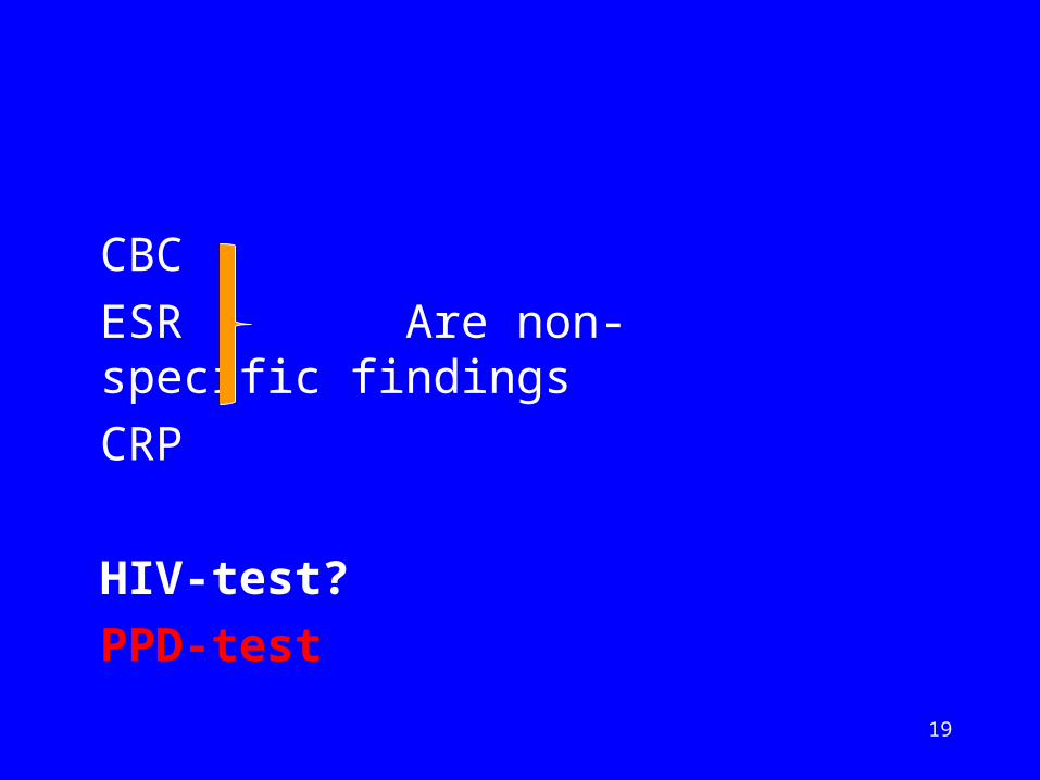
CBC
ESR Are non-specific findings
CRP
HIV-test?
PPD-test
19

SYNOVIAL FLUID
• Color
• WBC & diff: 10,000-30,000 cell/ml & PMN dominancy
(like to other chronic synovitis)
• Glucose, protein
AFB-smier (10-20% +)
Synovial fluid culture (79% +)20

Synovial biopsies :Rice bodies, pannus, necrosis, caseating or noncaseating granulomas ( 94% ) D.D: fungal, atypical mycobacterial infections, brucellosis& sarcoidosis
Synovial culture :confirms osteoarticular TB in ( 94% )
PCR 21

New Diagnostics:
Interferon Gamma Release Assays (IGRA’S)
Rapid Molecular Amplification
22

Diagnosis:
• The diagnosis of TB requires a high index of suspicion
• Diagnosis rests upon a combination of clinical presentation, pathologic findings, and culture results
• In endemic area?
23

Differential diagnosis?
24

25
Peripheral Arthritis Of
Brucellosis

Brucellosis is a zoonotic disease of domestic animals and humans.
The Many Names of Brucellosis: Malta Fever Undulant Fever Mediterranean Fever Gastric Fever
26

History:
• 1861: Marston published description of “Mediterranean gastric remittent fever”
• 1886: Sir David Bruce, microbe isolated from a soldier with Malta Fever; named it Micrococcus mellitensus
• 1897: Bernard Bang, first isolated B. abortus from cattle
27

Brucella species:
Four species can be pathogenic in humans: B. melitensis, B. abortus, B. canis and B. suis.
They are highly infectious, especially B. melitensis and B. suis.
28

BRUCELLANon-motile
Non-spore forming
Intracellular
Lack capsules
Gram-negative coccobacilliEnvironmental persistence
Temp, pH, humidityFrozen and aborted materials
29

Pathogenecity:
Infection of non-phagocytic cellsMechanism of invasion not clearly understood
Localize in rough endoplasmic reticulum
Infection of phagocytic cells Invasion of PMN or MN cells by suppression of
bactericidal responses
S-LPS (smooth) plays major role in intracellular survival; survive more effectively than rough
low toxicity for macrophages
30

Pathogenecity:
Virulence Elimination of virulent organisms depends on activated macrophages
and development of Th1 cell-mediated response to antigens
DeterminantsS-LPS is main antigen responsible for protection
Inhibition of phagolysosome fusion
Activation of myelo-peroxidase-halide system
Production of TNF
31

Epidemiology:
Worldwide, particularly Mediterranean, Africa, Middle East, Latin America
True incidence unknownEndemic areas: >200/100,000
U.S.: <1/100,000
In Iran, Brucellosis represents a major health problem
Iran J Radiol 2009, 6(1) 32

TRANSMISSION:
• Oral entry - most common route
Ingestion of contaminated animal products (often raw milk or its derivatives)
contact with contaminated fingers
• Aerosols
Inhalation of bacteria
Contamination of the conjunctivae
• Percutaneous :infection through skin
abrasions or by accidental inoculation
33

Human Disease• Incubation period:2-4 week
• Can affect any organ or organ system
• All patients have a cyclical fever
• clinical signs & symptoms:HeadacheWeaknessArthralgiaDepressionWeight lossFatigue Liver dysfunction 34

Human Disease:Duration of disease?days to months& year if untreated
Osteoarticular complications (In 20-60% of cases)
1. Reactive arthritis
2. Septic arthritis(38.8%)
3. Spondylitis (6.8%)
4. Sacroiliitis(46.6%)
5. Tendonitis&bursitis35

Sacroiliitis:
• Usually unilateral& nondestructive• Often Presents acutely& dramaticly• Severe low back& buttock pain• Difficulty in walking& standing• Fever(80%)• Direct tenderness on involve SIj• Bone scan is the most sensitive technique
36

37
Peripheral arthritis:
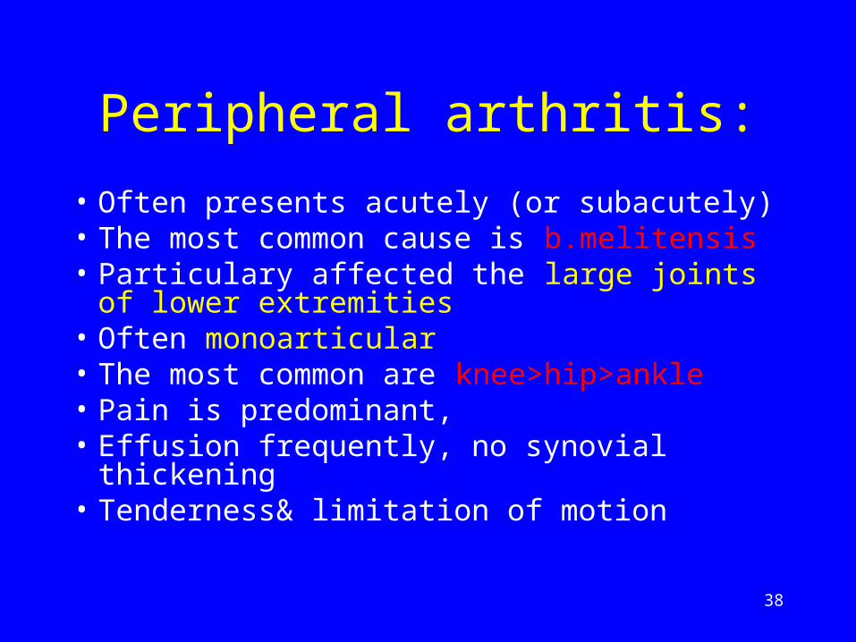
• Often presents acutely (or subacutely)• The most common cause is b.melitensis• Particulary affected the large joints of lower
extremities• Often monoarticular• The most common are knee>hip>ankle • Pain is predominant,• Effusion frequently, no synovial thickening• Tenderness& limitation of motion
38

Reactive arthritis
Sterile polyarthritis Non-destructive Intermittent & self-limited
39

Paraclinics:
• WBC: NL or & lymph • CRP, ESR:NL or
• Synovial fluid:• WBC:10,000-15,000/ml & lymph-dominancy• The yield of organism culture is low(50%)
• Plain x-ray is not diagnostic
40
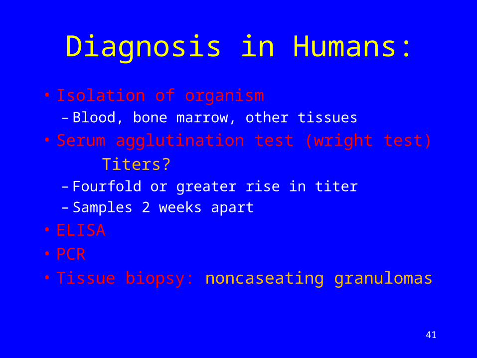
Diagnosis in Humans:
• Isolation of organism– Blood, bone marrow, other tissues
• Serum agglutination test (wright test)
Titers?– Fourfold or greater rise in titer
– Samples 2 weeks apart
• ELISA
• PCR• Tissue biopsy: noncaseating granulomas
41

42