1 Pathophysiology of GIT I Oral cavity and salivary glands Oesophagus Stomach and duodenum Small and...
68
1 Pathophysiology of GIT I Oral cavity and salivary glands Oesophagus Stomach and duodenum Small and large intestine
-
Upload
kathleen-heath -
Category
Documents
-
view
219 -
download
0
Transcript of 1 Pathophysiology of GIT I Oral cavity and salivary glands Oesophagus Stomach and duodenum Small and...
1
Pathophysiology of GIT I
Oral cavity and salivary glandsOesophagusStomach and duodenum Small and large intestine
2
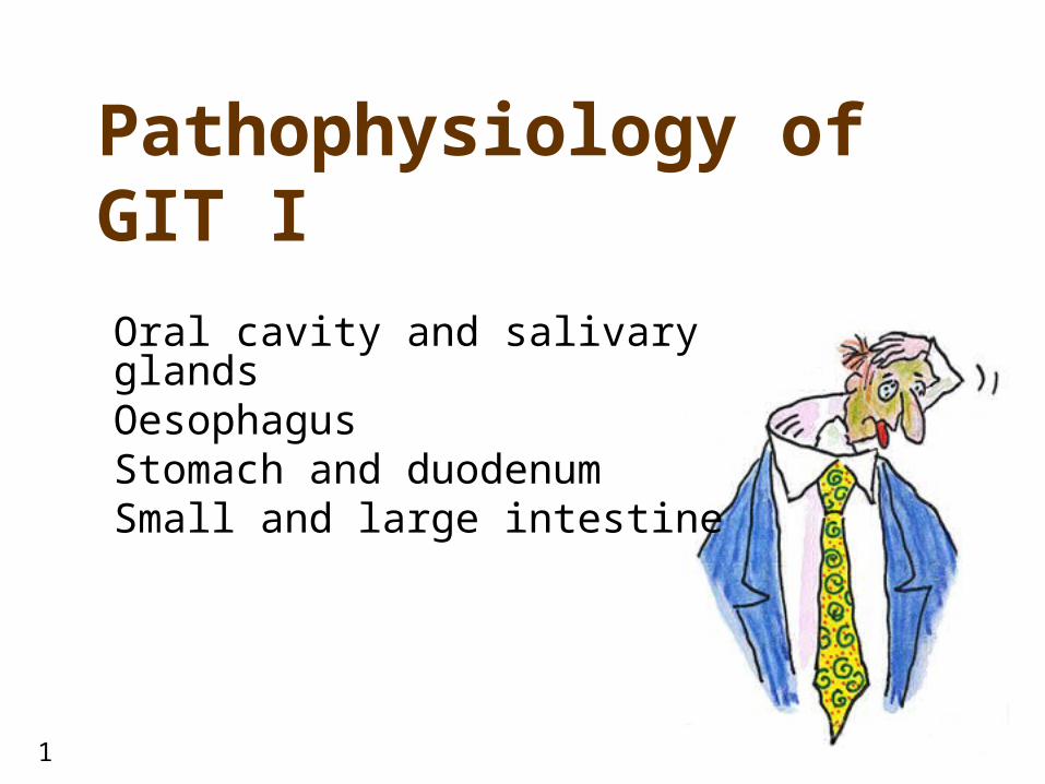
GIT• 1- oesophagus• 2- organs of peritoneal
cavity• 3- stomach (1.5l)• 4- gastroesophageal
junction• 5- pylorus• 6- small intestine (4.5 –
6m) • 7- duodenum• 8- jejunum• 9- ileum
• 10- ileocaecal valve• 11- large intestine
• ascendant• horizontal• descendant• rectum + anus

3
Pathophysiology of oral cavity

4
Pathophysiology of oral cavity• salivary glands - salivation (1 - 1.5l/day)
• continual production by small salivary glands• large glands secerns only upon stimulus
• centrum in medulla oblongata sal. glands (via n. facialis)• afferentation from upper centres (cortex, hypothalamus) upon
stimuli (taste, smell, chewing, …)• enzymes and ions of saliva
• -amylase (polysaccharides), lipase• lysozyme (bactericide)• K+, Na+, Cl-, HCO3
-
• disease of oral cavity• abnormal secretion of saliva
• - inflammation (e.g. tonsillitis), mechanical irritation • (xerostomy) - dehydration, Sjögren syndrome, drugs
• abnormal chewing• painful mandibular joint• injury of tongue• painful teeth• mucosal inflammation
• infections• herpetic (HSV-1), bacterial, candidiasis (in immune
compromised patients)• diseases of temporomandibular joint
• pain• dislocation (habitual)
• precanceroses and tumors of oral cavity• leucoplakia• carcinoma – smokers, alcoholics
• signs of systemic diseases in oral cavity• anaemia• vitamin and iron carrncy• malnutrition• cyanosis• Crohn’s disease
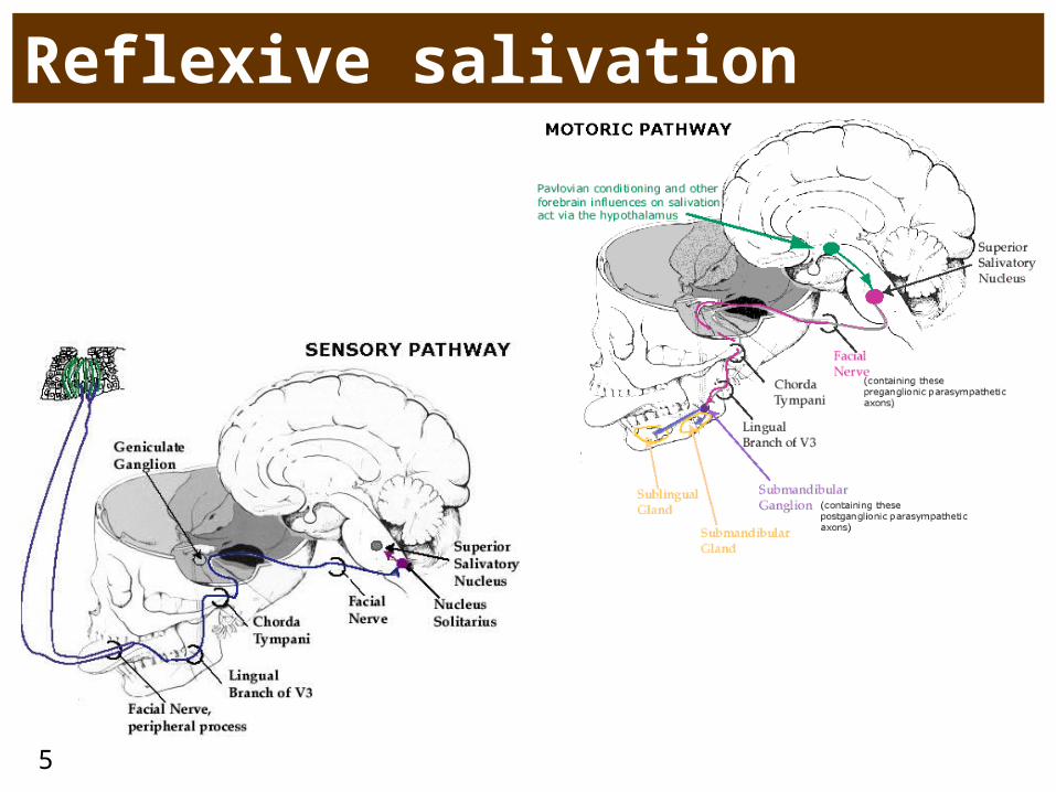
5
Reflexive salivation

6
Sjögren syndrome• syn. keratokonjunktivitis sicca• autoimmune reaction against salivary
(xerostomy) and tear glands (xerophtalmy)
• initiated by viral infection?
• symptoms• difficulties of chewing and swallowing• difficult talking• dry cough• irritation, eye burning, foreign body
feeling and reddening of eye • sometimes accompanied by joint and
muscle pain• SS can coexist with other autoimmune
diseases • rheumatoid arthritis• systemic lupus erythematodes• thyreopathy
7
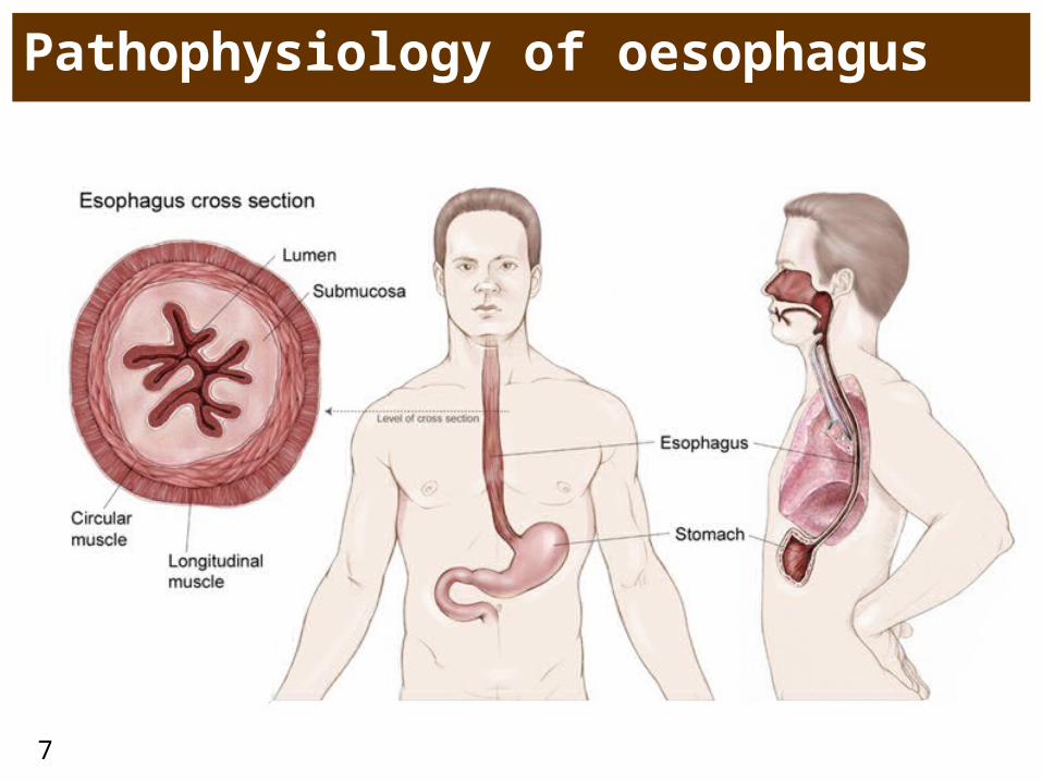
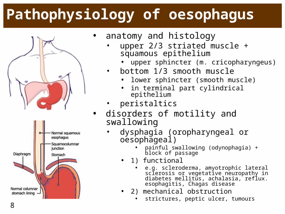
Pathophysiology of oesophagus
8
Pathophysiology of oesophagus• anatomy and histology
• upper 2/3 striated muscle + squamous epithelium• upper sphincter (m. cricopharyngeus)
• bottom 1/3 smooth muscle• lower sphincter (smooth muscle)• in terminal part cylindrical epithelium
• peristaltics• disorders of motility and swallowing
• dysphagia (oropharyngeal or oesophageal)
• painful swallowing (odynophagia) + block of passage
• 1) functional• e.g. scleroderma, amyotrophic lateral sclerosis
or vegetative neuropathy in diabetes mellitus, achalasia, reflux. esophagitis, Chagas disease
• 2) mechanical obstruction• strictures, peptic ulcer, tumours
9
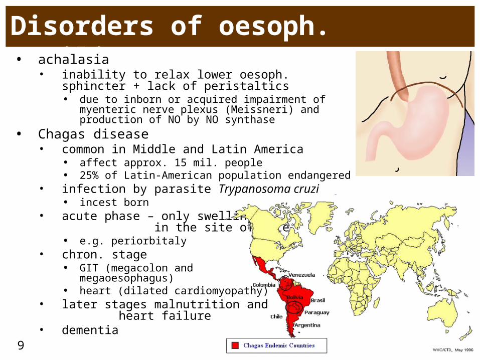
Disorders of oesoph. motility• achalasia
• inability to relax lower oesoph. sphincter + lack of peristaltics• due to inborn or acquired impairment of myenteric
nerve plexus (Meissneri) and production of NO by NO synthase
• Chagas disease• common in Middle and Latin America
• affect approx. 15 mil. people• 25% of Latin-American population endangered
• infection by parasite Trypanosoma cruzi• incest born
• acute phase – only swelling in the site of bite • e.g. periorbitaly
• chron. stage • GIT (megacolon and
megaoesophagus)• heart (dilated cardiomyopathy)
• later stages malnutrition and heart failure
• dementia
10
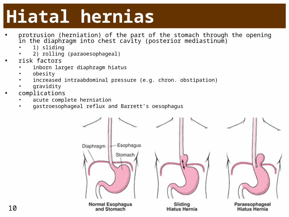
Hiatal hernias• protrusion (herniation) of the part of the stomach through the opening in the
diaphragm into chest cavity (posterior mediastinum)• 1) sliding • 2) rolling (paraoesophageal)
• risk factors• inborn larger diaphragm hiatus • obesity• increased intraabdominal pressure (e.g. chron. obstipation)• gravidity
• complications• acute complete herniation• gastroesophageal reflux and Barrett’s oesophagus
11
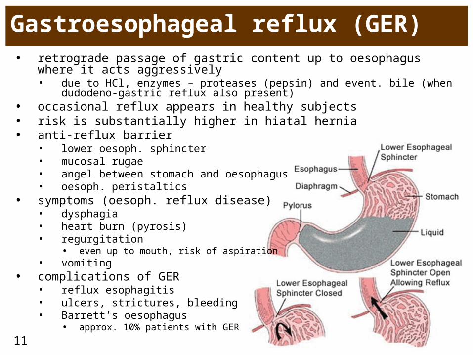
Gastroesophageal reflux (GER) • retrograde passage of gastric content up to oesophagus where it acts
aggressively• due to HCl, enzymes – proteases (pepsin) and event. bile (when dudodeno-
gastric reflux also present)• occasional reflux appears in healthy subjects• risk is substantially higher in hiatal hernia• anti-reflux barrier
• lower oesoph. sphincter• mucosal rugae• angel between stomach and oesophagus• oesoph. peristaltics
• symptoms (oesoph. reflux disease)• dysphagia• heart burn (pyrosis)• regurgitation
• even up to mouth, risk of aspiration• vomiting
• complications of GER• reflux esophagitis• ulcers, strictures, bleeding• Barrett’s oesophagus
• approx. 10% patients with GER
12
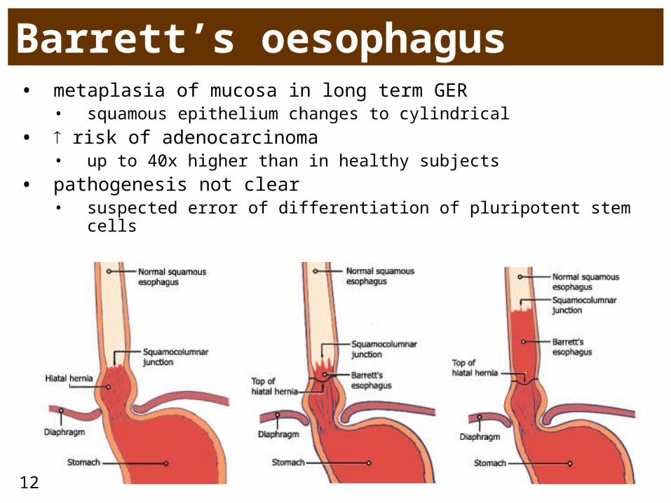
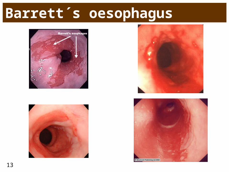
Barrett’s oesophagus• metaplasia of mucosa in long term GER
• squamous epithelium changes to cylindrical
• risk of adenocarcinoma • up to 40x higher than in healthy subjects
• pathogenesis not clear• suspected error of differentiation of pluripotent stem cells
13
Barrett´s oesophagus
14
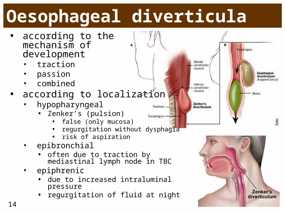
Oesophageal diverticula• according to the
mechanism of development • traction• passion• combined
• according to localization• hypopharyngeal
• Zenker’s (pulsion)• false (only mucosa)• regurgitation without dysphagia• risk of aspiration
• epibronchial• often due to traction by mediastinal
lymph node in TBC• epiphrenic
• due to increased intraluminal pressure • regurgitation of fluid at night
15
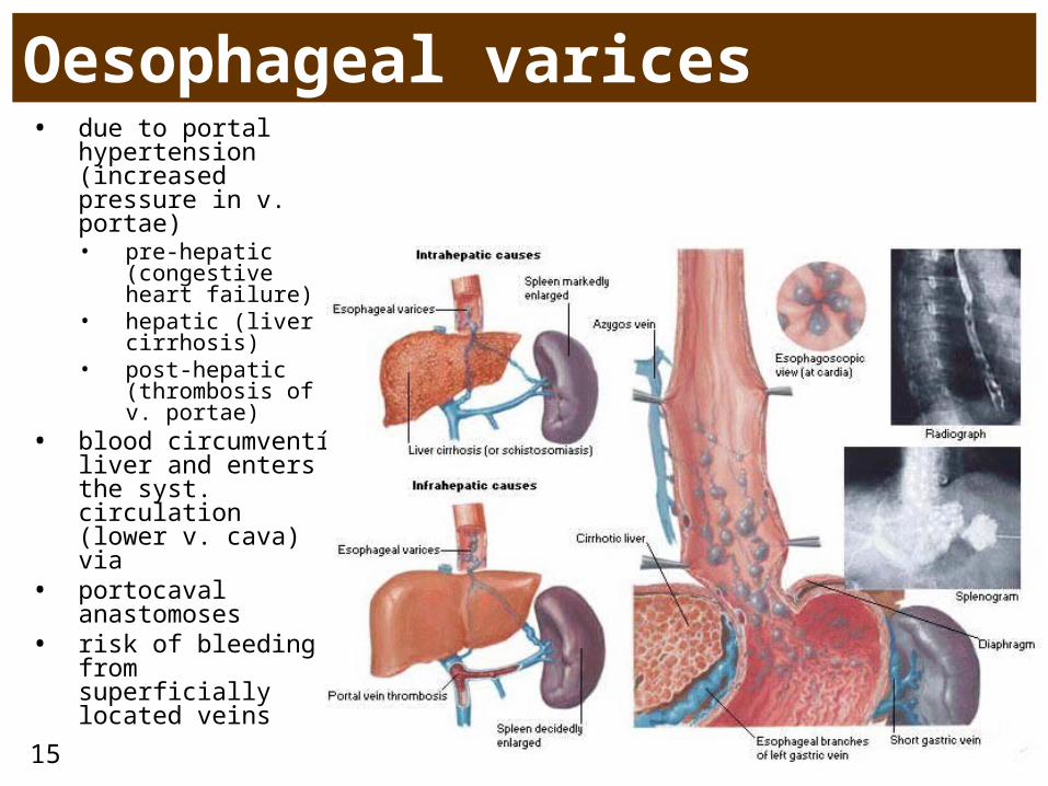
Oesophageal varices• due to portal
hypertension (increased pressure in v. portae)• pre-hepatic
(congestive heart failure)
• hepatic (liver cirrhosis)
• post-hepatic (thrombosis of v. portae)
• blood circumventí liver and enters the syst. circulation (lower v. cava) via
• portocaval anastomoses
• risk of bleeding from superficially located veins
16
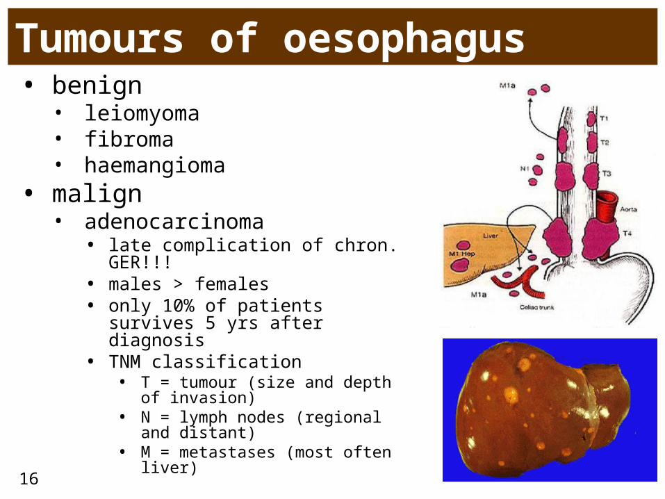
Tumours of oesophagus• benign• leiomyoma• fibroma• haemangioma
• malign• adenocarcinoma• late complication of chron.
GER!!!• males > females • only 10% of patients survives 5
yrs after diagnosis • TNM classification
• T = tumour (size and depth of invasion)
• N = lymph nodes (regional and distant)
• M = metastases (most often liver)
17
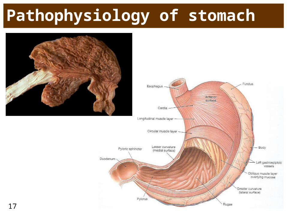
Pathophysiology of stomach
18
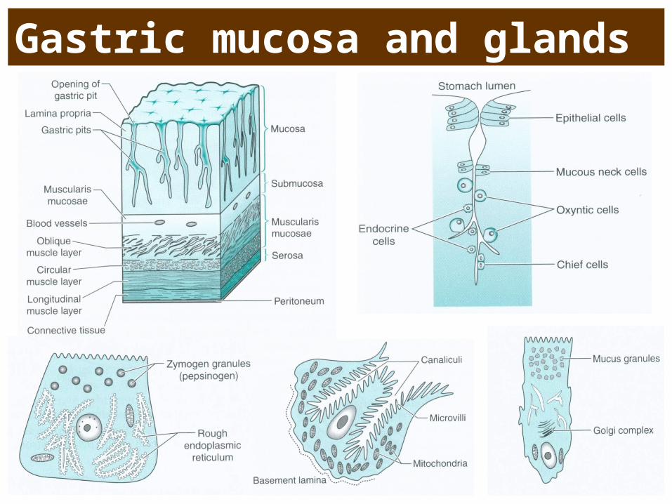
Gastric mucosa and glands
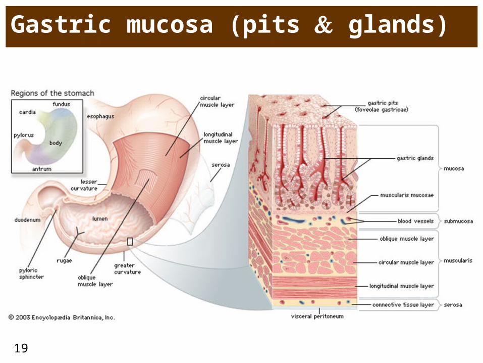
19
Gastric mucosa (pits glands)
20
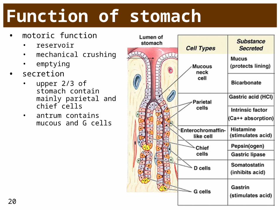
Function of stomach• motoric function
• reservoir• mechanical crushing• emptying
• secretion• upper 2/3 of stomach
contain mainly parietal and chief cells
• antrum contains mucous and G cells
21
22
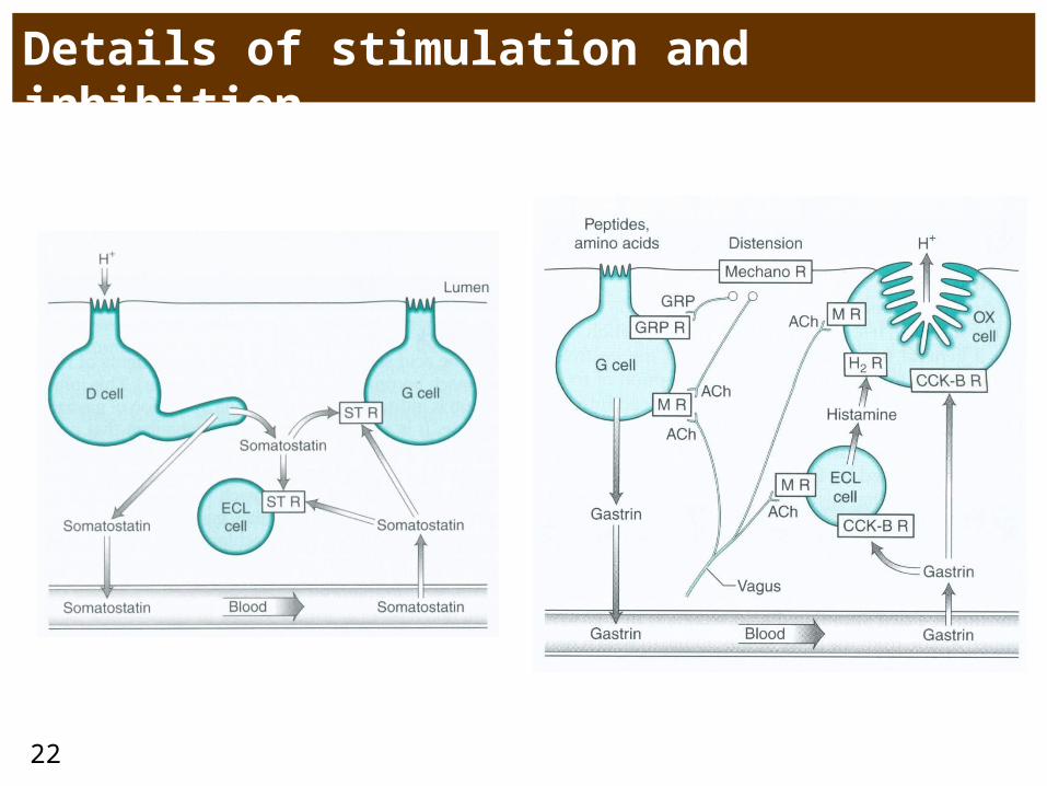
Details of stimulation and inhibition
23
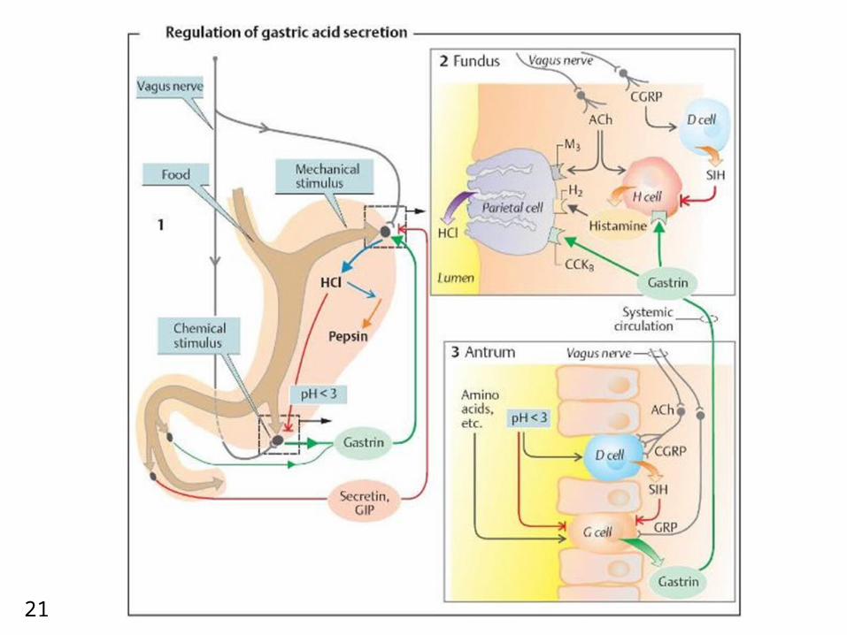
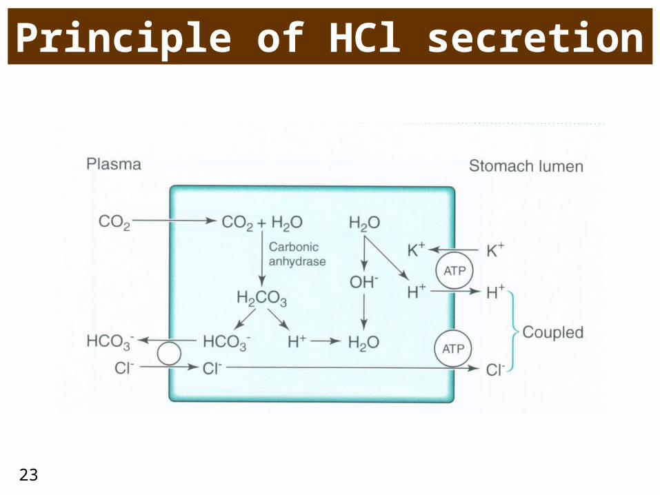
Principle of HCl secretion
24
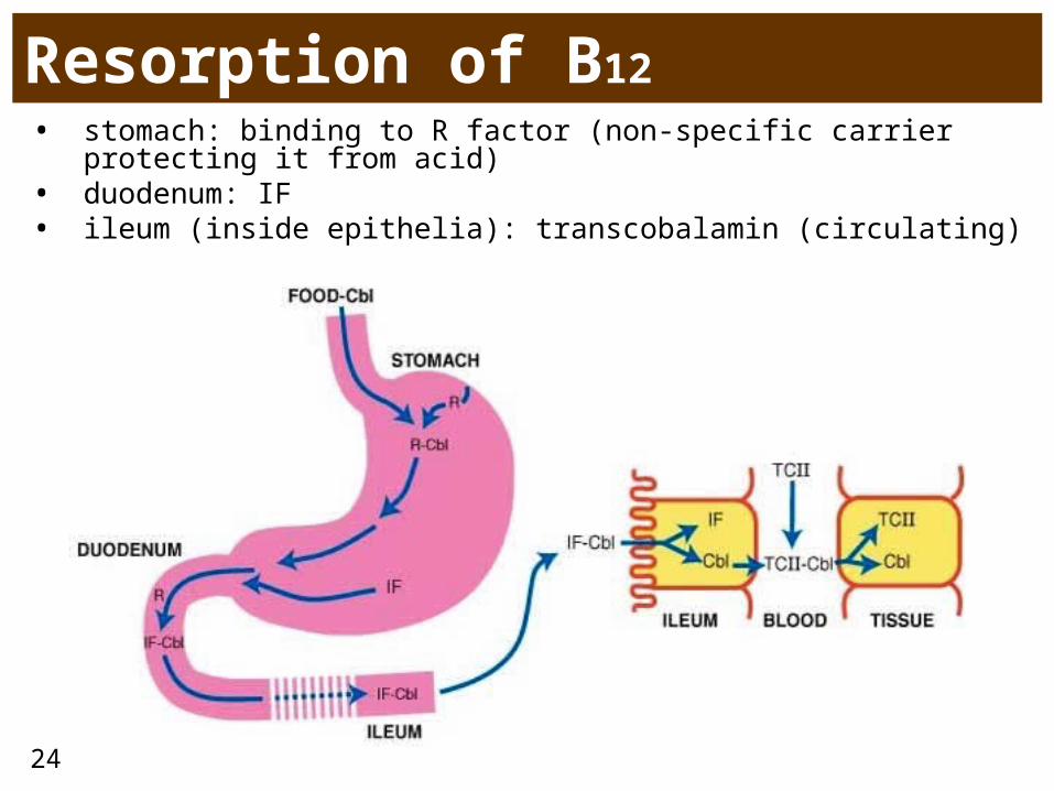
Resorption of B12• stomach: binding to R factor (non-specific carrier protecting it
from acid)• duodenum: IF• ileum (inside epithelia): transcobalamin (circulating)
25
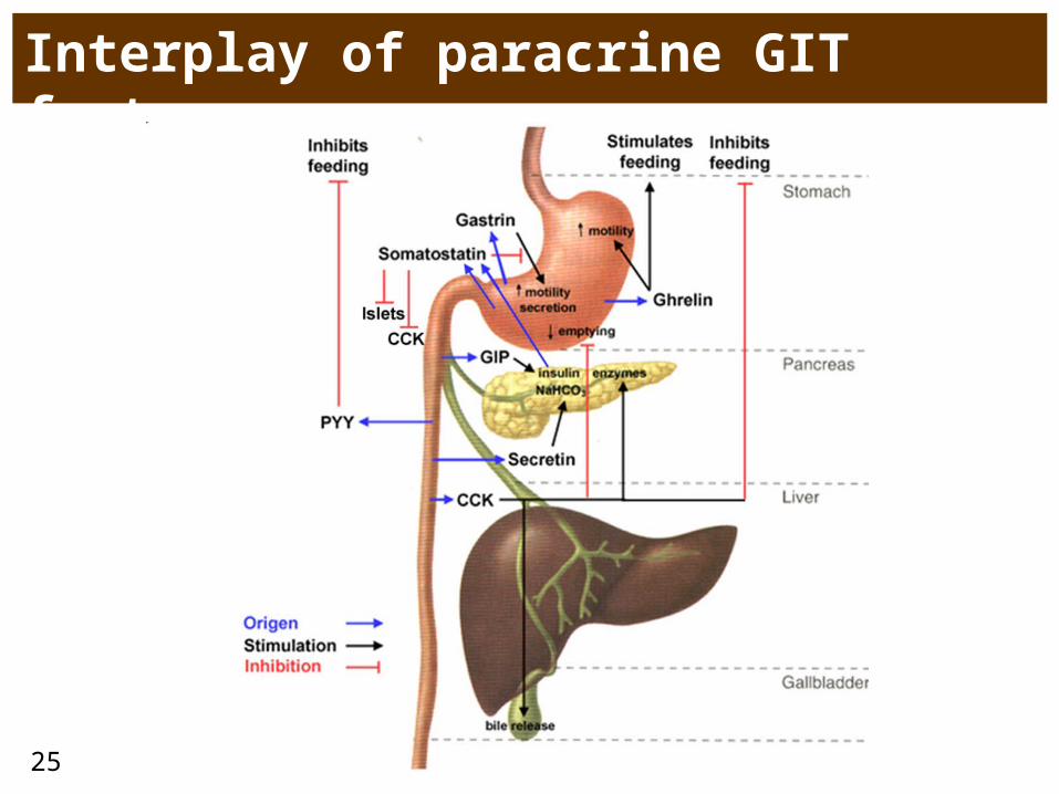
Interplay of paracrine GIT factors
26
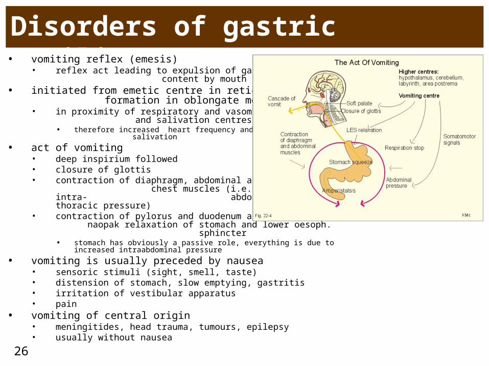
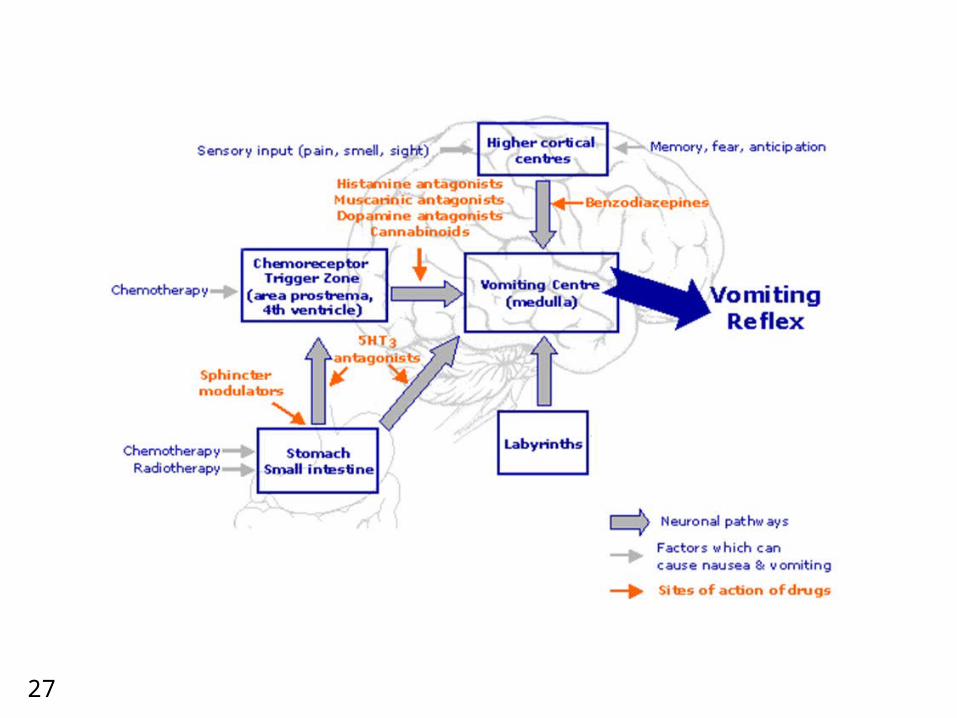
Disorders of gastric motility• vomiting reflex (emesis)
• reflex act leading to expulsion of gastric content by mouth
• initiated from emetic centre in reticular formation in oblongate medulla• in proximity of respiratory and vasomotor and
salivation centres• therefore increased heart frequency and
salivation
• act of vomiting• deep inspirium followed• closure of glottis• contraction of diaphragm, abdominal and
chest muscles (i.e. increase of intra- abdominal and intra-thoracic pressure)
• contraction of pylorus and duodenum and naopak relaxation of stomach and lower oesoph. sphincter• stomach has obviously a passive role, everything is due to increased
intraabdominal pressure
• vomiting is usually preceded by nausea• sensoric stimuli (sight, smell, taste)• distension of stomach, slow emptying, gastritis• irritation of vestibular apparatus• pain
• vomiting of central origin• meningitides, head trauma, tumours, epilepsy• usually without nausea
27
28
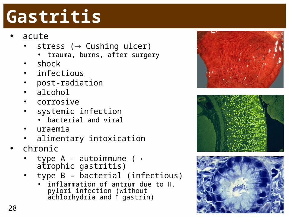
Gastritis• acute
• stress ( Cushing ulcer)• trauma, burns, after surgery
• shock• infectious• post-radiation• alcohol• corrosive• systemic infection
• bacterial and viral• uraemia• alimentary intoxication
• chronic • type A - autoimmune ( atrophic
gastritis)• type B – bacterial (infectious)
• inflammation of antrum due to H. pylori infection (without achlorhydria and gastrin)
29
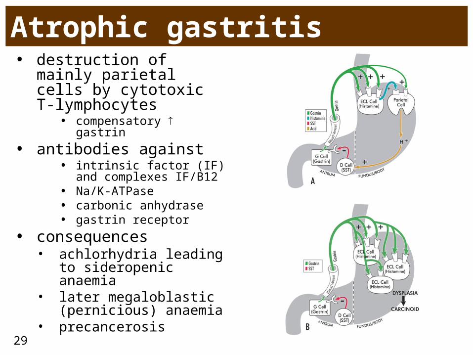
Atrophic gastritis prekanceróza • destruction of mainly
parietal cells by cytotoxic T-lymphocytes
• compensatory gastrin
• antibodies against• intrinsic factor (IF) and
complexes IF/B12 • Na/K-ATPase• carbonic anhydrase• gastrin receptor
• consequences • achlorhydria leading
to sideropenic anaemia
• later megaloblastic (pernicious) anaemia
• precancerosis
30
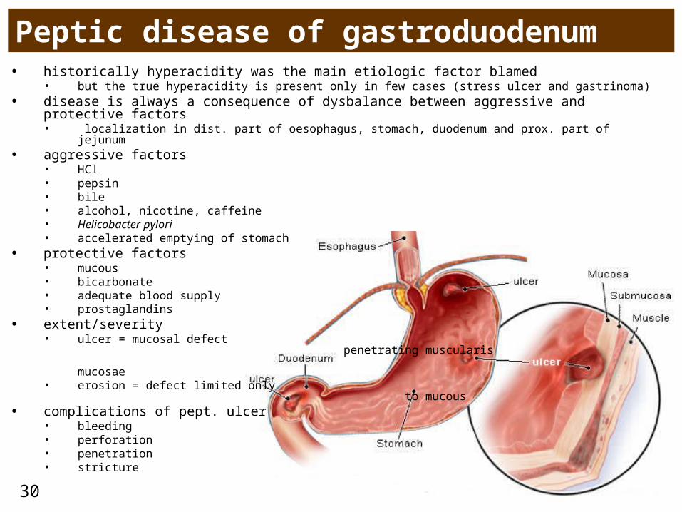
Peptic disease of gastroduodenum• historically hyperacidity was the main etiologic factor blamed
• but the true hyperacidity is present only in few cases (stress ulcer and gastrinoma)• disease is always a consequence of dysbalance between aggressive and protective factors
• localization in dist. part of oesophagus, stomach, duodenum and prox. part of jejunum• aggressive factors
• HCl• pepsin• bile• alcohol, nicotine, caffeine• Helicobacter pylori• accelerated emptying of stomach
• protective factors• mucous• bicarbonate• adequate blood supply• prostaglandins
• extent/severity• ulcer = mucosal defect penetrating
muscularis mucosae • erosion = defect limited only to
mucous • complications of pept. ulcer
• bleeding• perforation• penetration• stricture
31
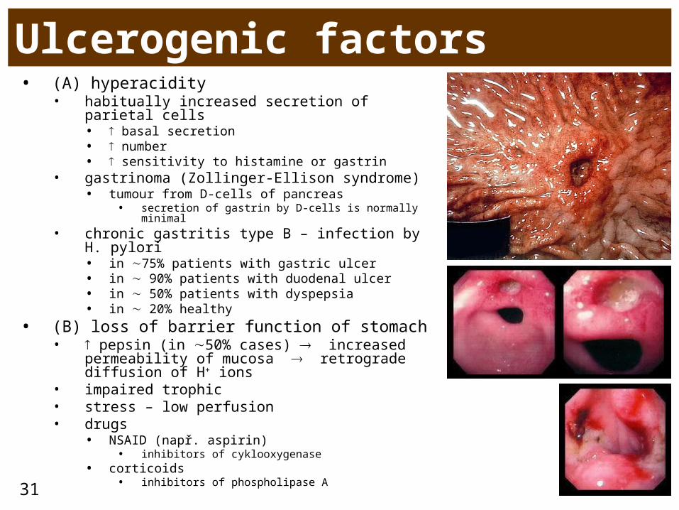
Ulcerogenic factors• (A) hyperacidity
• habitually increased secretion of parietal cells • basal secretion• number• sensitivity to histamine or gastrin
• gastrinoma (Zollinger-Ellison syndrome)• tumour from D-cells of pancreas
• secretion of gastrin by D-cells is normally minimal
• chronic gastritis type B – infection by H. pylori• in 75% patients with gastric ulcer• in 90% patients with duodenal ulcer• in 50% patients with dyspepsia • in 20% healthy
• (B) loss of barrier function of stomach• pepsin (in 50% cases) increased
permeability of mucosa retrograde diffusion of H+ ions
• impaired trophic • stress – low perfusion• drugs
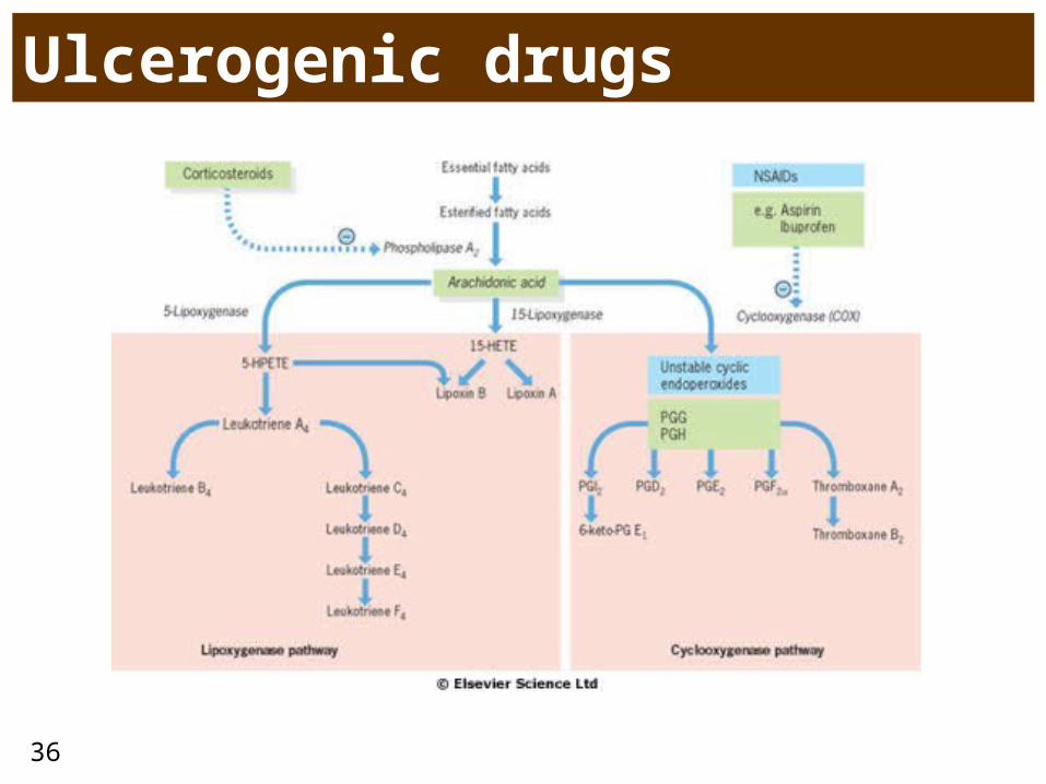
• NSAID (např. aspirin)• inhibitors of cyklooxygenase
• corticoids• inhibitors of phospholipase A
32
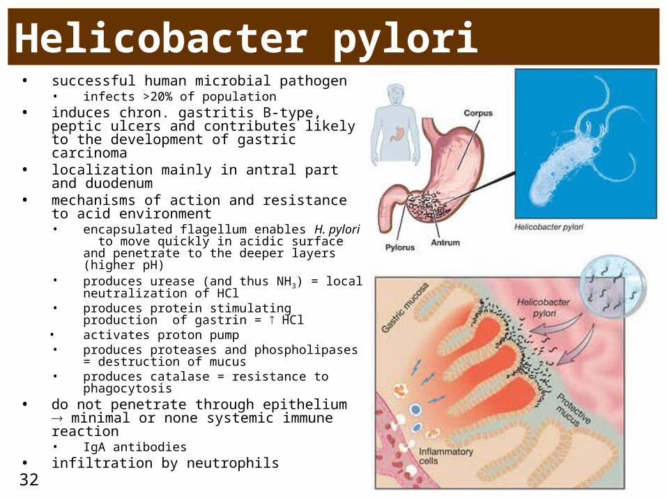
Helicobacter pylori• successful human microbial pathogen
• infects >20% of population• induces chron. gastritis B-type, peptic
ulcers and contributes likely to the development of gastric carcinoma
• localization mainly in antral part and duodenum
• mechanisms of action and resistance to acid environment• encapsulated flagellum enables H. pylori
to move quickly in acidic surface and penetrate to the deeper layers (higher pH)
• produces urease (and thus NH3) = local neutralization of HCl
• produces protein stimulating production of gastrin = HCl
• activates proton pump• produces proteases and phospholipases =
destruction of mucus• produces catalase = resistance to
phagocytosis• do not penetrate through epithelium
minimal or none systemic immune reaction• IgA antibodies
• infiltration by neutrophils

33
34
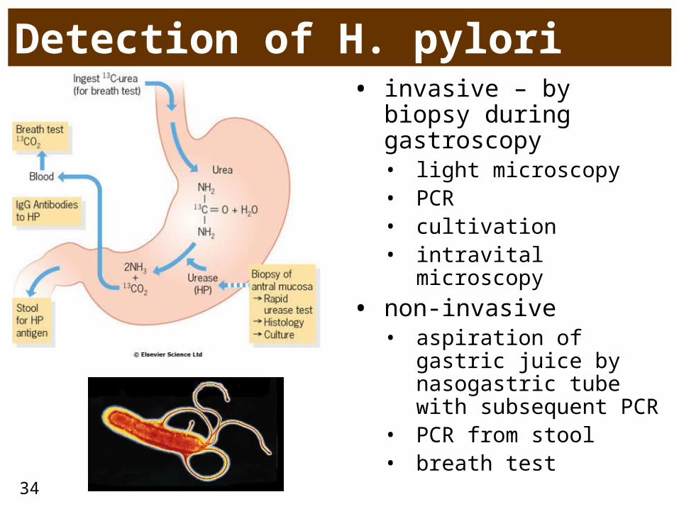
Detection of H. pylori• invasive – by biopsy
during gastroscopy• light microscopy • PCR• cultivation• intravital microscopy
• non-invasive• aspiration of gastric
juice by nasogastric tube with subsequent PCR
• PCR from stool• breath test
35
Symptoms of gastric vs. duodenal ulcer
• stomach• etiologically more often
contribution of loss of barrier function rather than true hyperacidity• chron. gastritis type B• duodenogastric reflux• drugs
• older people• painful in a fasting state,
relieved by meal• patients often put on weight
• duodenum• protection of duodenum
weak• Brunner’s glands secreting
alkalic mucus • coordinated peristaltics
mixing gastric content with pancreatic and biliary juices which then acidic content
• etiologically more often hyperacidity and infection by H. pylori
• genetic effects• often blood group 0• HLA-B5
• younger people• neurotics (faster gastric
motility)• painful after meal• seasonal manifestion
36
Ulcerogenic drugs
37
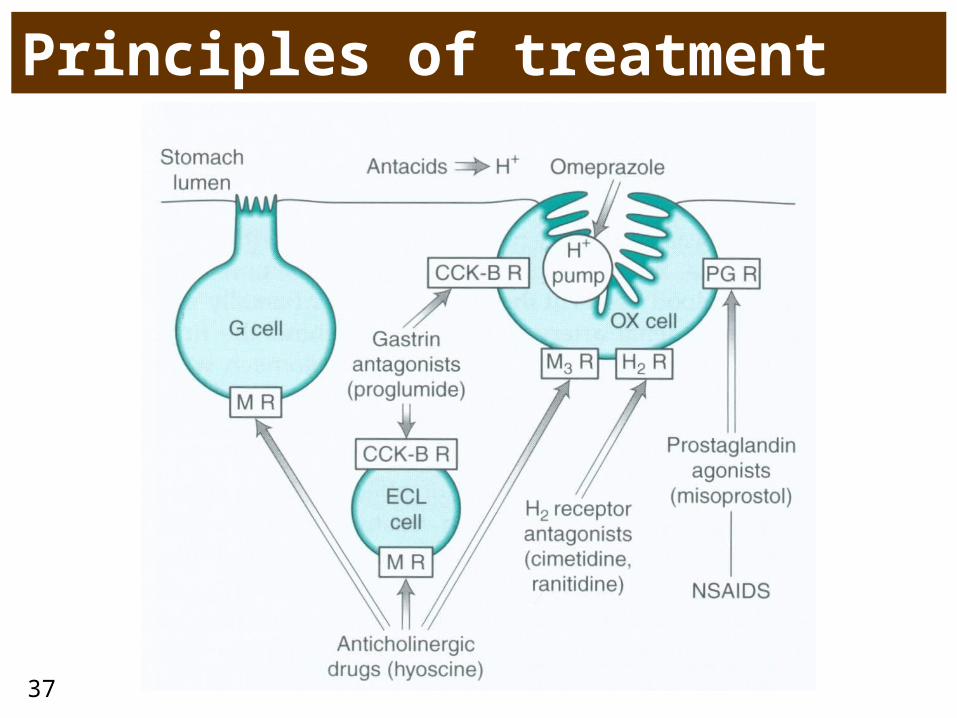
Principles of treatment
38
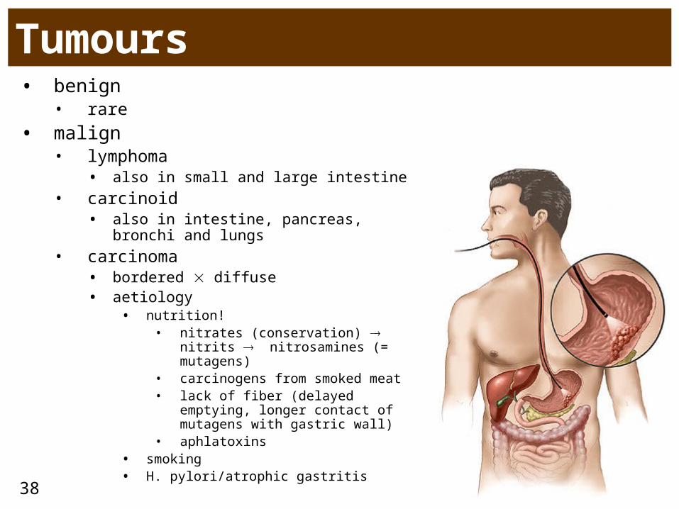
Tumours• benign
• rare
• malign• lymphoma
• also in small and large intestine• carcinoid
• also in intestine, pancreas, bronchi and lungs
• carcinoma• bordered diffuse• aetiology
• nutrition!• nitrates (conservation) nitrits
nitrosamines (= mutagens)• carcinogens from smoked meat• lack of fiber (delayed emptying,
longer contact of mutagens with gastric wall)
• aphlatoxins• smoking• H. pylori/atrophic gastritis
39
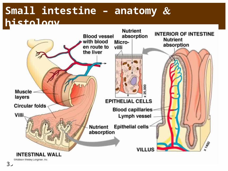
Small intestine – anatomy histology
40
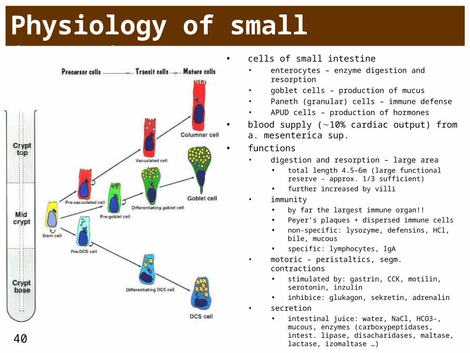
Physiology of small intestine• cells of small intestine
• enterocytes – enzyme digestion and resorption• goblet cells – production of mucus• Paneth (granular) cells – immune defense• APUD cells – production of hormones
• blood supply (10% cardiac output) from a. mesenterica sup.
• functions• digestion and resorption – large area
• total length 4.5–6m (large functional reserve - approx. 1/3 sufficient)
• further increased by villi
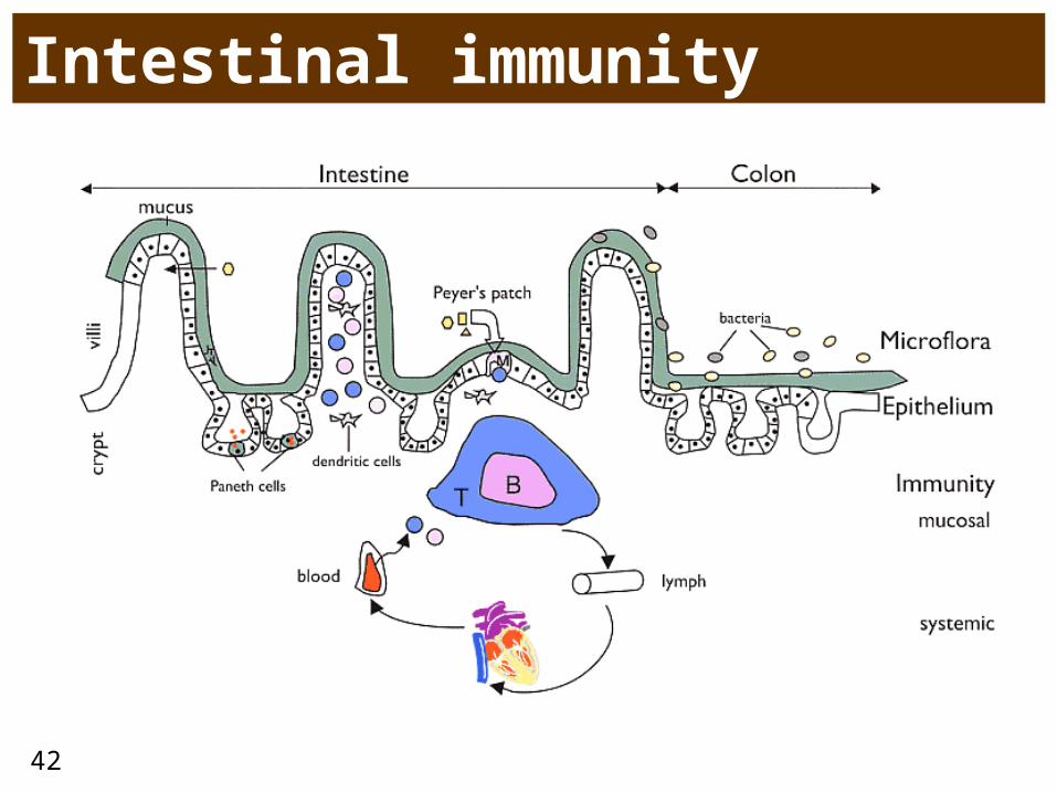
• immunity • by far the largest immune organ!!
• Peyer’s plaques + dispersed immune cells
• non-specific: lysozyme, defensins, HCl, bile, mucous
• specific: lymphocytes, IgA
• motoric – peristaltics, segm. contractions• stimulated by: gastrin, CCK, motilin, serotonin,
inzulin
• inhibice: glukagon, sekretin, adrenalin
• secretion• intestinal juice: water, NaCl, HCO3-, mucous,
enzymes (carboxypeptidases, intest. lipase, disacharidases, maltase, lactase, izomaltase …)
41
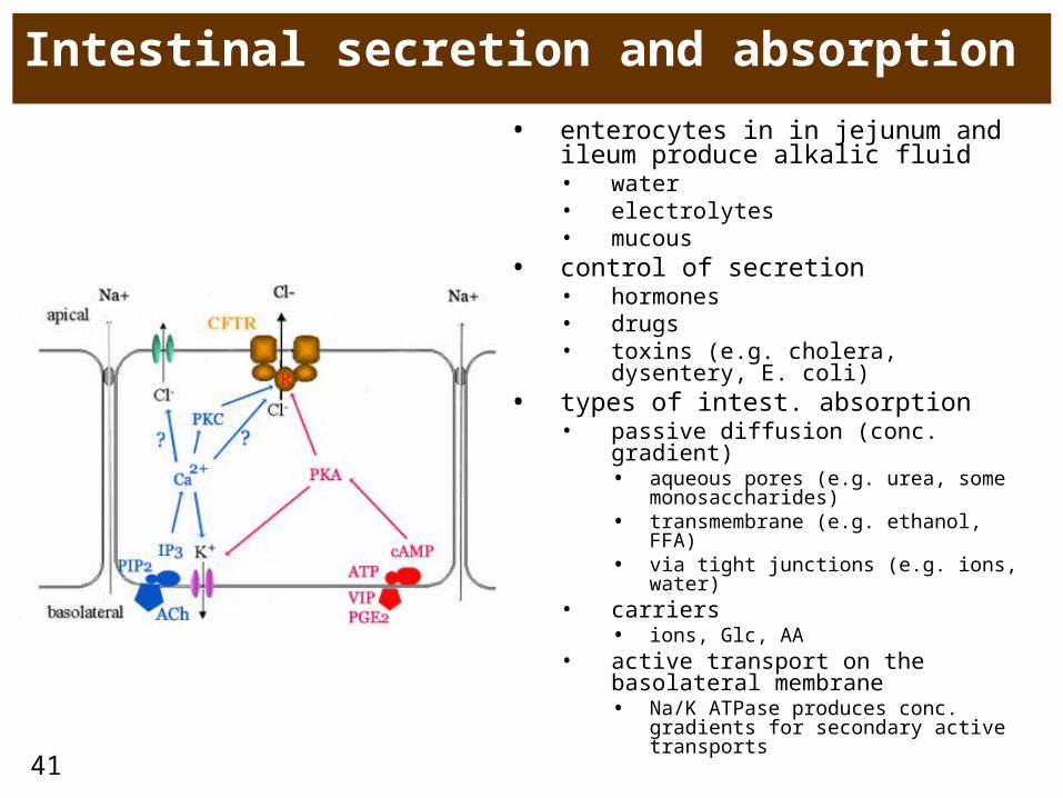
Intestinal secretion and absorption
• enterocytes in in jejunum and ileum produce alkalic fluid • water• electrolytes• mucous
• control of secretion• hormones• drugs • toxins (e.g. cholera, dysentery, E.
coli)• types of intest. absorption
• passive diffusion (conc. gradient)• aqueous pores (e.g. urea, some
monosaccharides)• transmembrane (e.g. ethanol, FFA) • via tight junctions (e.g. ions, water)
• carriers• ions, Glc, AA
• active transport on the basolateral membrane• Na/K ATPase produces conc.
gradients for secondary active transports
42
Intestinal immunity
43
Disorders of intestinal secretion and absorption = diarrhea• diarrhea = more frequent expulsion of stools (>3/day), often more liquid
consistence loss of fluid • due to imbalance between 3 main factors – secretion, resorption and motility
• acute• infection• dietary error• alimentary intoxication
• chronic• malabsorption (inflammatory bowel disease (Crohn disease, ulcerative colitis), chron. pancreatitis, liver
and biliary diseases)• colorectal carcinoma• neurogenic • metabolic (uremia, hyperthyreosis, adrenal insufficiency)
• etiology• infection, toxins, diet, neuropsychological (anxiety)
• pathogeneses• osmotic pressure (and thus water) in intest. lumen = osmotic
• typically when large amount of undigested nutrients stays in lumen• malabsorption syndrome (pancreatic insufficiency, biliary, disacharidaae deficiency – e.g. lactase)• ingestion (overdose) of salts (Mg, sulfates), antacids• bacterial overgrowth, resection, obstruction of lymphatics
• secretion of Cl (and thus water) into lumen = secretory• bacterial enterotoxins (Vibrio cholerae, Shigella dysenteriae, E. coli, Clostridium difficile, Salmonella typhi)• inflammatory exudation (Crohn d., ulcerative colitis)
• hypemotility• some regulatory peptides (VIP, serotonin, PGE)
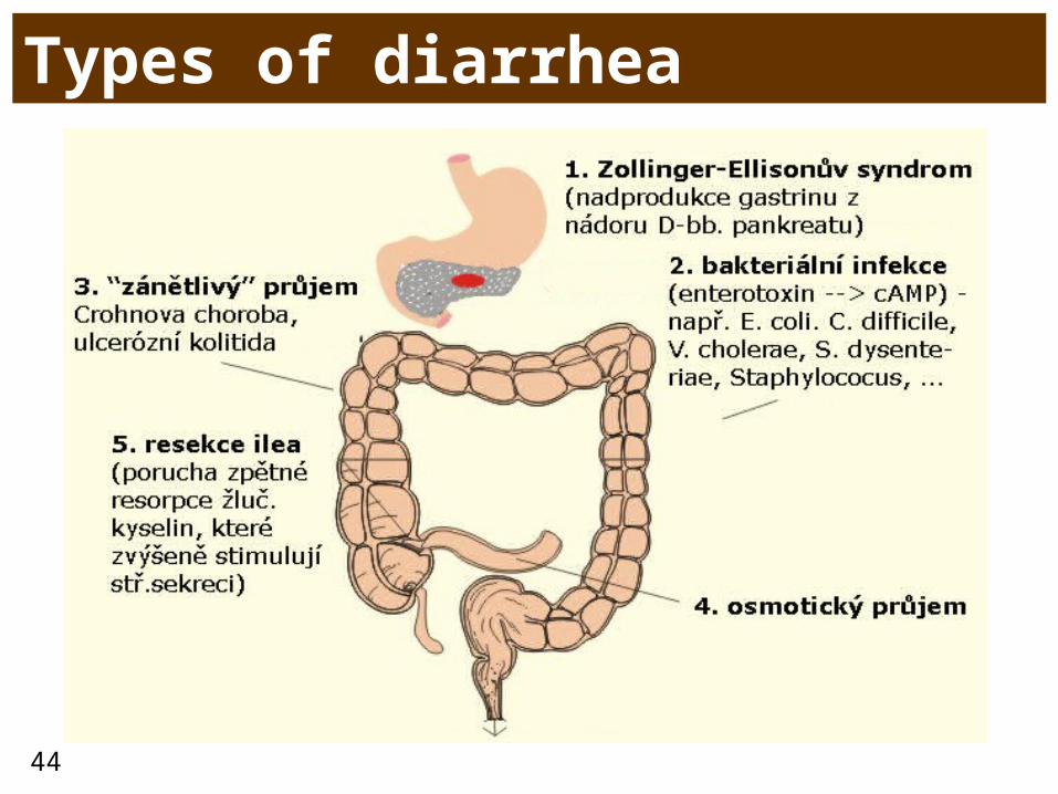
44
Types of diarrhea
45
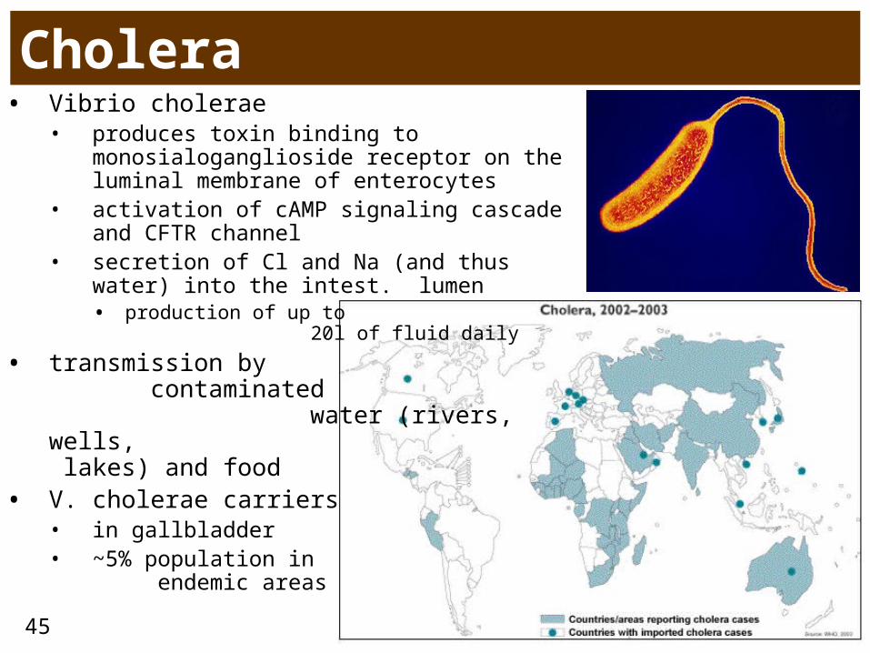
Cholera• Vibrio cholerae
• produces toxin binding to monosialoganglioside receptor on the luminal membrane of enterocytes
• activation of cAMP signaling cascade and CFTR channel
• secretion of Cl and Na (and thus water) into the intest. lumen• production of up to 20l
of fluid daily
• transmission by contaminated water (rivers, wells, lakes) and food
• V. cholerae carriers• in gallbladder • ~5% population in endemic
areas

46
Action of V. cholerae toxin
47
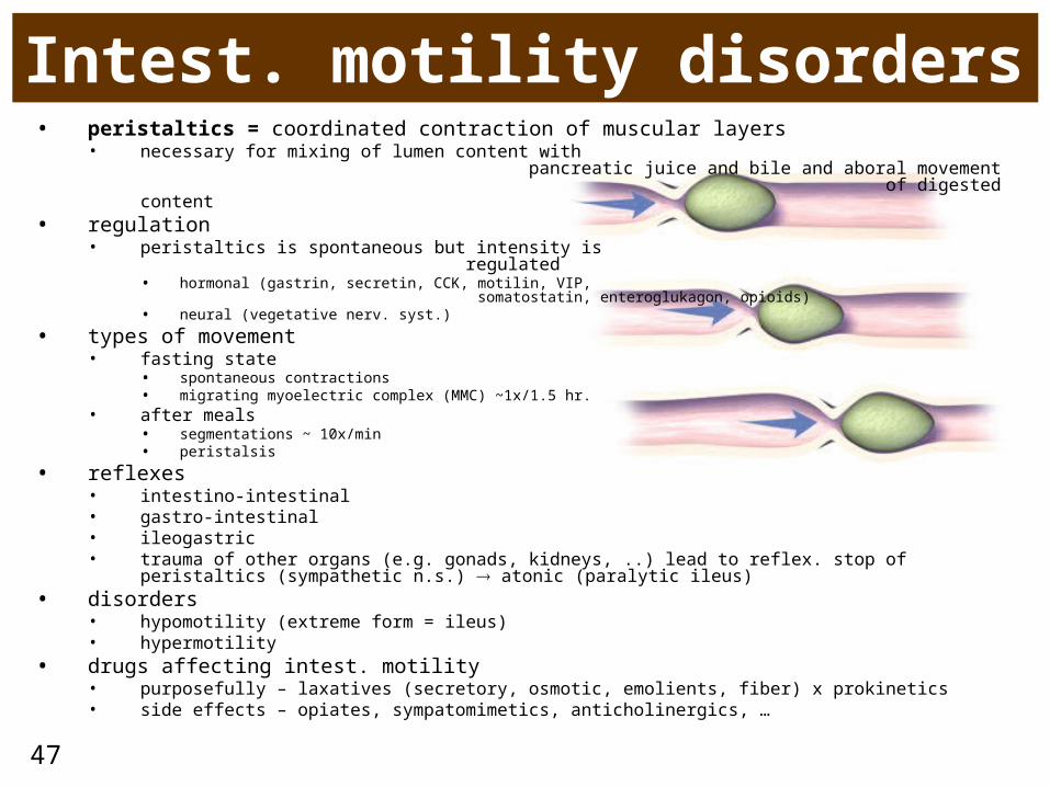
Intest. motility disorders• peristaltics = coordinated contraction of muscular layers
• necessary for mixing of lumen content with pancreatic juice and bile and aboral movement of digested content
• regulation • peristaltics is spontaneous but intensity is regulated
• hormonal (gastrin, secretin, CCK, motilin, VIP, somatostatin, enteroglukagon, opioids)
• neural (vegetative nerv. syst.)
• types of movement• fasting state
• spontaneous contractions• migrating myoelectric complex (MMC) ~1x/1.5 hr.
• after meals • segmentations ~ 10x/min• peristalsis
• reflexes• intestino-intestinal• gastro-intestinal• ileogastric• trauma of other organs (e.g. gonads, kidneys, ..) lead to reflex. stop of peristaltics (sympathetic
n.s.) atonic (paralytic ileus)• disorders
• hypomotility (extreme form = ileus)• hypermotility
• drugs affecting intest. motility• purposefully – laxatives (secretory, osmotic, emolients, fiber) x prokinetics• side effects – opiates, sympatomimetics, anticholinergics, …
48
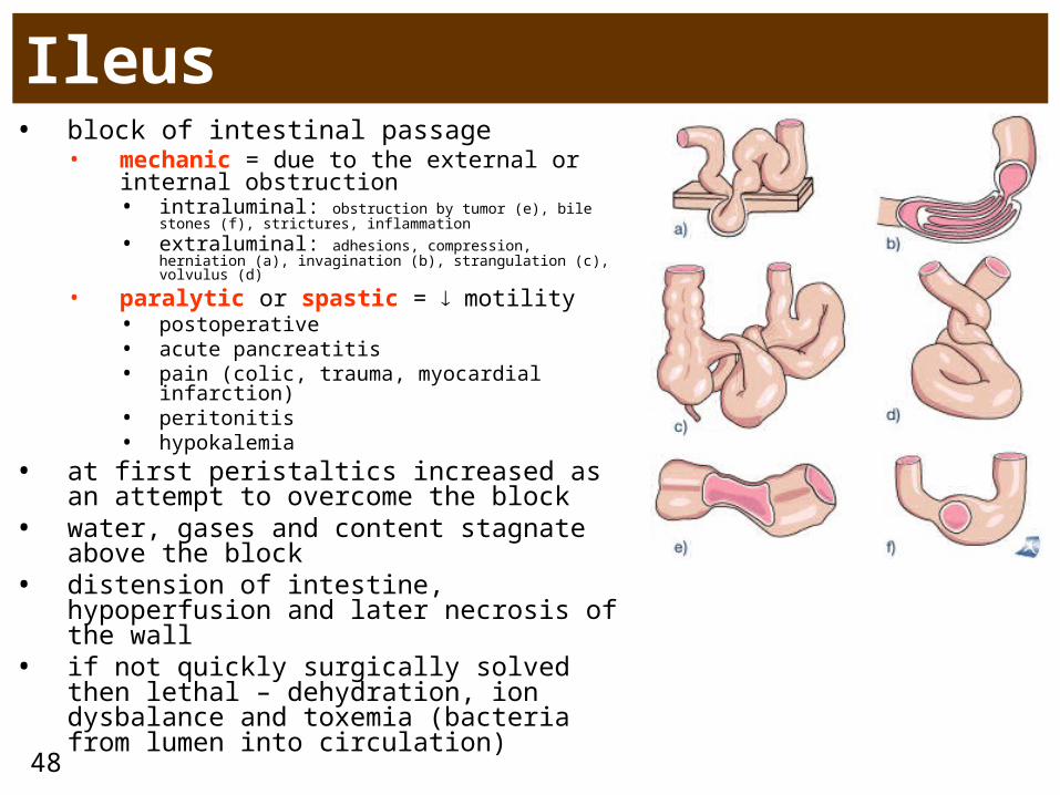
Ileus• block of intestinal passage
• mechanic = due to the external or internal obstruction • intraluminal: obstruction by tumor (e), bile stones (f),
strictures, inflammation
• extraluminal: adhesions, compression, herniation (a), invagination (b), strangulation (c), volvulus (d)
• paralytic or spastic = motility• postoperative• acute pancreatitis• pain (colic, trauma, myocardial infarction)• peritonitis• hypokalemia
• at first peristaltics increased as an attempt to overcome the block
• water, gases and content stagnate above the block
• distension of intestine, hypoperfusion and later necrosis of the wall
• if not quickly surgically solved then lethal – dehydration, ion dysbalance and toxemia (bacteria from lumen into circulation)
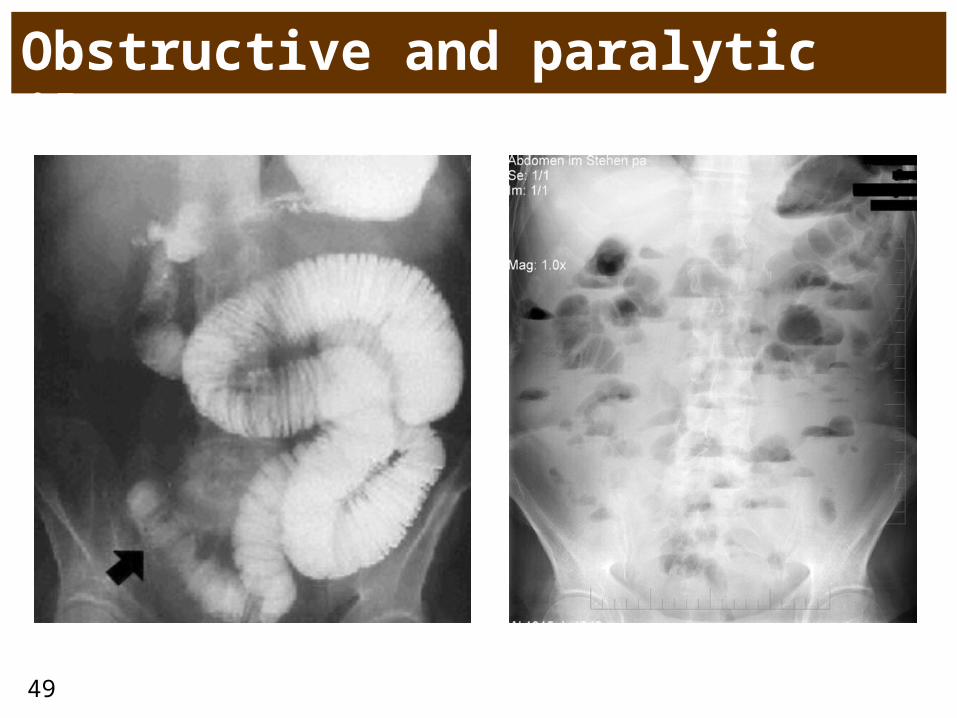
49
Obstructive and paralytic ileus
50
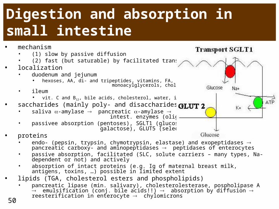
Digestion and absorption in small intestine• mechanism
• (1) slow by passive diffusion • (2) fast (but saturable) by facilitated transports
• localization • duodenum and jejunum
• hexoses, AA, di- and tripeptides, vitamins, FA, monoacylglycerols, cholesterol, Ca, Fe, water, ions
• ileum• vit. C and B12, bile acids, cholesterol, water, ions
• saccharides (mainly poly- and disaccharides)• saliva -amylase pancreatic -amylase intest.
enzymes (oligo- and disaccharides)• passivee absorption (pentoses), SGLT1 (glucose and
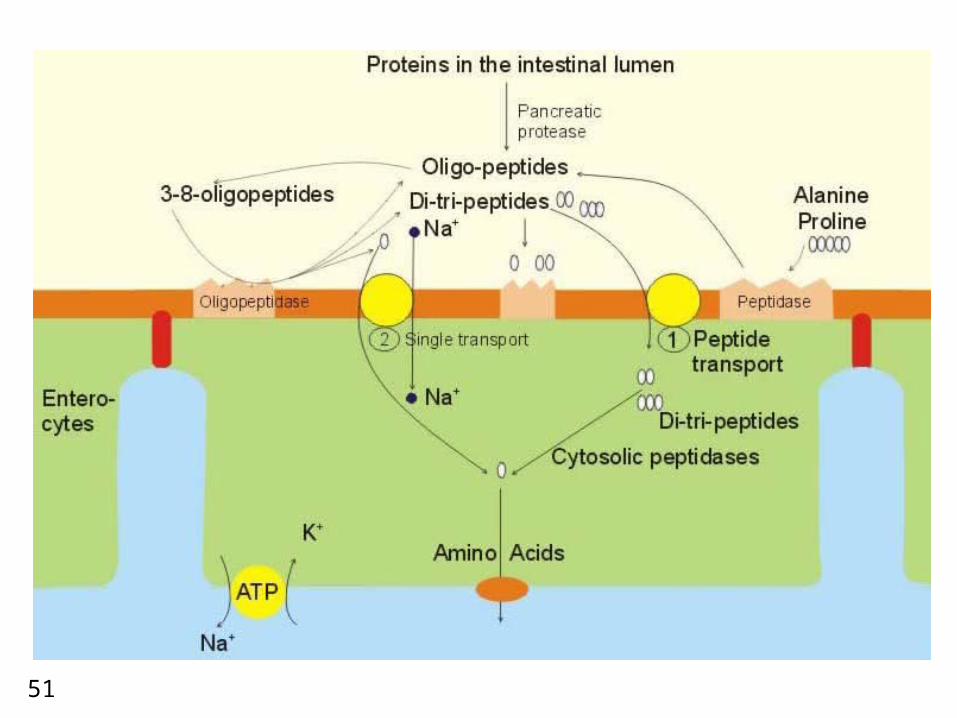
galactose), GLUT5 (selectively for fructose)• proteins
• endo- (pepsin, trypsin, chymotrypsin, elastase) and exopeptidases pancreatic carboxy- and aminopeptidases peptidases of enterocytes
• passive absorption, facilitated (SLC, solute carriers – many types, Na-dependent or not) and actively
• absorption of intact proteins (e.g. Ig of maternal breast milk, antigens, toxins, …) possible in limited extent
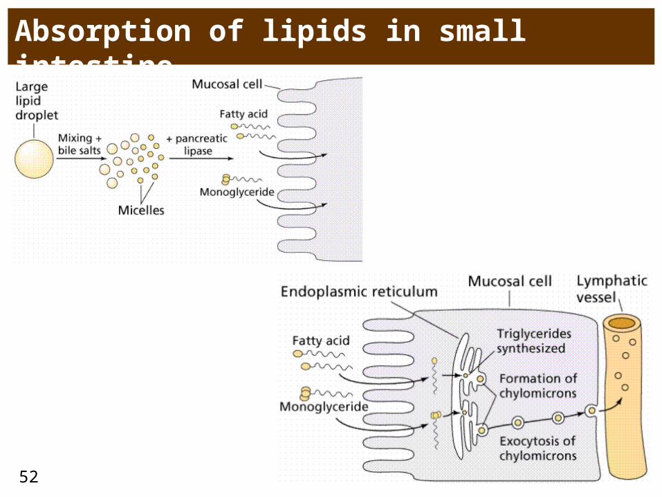
• lipids (TGA, cholesterol esters and phospholipids)• pancreatic lipase (min. salivary), cholesterolesterase, pospholipase A
emulsification (conj. bile acids!!) absorption by diffusion reesterification in enterocyte chylomicrons
51
52
Absorption of lipids in small intestine
53
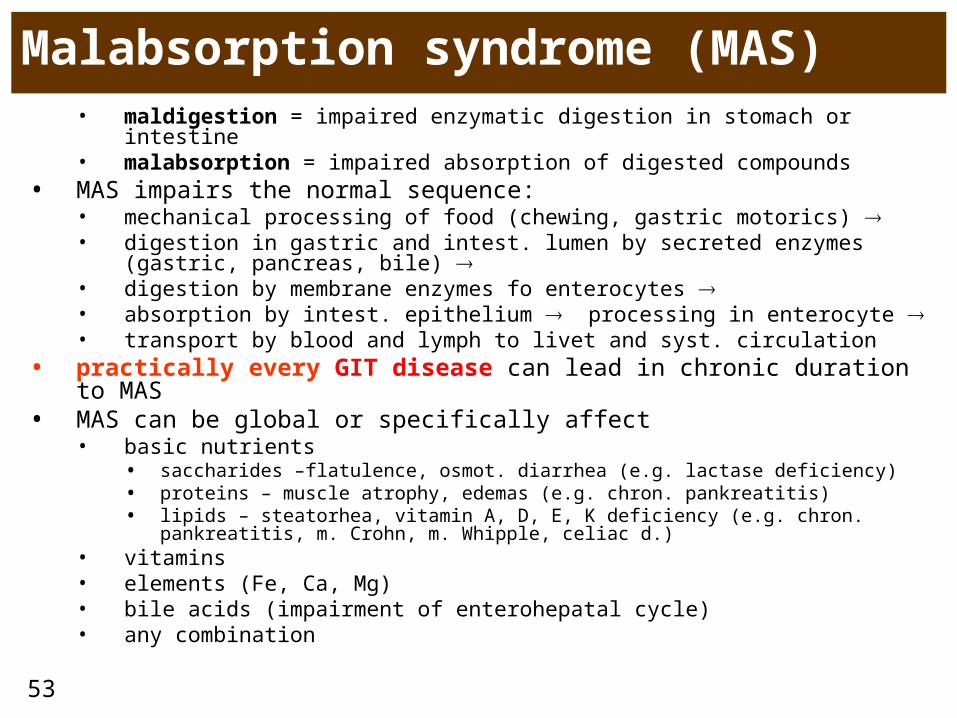
Malabsorption syndrome (MAS)• maldigestion = impaired enzymatic digestion in stomach or intestine• malabsorption = impaired absorption of digested compounds
• MAS impairs the normal sequence: • mechanical processing of food (chewing, gastric motorics) • digestion in gastric and intest. lumen by secreted enzymes (gastric,
pancreas, bile) • digestion by membrane enzymes fo enterocytes • absorption by intest. epithelium processing in enterocyte • transport by blood and lymph to livet and syst. circulation
• practically every GIT disease can lead in chronic duration to MAS• MAS can be global or specifically affect
• basic nutrients• saccharides –flatulence, osmot. diarrhea (e.g. lactase deficiency)• proteins – muscle atrophy, edemas (e.g. chron. pankreatitis)• lipids – steatorhea, vitamin A, D, E, K deficiency (e.g. chron. pankreatitis, m.
Crohn, m. Whipple, celiac d.)• vitamins• elements (Fe, Ca, Mg)• bile acids (impairment of enterohepatal cycle)• any combination
54
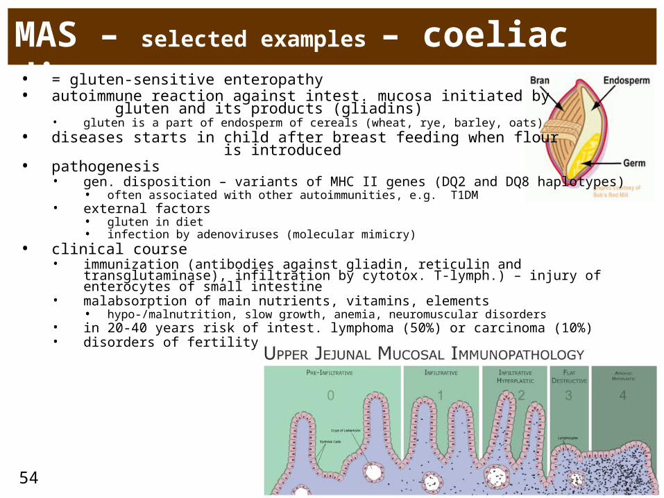
MAS – selected examples – coeliac dis.• = gluten-sensitive enteropathy• autoimmune reaction against intest. mucosa initiated by gluten
and its products (gliadins)• gluten is a part of endosperm of cereals (wheat, rye, barley, oats)
• diseases starts in child after breast feeding when flour is introduced
• pathogenesis• gen. disposition – variants of MHC II genes (DQ2 and DQ8 haplotypes)
• often associated with other autoimmunities, e.g. T1DM• external factors
• gluten in diet• infection by adenoviruses (molecular mimicry)
• clinical course• immunization (antibodies against gliadin, reticulin and transglutaminase),
infiltration by cytotox. T-lymph.) – injury of enterocytes of small intestine• malabsorption of main nutrients, vitamins, elements
• hypo-/malnutrition, slow growth, anemia, neuromuscular disorders• in 20-40 years risk of intest. lymphoma (50%) or carcinoma (10%) • disorders of fertility

55
56
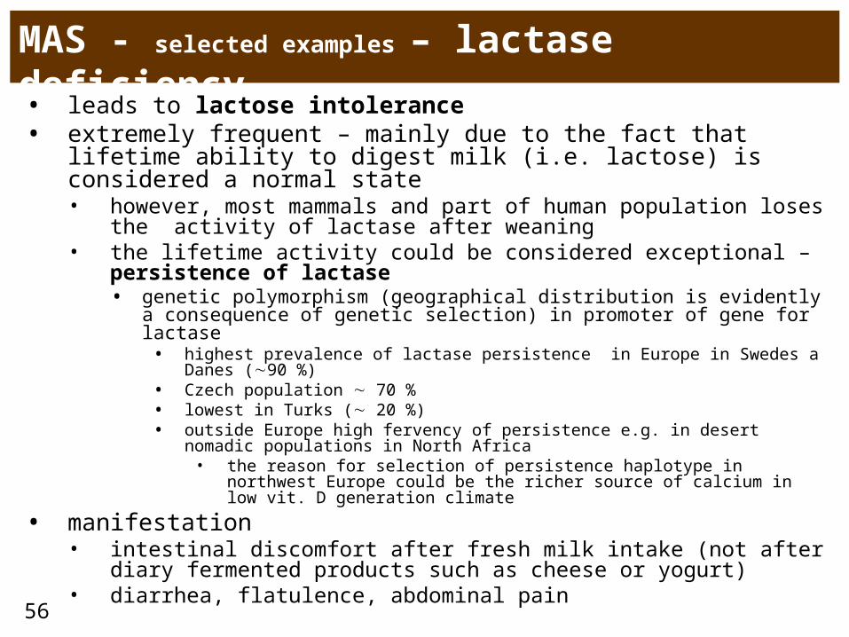
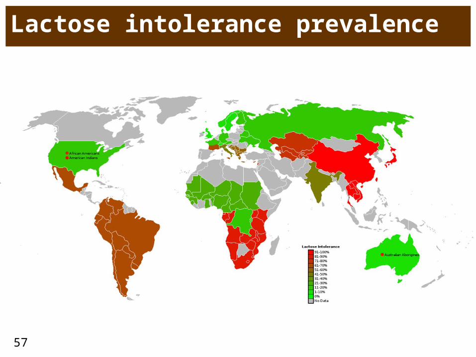
MAS - selected examples – lactase deficiency
• leads to lactose intolerance • extremely frequent – mainly due to the fact that lifetime ability
to digest milk (i.e. lactose) is considered a normal state• however, most mammals and part of human population loses the
activity of lactase after weaning• the lifetime activity could be considered exceptional –
persistence of lactase• genetic polymorphism (geographical distribution is evidently a
consequence of genetic selection) in promoter of gene for lactase• highest prevalence of lactase persistence in Europe in Swedes a Danes
(90 %)• Czech population 70 %• lowest in Turks ( 20 %)• outside Europe high fervency of persistence e.g. in desert nomadic
populations in North Africa• the reason for selection of persistence haplotype in northwest Europe
could be the richer source of calcium in low vit. D generation climate
• manifestation• intestinal discomfort after fresh milk intake (not after diary
fermented products such as cheese or yogurt)• diarrhea, flatulence, abdominal pain
57
Lactose intolerance prevalence
58
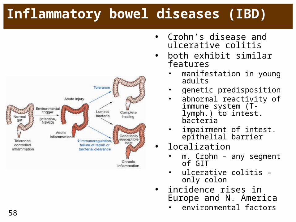
Inflammatory bowel diseases (IBD)
• Crohn’s disease and ulcerative colitis
• both exhibit similar features • manifestation in young
adults• genetic predisposition• abnormal reactivity of
immune system (T-lymph.) to intest. bacteria
• impairment of intest. epithelial barrier
• localization• m. Crohn – any segment
of GIT• ulcerative colitis – only
colon• incidence rises in Europe
and N. America• environmental factors
59
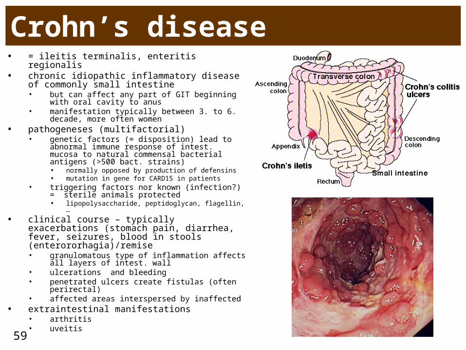
Crohn’s disease• = ileitis terminalis, enteritis regionalis• chronic idiopathic inflammatory disease of
commonly small intestine• but can affect any part of GIT beginning with
oral cavity to anus• manifestation typically between 3. to 6.
decade, more often women• pathogeneses (multifactorial)
• genetic factors (= disposition) lead to abnormal immune response of intest. mucosa to natural commensal bacterial antigens (>500 bact. strains)• normally opposed by production of defensins • mutation in gene for CARD15 in patients
• triggering factors nor known (infection?) = sterile animals protected • lipopolysaccharide, peptidoglycan, flagellin, …
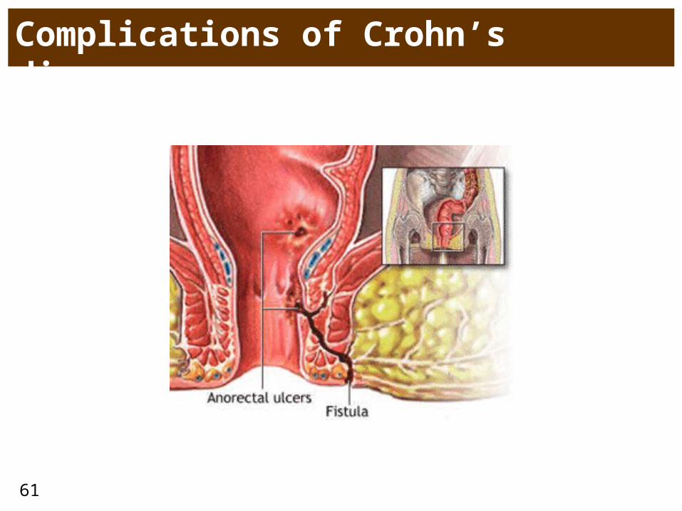
• clinical course – typically exacerbations (stomach pain, diarrhea, fever, seizures, blood in stools (enterororhagia)/remise• granulomatous type of inflammation affects all
layers of intest. wall• ulcerations and bleeding• penetrated ulcers create fistulas (often
perirectal)• affected areas interspersed by inaffected
• extraintestinal manifestations• arthritis• uveitis
60
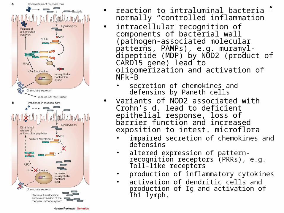
• reaction to intraluminal bacteria – normally “controlled inflammation”
• intracellular recognition of components of bacterial wall (pathogen-associated molecular patterns, PAMPs), e.g. muramyl-dipeptide (MDP) by NOD2 (product of CARD15 gene) lead to oligomerization and activation of NFk-B • secretion of chemokines and defensins
by Paneth cells• variants of NOD2 associated with
Crohn’s d. lead to deficient epithelial response, loss of barrier function and increased exposition to intest. microflora• impaired secretion of chemokines and
defensins• altered expression of pattern-
recognition receptors (PRRs), e.g. Toll-like receptors
• production of inflammatory cytokines• activation of dendritic cells and
production of Ig and activation of Th1 lymph.
61
Complications of Crohn’s disease
62
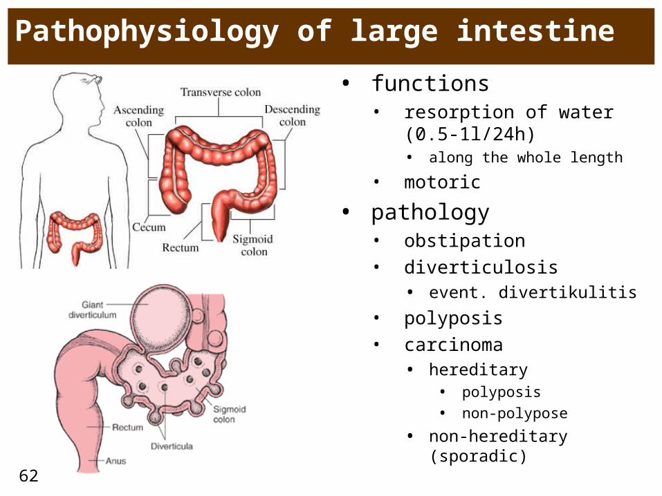
Pathophysiology of large intestine
• functions• resorption of water (0.5-
1l/24h)• along the whole length
• motoric
• pathology• obstipation• diverticulosis
• event. divertikulitis
• polyposis• carcinoma
• hereditary• polyposis
• non-polypose
• non-hereditary (sporadic)
63
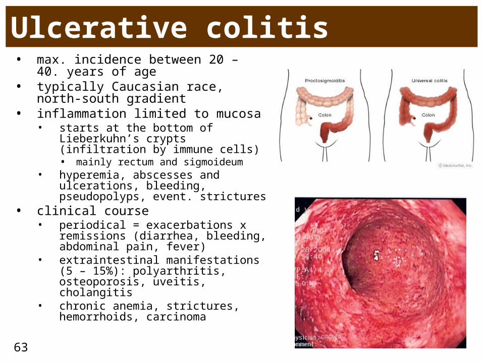
Ulcerative colitis• max. incidence between 20 – 40.
years of age• typically Caucasian race, north-
south gradient• inflammation limited to mucosa
• starts at the bottom of Lieberkuhn’s crypts (infiltration by immune cells)• mainly rectum and sigmoideum
• hyperemia, abscesses and ulcerations, bleeding, pseudopolyps, event. strictures
• clinical course• periodical = exacerbations x
remissions (diarrhea, bleeding, abdominal pain, fever)
• extraintestinal manifestations (5 – 15%): polyarthritis, osteoporosis, uveitis, cholangitis
• chronic anemia, strictures, hemorrhoids, carcinoma
64
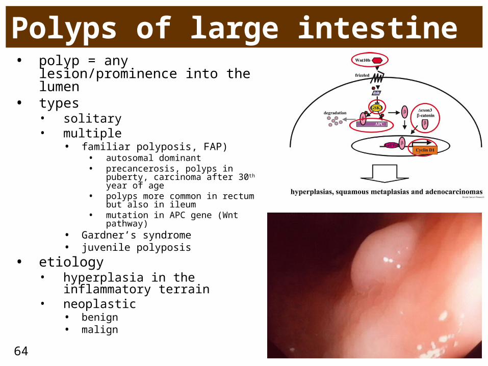
Polyps of large intestine• polyp = any lesion/prominence
into the lumen• types
• solitary• multiple
• familiar polyposis, FAP)• autosomal dominant• precancerosis, polyps in puberty,
carcinoma after 30th year of age• polyps more common in rectum
but also in ileum• mutation in APC gene (Wnt
pathway)• Gardner’s syndrome• juvenile polyposis
• etiology• hyperplasia in the
inflammatory terrain• neoplastic
• benign• malign
65
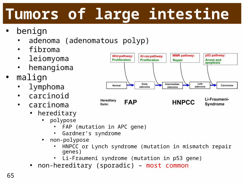
Tumors of large intestine• benign
• adenoma (adenomatous polyp)• fibroma• leiomyoma• hemangioma
• malign• lymphoma• carcinoid• carcinoma
• hereditary• polypose
• FAP (mutation in APC gene)• Gardner’s syndrome
• non-polypose • HNPCC or Lynch syndrome (mutation in mismatch repair
genes)• Li-Fraumeni syndrome (mutation in p53 gene)
• non-hereditary (sporadic) – most common

66
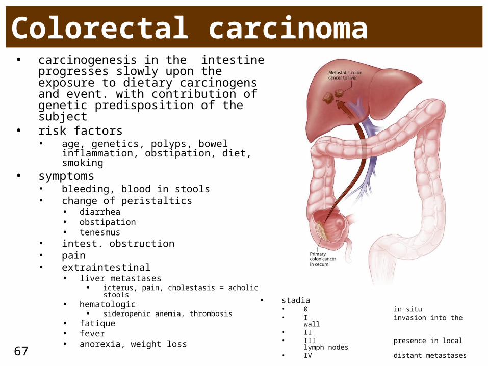
Colorectal carcinoma
67
Colorectal carcinoma• carcinogenesis in the intestine
progresses slowly upon the exposure to dietary carcinogens and event. with contribution of genetic predisposition of the subject
• risk factors• age, genetics, polyps, bowel
inflammation, obstipation, diet, smoking• symptoms
• bleeding, blood in stools• change of peristaltics
• diarrhea• obstipation• tenesmus
• intest. obstruction• pain• extraintestinal
• liver metastases• icterus, pain, cholestasis = acholic stools
• hematologic • sideropenic anemia, thrombosis
• fatique• fever• anorexia, weight loss
• stadia• 0 in situ• I invasion into the wall• II• III presence in local
lymph nodes• IV distant metastases
68