
1 Oligonucleotide interactionsFig 1: Principle of an oligonucleotide Funded cussion on different...
8
Joint Initiative of IITs and IISc – This lecture continues the dis 1 Oligonucleotide i The characteristic property o adenine and thymine, cytosin used for detection of oligon Such kind of sensors based ‘genosensor’. The oligonucl known as the ‘probe’ strand o exact complementarity of the sample strands. This hybridiz when the hybridization oc fluorophores that are linke hybridization. The specificit strand. If the probe strand is may arise. Hence an optimu threshold. One of the senso molecular beacon that emplo end and a quencher on the o will bend and the fluoresce presence of the sample stra sample strand resulting in str to be separated from the qu depicted in Figure 1. Fig 1: Prin – Funded scussion on different types of biosensors. interactions of complementary base pairing or hybridization ne and guanine moieties in oligonucleotide sequ nucleotide sequences in a sample with excellen d on oligonucleotide sequences is commonly r leotide sensors generally consist of an immob or ‘sense’ strand. On addition of the sample stra e sequences, hybridization will occur between t zation can be detected in the form of mass chang ccurs. Alternately, changes in fluorescence ed with the sequences can be monitored to ty of the binding will depend on the length s too short (~ 10 bp), then the probability of fa um length needs to be determined for fixing ors that is based on the complementary base oys a probe strand that is modified with a fluoro other. In the absence of the sample strand, the ence emission of the fluorophore will be quen and, hybridization will take place between th retching of the probe strand. This will cause the uencher resulting in fluorescence emission. Th nciple of an oligonucleotide-based capture sensor Page 3 of 10 n between the uences can be nt sensitivity. referred to as bilized strand and, if there is the probe and ges that occur emission of confirm the of the probe alse positives the detection pairing is a ophore at one probe strand nched. In the he probe and e fluorophore his concept is
Transcript of 1 Oligonucleotide interactionsFig 1: Principle of an oligonucleotide Funded cussion on different...
Joint Initiative of IITs and IISc –
This lecture continues the dis
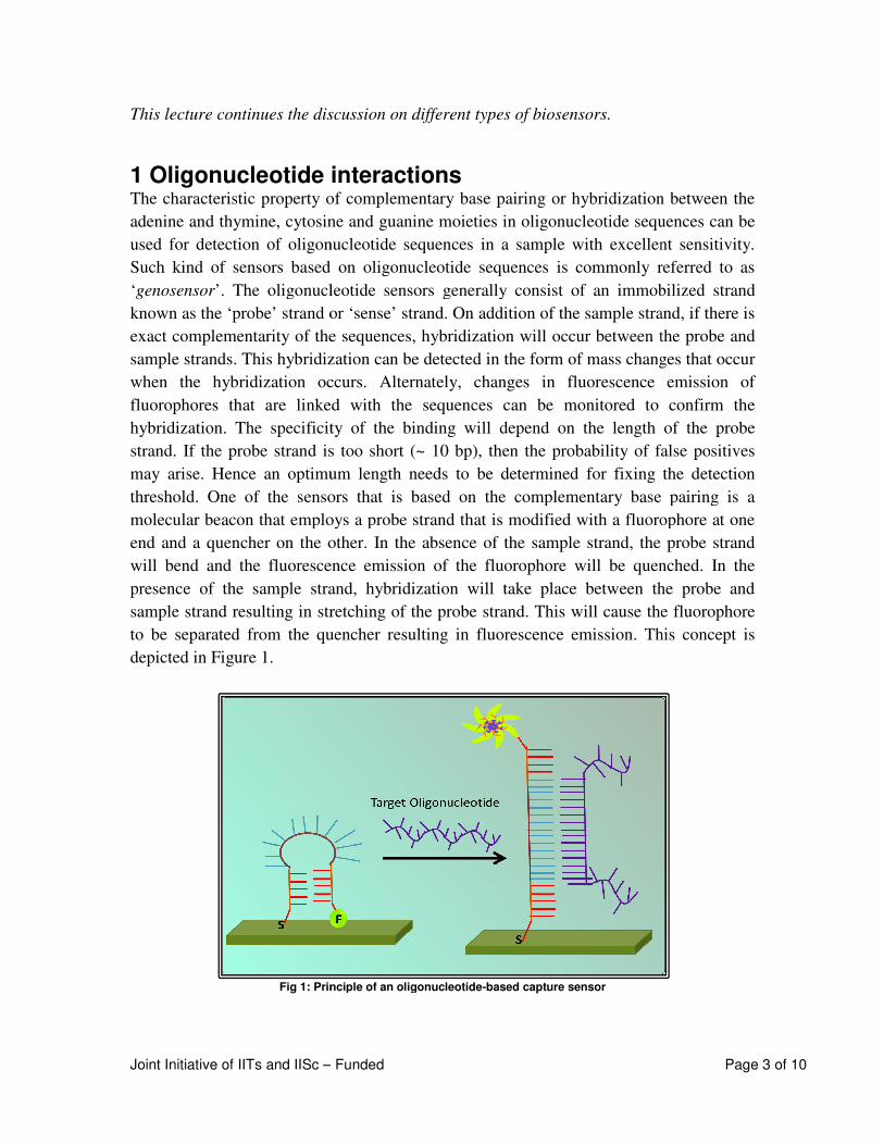
1 Oligonucleotide interactionsThe characteristic property of comple
adenine and thymine, cytosine and guanine
used for detection of oligonucleotide sequences in a sample with excellent sensitivity
Such kind of sensors based on oligonucleotide sequences is
‘genosensor’. The oligonucleotide sensors generally consist of an immobilized strand
known as the ‘probe’ strand or ‘sense’ strand. On addition of the sample strand, if the
exact complementarity of the sequences, hybridization will occur between the probe and
sample strands. This hybridization can be detected in the form of mass changes that occur
when the hybridization occurs. Alternately, changes in fluorescence emiss
fluorophores that are linked with the sequences can be monitored to confirm the
hybridization. The specificity of the binding will depend on the length of the probe
strand. If the probe strand is too short (~ 10 bp), then the probability of false po
may arise. Hence an optimum length needs to be determined for fixing the detection
threshold. One of the sensors that is based on the complementary base pairing is a
molecular beacon that employs a probe strand that is modified with a fluorophore a
end and a quencher on the other. In the absence of the sample strand, the probe strand
will bend and the fluorescence emission of the fluorophore will be quenched. In the
presence of the sample strand, hybridization will take place between the probe
sample strand resulting in stretching of the probe strand. This will cause the fluorophore
to be separated from the quencher resulting in fluorescence emission. This
depicted in Figure 1.
Fig 1: Principle of an oligonucleotide
– Funded
scussion on different types of biosensors.
Oligonucleotide interactions characteristic property of complementary base pairing or hybridization
adenine and thymine, cytosine and guanine moieties in oligonucleotide sequences can be
used for detection of oligonucleotide sequences in a sample with excellent sensitivity
Such kind of sensors based on oligonucleotide sequences is commonly referred
The oligonucleotide sensors generally consist of an immobilized strand
known as the ‘probe’ strand or ‘sense’ strand. On addition of the sample strand, if the
exact complementarity of the sequences, hybridization will occur between the probe and
sample strands. This hybridization can be detected in the form of mass changes that occur
when the hybridization occurs. Alternately, changes in fluorescence emiss
fluorophores that are linked with the sequences can be monitored to confirm the
hybridization. The specificity of the binding will depend on the length of the probe
strand. If the probe strand is too short (~ 10 bp), then the probability of false po
may arise. Hence an optimum length needs to be determined for fixing the detection
threshold. One of the sensors that is based on the complementary base pairing is a
molecular beacon that employs a probe strand that is modified with a fluorophore a
end and a quencher on the other. In the absence of the sample strand, the probe strand
will bend and the fluorescence emission of the fluorophore will be quenched. In the
presence of the sample strand, hybridization will take place between the probe
sample strand resulting in stretching of the probe strand. This will cause the fluorophore
to be separated from the quencher resulting in fluorescence emission. This
: Principle of an oligonucleotide-based capture sensor
Page 3 of 10
or hybridization between the
moieties in oligonucleotide sequences can be
used for detection of oligonucleotide sequences in a sample with excellent sensitivity.
commonly referred to as
The oligonucleotide sensors generally consist of an immobilized strand
known as the ‘probe’ strand or ‘sense’ strand. On addition of the sample strand, if there is
exact complementarity of the sequences, hybridization will occur between the probe and
sample strands. This hybridization can be detected in the form of mass changes that occur
when the hybridization occurs. Alternately, changes in fluorescence emission of
fluorophores that are linked with the sequences can be monitored to confirm the
hybridization. The specificity of the binding will depend on the length of the probe
strand. If the probe strand is too short (~ 10 bp), then the probability of false positives
may arise. Hence an optimum length needs to be determined for fixing the detection
threshold. One of the sensors that is based on the complementary base pairing is a
molecular beacon that employs a probe strand that is modified with a fluorophore at one
end and a quencher on the other. In the absence of the sample strand, the probe strand
will bend and the fluorescence emission of the fluorophore will be quenched. In the
presence of the sample strand, hybridization will take place between the probe and
sample strand resulting in stretching of the probe strand. This will cause the fluorophore
to be separated from the quencher resulting in fluorescence emission. This concept is
Deepa
Typewritten Text
Classification of biosensors based on transducers
Deepa
Typewritten Text

NTPEL – Nanotechnology - Nanobiotechnology
Joint Initiative of IITs and IISc – Funded Page 4 of 10
Similar to the complementary pairing exhibited by oligonucleotides that have been used
for sensing applications, a new category of peptide nucleic acids is now used as bio-
recognition elements. Peptide nucleic acids (PNA) areoligo-amides, which exhibit high
affinity to their complementary oligonucleotides. Capture-based sensors employing PNA
are a subject of active investigation.
2 Biomimetic receptors Receptors that are fabricated to mimic the bioreceptors are referred to as “biomimetic
receptors”. Genetic engineering techniques, artificial membrane fabrication and
molecular imprinting techniques have beenwidely used for fabricating biomimetic
receptors. Let us consider an example of a biomimetic receptor-based sensor using
artificial membrane fabrication for detectionof cholera toxins. Gangliosides (molecules
found in the ganglions of the brain and consist of glycosphingolipid with N-acetyl
muraminicacid) were incorporated into a matrix of diacetylenic lipids and were allowed
to self assemble to form a liposome. It is then exposed to UV radiations that provided
sufficient energy to cause photopolymerization. Figure 2 shows the reaction that occurs
during the photopolymerization process.
Fig 2: Photopolymerization reaction of diacetylenic lipids
It is observed that the product after photopolymerization has double bonds (‘ene’ as in
alkene) and triple bonds (‘yne’ as in alkyne). This ene-yne system causes absorption of
electromagnetic radiation in the visible region and hence the liposome appears blue or
purple. This liposome system has the ability to bind with cholera toxin as natural
membranes and hence forms the biomimetic receptor. Why does the cholera toxin bind to
the liposomal membrane? This is because of the presence of the ganglioside! The
ganglioside serves a recognition element for the cholera toxin, which is a protein that
NTPEL – Nanotechnology - Nanobiotechnology
Joint Initiative of IITs and IISc – Funded Page 5 of 10
specifically binds to gangliosides. Upon binding of the cholera toxin to the liposomal
membrane, a change of colour from blue to red occurs. This colour change can be
detected and quantified using spectrophotometry, as the absorption is directly
proportional to the concentration of toxin present in the system. Can you guess why the
colour change occurs? The binding of the protein results in reorientation of the lipid
chains as well as alters the conjugation causing the colour change!
One of the key aspects of biological systems is their specificity in binding to ligands. In
order to mimic this property, the concept of ‘molecular imprinting’ has been developed.
The molecular imprinting technique has a lot of potential in detecting toxins like
morphine, chemical warfare agents etc. Here, the molecule of interest (analyte) is mixed
with the monomers along with the cross linkers and allowed to polymerize. After
polymerization, the analyte molecules are extracted using an organic solvent thus leaving
an imprint for the particular analyte. Hence when the analyte is introduced for the next
time, it can simply bind to the binding site.
Detailed discussions on cell and tissue based sensors are given in Lecture 3 of the same
module. We will now move on to another type of classification.
3 Classification based on transducers Based on the transduction mechanism, biosensors are majorly classified into:
• Optical sensors
• Electrochemical sensors
• Mass-sensitive sensors
• Calorimetric sensors
3.1 Optical sensors
Optical sensors can convert any physical property of electromagnetic radiation into
electrical signals. Optical biosensors can use a variety of spectroscopy techniques, which
Did you know?....
A recent advancement in glucose sensors is contact lens based non-invasive glucose
detection, which detects glucose levels from the tear fluid. The sensor uses boronic
acid derivatives, which are synthetic molecules that exhibit great affinity to bind to
glucose. The binding causes a change in the fluorescence emission intensity of the
boronic acid derivative that can be correlated with the amount of glucose in the tear
sample!

NTPEL – Nanotechnology - Nanobiotechnology
Joint Initiative of IITs and IISc – Funded Page 6 of 10
includes absorption, reflectance, polarization, luminescence, fluorescence,surface
enhanced plasmon resonance (SERS), refraction, dispersion, etc. The quantification is
carried out based on the intensity or amplitudeof the optical signal,as a direct correlation
exists between the amplitude and the concentration of the analyte.
A typical optical biosensor design will involvebinding of the analyte molecule to an
optical probe resulting in change in the optical properties of the probe,which can be
detected directly. Alternately, an additional ‘capture agent’ may be added to bind with
theanalyte-probe complex. This binding in turn canlead to the formation of a coloured
product that can be detected and quantified. So what are these ‘capture agents’? They
may be single stranded DNA specific to the complementary strand, or may be antibodies
against a specific antigen. The concept of capture agents is employed in ELISA
techniques that have been elaborated in Lecture 1 of the same module.
3.2 Gene chip Introduction of more than one type of sensing probecan lead to detection of more than
one type of analyte. This can be invaluable in the case of medical diagnostics as well as
environmental pollution monitoring where detection of multiple analytes can be done at
the same time. Such sensors have their sensing strands specific for each analyte arranged
in an array. One such example of a sensor array isthe ‘gene chip’. The gene chip is a
DNA microarray commonly also known as the ‘DNA chip’. It is a collection of either
oligonucleotides or cDNA-based fragments (referred as probe sequences) attached to a
solid support. (A cDNA or complementary DNA is complementary to the mRNA sequence
and does not contain the introns. It in fact contains the complementary gene sequence
leading to the protein!).The density of the oligonucleotide sequence in each spot as well
as the base pair length are factors that will influence the sensor performance. Typically,
each spot is composed of approximately 40 oligonucleotides each with a base pair length
of about 25.A matching control spot with similar density and base pair length but with
one central base changed is also available. Spotting of the oligonucleotide sequences has
been achieved through many techniques such as atomic force microscopy, ink jetting,
laser-assisted bioprinting etc.
It is necessary to know the sequence that is being detected in order to design the probe
strands. The length of the probe strand must be carefully chosen. One major challenge
involved in the DNA microarray is the need of different permutations for each gene in
order to avoid erroneous false positives due to short probe sequences. In the case of a
cDNA microarray, the cDNA-sized fragmentsare usually produced by polymerase chain
reaction. The cDNA are long fragments that uniquely identify a specific gene. The
advantages of such microarrays are:
• Small size (A 1.3 x 1.3 cm slide can pack up to 6000 genes!)

NTPEL – Nanotechnology - Nanobiotechnology
Joint Initiative of IITs and IISc –
• High sensitivity
• High throughput (Can detect thousands of different sequences at the same time!
• Small analyte volume
This microarray technology is widely
cell samples obtained from patients,
chip containing the probe strands. H
strands, if present. Upon hyb
quantified by scanning the glass slide
ability to detect mRNA present at <1 molecule in 100,000.
introduced into a single chip!
a pathogen.
Fig 3: Schematic representation of
3.3 Optical barcodingOptical barcoding is a recently evolved technology making use of
microsphere capture reagents.
Nanobiotechnology
– Funded
Can detect thousands of different sequences at the same time!
Small analyte volume
This microarray technology is widely used in clinics, where mRNA is isolated fro
obtained from patients,labeled with a fluorescent probe and applied to the
containing the probe strands. Hybridization takes place between the complementary
. Upon hybridization, the fluorescence intensity is detected
scanning the glass slide containing the microarray. Gene chips have
ability to detect mRNA present at <1 molecule in 100,000. Entire genomes can be now
introduced into a single chip! Figure 3 shows the principle of a gene chip for detection of
Fig 3: Schematic representation of a gene chip for detection of pathogens
Optical barcoding is a recently evolved technology making use of quantum dot
microsphere capture reagents. Quantum dots are semi-conductor nanoparticles that
Page 7 of 10
Can detect thousands of different sequences at the same time!)
nics, where mRNA is isolated from the
and applied to the
ybridization takes place between the complementary
is detected and
. Gene chips have the
Entire genomes can be now
Figure 3 shows the principle of a gene chip for detection of
quantum dot–loaded
conductor nanoparticles that

NTPEL – Nanotechnology - Nanobiotechnology
Joint Initiative of IITs and IISc – Funded Page 8 of 10
exhibit size-dependent fluorescence emission. (More detailed discussions on quantum
dots is given in Module 12). Taking into account of the disadvantage of fluorescent
probes as they are susceptible to photo-bleaching, researchers have come up with the use
of quantum dot technology. The most important advantage of quantum dot is its size
dependent luminescence and narrow emission bands from a common excitation
wavelength. Moreover, it is also stable against photobleaching. Optical barcoding
involves loading the polymeric microspheres with different amounts of several colors of
quantum dots to obtain a unique fluorescence signature. Six colors at 10 possible
intensities allows for >106 possible codes. Capture molecule on surface of beads grabs the
labeled analyte and it can be detected.
3.4 Optic fiber sensor One of the emerging trends in clinical diagnosis is to detect an anomaly even before the
onset of symptoms. Any disease or disorder is initiated at a molecular level, which later is
amplified into a cellular level and then spreads to multiple cells. If a considerable amount
of the tissue/organ is impaired, then the individual starts manifesting the symptoms. In
many cases, the onset of symptoms signifies an advanced stage of the disease, which
makes it difficult to treat thereby reducing the survival rate. Hence, diagnosis at the
molecular level has a major implication on the success of the treatment. This aspect has
been realized with the advent of nanotechnology where analyte detection in the
zeptomolar range (10-21
M) has been reported. This means that a few hundred molecules
of the analyte can be detected!
An example of a super ultra-sensitive sensor employs a fiber optic probe whose tip is
etched to obtain a tapered tip about 40 nm wide at its narrowest point. The nano-
dimensions of the tip and the tapered geometry facilitate the penetration of the probe into
a cell. On insertion into the cell, the membrane bilayer self-seals around the fiber optic
probe and hence no permanent damage to the cell membrane occurs. Why is a fiber optic
probe used? An optic fiber is one in which the intensity of the light radiation is
transmitted without any loss and hence can be used for high sensitivity optical detection
without any loss in signal intensity. The tip of the optic fiber is chemically functionalized
with an antibody againstbenzopyrenetetrol. The chemical functionalization is essential
because if the antibody were to be physisorbed on the probe, the risk of the antibody
being desorbed will be high. Benzopyrenetetrol is the metabolic product of benzopyrene,
a chemical carcinogen. How doesbenzopyrene gain entry into the biological system?
Benzopyrene is a polycyclic aromatic hydrocarbon that is found in automobile emissions
and smoke emissions due to burning of wood. Yet another common source of
benzopyrene is grilled and/or deep fried food items!

NTPEL – Nanotechnology - Nanobiotechnology
Joint Initiative of IITs and IISc –
(OOPS!!! Most of my favourite fast food items might be
health!!!)
Once the benzopyrene enters the system, it can undergo a series of
reactions resulting in the formation of benzopyrenetetrol. The reactions leading to the
formation of benzopyrenetetrol from benzopyrene
OH
OH
O2 2e ;2
O2 2e ;2
CYP1A1/CYP1B
benzo[a]pyrene
(-)benzo[a]pyrene-
7,8-dihydrodiol
Fig 4: Reactions leading to the formation of benzopyrenetetrol from benzopyrene
The tetrol derivative can interact with DNA, which in turn can transform the normal cell
into a cancerous cell. This is because the tetrol
can interact with the sugar and phosphate groups of the DNA.
benzopyrenetetrol in the cell therefore indicates a potential risk of cancer development.
Thus, quantification of this analyte can be a measu
an individual.The probe when inserted into the cell exposes the antibody to the cytosol. If
benzopyrenetetrol is present in the cell it will bind with the antibody, forming an immune
complex, which can be detected fro
immune complex.
Nanobiotechnology
– Funded
OOPS!!! Most of my favourite fast food items might be detrimental to my
Once the benzopyrene enters the system, it can undergo a series of enzyme
reactions resulting in the formation of benzopyrenetetrol. The reactions leading to the
formation of benzopyrenetetrol from benzopyreneis shown in Figure 4.
O
OH
OH
O2H
2H
H2O
H2O
H2O
B1
(+)benzo[a]pyrene-
7,8-epoxide
Epoxide Hydrolase
(+)benzo[a]pyrene-7,8-
dihydrodiol; 9,10-epoxide(+)benzo[
7,8, 9,10-
Hydrolase
H2O
Reactions leading to the formation of benzopyrenetetrol from benzopyrene
The tetrol derivative can interact with DNA, which in turn can transform the normal cell
This is because the tetrol molecule has four hydroxyl groups that
can interact with the sugar and phosphate groups of the DNA.The presence of
benzopyrenetetrol in the cell therefore indicates a potential risk of cancer development.
Thus, quantification of this analyte can be a measure of the risk of developing cancer in
an individual.The probe when inserted into the cell exposes the antibody to the cytosol. If
benzopyrenetetrol is present in the cell it will bind with the antibody, forming an immune
complex, which can be detected from the unique fluorescence emission wavelength of the
Page 9 of 10
detrimental to my
enzyme-catalyzed
reactions resulting in the formation of benzopyrenetetrol. The reactions leading to the
OH
OH
OH
OH
[a]pyrene-
-tetrol
Reactions leading to the formation of benzopyrenetetrol from benzopyrene
The tetrol derivative can interact with DNA, which in turn can transform the normal cell
four hydroxyl groups that
The presence of
benzopyrenetetrol in the cell therefore indicates a potential risk of cancer development.
re of the risk of developing cancer in
an individual.The probe when inserted into the cell exposes the antibody to the cytosol. If
benzopyrenetetrol is present in the cell it will bind with the antibody, forming an immune
m the unique fluorescence emission wavelength of the
NTPEL – Nanotechnology - Nanobiotechnology
Joint Initiative of IITs and IISc – Funded Page 10 of 10
4 Reference
Biosensors and biochips: advances in biological and medical diagnosis,Tuan vo-Dinh,
Drian Cullum, Freselaus J Analytical Chemistry, 2000, 366, 540-551.
Deepa
Typewritten Text
Deepa
Typewritten Text
Source: http://nptel.ac.in/courses/118106019/37