1 Drug Absorption, Distribution and Elimination · BLUK100-Calvey November 27, 2007 13:44 Drug...
22
BLUK100-Calvey November 27, 2007 13:44 1 Drug Absorption, Distribution and Elimination Drugs can be defined as agents that modify normal biolog- ical responses and thus produce pharmacological effects. These are frequently dependent on the transfer of drugs across one or more cellular membranes, whose structure and physicochemical properties govern the rate and extent of drug transfer. Cellular membranes are usually about 10 nm wide and consist of a bimolecular layer of phospholipid and protein (Fig. 1.1). The lipid layer is relatively fluid, and individ- ual phospholipid molecules can move laterally within the membrane. Extrinsic (peripheral) proteins are present on the external or internal aspect of the membrane. In con- trast, intrinsic (integral) proteins traverse the entire width of the cell membrane and may form an annulus surround- ing small pores or ion channels approximately 0.5 nm in diameter (Fig. 1.1). Both intrinsic and extrinsic proteins can act as enzymes or receptors and may mediate the active transport of drugs. Approximately 5–10% of the cell membrane consists of carbohydrates, mainly glycolipids or glycoproteins. They are believed to be responsible for the immunological char- acteristics of cells and play an important part in molecular recognition. Many cell membranes also contain inorganic ions (e.g. Ca 2+ ). Lipid cell membranes are excellent electrical insulators. Consequently, there may be differences in electrical poten- tial across cellular membranes, which can facilitate or im- pede the passive transport of charged molecules through ion channels. Transfer of drugs across cell membranes In general, drugs may cross cell membranes by Passive diffusion Carrier transport Passive diffusion In most cases, drugs cross cell membranes by passive dif- fusion down a concentration gradient due to random molecular movements produced by thermal energy. The rate of drug transfer is directly proportional to the dif- ference in concentration, and to the solubility of drugs in membranes, which is extremely variable. Highly po- lar substances (e.g. quaternary amines) are insoluble in membrane lipids and are unable to penetrate cellular membranes. In contrast, drugs with a high lipid solubil- ity (e.g. diazepam, fentanyl) readily dissolve in membrane phospholipids and rapidly diffuse across cellular mem- branes. Other less lipid-soluble drugs (e.g. morphine) dif- fuse more slowly and their onset of action is often delayed. Molecular size is a less important factor in the pas- sive diffusion of drugs. Some low molecular weight com- pounds may diffuse through ion channels, or penetrate small intercellular or paracellular channels (particularly in ‘leaky’ epithelial membranes). In contrast, molecules larger than 100–200 Da are usually unable to cross cell membranes. The permeability of vascular endothelium is greater than other tissues, and most ionized compounds can readily cross capillary membranes. Most drugs are weak acids or weak bases and are thus present in physiological conditions in both an ionized and a non-ionized form. Their ionization or dissociation can be represented by the equations: AH A − + H + (for acids) BH + B + H + (for bases) Weak acids and bases are predominantly present as the species AH and BH + in acidic conditions, but as A − and B in alkaline conditions. The non-ionized forms AH and B are lipid soluble and can readily diffuse across cell mem- branes, while the ionized forms A − and BH + are effectively impermeable. As the proportion of the drug that is present in the non-ionized form is dependent on pH, differences 1
Transcript of 1 Drug Absorption, Distribution and Elimination · BLUK100-Calvey November 27, 2007 13:44 Drug...
BLUK100-Calvey November 27, 2007 13:44
1 Drug Absorption, Distributionand Elimination
Drugs can be defined as agents that modify normal biolog-
ical responses and thus produce pharmacological effects.
These are frequently dependent on the transfer of drugs
across one or more cellular membranes, whose structure
and physicochemical properties govern the rate and extent
of drug transfer.
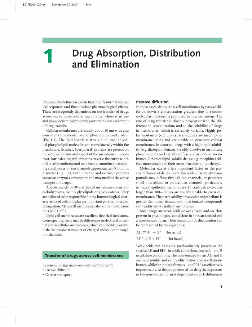
Cellular membranes are usually about 10 nm wide and
consist of a bimolecular layer of phospholipid and protein
(Fig. 1.1). The lipid layer is relatively fluid, and individ-
ual phospholipid molecules can move laterally within the
membrane. Extrinsic (peripheral) proteins are present on
the external or internal aspect of the membrane. In con-
trast, intrinsic (integral) proteins traverse the entire width
of the cell membrane and may form an annulus surround-
ing small pores or ion channels approximately 0.5 nm in
diameter (Fig. 1.1). Both intrinsic and extrinsic proteins
can act as enzymes or receptors and may mediate the active
transport of drugs.
Approximately 5–10% of the cell membrane consists of
carbohydrates, mainly glycolipids or glycoproteins. They
are believed to be responsible for the immunological char-
acteristics of cells and play an important part in molecular
recognition. Many cell membranes also contain inorganic
ions (e.g. Ca2+).
Lipid cell membranes are excellent electrical insulators.
Consequently, there may be differences in electrical poten-
tial across cellular membranes, which can facilitate or im-
pede the passive transport of charged molecules through
ion channels.
Transfer of drugs across cell membranes
In general, drugs may cross cell membranes by� Passive diffusion� Carrier transport
Passive diffusionIn most cases, drugs cross cell membranes by passive dif-
fusion down a concentration gradient due to random
molecular movements produced by thermal energy. The
rate of drug transfer is directly proportional to the dif-
ference in concentration, and to the solubility of drugs
in membranes, which is extremely variable. Highly po-
lar substances (e.g. quaternary amines) are insoluble in
membrane lipids and are unable to penetrate cellular
membranes. In contrast, drugs with a high lipid solubil-
ity (e.g. diazepam, fentanyl) readily dissolve in membrane
phospholipids and rapidly diffuse across cellular mem-
branes. Other less lipid-soluble drugs (e.g. morphine) dif-
fuse more slowly and their onset of action is often delayed.
Molecular size is a less important factor in the pas-
sive diffusion of drugs. Some low molecular weight com-
pounds may diffuse through ion channels, or penetrate
small intercellular or paracellular channels (particularly
in ‘leaky’ epithelial membranes). In contrast, molecules
larger than 100–200 Da are usually unable to cross cell
membranes. The permeability of vascular endothelium is
greater than other tissues, and most ionized compounds
can readily cross capillary membranes.
Most drugs are weak acids or weak bases and are thus
present in physiological conditions in both an ionized and
a non-ionized form. Their ionization or dissociation can
be represented by the equations:
AH � A− + H+ (for acids)
BH+ � B + H+ (for bases)
Weak acids and bases are predominantly present as the
species AH and BH+ in acidic conditions, but as A− and B
in alkaline conditions. The non-ionized forms AH and B
are lipid soluble and can readily diffuse across cell mem-
branes, while the ionized forms A− and BH+ are effectively
impermeable. As the proportion of the drug that is present
in the non-ionized form is dependent on pH, differences
1
BLUK100-Calvey November 27, 2007 13:44
2 Chapter 1
Intrinsic or integral protein Extrinsic or peripheral protein(enclosing an ion channel)
Phosphorylated Fatty acid tail of head of phospholipid of phospholipid
Fig. 1.1 The phospholipid and protein
structure of a typical cell membrane.
in H+ concentration across cellular membranes can pro-
vide a diffusion gradient for the passive transfer of the
non-ionized form.
Consider a weak acidic drug that dissociates in the
manner:
AH � A− + H+
From the Henderson–Hasselbalch equation, it can be
shown that
pKa−pH � log[AH]
[A−],
where [AH] and [A−] are the concentrations of non-
ionized and ionized forms and pKa (the negative log-
arithm of the dissociation constant) is the pH value at
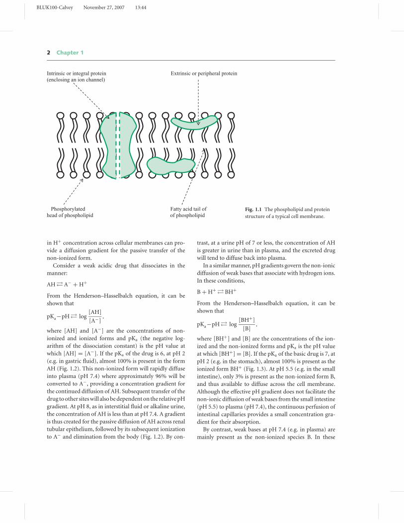
which [AH] = [A−]. If the pKa of the drug is 6, at pH 2
(e.g. in gastric fluid), almost 100% is present in the form
AH (Fig. 1.2). This non-ionized form will rapidly diffuse
into plasma (pH 7.4) where approximately 96% will be
converted to A−, providing a concentration gradient for
the continued diffusion of AH. Subsequent transfer of the
drug to other sites will also be dependent on the relative pH
gradient. At pH 8, as in interstitial fluid or alkaline urine,
the concentration of AH is less than at pH 7.4. A gradient
is thus created for the passive diffusion of AH across renal
tubular epithelium, followed by its subsequent ionization
to A− and elimination from the body (Fig. 1.2). By con-
trast, at a urine pH of 7 or less, the concentration of AH
is greater in urine than in plasma, and the excreted drug
will tend to diffuse back into plasma.
In a similar manner, pH gradients govern the non-ionic
diffusion of weak bases that associate with hydrogen ions.
In these conditions,
B + H+ � BH+
From the Henderson–Hasselbalch equation, it can be
shown that
pKa−pH � log[BH+]
[B],
where [BH+] and [B] are the concentrations of the ion-
ized and the non-ionized forms and pKa is the pH value
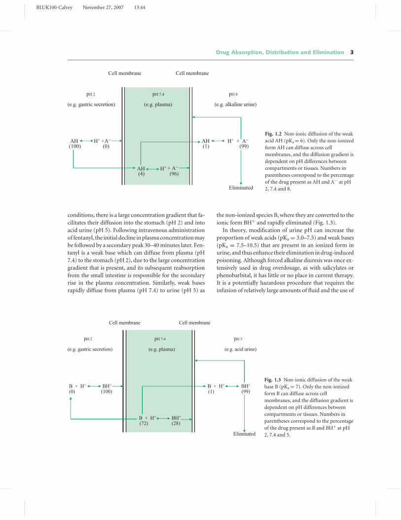
at which [BH+] = [B]. If the pKa of the basic drug is 7, at
pH 2 (e.g. in the stomach), almost 100% is present as the
ionized form BH+ (Fig. 1.3). At pH 5.5 (e.g. in the small
intestine), only 3% is present as the non-ionized form B,
and thus available to diffuse across the cell membrane.
Although the effective pH gradient does not facilitate the
non-ionic diffusion of weak bases from the small intestine
(pH 5.5) to plasma (pH 7.4), the continuous perfusion of
intestinal capillaries provides a small concentration gra-
dient for their absorption.
By contrast, weak bases at pH 7.4 (e.g. in plasma) are
mainly present as the non-ionized species B. In these
BLUK100-Calvey November 27, 2007 13:44
Drug Absorption, Distribution and Elimination 3
Cell membrane Cell membrane
pH 2 pH 7.4 pH 8
(e.g. gastric secretion) (e.g. plasma) (e.g. alkaline urine)
AH H+ +A− AH H+ + A−
(100) (0) (1) (99)
AH H+ + A−
(4) (96)
Eliminated
Fig. 1.2 Non-ionic diffusion of the weak
acid AH (pKa = 6). Only the non-ionized
form AH can diffuse across cell
membranes, and the diffusion gradient is
dependent on pH differences between
compartments or tissues. Numbers in
parentheses correspond to the percentage
of the drug present as AH and A− at pH
2, 7.4 and 8.
conditions, there is a large concentration gradient that fa-
cilitates their diffusion into the stomach (pH 2) and into
acid urine (pH 5). Following intravenous administration
of fentanyl, the initial decline in plasma concentration may
be followed by a secondary peak 30–40 minutes later. Fen-
tanyl is a weak base which can diffuse from plasma (pH
7.4) to the stomach (pH 2), due to the large concentration
gradient that is present, and its subsequent reabsorption
from the small intestine is responsible for the secondary
rise in the plasma concentration. Similarly, weak bases
rapidly diffuse from plasma (pH 7.4) to urine (pH 5) as
the non-ionized species B, where they are converted to the
ionic form BH+ and rapidly eliminated (Fig. 1.3).
In theory, modification of urine pH can increase the
proportion of weak acids (pKa = 3.0–7.5) and weak bases
(pKa = 7.5–10.5) that are present in an ionized form in
urine, and thus enhance their elimination in drug-induced
poisoning. Although forced alkaline diuresis was once ex-
tensively used in drug overdosage, as with salicylates or
phenobarbital, it has little or no place in current therapy.
It is a potentially hazardous procedure that requires the
infusion of relatively large amounts of fluid and the use of
Cell membrane Cell membrane
pH 2 pH 7.4 pH 5
(e.g. gastric secretion) (e.g. plasma) (e.g. acid urine)
B + H+ BH+ B + H+ BH+
(0) (100) (1) (99)
B + H+ BH+
(72) (28)
Eliminated
Fig. 1.3 Non-ionic diffusion of the weak
base B (pKa = 7). Only the non-ionized
form B can diffuse across cell
membranes, and the diffusion gradient is
dependent on pH differences between
compartments or tissues. Numbers in
parentheses correspond to the percentage
of the drug present as B and BH+ at pH
2, 7.4 and 5.
BLUK100-Calvey November 27, 2007 13:44
4 Chapter 1
loop diuretics or mannitol. In addition, pulmonary and
cerebral oedema are possible complications, particularly
in the elderly.
Carrier transportCarrier transport can be divided into two main types:� Facilitated diffusion� Active transport
Facilitated diffusionFacilitated diffusion is a form of carrier transport that
does not require the expenditure of cellular energy. Many
physiological substrates combine with specific sites on in-
trinsic proteins, resulting in conformational (allosteric)
changes in protein structure. These changes facilitate the
transcellular transport of many endogenous compounds.
In these conditions, physiological substrates enter cells
down a concentration gradient, but at a faster rate than
anticipated from their lipid solubility or molecular size.
Facilitated diffusion mediates the absorption of some sim-
ple sugars, steroids, amino acids and pyrimidines from the
small intestine and their subsequent transfer across cell
membranes.
Active transportIn contrast, active transport requires cellular or metabolic
energy and can transfer drugs against a concentration
gradient. In some instances, metabolic energy is directly
produced from the hydrolysis of ATP (primary active
transport). More commonly, metabolic energy is provided
by the active transport of Na+, or is dependent on the
electrochemical gradient produced by the sodium pump,
Na+/K+ ATPase (secondary active transport). It is gener-
ally considered that the drug or substrate initially com-
bines with an intrinsic carrier protein (which may be an
ion channel or Na+/K+ ATPase). The drug–protein com-
plex is then transferred across the cell membrane, where
the drug is released and the carrier protein returns to the
opposite side of the membrane.
Active transport systems are saturable and specific and
can be inhibited by other drugs (Chapter 4). They play a
crucial role in the transfer of drugs across cell membranes
at many sites, including the small intestine, the proxi-
mal renal tubule, the biliary canaliculus and the choroid
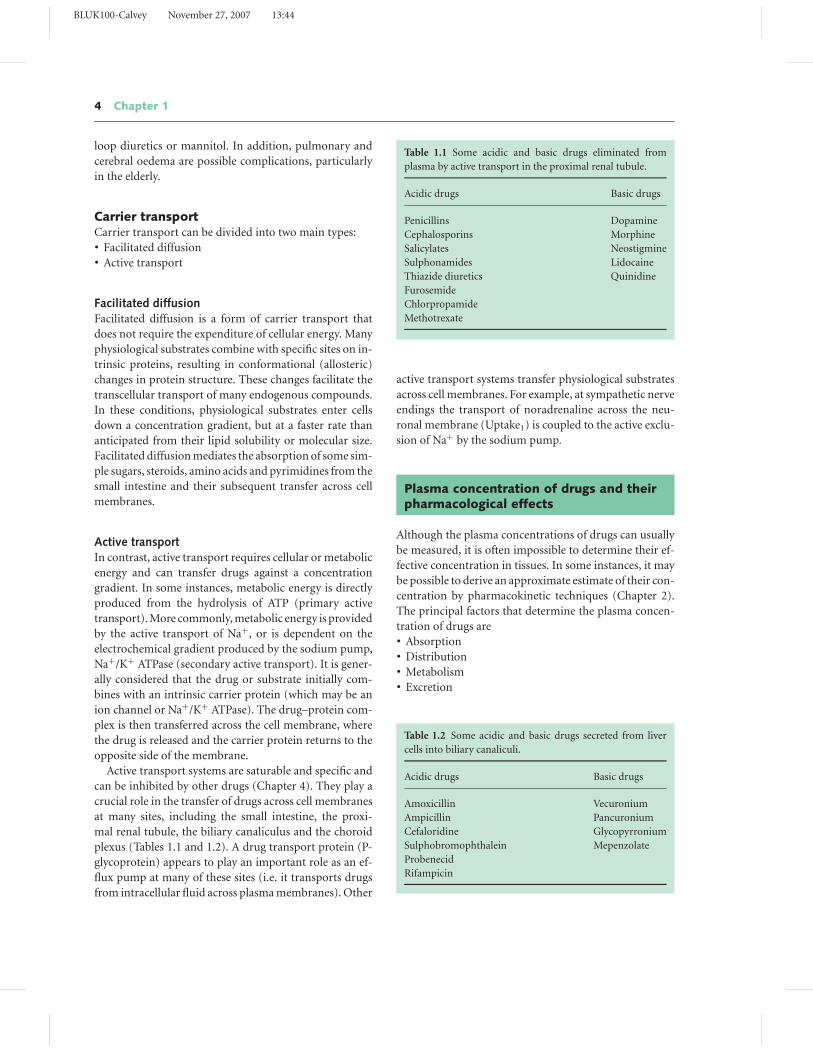
plexus (Tables 1.1 and 1.2). A drug transport protein (P-
glycoprotein) appears to play an important role as an ef-
flux pump at many of these sites (i.e. it transports drugs
from intracellular fluid across plasma membranes). Other
Table 1.1 Some acidic and basic drugs eliminated from
plasma by active transport in the proximal renal tubule.
Acidic drugs Basic drugs
Penicillins Dopamine
Cephalosporins Morphine
Salicylates Neostigmine
Sulphonamides Lidocaine
Thiazide diuretics Quinidine
Furosemide
Chlorpropamide
Methotrexate
active transport systems transfer physiological substrates
across cell membranes. For example, at sympathetic nerve
endings the transport of noradrenaline across the neu-
ronal membrane (Uptake1) is coupled to the active exclu-
sion of Na+ by the sodium pump.
Plasma concentration of drugs and theirpharmacological effects
Although the plasma concentrations of drugs can usually
be measured, it is often impossible to determine their ef-
fective concentration in tissues. In some instances, it may
be possible to derive an approximate estimate of their con-
centration by pharmacokinetic techniques (Chapter 2).
The principal factors that determine the plasma concen-
tration of drugs are� Absorption� Distribution� Metabolism� Excretion
Table 1.2 Some acidic and basic drugs secreted from liver
cells into biliary canaliculi.
Acidic drugs Basic drugs
Amoxicillin Vecuronium
Ampicillin Pancuronium
Cefaloridine Glycopyrronium
Sulphobromophthalein Mepenzolate
Probenecid
Rifampicin
BLUK100-Calvey November 27, 2007 13:44
Drug Absorption, Distribution and Elimination 5
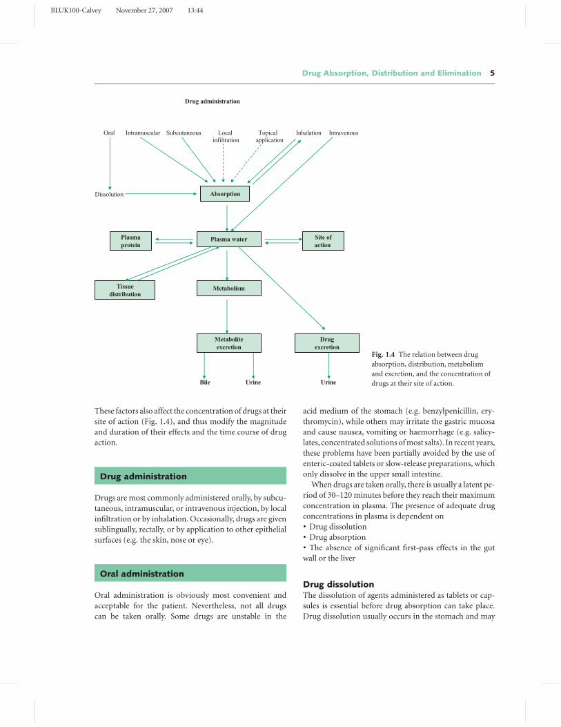
Drug administration
Oral Intramuscular Subcutaneous Local Topical Inhalation Intravenous infiltration application
Dissolution
Bile Urine Urine
Absorption
Plasma water
Metabolism
Metabolite excretion
Site of action
Plasma protein
Tissue distribution
Drug excretion
Fig. 1.4 The relation between drug
absorption, distribution, metabolism
and excretion, and the concentration of
drugs at their site of action.
These factors also affect the concentration of drugs at their
site of action (Fig. 1.4), and thus modify the magnitude
and duration of their effects and the time course of drug
action.
Drug administration
Drugs are most commonly administered orally, by subcu-
taneous, intramuscular, or intravenous injection, by local
infiltration or by inhalation. Occasionally, drugs are given
sublingually, rectally, or by application to other epithelial
surfaces (e.g. the skin, nose or eye).
Oral administration
Oral administration is obviously most convenient and
acceptable for the patient. Nevertheless, not all drugs
can be taken orally. Some drugs are unstable in the
acid medium of the stomach (e.g. benzylpenicillin, ery-
thromycin), while others may irritate the gastric mucosa
and cause nausea, vomiting or haemorrhage (e.g. salicy-
lates, concentrated solutions of most salts). In recent years,
these problems have been partially avoided by the use of
enteric-coated tablets or slow-release preparations, which
only dissolve in the upper small intestine.
When drugs are taken orally, there is usually a latent pe-
riod of 30–120 minutes before they reach their maximum
concentration in plasma. The presence of adequate drug
concentrations in plasma is dependent on� Drug dissolution� Drug absorption� The absence of significant first-pass effects in the gut
wall or the liver
Drug dissolutionThe dissolution of agents administered as tablets or cap-
sules is essential before drug absorption can take place.
Drug dissolution usually occurs in the stomach and may
BLUK100-Calvey November 27, 2007 13:44
6 Chapter 1
be dependent on gastric acidity. Variations in the speed
of dissolution and the rate and extent of gastric emptying
can thus affect the amount of drug in solution in the up-
per part of the small intestine (where absorption mainly
occurs).
Many pharmaceutical factors influence the dissolution
of tablets and capsules, including particle size, chemical
formulation, the inclusion of inert fillers and the outer
coating of the tablet. In these circumstances, proprietary
or generic preparations of the same drug may have differ-
ent dissolution characteristics and thus produce a range of
plasma concentrations after oral administration. At one
time, differences in the potency of digoxin tablets sus-
pected from clinical observations were eventually traced
to variations in the dissolution of different preparations
of the drug. Similarly, toxic effects were produced by
diphenylhydantoin (phenytoin) tablets when an excipi-
ent (calcium sulphate) was replaced by lactose. In these
conditions, dissolution was more rapid, resulting in faster
and more extensive absorption, and higher blood levels of
the drugs.
Sustained release preparationsSustained release oral preparations usually consist of
multi-lamellated erodable polymers and are designed to
allow the slow continuous release of drugs. Other formula-
tions may permit the release of fixed doses of a drug at reg-
ular intervals. Some preparations are osmotically active,
or incorporate an ion-exchange resin that allows drugs
to be released in solution at a defined ionic concentration
and pH. Their use often results in greater convenience and
safety, improves bioavailability and causes less variability
in plasma concentrations. They may also reduce side ef-
fects, drug dosage, frequency of administration and cost.
Many drugs may be administered in this manner, in-
cluding some opioid analgesics, NSAIDs, bronchodilators,
antihypertensive drugs, antiarrhythmic agents and potas-
sium salts.
Drug absorptionThe absorption of drugs in the stomach and the small in-
testine is primarily dependent on their physicochemical
properties, particularly their lipid solubility. Non-ionized
compounds (e.g. ethyl alcohol) and low molecular weight
substances (e.g. urea) readily cross cell membranes by pas-
sive diffusion and are easily and rapidly absorbed from
the gut. Drugs that are weak acids (e.g. aspirin) are pre-
dominantly non-ionized and lipid-soluble in acidic con-
ditions and partially diffuse into plasma from the stom-
ach. In contrast, basic drugs (e.g. propranolol, most ben-
zodiazepines) are less ionized and more lipid-soluble in
alkaline conditions and are preferentially absorbed from
the duodenum (pH 5–6). Strong bases (e.g. quaternary
amines) are always ionized in solution and are not signif-
icantly absorbed from the gut.
In practice, other factors influence the site of drug ab-
sorption. Mucosal surface area is more extensive in the
upper small intestine than the stomach, and most drugs,
whether acids or bases, are predominantly absorbed from
the duodenum. Nevertheless, some non-ionized com-
pounds and acidic drugs may be partially absorbed from
the stomach and may produce a rapid increase in plasma
concentration after oral administration.
Drugs that affect gastric motilityCompounds affecting gastric motility can modify drug
dissolution, and influence the rate, but not the extent,
of drug absorption. In particular, drugs that slow gas-
tric emptying (e.g. atropine, morphine) decrease the rate
of drug absorption. Other drug interactions, as between
tetracyclines and iron, or colestyramine and digoxin, may
affect the extent of drug absorption and thus modify sys-
temic bioavailability. Drug absorption may be reduced
in pathological conditions affecting the gastrointestinal
tract, particularly in coeliac disease, Crohn’s disease, ob-
structive jaundice, or after extensive resection of the small
intestine.
Drug absorption and carrier transportAlthough most drugs are absorbed from the stomach
and small intestine by passive diffusion, occasionally ab-
sorption is dependent on carrier transport. Levodopa is
absorbed by a carrier protein that normally transports
amino acids, and fluorouracil is absorbed by the car-
rier that transports pyrimidine bases. In contrast, a car-
rier protein (P-glycoprotein) can transport many drugs
from the intracellular environment to the intestinal lu-
men and actively opposes drug absorption. It is constitu-
tively expressed on the luminal surface of most intestinal
cells.
Subcutaneous and intramuscularadministration
Some drugs do not produce adequate plasma concentra-
tions or pharmacological effects after oral administration
and are usually given subcutaneously or intramuscularly.
BLUK100-Calvey November 27, 2007 13:44
Drug Absorption, Distribution and Elimination 7
In particular, drugs broken down in the gut (e.g. ben-
zylpenicillin, polypeptide hormones), are poorly or un-
predictably absorbed (e.g. aminoglycosides), or drugs that
have significant first-pass effects (e.g. opioid analgesics),
are often given by these routes. Drugs are sometimes given
by the intramuscular route when patients are intolerant
of oral preparations (e.g. iron salts) or when patient com-
pliance is known to be poor (e.g. in schizophrenia).
Absorption of drugs by subcutaneous or intramuscu-
lar administration is not usually dependent on the dis-
sociation constant of the drug or its pH, but is often de-
termined by regional blood flow. The onset of action of
a drug given by intramuscular injection is usually more
rapid and the duration of action shorter than when the
subcutaneous route is used because of differences in the
perfusion of muscle and subcutaneous tissues. The sub-
cutaneous administration of relatively insoluble drugs or
drug complexes is sometimes used to slow the rate of ab-
sorption and prolong the duration of action (e.g. with
preparations of insulin or penicillin). In these conditions,
the rate of dissolution of the drug from the complex
and its subsequent absorption governs the duration of
action.
Implantable subcutaneous preparations are sometimes
used in hormone replacement therapy (e.g. estradiol and
testosterone implants). Controlled release systems capable
of an increased release rate on demand (by the external
application of magnetic or ultrasonic fields, or the use of
enzymes) are being developed.
Intravenous administration
Drugs are usually given intravenously when a rapid or
an immediate onset of action is necessary. When given
by this route, their effects are usually dependable and re-
producible. This method of administration often permits
the dose to be accurately related to its effects, and thus
eliminates some of the problems associated with interindi-
vidual variability in drug response. Although most drugs
can be safely given as a rapid intravenous bolus, in some
instances (e.g. aminophylline) they must be given slowly
to avoid the cardiac complications associated with high
plasma concentrations. Irritant drugs must be given in-
travenously in order to avoid local tissue or vascular com-
plications. Some drugs (e.g. diazepam) can cause local
complications such as superficial thrombophlebitis after
intravenous administration. It is uncertain if this is re-
lated to the pH of the injected solution. When drugs that
release histamine from mast cells are given intravenously
(e.g. vancomycin, morphine) local or generalized vasodi-
latation and oedema (‘flare and weal’) in the surrounding
tissues may be observed.
Mini-infusion pumps and syringe driversThe development of mini-infusion pumps for intermit-
tent intravenous drug delivery is particularly valuable
in pain relief. Some of these devices incorporate elec-
tronic pumps to provide ‘on-demand’ bolus release of
the drug according to the patient’s needs. Alternatively,
the use of gravity methods and balloon reservoir devices
provide accurate mechanical control of drug administra-
tion. Battery-operated syringe drivers for the continuous
administration of opioid analgesics are particularly valu-
able in the domiciliary management of patients with in-
tractable pain associated with malignant disease.
Targeted drug deliverySome delivery systems have been designed to selectively
target drugs to their desired site of action, thus avoid-
ing excessive toxicity and rapid inactivation. For instance,
microparticulate carrier systems (e.g. liposomes, red cells,
microspherical beads) have been occasionally used in the
treatment of infectious and neoplastic diseases. Similarly,
drugs conjugated with antibodies are sometimes used in
the management of malignant disease. In these conditions,
the more extensive use of ‘pro-drugs’ often leads to greater
target specificity.
Other routes of drug administration
Transmucosal administrationDrugs are frequently applied to mucous membranes at
various sites, including the conjunctiva, nose, larynx and
the mucosal surfaces of the genitourinary tract, to pro-
duce topical effects. Antibiotics, steroids and local anaes-
thetic agents are commonly used for this purpose. Sys-
temic absorption readily occurs due to the high vascularity
of mucous areas, and local anaesthetics may produce toxic
effects.
Alternatively, drugs may be administered to mucosal ar-
eas in order to provide a more rapid onset of systemic ac-
tion and to avoid first-pass metabolism. The buccal route
(i.e. the positioning of tablets between the teeth and the
gum) may be used for the administration of glyceryl trini-
trate, hyoscine and prochlorperazine. Similarly, oral trans-
mucosal administration of opioid analgesics using drug
BLUK100-Calvey November 27, 2007 13:44
8 Chapter 1
impregnated ‘lollipops’ has been employed in the man-
agement of postoperative pain.
Nasal administrationCertain hypothalamic and pituitary polypeptides that are
destroyed in the gut are given by nasal administration.
A number of other drugs, including opioid analgesics,
steroids, histamine antagonists, propranolol and vitamin
B12, can also be given by this route. These drugs may
be partly absorbed from vascular lymphoid tissue on the
nasal mucosa. Other evidence suggests that some drugs
are rapidly absorbed from the nasal mucosa to the CSF
and the cerebral circulation, since the submucous space of
the nose is in direct contact with the subarachnoid space
adjacent to the olfactory lobes. After nasal administration,
the concentration of some drugs in the CSF may be sig-
nificantly higher than in plasma.
Transdermal administrationMost drugs are poorly absorbed through intact skin. The
stratum corneum is the main barrier to the diffusion of
drugs, and its lipid lamellar bilayers prevent the penetra-
tion of polar compounds. Nevertheless, some extremely
potent drugs with a high lipid solubility (e.g. glyceryl trini-
trate, hyoscine) are absorbed transdermally and can pro-
duce systemic effects when applied to the skin. In these
conditions, the stratum corneum may act as a reservoir
for lipid-soluble drugs for several days after administra-
tion is stopped. The absorption of drugs from the skin
may be influenced by the vehicle used for administration
and can be increased by the use of various penetration
enhancers (e.g. dimethyl sulfoxide).
Local anaesthetic preparations (EMLA, tetracaine gel)
are frequently used to produce analgesia prior to
venepuncture. An occlusive dressing and a relatively long
contact time (30–45 min) are required to produce effective
analgesia.
Infiltration techniquesLocal anaesthetics are commonly infiltrated into the skin
or mucous membranes when it is important to confine
their action to a region or an area of the body; they are often
combined with vasoconstrictors in order to restrict their
absorption and prolong the duration of drug action. Al-
ternatively, they may be injected at various sites on nerves
and nerve plexuses to produce conduction anaesthesia.
Local anaesthetics, analgesics and occasionally antibiotics
may also be given by intrathecal injection.
InhalationThe uptake and distribution of inhalational anaesthetic
agents is dependent on their transfer from alveoli to
pulmonary capillaries. Many factors, which include the
inspired concentration, adequacy of pulmonary venti-
lation, lipid solubility and blood–gas partition coeffi-
cient of individual agents determine the rate of transfer
(Chapter 8).
Corticosteroids and some bronchodilators are given to
produce a local action on respiratory bronchioles and to
avoid systemic effects. Particle size may influence their
distribution to the site of action. In general, particles with
a diameter greater than 10 �m are deposited in the upper
respiratory tract. Particles with a diameter of 2–10 �m are
deposited in bronchioles, while those with a diameter less
that 2 �m reach the alveoli.
Drug distribution
After administration and absorption, drugs are initially
present in plasma and may be partly bound to plasma
proteins. They may subsequently gain access to interstitial
fluid and intracellular water, depending on their physic-
ochemical properties (in particular, their lipid solubility
and ionic dissociation). Consequently, they may be rapidly
distributed in other tissues and organs. When distribution
is complete their concentration in plasma water and ex-
tracellular fluid is approximately equal.
The distribution of drugs in the body is extremely vari-
able (Table 1.3). It may be assessed by preclinical stud-
ies in experimental animals or by pharmacokinetic meth-
ods. Some drugs are extensively protein-bound and are
predominantly present in plasma. Similarly, ionized com-
pounds cannot readily penetrate most cell membranes and
are largely distributed in extracellular fluid. Consequently,
these drugs usually have a low apparent volume of distri-
bution. In contrast, lipid-soluble drugs with a relatively
low molecular weight are widely distributed in tissues. For
instance, ethyl alcohol, urea and some sulphonamides are
evenly distributed throughout body water. These drugs
usually have a volume of distribution similar to total
body water. Other drugs penetrate cells and are extensively
bound to tissue proteins, or are sequestered in fat. In these
conditions, the volume of distribution is characteristically
greater than total body water.
Following intravenous administration, some drugs are
initially sequestered by well-perfused tissues, but are sub-
sequently redistributed to other organs as the plasma
BLUK100-Calvey November 27, 2007 13:44
Drug Absorption, Distribution and Elimination 9
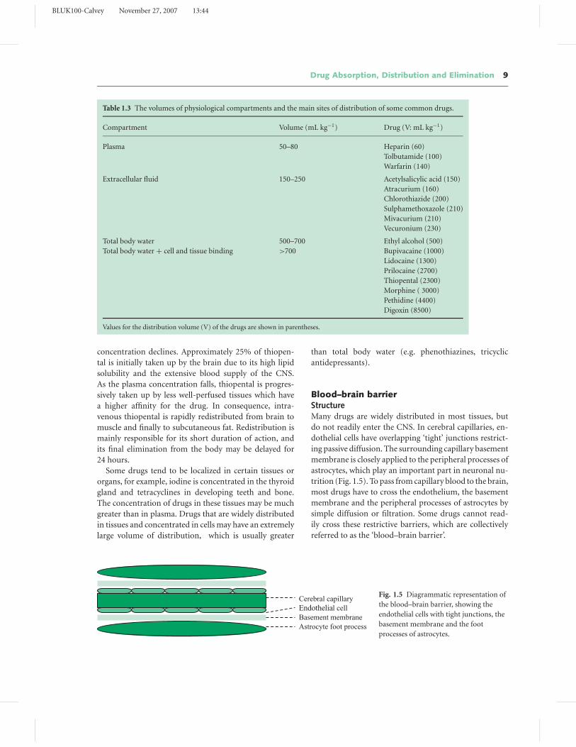
Table 1.3 The volumes of physiological compartments and the main sites of distribution of some common drugs.
Compartment Volume (mL kg−1) Drug (V: mL kg−1)
Plasma 50–80 Heparin (60)
Tolbutamide (100)
Warfarin (140)
Extracellular fluid 150–250 Acetylsalicylic acid (150)
Atracurium (160)
Chlorothiazide (200)
Sulphamethoxazole (210)
Mivacurium (210)
Vecuronium (230)
Total body water 500–700 Ethyl alcohol (500)
Total body water + cell and tissue binding >700 Bupivacaine (1000)
Lidocaine (1300)
Prilocaine (2700)
Thiopental (2300)
Morphine ( 3000)
Pethidine (4400)
Digoxin (8500)
Values for the distribution volume (V) of the drugs are shown in parentheses.
concentration declines. Approximately 25% of thiopen-
tal is initially taken up by the brain due to its high lipid
solubility and the extensive blood supply of the CNS.
As the plasma concentration falls, thiopental is progres-
sively taken up by less well-perfused tissues which have
a higher affinity for the drug. In consequence, intra-
venous thiopental is rapidly redistributed from brain to
muscle and finally to subcutaneous fat. Redistribution is
mainly responsible for its short duration of action, and
its final elimination from the body may be delayed for
24 hours.
Some drugs tend to be localized in certain tissues or
organs, for example, iodine is concentrated in the thyroid
gland and tetracyclines in developing teeth and bone.
The concentration of drugs in these tissues may be much
greater than in plasma. Drugs that are widely distributed
in tissues and concentrated in cells may have an extremely
large volume of distribution, which is usually greater
than total body water (e.g. phenothiazines, tricyclic
antidepressants).
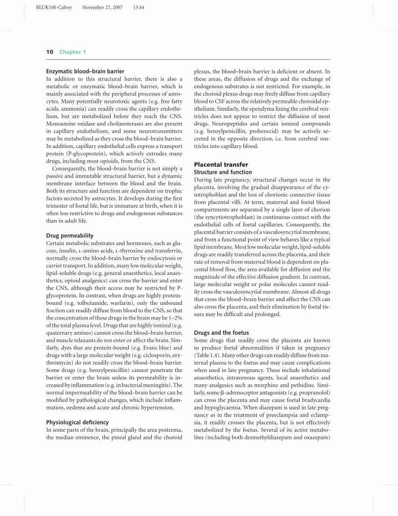
Blood–brain barrierStructureMany drugs are widely distributed in most tissues, but
do not readily enter the CNS. In cerebral capillaries, en-
dothelial cells have overlapping ‘tight’ junctions restrict-
ing passive diffusion. The surrounding capillary basement
membrane is closely applied to the peripheral processes of
astrocytes, which play an important part in neuronal nu-
trition (Fig. 1.5). To pass from capillary blood to the brain,
most drugs have to cross the endothelium, the basement
membrane and the peripheral processes of astrocytes by
simple diffusion or filtration. Some drugs cannot read-
ily cross these restrictive barriers, which are collectively
referred to as the ‘blood–brain barrier’.
Cerebral capillary
Endothelial cell
Basement membrane
Astrocyte foot process
Fig. 1.5 Diagrammatic representation of
the blood–brain barrier, showing the
endothelial cells with tight junctions, the
basement membrane and the foot
processes of astrocytes.
BLUK100-Calvey November 27, 2007 13:44
10 Chapter 1
Enzymatic blood–brain barrierIn addition to this structural barrier, there is also a
metabolic or enzymatic blood–brain barrier, which is
mainly associated with the peripheral processes of astro-
cytes. Many potentially neurotoxic agents (e.g. free fatty
acids, ammonia) can readily cross the capillary endothe-
lium, but are metabolized before they reach the CNS.
Monoamine oxidase and cholinesterases are also present
in capillary endothelium, and some neurotransmitters
may be metabolized as they cross the blood–brain barrier.
In addition, capillary endothelial cells express a transport
protein (P-glycoprotein), which actively extrudes many
drugs, including most opioids, from the CNS.
Consequently, the blood–brain barrier is not simply a
passive and immutable structural barrier, but a dynamic
membrane interface between the blood and the brain.
Both its structure and function are dependent on trophic
factors secreted by astrocytes. It develops during the first
trimester of foetal life, but is immature at birth, when it is
often less restrictive to drugs and endogenous substances
than in adult life.
Drug permeabilityCertain metabolic substrates and hormones, such as glu-
cose, insulin, l-amino acids, l-thyroxine and transferrin,
normally cross the blood–brain barrier by endocytosis or
carrier transport. In addition, many low molecular weight,
lipid-soluble drugs (e.g. general anaesthetics, local anaes-
thetics, opioid analgesics) can cross the barrier and enter
the CNS, although their access may be restricted by P-
glycoprotein. In contrast, when drugs are highly protein-
bound (e.g. tolbutamide, warfarin), only the unbound
fraction can readily diffuse from blood to the CNS, so that
the concentration of these drugs in the brain may be 1–2%
of the total plasma level. Drugs that are highly ionized (e.g.
quaternary amines) cannot cross the blood–brain barrier,
and muscle relaxants do not enter or affect the brain. Sim-
ilarly, dyes that are protein-bound (e.g. Evans blue) and
drugs with a large molecular weight (e.g. ciclosporin, ery-
thromycin) do not readily cross the blood–brain barrier.
Some drugs (e.g. benzylpenicillin) cannot penetrate the
barrier or enter the brain unless its permeability is in-
creased by inflammation (e.g. in bacterial meningitis). The
normal impermeability of the blood–brain barrier can be
modified by pathological changes, which include inflam-
mation, oedema and acute and chronic hypertension.
Physiological deficiencyIn some parts of the brain, principally the area postrema,
the median eminence, the pineal gland and the choroid
plexus, the blood–brain barrier is deficient or absent. In
these areas, the diffusion of drugs and the exchange of
endogenous substrates is not restricted. For example, in
the choroid plexus drugs may freely diffuse from capillary
blood to CSF across the relatively permeable choroidal ep-
ithelium. Similarly, the ependyma lining the cerebral ven-
tricles does not appear to restrict the diffusion of most
drugs. Neuropeptides and certain ionized compounds
(e.g. benzylpenicillin, probenecid) may be actively se-
creted in the opposite direction, i.e. from cerebral ven-
tricles into capillary blood.
Placental transferStructure and functionDuring late pregnancy, structural changes occur in the
placenta, involving the gradual disappearance of the cy-
totrophoblast and the loss of chorionic connective tissue
from placental villi. At term, maternal and foetal blood
compartments are separated by a single layer of chorion
(the syncytiotrophoblast) in continuous contact with the
endothelial cells of foetal capillaries. Consequently, the
placental barrier consists of a vasculosyncytial membrane,
and from a functional point of view behaves like a typical
lipid membrane. Most low molecular weight, lipid-soluble
drugs are readily transferred across the placenta, and their
rate of removal from maternal blood is dependent on pla-
cental blood flow, the area available for diffusion and the
magnitude of the effective diffusion gradient. In contrast,
large molecular weight or polar molecules cannot read-
ily cross the vasculosyncytial membrane. Almost all drugs
that cross the blood–brain barrier and affect the CNS can
also cross the placenta, and their elimination by foetal tis-
sues may be difficult and prolonged.
Drugs and the foetusSome drugs that readily cross the placenta are known
to produce foetal abnormalities if taken in pregnancy
(Table 1.4). Many other drugs can readily diffuse from ma-
ternal plasma to the foetus and may cause complications
when used in late pregnancy. These include inhalational
anaesthetics, intravenous agents, local anaesthetics and
many analgesics such as morphine and pethidine. Simi-
larly, some �-adrenoceptor antagonists (e.g. propranolol)
can cross the placenta and may cause foetal bradycardia
and hypoglycaemia. When diazepam is used in late preg-
nancy as in the treatment of preeclampsia and eclamp-
sia, it readily crosses the placenta, but is not effectively
metabolized by the foetus. Several of its active metabo-
lites (including both desmethyldiazepam and oxazepam)
BLUK100-Calvey November 27, 2007 13:44
Drug Absorption, Distribution and Elimination 11
Table 1.4 Drugs that may cause foetal damage or malforma-
tion (teratogenic effects) if taken during pregnancy.
Drug Effect on foetus
Methotrexate Hydrocephalus; neural tube defects
Tretinoin Hydrocephalus
Phenytoin Cleft lip and palate; cardiac defects
Sodium valproate Neural tube defects
Oestrogens Vaginal adenosis; testicular atrophy
Aminoglycosides Cochlear and vestibular damage
Tetracyclines Dental pigmentation; enamel
hypoplasia
Carbimazole Goitre; hypothyroidism
Propylthiouracil Goitre; hypothyroidism
Warfarin Nasal hypoplasia; epiphyseal
calcification
accumulate in foetal tissues and can cause neonatal hypo-
tonia and hypothermia. By contrast, ionized compounds
(e.g. muscle relaxants) cannot readily cross the placenta.
Protein binding
Plasma protein binding plays an essential role in the trans-
port and distribution of drugs. Most drugs are relatively
lipid-soluble, but are only poorly soluble in plasma water.
Consequently, binding to plasma proteins is essential for
their transport in plasma.
Most drugs are reversibly bound to plasma proteins,
according to the reaction:
unbound drug + protein � drug − protein complex
During perfusion, the unbound drug diffuses into tissues,
and as its concentration in plasma falls, protein-bound
drug rapidly dissociates. Consequently, a continuous con-
centration gradient is present for the diffusion of drugs
from plasma to tissues.
Binding by albumin and globulinsAlbumin usually plays the most important role in the
binding of drugs. It has a number of distinct binding
sites with a variable affinity for drugs, and mainly binds
neutral or acidic compounds, including salicylates, in-
dometacin, tolbutamide, carbenoxolone and oral antico-
agulants. Some basic drugs and physiological substrates
such as bilirubin, fatty acids and tryptophan are also
bound by albumin.
Globulins bind many basic drugs (e.g. bupivacaine,
opioid analgesics). These drugs are mainly bound by �-
globulins or by �1-acid glycoprotein. Plasma globulins
also play an important part in the binding of minerals, vi-
tamins and hormones. Hydrocortisone (cortisol) is mainly
transported in plasma by a specific globulin (transcortin)
for which it has a high affinity.
Some drugs (e.g. pancuronium) are bound by both
globulins and albumin. Indeed, the resistance to muscle
relaxants that often occurs in liver disease may be due to
their increased binding by plasma globulins.
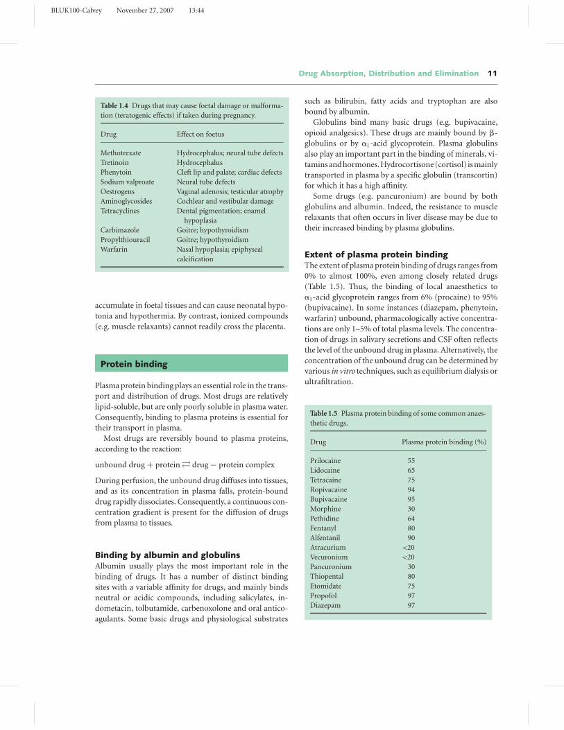
Extent of plasma protein bindingThe extent of plasma protein binding of drugs ranges from
0% to almost 100%, even among closely related drugs
(Table 1.5). Thus, the binding of local anaesthetics to
�1-acid glycoprotein ranges from 6% (procaine) to 95%
(bupivacaine). In some instances (diazepam, phenytoin,
warfarin) unbound, pharmacologically active concentra-
tions are only 1–5% of total plasma levels. The concentra-
tion of drugs in salivary secretions and CSF often reflects
the level of the unbound drug in plasma. Alternatively, the
concentration of the unbound drug can be determined by
various in vitro techniques, such as equilibrium dialysis or
ultrafiltration.
Table 1.5 Plasma protein binding of some common anaes-
thetic drugs.
Drug Plasma protein binding (%)
Prilocaine 55
Lidocaine 65
Tetracaine 75
Ropivacaine 94
Bupivacaine 95
Morphine 30
Pethidine 64
Fentanyl 80
Alfentanil 90
Atracurium <20
Vecuronium <20
Pancuronium 30
Thiopental 80
Etomidate 75
Propofol 97
Diazepam 97
BLUK100-Calvey November 27, 2007 13:44
12 Chapter 1
Drug competition and displacementDrugs and endogenous substrates that are extensively
bound to proteins may compete for (and be displaced
from) their binding sites. In most instances, binding of
drugs at clinical concentrations only occupies a small pro-
portion of the available binding sites and does not ap-
proach saturation. Consequently, competition between
drugs resulting in clinically significant displacement from
plasma protein binding is extremely rare (Chapter 4).
Protein binding and drug eliminationThe hepatic clearance of many drugs is limited by liver
blood flow and is not restricted by plasma protein bind-
ing (which is a rapidly reversible process). Drug disso-
ciation from binding to plasma proteins probably occurs
within microseconds or milliseconds. By contrast, the hep-
atic perfusion time may be several seconds or more. Thus,
extensive protein binding only decreases hepatic clearance
when the ability of the liver to extract, metabolize or ex-
crete the drug is low. Similarly, protein binding is unlikely
to restrict the renal elimination of drugs, either by the
glomerulus or the renal tubule. Only the unbound drug
is secreted by the proximal tubule, but the resultant de-
crease in its plasma concentration leads to the immediate
dissociation of protein-bound drug in order to maintain
equilibrium. Indeed, a number of protein-bound drugs
are completely cleared in a single passage through the kid-
ney (e.g. benzylpenicillin).
Protein binding in pathological conditionsBinding to plasma proteins is modified in pathological
conditions associated with hypoalbuminaemia, as in hep-
atic cirrhosis, nephrosis, trauma or burns. In these con-
ditions, the concentration of the unbound drug tends to
increase and may result in toxic effects (e.g. with pheny-
toin or prednisolone). Significant changes are particularly
likely when high doses of drugs are used, or when drugs are
given intravenously. In these conditions, binding to albu-
min and other plasma proteins may be saturated, causing
a disproportionate increase in the concentration of the
unbound drug. Tissues and organs that are well perfused
(e.g. brain, heart, abdominal viscera) may receive a higher
proportion of the dose, predisposing them to potential
toxic effects. Similar effects may occur in elderly patients
and in subjects with renal impairment, possibly due to al-
terations in the affinity of drugs for albumin. The plasma
concentration of �1-acid glycoprotein can also be modi-
fied by a number of pathological conditions including my-
ocardial infarction, rheumatoid arthritis, Crohn’s disease,
renal failure and malignant disease, as well as operative
surgery. In these conditions, the binding of basic drugs
(e.g. propranolol, chlorpromazine) is increased, and the
concentration of the free, unbound drug is reduced.
Drug metabolism
Most drugs are eliminated by drug metabolism, which
mainly occurs in the liver. Nevertheless, certain drugs are
partly or completely broken down by other tissues. Some
esters that are used in anaesthesia are hydrolysed by plasma
cholinesterase (e.g. suxamethonium, mivacurium) or red
cell acetylcholinesterase (e.g. esmolol, remifentanil). In
addition, drugs may be partly or completely metabolized
by the gut (e.g. morphine, chlorpromazine), the kidney
(e.g. midazolam, dopamine) or the lung (e.g. angiotensin
I, prilocaine).
Nevertheless, the liver is mainly responsible for the
breakdown of drugs. Hepatic metabolism decreases the
concentration of the active drug in plasma, and thus pro-
motes its removal from the site of action. This mainly
involves the enzymatic conversion of lipid-soluble non-
polar drugs into water-soluble polar compounds, which
can be filtered by the renal glomerulus or secreted into
urine or bile.
Metabolism usually reduces the biological activity of
drugs, and most metabolites have less inherent activity
than their parent compounds. In addition, their ability to
penetrate to receptor sites is limited because of their poor
lipid solubility. Nevertheless, some drugs are relatively
inactive when administered, and require metabolism to
produce or enhance their pharmacological effects (Ta-
ble 1.6). Other drugs may be metabolized to compounds
Table 1.6 Drugs that require metabolism to produce their
pharmacological effects.
Drug Active metabolite
Prontosil red Sulphanilamide
Chloral hydrate Trichlorethanol
Cyclophosphamide Phosphoramide mustard
Cortisone Hydrocortisone
Prednisone Prednisolone
Methyldopa Methylnoradrenaline
Proguanil Cycloguanil
Enalapril Enalaprilat
BLUK100-Calvey November 27, 2007 13:44
Drug Absorption, Distribution and Elimination 13
Table 1.7 Phase 1 reactions resulting in drug oxidation, reduction and hydrolysis.
Reaction Site Enzyme Example
Oxidation Hepatic endoplasmic Cytochrome P450 Thiopental → pentobarbital
reticulum
Mitochondria Monoamine oxidase Dopamine → dihydroxyphenylacetaldehyde
Hepatic cell cytoplasm Alcohol dehydrogenase Alcohol → acetaldehyde
Reduction Hepatic endoplasmic Cytochrome P450 Halothane → chlorotrifluoroethane
reticulum
Hepatic cell cytoplasm Alcohol dehydrogenase Chloral hydrate → trichlorethanol
Hydrolysis Hepatic endoplasmic Carboxyesterase Pethidine → pethidinic acid
reticulum
Plasma Cholinesterase Suxamethonium → succinate + choline
Erythrocyte Acetylcholinesterase Remifentanil → carboxylated derivatives
Neuromuscular junction Acetylcholinesterase Acetylcholine → acetate + choline
Hepatic cell cytoplasm Amidase Lidocaine → 2,6-xylidine + diethylglycine
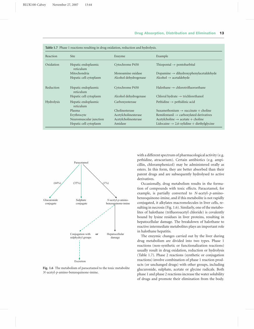
Paracetamol
(64%) (35%) (1%)
Glucuronide Sulphate N-acetyl-p-amino-conjugate conjugate benzoquinone-imine
Conjugation with or Hepatocellularsulphydryl groups damage
Excretion
Fig. 1.6 The metabolism of paracetamol to the toxic metabolite
N-acetyl-p-amino-benzoquinone-imine.
with a different spectrum of pharmacological activity (e.g.
pethidine, atracurium). Certain antibiotics (e.g. ampi-
cillin, chloramphenicol) may be administered orally as
esters. In this form, they are better absorbed than their
parent drugs and are subsequently hydrolysed to active
derivatives.
Occasionally, drug metabolism results in the forma-
tion of compounds with toxic effects. Paracetamol, for
example, is partially converted to N-acetyl- p-amino-
benzoquinone-imine, and if this metabolite is not rapidly
conjugated, it alkylates macromolecules in liver cells, re-
sulting in necrosis (Fig. 1.6). Similarly, one of the metabo-
lites of halothane (trifluoroacetyl chloride) is covalently
bound by lysine residues in liver proteins, resulting in
hepatocellular damage. The breakdown of halothane to
reactive intermediate metabolites plays an important role
in halothane hepatitis.
The enzymic changes carried out by the liver during
drug metabolism are divided into two types. Phase 1
reactions (non-synthetic or functionalization reactions)
usually result in drug oxidation, reduction or hydrolysis
(Table 1.7). Phase 2 reactions (synthetic or conjugation
reactions) involve combination of phase 1 reaction prod-
ucts (or unchanged drugs) with other groups, including
glucuronide, sulphate, acetate or glycine radicals. Both
phase 1 and phase 2 reactions increase the water solubility
of drugs and promote their elimination from the body.
BLUK100-Calvey November 27, 2007 13:44
14 Chapter 1
Fig. 1.7 Electron micrography of part of a
mouse liver cell, showing mitochondria
(M), endoplasmic reticulum (ER) and the
nuclear membrane enclosing the nucleus
(N) (×30,000).
Some drugs (e.g. sodium salicylate) are almost entirely
metabolized by phase 2 reactions.
Phase 1 reactionsMost phase 1 reactions and glucuronide conjugation are
carried out by the smooth endoplasmic reticulum or the
microsomes (Fig. 1.7). Most drug oxidation and reduc-
tion, and some hydrolysis, is carried out by a non-specific
microsomal enzyme system (cytochrome P450 or the
‘mixed function oxidase system’). Cytochrome P450 con-
sists of many distinct but genetically related forms of a
superfamily of haem proteins. Their name is derived from
their ability, in the reduced state, to combine with carbon
monoxide and form a complex that maximally absorbs
light at a wavelength of 450 nm.
Cytochrome P450Drug oxidation by cytochrome P450 depends on the
flavoprotein NADPH-CYP reductase, the electron donor
NADPH and molecular oxygen (as well as cytochrome b5
and NADPH-cytochrome b5 reductase). The reaction in-
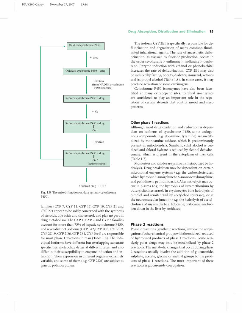
volves a complex enzymatic cycle (Fig. 1.8), which results
in the breakdown of molecular oxygen. A single oxygen
atom (from O2) is released as H2O and the other is trans-
ferred to the substrate (D), according to the equation:
DH + O2 + NADPH + H+ → DOH + H2O + NADP+
Cytochrome P450 enzymes may also mediate the reduc-
tive metabolism of certain drugs, such as halothane (Table
1.7). This is dependent on the ability of drugs to directly
accept electrons from the reduced cytochrome P450 drug
complex (Fig. 1.8) and is enhanced by hypoxia.
Isoforms of cytochrome P450Different forms of human cytochrome P450 are classi-
fied by the similarity in their amino acid sequences into
gene families and gene subfamilies. The members of each
gene family (CYP 1, CYP 2 etc.) have a common amino
acid sequence of 40% or more, while members of each
subfamily (CYP 1A, CYP 1B etc.) have a sequence simi-
larity of more than 55%. At the present time, 17 different
gene families have been identified, and at least six of these
BLUK100-Calvey November 27, 2007 13:44
Drug Absorption, Distribution and Elimination 15
+ drug
+ electron (from NADPH cytochrome
P450 reductase)
+ O2
+ electron
Oxidized drug + H2O
Oxidized cytochrome P450
Oxidized cytochrome P450 − drug
Reduced cytochrome P450 − drug
Reduced cytochrome P450 − drug
⏐⏐⏐⏐ O2
Reduced cytochrome P450 − drug
⏐⏐O2 *
(active electron)
Fig. 1.8 The mixed-function oxidase system (cytochrome
P450).
families (CYP 7, CYP 11, CYP 17, CYP 19, CYP 21 and
CYP 27) appear to be solely concerned with the synthesis
of steroids, bile acids and cholesterol, and play no part in
drug metabolism. The CYP 1, CYP 2 and CYP 3 families
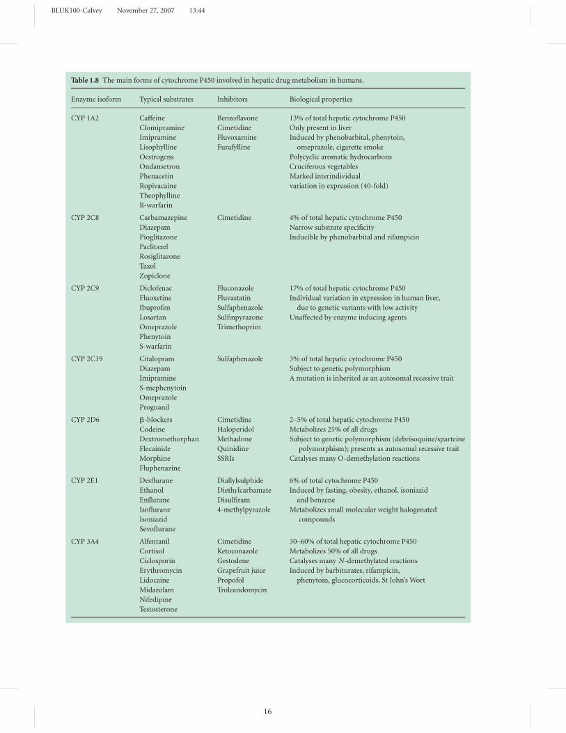
account for more than 75% of hepatic cytochrome P450,
and seven distinct isoforms (CYP 1A2, CYP 2C8, CYP 2C9,
CYP 2C19, CYP 2D6, CYP 2E1, CYP 3A4) are responsible
for most phase 1 reactions in man (Table 1.8). The indi-
vidual isoforms have different but overlapping substrate
specificities, metabolize drugs at different rates, and also
differ in their susceptibility to enzyme induction and in-
hibition. Their expression in different organs is extremely
variable, and some of them (e.g. CYP 2D6) are subject to
genetic polymorphism.
The isoform CYP 2E1 is specifically responsible for de-
fluorination and degradation of many common fluori-
nated inhalational agents. The rate of anaesthetic deflu-
orination, as assessed by fluoride production, occurs in
the order sevoflurane > enflurane > isoflurane > desflu-
rane. Enzyme induction with ethanol or phenobarbital
increases the rate of defluorination. CYP 2E1 may also
be induced by fasting, obesity, diabetes, isoniazid, ketones
and isopropyl alcohol (Table 1.8). In some cases, it may
produce activation of some carcinogens.
Cytochrome P450 isoenzymes have also been iden-
tified at many extrahepatic sites. Cerebral isoenzymes
are considered to play an important role in the regu-
lation of certain steroids that control mood and sleep
patterns.
Other phase 1 reactionsAlthough most drug oxidation and reduction is depen-
dent on isoforms of cytochrome P450, some endoge-
nous compounds (e.g. dopamine, tyramine) are metab-
olized by monoamine oxidase, which is predominantly
present in mitochondria. Similarly, ethyl alcohol is oxi-
dized and chloral hydrate is reduced by alcohol dehydro-
genase, which is present in the cytoplasm of liver cells
(Table 1.7).
Most esters and amides are primarily metabolized by hy-
drolysis. Drug breakdown may be dependent on certain
microsomal enzyme systems (e.g. the carboxylesterases,
which hydrolyse diamorphine to 6-monoacetylmorphine,
and pethidine to pethidinic acid). Alternatively, it may oc-
cur in plasma (e.g. the hydrolysis of suxamethonium by
butyrylcholinesterase), in erythrocytes (the hydrolysis of
esmolol and remifentanil by acetylcholinesterase), or at
the neuromuscular junction (e.g. the hydrolysis of acetyl-
choline). Many amides (e.g. lidocaine, prilocaine) are bro-
ken down in the liver by amidases.
Phase 2 reactionsPhase 2 reactions (synthetic reactions) involve the conju-
gation of other chemical groups with the oxidized, reduced
or hydrolysed products of phase 1 reactions. Some rela-
tively polar drugs may only be metabolized by phase 2
reactions. The metabolic changes that occur during phase
2 reactions usually involve the addition of glucuronide,
sulphate, acetate, glycine or methyl groups to the prod-
ucts of phase 1 reactions. The most important of these
reactions is glucuronide conjugation.
BLUK100-Calvey November 27, 2007 13:44
Table 1.8 The main forms of cytochrome P450 involved in hepatic drug metabolism in humans.
Enzyme isoform Typical substrates Inhibitors Biological properties
CYP 1A2 Caffeine Benzoflavone 13% of total hepatic cytochrome P450
Clomipramine Cimetidine
Imipramine Fluvoxamine
Only present in liver
Lisophylline Furafylline
Induced by phenobarbital, phenytoin,
Oestrogens
omeprazole, cigarette smoke
Ondansetron
Polycyclic aromatic hydrocarbons
Phenacetin
Cruciferous vegetables
Ropivacaine
Marked interindividual
Theophylline
variation in expression (40-fold)
R-warfarin
CYP 2C8 Carbamazepine Cimetidine 4% of total hepatic cytochrome P450
Diazepam Narrow substrate specificity
Pioglitazone Inducible by phenobarbital and rifampicin
Paclitaxel
Rosiglitazone
Taxol
Zopiclone
CYP 2C9 Diclofenac Fluconazole 17% of total hepatic cytochrome P450
Fluoxetine Fluvastatin
Ibuprofen Sulfaphenazole
Individual variation in expression in human liver,
Losartan Sulfinpyrazone
due to genetic variants with low activity
Omeprazole Trimethoprim
Unaffected by enzyme inducing agents
Phenytoin
S-warfarin
CYP 2C19 Citalopram Sulfaphenazole 3% of total hepatic cytochrome P450
Diazepam
Imipramine
Subject to genetic polymorphism
S-mephenytoin
A mutation is inherited as an autosomal recessive trait
Omeprazole
Proguanil
CYP 2D6 �-blockers Cimetidine 2–5% of total hepatic cytochrome P450
Codeine Haloperidol
Dextromethorphan Methadone
Metabolizes 25% of all drugs
Flecainide Quinidine
Subject to genetic polymorphism (debrisoquine/sparteine
Morphine SSRIs
polymorphism); presents as autosomal recessive trait
Fluphenazine
Catalyses many O-demethylation reactions
CYP 2E1 Desflurane Diallylsulphide 6% of total cytochrome P450
Ethanol Diethylcarbamate Induced by fasting, obesity, ethanol, isoniazid
Enflurane Disulfiram and benzene
Isoflurane 4-methylpyrazole Metabolizes small molecular weight halogenated
Isoniazid compounds
Sevoflurane
CYP 3A4 Alfentanil Cimetidine 30–60% of total hepatic cytochrome P450
Cortisol Ketoconazole
Ciclosporin Gestodene
Metabolizes 50% of all drugs
Erythromycin Grapefruit juice
Catalyses many N-demethylated reactions
Lidocaine Propofol
Induced by barbiturates, rifampicin,
Midazolam Troleandomycin
phenytoin, glucocorticoids, St John’s Wort
Nifedipine
Testosterone
16
BLUK100-Calvey November 27, 2007 13:44
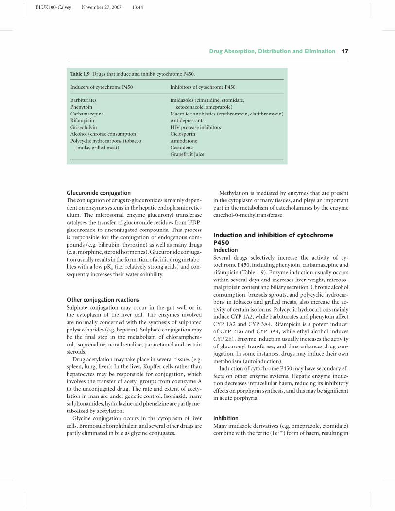
Drug Absorption, Distribution and Elimination 17
Table 1.9 Drugs that induce and inhibit cytochrome P450.
Inducers of cytochrome P450 Inhibitors of cytochrome P450
Barbiturates Imidazoles (cimetidine, etomidate,
Phenytoin ketoconazole, omeprazole)
Carbamazepine Macrolide antibiotics (erythromycin, clarithromycin)
Rifampicin Antidepressants
Griseofulvin HIV protease inhibitors
Alcohol (chronic consumption) Ciclosporin
Polycyclic hydrocarbons (tobacco Amiodarone
smoke, grilled meat) Gestodene
Grapefruit juice
Glucuronide conjugationThe conjugation of drugs to glucuronides is mainly depen-
dent on enzyme systems in the hepatic endoplasmic retic-
ulum. The microsomal enzyme glucuronyl transferase
catalyses the transfer of glucuronide residues from UDP-
glucuronide to unconjugated compounds. This process
is responsible for the conjugation of endogenous com-
pounds (e.g. bilirubin, thyroxine) as well as many drugs
(e.g. morphine, steroid hormones). Glucuronide conjuga-
tion usually results in the formation of acidic drug metabo-
lites with a low pKa (i.e. relatively strong acids) and con-
sequently increases their water solubility.
Other conjugation reactionsSulphate conjugation may occur in the gut wall or in
the cytoplasm of the liver cell. The enzymes involved
are normally concerned with the synthesis of sulphated
polysaccharides (e.g. heparin). Sulphate conjugation may
be the final step in the metabolism of chlorampheni-
col, isoprenaline, noradrenaline, paracetamol and certain
steroids.
Drug acetylation may take place in several tissues (e.g.
spleen, lung, liver). In the liver, Kupffer cells rather than
hepatocytes may be responsible for conjugation, which
involves the transfer of acetyl groups from coenzyme A
to the unconjugated drug. The rate and extent of acety-
lation in man are under genetic control. Isoniazid, many
sulphonamides, hydralazine and phenelzine are partly me-
tabolized by acetylation.
Glycine conjugation occurs in the cytoplasm of liver
cells. Bromosulphonphthalein and several other drugs are
partly eliminated in bile as glycine conjugates.
Methylation is mediated by enzymes that are present
in the cytoplasm of many tissues, and plays an important
part in the metabolism of catecholamines by the enzyme
catechol-0-methyltransferase.
Induction and inhibition of cytochromeP450InductionSeveral drugs selectively increase the activity of cy-
tochrome P450, including phenytoin, carbamazepine and
rifampicin (Table 1.9). Enzyme induction usually occurs
within several days and increases liver weight, microso-
mal protein content and biliary secretion. Chronic alcohol
consumption, brussels sprouts, and polycyclic hydrocar-
bons in tobacco and grilled meats, also increase the ac-
tivity of certain isoforms. Polycyclic hydrocarbons mainly
induce CYP 1A2, while barbiturates and phenytoin affect
CYP 1A2 and CYP 3A4. Rifampicin is a potent inducer
of CYP 2D6 and CYP 3A4, while ethyl alcohol induces
CYP 2E1. Enzyme induction usually increases the activity
of glucuronyl transferase, and thus enhances drug con-
jugation. In some instances, drugs may induce their own
metabolism (autoinduction).
Induction of cytochrome P450 may have secondary ef-
fects on other enzyme systems. Hepatic enzyme induc-
tion decreases intracellular haem, reducing its inhibitory
effects on porphyrin synthesis, and this may be significant
in acute porphyria.
InhibitionMany imidazole derivatives (e.g. omeprazole, etomidate)
combine with the ferric (Fe3+) form of haem, resulting in
BLUK100-Calvey November 27, 2007 13:44
18 Chapter 1
reversible non-competitive inhibition of CYP 3A4 and var-
ious other isoforms. Quinidine is a competitive inhibitor
of CYP 2D6 (although it is not metabolized by this iso-
form). In addition, some synthetic corticosteroids (e.g.
gestodene) are oxidized by CYP 3A4 and combine with
it covalently (‘suicide inhibition’). Furafylline affects CYP
1A2 in a similar manner. Many other drugs also inhibit
some cytochrome P450 isoforms, particularly CYP 3A4
(Table 1.9). Enzyme inhibition may increase plasma con-
centrations of other concurrently used drugs, resulting in
drug interactions (Chapters 4 and 5).
First-pass metabolismAfter oral administration, some drugs are extensively me-
tabolized by the gut wall (e.g. chlorpromazine, dopamine)
or by the liver (e.g. lidocaine, pethidine) before they en-
ter the systemic circulation (‘presystemic’ or ‘first-pass
metabolism’). In these conditions, oral administration
may not produce adequate plasma concentrations in the
systemic circulation and may result in an impaired re-
sponse to drugs. First-pass metabolism by the liver is rela-
tively common with drugs that have a high hepatic extrac-
tion ratio (i.e. when the concentration in the hepatic vein
is less than 50% of that in the portal vein). In these condi-
tions, clearance is primarily dependent on liver blood flow
rather than the activity of drug-metabolizing enzymes,
and drugs that reduce hepatic blood flow (e.g. propra-
nolol) may influence the magnitude of the first-pass ef-
fect. Drugs are sometimes given by sublingual or rectal
administration in order to avoid first-pass metabolism in
the liver.
Individual differences in drug metabolismWhen some drugs are administered in the same dose to
different patients, plasma concentrations may vary over
a 10-fold range. The phenomenon is sometimes due to
interindividual differences in drug metabolism, which is
an important cause of the variability in response to drugs
(Chapter 5). Most of the available evidence suggests that
the rate and the pattern of drug metabolism are mainly
controlled by genetic factors, including sex, race and eth-
nicity. Some metabolic pathways are subject to genetic
polymorphism (e.g. drug acetylation, ester hydrolysis).
For example, individuals who are deficient in CYP 2D6
(Table 1.8) may have an impaired analgesic response to
codeine, since they convert little or none of the drug to
morphine.
Environmental factors, including diet, cigarette smok-
ing, alcohol consumption and exposure to insecticides,
are probably of lesser importance. However, interindi-
vidual differences in plasma concentrations and vari-
able responses are sometimes related to drug interactions
(Chapter 4), particularly with agents that induce or in-
hibit hepatic enzyme systems. Interactions with enzyme
inducers or inhibitors are commoner with low extraction,
extensively protein-bound drugs whose clearance is de-
pendent on metabolism rather than hepatic blood flow.
High extraction drugs whose clearance is dependent on
hepatic blood flow are unlikely to be involved in signifi-
cant metabolic reactions.
Drug metabolism may be related to age, and the hepatic
metabolism of many drugs is modified in childhood and
in the elderly. Neonates have impaired drug metaboliz-
ing systems, and some isoforms of cytochrome P450 and
glucuronyl transferase may be relatively immature. In the
elderly, drug metabolism is also modified, although altered
environmental influences may be of more importance.
Pathological changesPathological changes may affect the metabolism and clear-
ance of drugs in an unpredictable manner. In severe hep-
atic disease (e.g. cirrhosis or hepatitis), the elimination of
drugs that are primarily metabolized may be impaired.
The reduction in clearance may result in drug cumu-
lation, and the urinary elimination of metabolites may
be decreased. Liver disease may also enhance and pro-
long the effects of drugs that are metabolized by plasma
cholinesterase. Any decrease in cardiac output (e.g. due to
heart block, myocardial infarction or hypertension) may
reduce the elimination of drugs whose clearance is depen-
dent on hepatic blood flow. Renal disease usually has little
or no effect on drug metabolism, although polar metabo-
lites may accumulate in plasma and produce toxic effects.
Thus, norpethidine (a demethylated metabolite of pethi-
dine) is normally eliminated in urine, but in renal failure
its excretion is impaired, and may sometimes cause cere-
bral excitation and convulsions.
Hepatic, renal and cardiac diseases are important fac-
tors affecting the variable response to drugs (Chapter 5).
Drug excretion
Almost all drugs and their metabolites are eventually elim-
inated from the body in urine or in bile. Small amounts
of some drugs are excreted in saliva and in milk.
The molecular weight of drugs and their metabolites
plays an important part in determining their route of
BLUK100-Calvey November 27, 2007 13:44
Drug Absorption, Distribution and Elimination 19
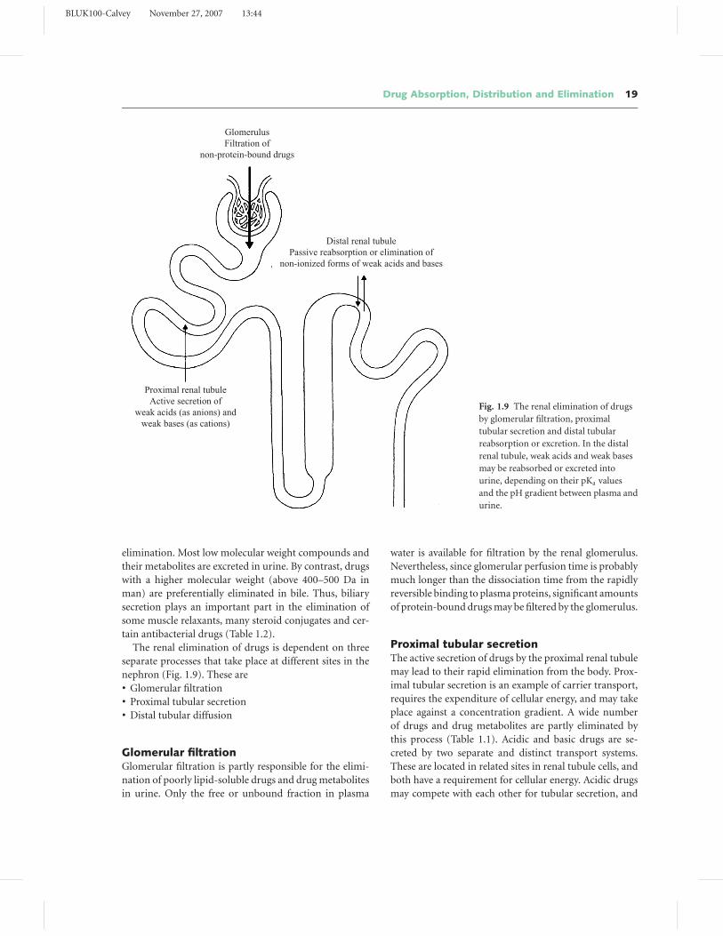
GlomerulusFiltration of
non-protein-bound drugs
Distal renal tubulePassive reabsorption or elimination of
non-ionized forms of weak acids and bases
Proximal renal tubuleActive secretion of
weak acids (as anions) andweak bases (as cations)
Fig. 1.9 The renal elimination of drugs
by glomerular filtration, proximal
tubular secretion and distal tubular
reabsorption or excretion. In the distal
renal tubule, weak acids and weak bases
may be reabsorbed or excreted into
urine, depending on their pKa values
and the pH gradient between plasma and
urine.
elimination. Most low molecular weight compounds and
their metabolites are excreted in urine. By contrast, drugs
with a higher molecular weight (above 400–500 Da in
man) are preferentially eliminated in bile. Thus, biliary
secretion plays an important part in the elimination of
some muscle relaxants, many steroid conjugates and cer-
tain antibacterial drugs (Table 1.2).
The renal elimination of drugs is dependent on three
separate processes that take place at different sites in the
nephron (Fig. 1.9). These are� Glomerular filtration� Proximal tubular secretion� Distal tubular diffusion
Glomerular filtrationGlomerular filtration is partly responsible for the elimi-
nation of poorly lipid-soluble drugs and drug metabolites
in urine. Only the free or unbound fraction in plasma
water is available for filtration by the renal glomerulus.
Nevertheless, since glomerular perfusion time is probably
much longer than the dissociation time from the rapidly
reversible binding to plasma proteins, significant amounts
of protein-bound drugs may be filtered by the glomerulus.
Proximal tubular secretionThe active secretion of drugs by the proximal renal tubule
may lead to their rapid elimination from the body. Prox-
imal tubular secretion is an example of carrier transport,
requires the expenditure of cellular energy, and may take
place against a concentration gradient. A wide number
of drugs and drug metabolites are partly eliminated by
this process (Table 1.1). Acidic and basic drugs are se-
creted by two separate and distinct transport systems.
These are located in related sites in renal tubule cells, and
both have a requirement for cellular energy. Acidic drugs
may compete with each other for tubular secretion, and
BLUK100-Calvey November 27, 2007 13:44
20 Chapter 1
basic drugs may interfere with the elimination of other
bases or cations. Acids do not usually compete with or
affect the secretion of bases. Occasionally, the competitive
inhibition of the tubular transport of acids or bases is of
practical significance (e.g. the inhibition of penicillin se-
cretion by probenecid, or the reduction of urate transport
by thiazide diuretics).
During tubular secretion, only the unbound drug is
transferred from plasma to tubular cells. Nevertheless,
protein or red cell binding does not apparently restrict
tubular secretion, and some drugs that are significantly
bound to plasma proteins (e.g. phenol red, some peni-
cillins) are completely cleared by the kidney in a single
circulation. As discussed above, this probably reflects the
rapid dissociation from plasma protein in relation to the
time required for renal tubular perfusion.
Distal tubular diffusionIn the distal renal tubule, non-ionic diffusion is partly re-
sponsible for the reabsorption and elimination of acids
and bases. In this region of the nephron, there is a con-
siderable H+ gradient between plasma and acid urine.
Most acidic drugs are preferentially excreted in alkaline
urine, where they are present as non-diffusible anions.
In acid urine, they are usually present as non-ionized
molecules that can readily diffuse back into plasma. In
these conditions, they are slowly eliminated from the body
and their half-lives may be prolonged. For instance, the
weak acid probenecid is actively secreted in the prox-
imal renal tubule as an anion, i.e. R-COO−. In acidic
conditions (e.g. in the distal renal tubule), it is partially
present in the non-ionized form R-COOH and is exten-
sively reabsorbed. In consequence, its elimination from
the body is relatively slow and its half-life is approximately
6–12 hours.
By contrast, basic drugs (e.g. secondary and tertiary
amines) are preferentially excreted in acid urine (Fig. 1.3).
In these conditions they can readily diffuse from plasma
to urine where they are trapped as cations. This provides
a gradient for the diffusion of the non-ionized drug from
plasma to urine. Many basic drugs are highly lipid-soluble
and extensively bound to plasma proteins and may not
be significantly eliminated by glomerular filtration or by
tubular secretion. Diffusion of the non-ionized fraction
from the relatively alkaline plasma to acid urine (Fig. 1.3)
may be the only method responsible for the elimination of
these drugs. At one time, the effects of changes in urine pH
on the elimination of weak acids and bases were sometimes
utilized in the treatment of drug overdosage.
Biliary excretionThe biliary excretion of drugs and drug metabolites is usu-
ally less important than their renal elimination. Neverthe-
less, almost all drugs or their metabolites can be identified
in bile after oral or parenteral administration (although
only trace amounts of many compounds may be detected).
Biliary excretion is usually the major route of elimination
of compounds with a molecular weight of more than 400–
500 Da.
Ionized or partly ionized drugs and their metabolites
are usually eliminated from liver cells by active transport.
High molecular weight anions (including glucuronide and
sulphate conjugates) and cations (including quaternary
amines) are actively transferred from hepatocytes to the
biliary canaliculus by separate transport systems, which
are dependent on Na+/K+ ATPase. Biliary secretion is rel-
atively non-specific, saturable and can be competitively or
non-competitively inhibited by other drugs. Thus, anions
compete with each other for canalicular transport, while
basic drugs interfere with the elimination of other bases
or cations. In many respects, the biliary secretion of an-
ions and cations is similar to their active transport in the
proximal renal tubule, and accounts for the high concen-
trations of certain drugs in bile (in some instances, more
than 100 times their plasma level).
The phenomenon is sometimes of practical signifi-
cance. The visualization of contrast media during radi-
ological examination of the biliary tract is dependent on
their active secretion and concentration in bile. Similarly,
the high concentrations of ampicillin and rifampicin that
are eliminated in bile may account for their effective-
ness in enteric infections. Many muscle relaxants are also
present in high concentrations in bile. Monoquaternary
compounds (e.g. vecuronium) are more extensively elimi-
nated than their bisquaternary analogues (pancuronium),
and this may partly account for the differences in their du-
ration of action.
Enterohepatic circulationMany compounds that are eliminated in bile as glu-
curonide conjugates are hydrolysed in the small intestine
by bacterial flora that secrete the enzyme glucuronidase.
After hydrolysis the unchanged drug is reabsorbed, me-
tabolized and re-excreted as a glucuronide conjugate
(Fig. 1.10). This ‘enterohepatic circulation’ of drugs
may occur many times before compounds are finally
eliminated from the body and is often associated with
a substantial first-pass effect and a prolonged plasma
half-life.
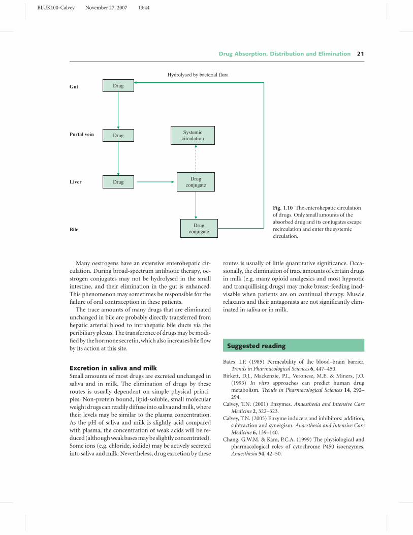
BLUK100-Calvey November 27, 2007 13:44
Drug Absorption, Distribution and Elimination 21
Hydrolysed by bacterial flora
Gut
Portal vein
Liver
Bile
Drug
Drug
Drug
Systemic circulation
Drug conjugate
Drug conjugate
Fig. 1.10 The enterohepatic circulation
of drugs. Only small amounts of the
absorbed drug and its conjugates escape
recirculation and enter the systemic
circulation.
Many oestrogens have an extensive enterohepatic cir-
culation. During broad-spectrum antibiotic therapy, oe-
strogen conjugates may not be hydrolysed in the small
intestine, and their elimination in the gut is enhanced.
This phenomenon may sometimes be responsible for the
failure of oral contraception in these patients.
The trace amounts of many drugs that are eliminated
unchanged in bile are probably directly transferred from
hepatic arterial blood to intrahepatic bile ducts via the
peribiliary plexus. The transference of drugs may be modi-
fied by the hormone secretin, which also increases bile flow
by its action at this site.
Excretion in saliva and milkSmall amounts of most drugs are excreted unchanged in
saliva and in milk. The elimination of drugs by these
routes is usually dependent on simple physical princi-
ples. Non-protein bound, lipid-soluble, small molecular
weight drugs can readily diffuse into saliva and milk, where
their levels may be similar to the plasma concentration.
As the pH of saliva and milk is slightly acid compared
with plasma, the concentration of weak acids will be re-
duced (although weak bases may be slightly concentrated).
Some ions (e.g. chloride, iodide) may be actively secreted
into saliva and milk. Nevertheless, drug excretion by these
routes is usually of little quantitative significance. Occa-
sionally, the elimination of trace amounts of certain drugs
in milk (e.g. many opioid analgesics and most hypnotic
and tranquillising drugs) may make breast-feeding inad-
visable when patients are on continual therapy. Muscle
relaxants and their antagonists are not significantly elim-
inated in saliva or in milk.
Suggested reading
Bates, I.P. (1985) Permeability of the blood–brain barrier.
Trends in Pharmacological Sciences 6, 447–450.
Birkett, D.J., Mackenzie, P.I., Veronese, M.E. & Miners, J.O.
(1993) In vitro approaches can predict human drug
metabolism. Trends in Pharmacological Sciences 14, 292–
294.
Calvey, T.N. (2001) Enzymes. Anaesthesia and Intensive Care
Medicine 2, 322–323.
Calvey, T.N. (2005) Enzyme inducers and inhibitors: addition,
subtraction and synergism. Anaesthesia and Intensive Care
Medicine 6, 139–140.
Chang, G.W.M. & Kam, P.C.A. (1999) The physiological and
pharmacological roles of cytochrome P450 isoenzymes.
Anaesthesia 54, 42–50.
BLUK100-Calvey November 27, 2007 13:44
22 Chapter 1
Cholerton, S., Daly, A.K. & Idle, J.R. (1992) The role of individ-
ual human cytochromes P450 in drug metabolism and clin-
ical response. Trends in Pharmacological Sciences 13, 434–
439.
Fehrenbach, A. (1987) Drugs in breast milk. British Journal of
Pharmaceutical Practice 9, 288–290.
George, C.F. (1981) Drug metabolism by the gastro-intestinal
mucosa. Clinical Pharmacokinetics 6, 259–274.
Gonzalez, F.J. (1992) Human cytochromes P450: problems and
prospects. Trends in Pharmacological Sciences 13, 346–352.
Guengerich, F.P. (1989) Characterization of human microso-
mal cytochrome P-450 enzymes. Annual Review of Pharma-
cology and Toxicology 29, 241–264.
Iohom, G., Fitzgerald, D. & Cunningham, A.J. (2004) Principles
of pharmacogenetics – implications for the anaesthetist.
British Journal of Anaesthesia 93, 440–450.
Kharasch, E.D. & Thummel, K.E. (1993) Identification of
cytochrome P450 2E1 as the predominant enzyme
catalysing human liver microsomal defluorination of
sevoflurane, isoflurane and methoxyflurane. Anesthesiology
79, 795–807.
Krishna, D.R. & Klotz, U. (1994) Extrahepatic metabolism
of drugs in humans. Clinical Pharmacokinetics 26, 144–
160.
Lieber, C.S. (1997) Cytochrome P-450 2E1: its physiological
and pathological role. Physiological Reviews 77, 517–544.
Lin, J.H & Lu, A.Y. (2001) Interindividual variability in inhibi-
tion and induction of cytochrome P450 enzymes. Annual
Review of Pharmacology and Toxicology 41, 535–567.
Okey, A.B. (1990) Enzyme induction in the cytochrome P-450
system. Pharmacology and Therapeutics 45, 241–298.
Pardridge, W.M. (1988) Recent advances in blood–brain barrier
transport. Annual Review of Pharmacology and Toxicology
28, 25–39.
Pleuvry, B.J. (2002) Body compartments and drug distribution.
Anaesthesia and Intensive Care Medicine 3, 256–260.
Pleuvry, B.J. (2005) Factors affecting drug absorption and dis-
tribution. Anaesthesia and Intensive Care Medicine 6, 135–
138.
Routledge, P.A. (1986) The plasma protein binding of basic
drugs. British Journal of Clinical Pharmacology 22, 499–506.
Schinkel, A.H. (1997) The physiological function of drug-
transporting P-glycoproteins. Seminars in Cancer Biology
3, 161–170.
Somogyi, A. (1996) Renal transport of drugs: specificity and
molecular mechanisms. Clinical and Experimental Pharma-
cology and Physiology 23, 986–989.
Spatzeneger, M. & Jaeger, W. (1995) Clinical importance of hep-
atic cytochrome P450 in drug metabolism. Drug Metabolism
Reviews 27, 397–417.
Stoughton, R.B. (1989) Percutaneous absorption of drugs. An-
nual Review of Pharmacology and Toxicology 29, 55–69.
Tamai, I. & Tsuji, A. (2000) Transporter-mediated permeation
of drugs across the blood–brain barrier. Journal of Pharma-
ceutical Sciences 89, 1371–1388.
Tanaka, E. (1999) Update: genetic polymorphism of drug me-
tabolizing enzymes in humans. Journal of Clinical Pharmacy
and Therapeutics 24, 323–329.
Tucker, G.T. (1979) Drug metabolism. British Journal of Anaes-
thesia 51, 603–618.
Tucker, G.T. (1994) Clinical implications of genetic polymor-
phism in drug metabolism. Journal of Pharmacy and Phar-
macology 46, 417–424.
Wood, M. (1997) Drug distribution: less passive, more active?
Anesthesiology 87, 1274–1276.
Zhang, Y. & Benet, L.Z. (2001) The gut as a barrier to drug
absorption: combined role of cytochrome P450 3A and P-
glycoprotein. Clinical Pharmacokinetics 40, 159–168.


![Cours de Pharmacologie - Extranets du CHU de Nice · Pharmacologie générale Pharmacocinétique Pharmacodynamie [ ] Absorption Elimination Métabolisme Distribution effets Forme](https://static.fdocuments.net/doc/165x107/5b9b409309d3f21b2f8ce59b/cours-de-pharmacologie-extranets-du-chu-de-nice-pharmacologie-generale-pharmacocinetique.jpg)


