Frog Lab: Anatomy and Contraction Frog Heart Anatomy/Function

1 Cutaneous anatomyand function
Robert P. Chilcott
Chemical Hazards and Poisons Division, Centre for Radiation, Chemical and EnvironmentalHazards, Chilton, Oxfordshire OX11 0RQ, UK
Primary Learning Objectives
• Appreciation of the highly variable morphology of the skin, particularly between anatomicalregions (intra-individual variation) and between species.
• Basic understanding of the functional anatomy of the epidermis in relation to skin barrierproperties.
1.1 Introduction and scope
In terrestrial mammals, the integument contributes to a variety of physiological functionsincluding thermoregulation, immune defence and the prevention of catastrophic waterloss. It is the barrier property of skin that is of specific relevance to dermal toxicology(dermatotoxicology), so the purpose of this chapter is to outline the anatomical and histologicalfeatures that contribute to skin barrier function. Therefore, this chapter concentrates onthe outermost (epidermal) layers associated with protecting the skin from the ingress ofxenobiotics. More detailed information on the structure and function of the dermis andhypodermis may be found elsewhere (Forslind et al. 2004; Freinkel and Woodley 2001;Montagna 1962).
1.2 Surface features
The skin is not a homogenous covering. Its structure and function vary considerably, resulting inregional variations in permeability that may span several orders of magnitude.
The protective function of the human integument is reflected by its relatively small surface area(∼2 m2). In contrast, the lung and gastrointestinal tract have evolved to facilitate absorptionand so have much higher surface areas (∼150 and 200 m2, respectively).
Principles and Practice of Skin Toxicology Edited by Robert P. Chilcott and Shirley Price© 2008 John Wiley & Sons, Ltd
COPYRIG
HTED M
ATERIAL
4 CH01: CUTANEOUS ANATOMY AND FUNCTION
Human skin can essentially be divided into two types; glabrous (non-hairy) and non-glabrous. The former is generally thicker and less permeable than the latter and is limited toareas such as the palms of the hand, soles of the feet and lips.
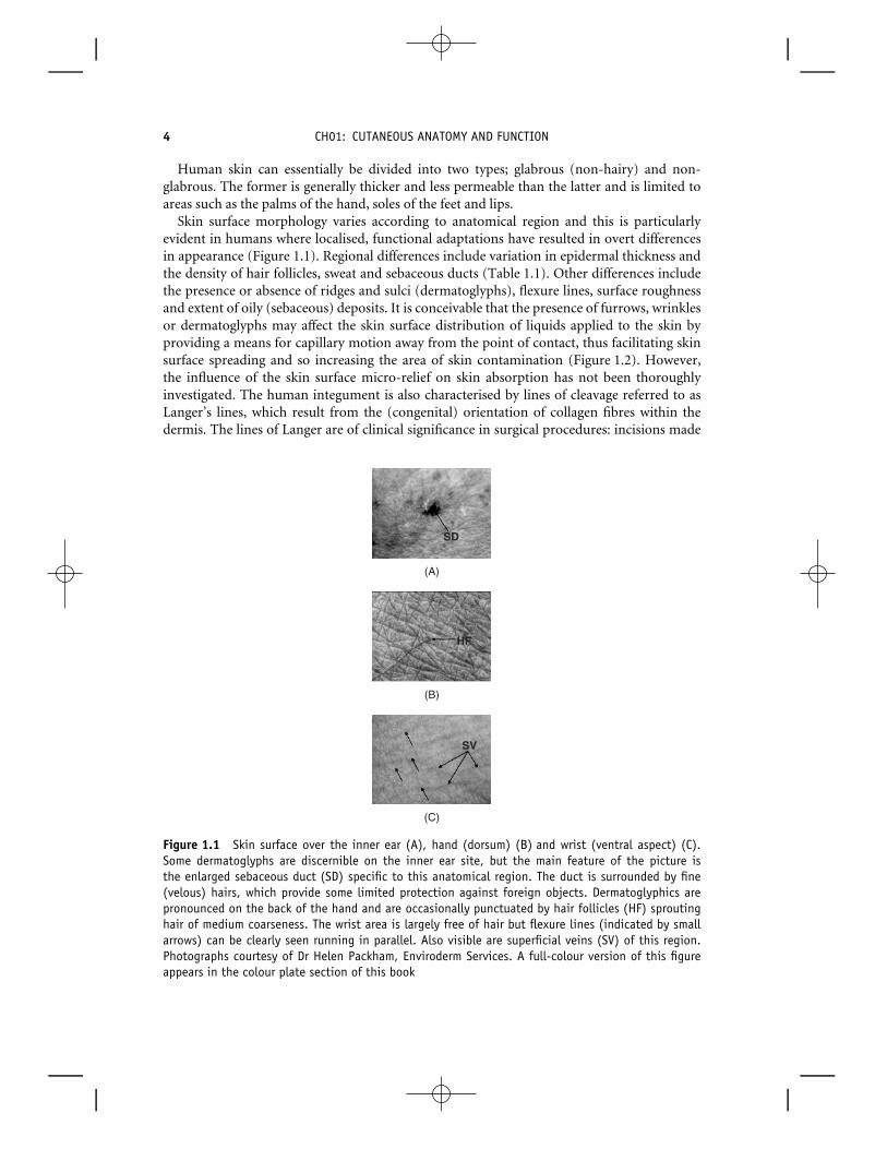
Skin surface morphology varies according to anatomical region and this is particularlyevident in humans where localised, functional adaptations have resulted in overt differencesin appearance (Figure 1.1). Regional differences include variation in epidermal thickness andthe density of hair follicles, sweat and sebaceous ducts (Table 1.1). Other differences includethe presence or absence of ridges and sulci (dermatoglyphs), flexure lines, surface roughnessand extent of oily (sebaceous) deposits. It is conceivable that the presence of furrows, wrinklesor dermatoglyphs may affect the skin surface distribution of liquids applied to the skin byproviding a means for capillary motion away from the point of contact, thus facilitating skinsurface spreading and so increasing the area of skin contamination (Figure 1.2). However,the influence of the skin surface micro-relief on skin absorption has not been thoroughlyinvestigated. The human integument is also characterised by lines of cleavage referred to asLanger’s lines, which result from the (congenital) orientation of collagen fibres within thedermis. The lines of Langer are of clinical significance in surgical procedures: incisions made
(B)
(C)
(A)
SD
HF
SV
Figure 1.1 Skin surface over the inner ear (A), hand (dorsum) (B) and wrist (ventral aspect) (C).Some dermatoglyphs are discernible on the inner ear site, but the main feature of the picture isthe enlarged sebaceous duct (SD) specific to this anatomical region. The duct is surrounded by fine(velous) hairs, which provide some limited protection against foreign objects. Dermatoglyphics arepronounced on the back of the hand and are occasionally punctuated by hair follicles (HF) sproutinghair of medium coarseness. The wrist area is largely free of hair but flexure lines (indicated by smallarrows) can be clearly seen running in parallel. Also visible are superficial veins (SV) of this region.Photographs courtesy of Dr Helen Packham, Enviroderm Services. A full-colour version of this figureappears in the colour plate section of this book

1.2: SURFACE FEATURES 5
Tabl
e1.
1Se
lect
ion
ofqu
anti
tati
veda
taof
hum
ansk
inch
arac
teri
stic
s
An
atom
ical
Th
ickn
ess
Kin
etic
sA
ppen
dage
alD
ensi
tyT
empe
ratu
reSu
rfac
eLi
pids
Loca
tion
Epi
derm
isD
erm
isT
urn
over
Des
quam
atio
nG
lan
dsFo
llicl
es(◦ C
)(μ
gcm
−2)
(μm
)(μ
m)
(day
s)(g
m−2
day−1
)(c
m−2
)(c
m−2
)
Fin
ger
547
1207
0–
50T
hig
h61
1298
0.3
5531
.35
Fore
arm
5311
1813
0.1
32.1
6A
bdom
en42
2163
9.6
7032
.6T
hor
ax51
1676
100
7532
.710
Axi
lla44
1186
65B
ack
7123
2615
657
Pu
bis
4510
14So
le11
5915
34
Face
5222
71
400
–90
0
700
12Fo
reh
ead
8215
006.
31.
776
534
.324
Pal
ms
575
1100
303.
5Sc
alp
9.6
2.1
350
Tu
rnov
er(k
inet
ics)
refe
rsto
the
aver
age
tim
eta
ken
for
ace
llin
the
stra
tum
basa
leto
reac
hth
est
ratu
mco
rneu
m.

6 CH01: CUTANEOUS ANATOMY AND FUNCTION
Figure 1.2 Surface autoradiograph of pig skin exposed to a single, discrete droplet (100 μl) of14C-radiolabelled benzene under unoccluded conditions. Radioactive material (indicated by the darkareas) can be seen to preferentially partition into hair follicles (F) and hair shafts (S). Dermatoglyphicscan be seen radiating from (and interconnecting) adjacent hair follicles (RD), indicative of capillarymovement along the sulci
parallel to Langer’s lines generally heal more readily and are less likely to form scar tissue(Monaco and Grumbine 1986).
Numerous studies have demonstrated that skin permeability is also subject to anatomicalvariation (Feldmann and Maibach 1967, Maibach et al. 1971, Rougier et al. 1986). Whilstepidermal thickness is commonly considered to be a prime determinant of regional skinpermeability, such generalisations should be interpreted with caution (for example, seeFigure 1.3) as other factors such as the regional lipid content (Table 1.1) or morphology of thestratum corneum may be implicated (Rougier et al. 1988).
There is a superficial ‘layer’ of skin that is often overlooked in dermal toxicology: the ‘acidmantle’. This forms a thin film on the skin surface and is comprised of sebum, corneocytedebris and residual material from sweat. This mixture of substances generally imparts a lowpH on the skin surface owing to the presence of free fatty acids and, being predominantlylipophilic, may conceivably influence the partitioning of substances into the skin or act as anadsorbent matrix to trap microscopic particles such as dirt, dust or powders.
The predominant component of the acid mantle is sebum, considered by some to bevestigial (Kligman 1963). Sebum is mainly composed of triglycerides, wax esters and squalene,with the actual composition (and amount being secreted) varying according to anatomicallocation (Figure 1.4).
The evolutionary significance of sebum has been subject to much debate and several putativefunctions including anti-microbial activity, ‘water-proofing’ and ‘sweat-sheet’ formation havebeen proposed (Porter 2001). However, sebum may represent a significant route of excretionfor lipophilic substances (Faergemann et al. 1993; Iida et al. 1999) and may be of physiologicalsignificance for the delivery of vitamin E to the skin surface where it could act as a superficialantioxidant (Thiele et al. 1999).

1.2: SURFACE FEATURES 7
1.0
10.0
100.0
1000.0
Rel
ativ
e sk
in p
erm
eabi
lity
0 200 400 600 800 1000 1200 1400
Epidermal thickness (µm)
AB
C
D
E
F
G
H
I
J
K
L
Figure 1.3 Epidermal thickness as a function of skin permeability (expressed relative to the leastpermeable site, the back of the hand) measured in human volunteers to the nerve agent VX(O-ethyl-S-[2(diisopropylamino)ethyl] methylphosphonothioate). Anatomical regions (in order thick-est to thinnest): A = plantar; B = palmar; C = cheek; D = nape of neck; E = forehead; F = back;G = groin; H = forearm (ventral aspect); I = forearm (dorsal aspect); J = scrotum; K = axilla;L = abdomen
0
20
40
60
80
100
120
140
160
180
forehead cheek chest back arm leg
Anatomical Region
Mas
s (µ
g cm
−2)
squalene
wax esters
fatty acids
diglycerides
triglycerides
cholesterol ester
cholesterol
Figure 1.4 Quantity and composition of sebum, according to anatomical location (Greene et al.1970, Reprinted by permission from Macmillan Publishers Ltd)

8 CH01: CUTANEOUS ANATOMY AND FUNCTION
Clearly, the distribution and composition of the acid mantle will be dictated to some degreeby the regional distribution of sweat and sebaceous glands. The former are found in highestabundance on palmar–plantar regions where the latter are absent. Sebaceous glands aregenerally associated with hair follicles, though in some areas such as the nipples, labia minoraand prepuce, they open directly onto the skin surface. The highest densities of sebaceousglands are found on the scalp and face, with the forehead secreting the largest quantity ofsebum per unit area of skin (Snyder et al. 1981).
It is possible that certain protocols involved in preparing skin tissue for in vitro absorptionstudies may alter the characteristics of the acid mantle. For example, the practice of brieflyimmersing skin in hot water (a standard method for the preparation of epidermal membranes)may perturb or remove the acid mantle from the skin surface. Consequently, this could affectpartitioning of chemicals into the skin and so alter skin absorption kinetics.
1.3 Functional histology of the epidermisand associated structures
The upper layer of the skin (epidermis) is mainly responsible for providing protection against theingress of chemicals and is subject to a cycle of renewal which takes 5–30 days.
The skin is a multi-layered (veneered or stratified) structure comprising three principallayers, namely, the epidermis, dermis (corium) and hypodermis (Figure 1.5). In general, theepidermis accounts for ∼5% of the combined thickness of human epidermis and dermis exceptin regions that are exposed to physical stress such as palmar-plantar skin where the proportionof epidermis is ∼60% (Table 1.1). The epidermis provides protection against xenobiotics,
DERMIS
EPIDERMIS
HYPODERMIS
Protection against xenobiotics, radiation, micro-organisms & physical trauma.
Provides elasticity, plasticity, structural support, tensile strength,“sensing”abilities & biochemical / immunological support to epidermis.
Insulation, energy metabolism, paddingand lubricant.
“OUTSIDE”
“INSIDE”
H
SD N
SG
SP
AM
Figure 1.5 Schematic representation of skin structure and associated functions. Note that therelative thickness of each layer is not to scale (see text). Several adnexal structures are shown(SP = superficial plexus; SG = sebaceous gland; SD = sweat duct; N = Pacinian corpuscle; H = hair).In humans the skin is covered with a thin layer of lipids known as the acid mantle (AM), whichcomprises sebum, cell debris and sweat residua. A full-colour version of this figure appears in thecolour plate section of this book

1.3: FUNCTIONAL HISTOLOGY OF THE EPIDERMIS AND ASSOCIATED STRUCTURES 9
Stratum Basale
Stratum Spinosum
Stratum Granulosum
Stratum Corneum
Free corneocytes
AP
ICA
L MIG
RA
TIO
N5 –30 D
AY
S
TE
RM
INA
L D
IFF
ER
EN
TIA
TIO
N24 H
OU
RS
Dermo-epidermal junction
hemidesmosomes
desmosomes
corneodesmosomes
Lamellar Bodies
Keratohyalin granules
Corneocyte envelope
Figure 1.6 Schematic representation of individual cells of the epidermis. The basal cells (anchoredto the dermo–epidermal junction via hemidesmosomes) undergo apical migration towards the skinsurface whilst undergoing a process of differentiation. The first stage of differentiation results in theappearance of spinous cells (stratum spinosum) in which adjacent cells are interconnected by tightjunctions (desmosomes). Keratohyaline granules, which contain profilaggrin (which facilitates thebundling of keratin in later stages of terminal differentiation) and filaggrin (the putative precursorof natural moisturising factor, NMF), begin to appear. The production of lamellar bodies is consistentwith the formation of the stratum granulosum, exocytosis of which forms the lipid matrix in whichcorneocytes are embedded. During apical migration, cohesion of desmosomes is gradually degradedby the action of enzymes culminating the loss (sloughing) of free corneocytes thereby regulating thethickness of the stratum corneum
micro-organisms, some forms of radiation and, to a limited extent, mechanical trauma. Mostof these functions are fulfilled by the stratum corneum, the outermost layer of the skin.
The epidermis is predominantly (>90%) populated by keratinocytes that continuouslyundergo apical migration from the stratum basale. During migration, keratinocytes undergoseveral stages of differentiation, which can be identified histologically as the stratum spinosum,stratum granulosum and stratum corneum (Figure 1.6). In regions where the epidermis isthicker, an additional layer (between the stratum granulosum and the stratum corneum)termed the stratum lucidum may be observed.
The nomenclature of the different epidermal layers reflects position or cellular morphology(Figure 1.6). Basal cells are sited at the base of the epidermis. Cells of the stratum spinosumradiate small spines, though this appearance is now thought to be an artefact of the lightmicroscope rather than a definitive structural feature. Cells of the stratum granulosum haveinclusion bodies (precursors of the lipid matrix of the stratum corneum) that impart a granularappearance.
Occasionally, older terminology may be found in the literature (Table 1.2). For example,the basal and spinosum layers may be referred to as the stratum Malpighii (after the Italianphysician Marcello Malpighi, circa 1628–1694).
Apical migration and differentiation, from basal cell to fully formed corneocyte, takesapproximately 5–30 days (Figure 1.6), depending on anatomical region (Table 1.1). In

10 CH01: CUTANEOUS ANATOMY AND FUNCTION
Table 1.2 Alternative histological nomenclature of the epidermal layers, withtypical thickness measurement (for human skin)
Current Nomenclature Thickness (μm) Alternative Nomenclature
Stratum corneum 10–20 Horny layerStratum granulosum Granular layerStratum spinosum 50–100 Prickle cell/spinous layer,
acanthocyte (refers toindividual cell)
Stratum basal Stratum germinativum, rootlets
contrast, the final stage of (terminal) differentiation may occur in less than 24 hours andenables prompt repair of superficial damage to the stratum corneum. The gradual degradationof cell–cell adhesion (mediated via desmosomes) ultimately leads to loss of corneocytes(sloughing) and can account for up to one gramme of material (the main constituent of ‘housedust’) per adult per day (Snyder et al. 1981).
Other types of cell present in the epidermis include Langerhans cells (involved withantigen presentation) and melanocytes (which synthesise the photo-protectant, melanin).The mobile nature of these two (dendritic) cell types enables them to migrate and pop-ulate the interstitial space between keratinocytes, and there is growing evidence thatmelanocytes, Langerhans and keratinocytes form functional units within the epidermis(Nordlund and Boissy 2001). Indeed, melanocytes interact with a predefined number of ker-atinocytes within the basal epidermis (the so-called melanocyte–keratinocyte unit) accordingto set ratios depending on constitutive (normal) skin colour (Seiberg 2001). The role ofmelanocytes and Langerhan’s cells are considered in more detail in Chapters 3 and 10,respectively.
The outmost layer of the epidermis, the stratum corneum, is the predominant barrier layer. Thisproperty arises from the arrangement of cornified cells embedded in a lipid matrix known as the‘brick and mortar’ structure.
Terminally differentiated keratinocytes of the stratum corneum are known as corneocytes andare largely devoid of normal cellular functions, being predominantly composed of protein(keratin) and a remnant of the original cell wall (‘corneocyte envelope’).
The ultrastructure of the stratum corneum is described by the ‘brick and mortar model’(Michaels et al. 1975). The functional implication of this architecture is that some skinpenetrants must diffuse via a long and tortuous route between adjacent corneocytes, thusreducing their rate of absorption. This is known as the intercellular route (Figure 1.7). Incontrast, some chemicals may diffuse equally through both corneocytes and the lipid mortar,resulting in a transcellular route (Figure 1.7). Both inter and intracellular routes are collectivelyknown as bulk pathways. A third, potential route of entry across the skin involves diffusiondown hair follicles and into sebaceous glands or via sweat ducts (Figure 1.7). These are referredto as ‘shunt pathways’ and their contribution to skin absorption is currently a contentiousissue. Historically, the relative role of the shunt and bulk transport pathways have been likenedto an army crossing marshland that contains a few narrow bridges: whilst a small number of

1.3: FUNCTIONAL HISTOLOGY OF THE EPIDERMIS AND ASSOCIATED STRUCTURES 11
See Figure 8
Topically applied
substanceTRANSFOLICULAR
mouse human
INTERCELLULAR TRANSCELLULAR
hair
Corneocyte‘brick’
Lipid ‘mortar’
Figure 1.7 Schematic representation of arrangement of corneocytes in mouse and human stratumcorneum (‘brick and mortar’ model). The stacked (columnar) arrangement of corneocytes in mousestratum corneum facilitates a relatively short route for diffusion. In contrast, the oblique arrangementof corneocytes in human stratum corneum compels molecules (diffusing via the intercellular route) totake a long and tortuous route. The two other routes of entry (transcellular and transfollicular) areshown for comparison. The structure of the lipid mortar is detailed in Figure 1.8
soldiers can rapidly march across the bridges in single file, the majority have to trudge slowlythrough the boggy ground (Scheuplein 1976). This analogy pertains to the relatively smallsurface area occupied by hair follicles. For example, the average width of a scalp hair is ∼50 μmand this region contains ∼300 hair follicles per cm2. Thus, the total surface area occupied byhair follicles per cm2 of scalp skin is exceedingly small: approximately 0.007 cm2. However,this does not take into account the fact that a hair follicle is a three dimensional structure thatpenetrates deep into the dermis. Assuming that an average hair follicle is 500 μm deep (andapproximates in shape to a cylinder), then the total surface area of hair follicles per cm2 can becalculated to be ∼0.95 cm2. Thus, on the scalp at least, the presence of hair follicles essentiallydoubles the surface area available for skin absorption.
Skin appendages such as hair follicles provide a potential ‘short-cut’ for skin absorption bypenetrating directly into the dermis. However, the practical relevance of such shunt pathways is ofsome considerable debate.
It is important to note that such shunt pathways are not the biological equivalent of intergalacticwormholes and do not provide a paranormal route of entry into the skin. Hair follicles andother appendageal structures are generally lined with cornified cells and so diffusion from thefollicle into the dermis is still subject to the same barrier layer as is present on the skin surface.

12 CH01: CUTANEOUS ANATOMY AND FUNCTION
Furthermore, the follicles are usually full of very lipophilic material (sebum) and so effectivelyexclude hydrophilic substances or partition and bind very lipophilic materials. Thus, theappearance of a chemical within hair follicles in the dermal region of skin does not equate todermal delivery: the substance is still on the outside of the body! However, it should be notedthat for some chemicals (hydrophilic, charged molecules; Chapter 6), the shunt pathways mayrepresent the predominant route of penetration, although the overall rate of absorption ofsuch compounds is generally very low. The relative contribution of each transport pathway isdiscussed in more detail in Chapter 5.
Whilst corneocytes can be considered to be hydrophilic domains, they are surrounded bya lipid-rich matrix mainly comprising ceramides, free fatty acids and cholesterol (Downinget al. 1987). Thus, the intercellular domain is predominantly a lipophilic environment. Thiscombination imparts a degree of ‘amphiphobicity’ upon the stratum corneum, providinglimited protection against both lipophilic and hydrophilic penetrants. The composition andunderlying metabolism of stratum corneum lipids (as opposed to the skin surface lipidsdiscussed above) is reviewed in Chapter 2.
The molecular packing of the lipid matrix within the inter-corneocyte spaces effectivelysets an upper limit on the physical size of molecules that may penetrate the stratum corneum(Figure 1.8). This is referred to as the ‘rule of 500’ (Bos and Meinardi 2000) since fewsubstances with a molecular weight above 500 Da are capable of passive diffusion throughthe skin. However, recent studies suggest that ultra-fine particles (also termed nanoparticles)
(A) (B)
(Inset, Figure 7)
20-40 nmLipid lamellae
Corneocyte envelope
Small molecules diffuse freely
Large molecules physically excluded
Direction of flow
LCC
Figure 1.8 Arrangement of lipid lamellae within the inter-corneocyte space of the stratum corneum.(A) Empirical representation of adjacent lipid layers showing the physical exclusion of large molecules.The lamellae are ‘riveted’ to the outer corneocyte envelope by a long-chain ceramide (LCC). (B) Electronmicrograph of the inter-corneocyte domain, demonstrating the lipid lamellar packing (courtesy ofProfessor Joke Bouwstra, University of Leiden, The Netherlands)

1.4: SPECIES DIFFERENCES 13
have the potential to penetrate the stratum corneum (Ryman-Rasmussen et al. 2006). Whilstthis is largely unexpected in terms of molecular weight, the diameter of such particles is lessthan the distance that separates adjacent corneocytes and thus diffusion through the stratumcorneum is plausible. Given current health concerns over the increasing use of nanoparticlesin consumer products, it is likely that a great deal more research will be conducted in thisrelatively new area.
The epidermis is anchored to the dermis via a continuous, protein-rich region termedthe dermo-epidermal junction. This structure is highly invaginated and forms characteristic(‘rete’) ridges on skin sections that are readily discernible under the light microscope.The underlying blood supply (superficial plexus) interdigitates with the rete ridges, thusproviding a large surface area for the bi-directional transfer of nutrients, oxygen and wasteproducts. Chemicals that are able to traverse the epidermis are generally subject to systemicabsorption by the superficial (papillary) plexus at this anatomical region (Figure 1.5) and sothe dermis and hypodermis are not generally relevant to the percutaneous absorption kineticsof many substances. However, if the peripheral blood supply (i.e. the superficial plexus) isreduced by vasoconstriction, systemic uptake may be diminished, resulting in accumulationof penetrant within the dermal tissue; conversely, vasodilation may increase systemic uptakefrom the superficial plexus (Brain et al. 2006, Rommen et al. 1999. Alternatively, the ‘groundsubstance’ of the dermis essentially represents an aqueous gel environment and this willprovide an additional barrier to the ingress of strongly lipophilic substances (Flynn et al.1981). Therefore, it is important when conducting in vitro skin absorption studies to select themost appropriate tissue preparation: epidermal membranes are arguably the most relevantmodel since penetration through this layer in vivo results in contact with the circulatorysystem (superficial plexus; see Figure 1.5). The presence of dermal tissue in dermatomed skinis therefore representative of an additional barrier that is not normally present in vivo and maylead to an underestimate of skin absorption for lipophilic substances (Chapter 9).
1.4 Species differences
Human skin is remarkable in many respects from most other mammals and this is of relevance tothe interpretation of toxicological data obtained from animal models such as the rat, mouse andguinea pig.
The most obvious difference between human and animal models is pelage density (Figure 1.9):a thick coat of hair provides a substantial degree of protection against the ingress of xenobioticsand exposure to radiation. As a possible consequence of this evolutionary divergence, thestratum corneum of rodents and lagomorphs is generally more permeable and considerablymore fragile than ‘naked’ species such as pig and human (see legend, Figure 1.9). This differenceis manifest when preparing tissue samples for in vitro skin absorption studies: human and(to some extent) pig skin can be used to prepare strong, coherent sheets of stratum corneumor epidermis that retain their physical durability for several months at room temperature. Incontrast, it is practically impossible to produce similar tissue preparations for rodent skin,although limited success can be achieved with sodium bromide separation of neonatal rat skin(Scott et al. 1986). This species difference in pelage density between human and rodent skin is

14 CH01: CUTANEOUS ANATOMY AND FUNCTION
E
D
SCE
D
500μm
SC
E
D
SC
H
E
D
SC
H
SCE
DH
H
H
H SCE
DH
H
H
H
(A)
(B)
(C)
Figure 1.9 Representative sections of dermatomed guinea pig (A), pig (B) and human (C) skin. Twoprincipal layers are discernible in each section: the epidermis (E) and dermis (D). Note that guineapig stratum corneum (SC) appears as an incoherent, flaky layer whereas SC of pig and human retains aflatter, more compact appearance. A large number of hairs (H) are present in the guinea pig section.A full-colour version of this figure appears in the colour plate section of this book
of particular relevance when interpreting toxicological studies, especially if the test substancehas demonstrable affinity for hair or associated (appendageal) structures.
Animal skin also contains a layer of muscle (panniculus carnosus), which is largely absent inhumans with the exception of the platysma, situated over the ventral aspect of the neck. This isof relevance when conducting in vitro skin absorption studies with full thickness animal skin,as the panniculus carnosus represents an additional barrier layer to diffusion (although thiscan be avoided by the use of skin dermatomed to an appropriate thickness).
Mouse skin is generally more permeable than human and most other species. This mayin part be attributable to the arrangement of corneocytes within the stratum corneum(Bergstresser and Chapman 1980). In human skin, corneocytes are normally offset betweenadjacent rows and this provides a tortuous route for intercellular transport. In contrast,murine corneocytes are arranged in columns (stacks) and so may offer a more direct route forthe ingress of xenobiotics (Figure 1.7).
From a histological perspective, the pig (sus scrofa) is the species that bears most resemblanceto human (Figure 1.9) and so the use of strains with reduced growth rates (such as the Gottingenminipig®) are becoming increasingly common in toxicological and pharmacological studies.

REFERENCES 15
Summary
• Human skin presents a barrier to the ingress of many xenobiotics and has a correspondingly lowsurface area in comparison with other externalised organs such as the lung and gastrointestinaltract.
• The integument cannot be considered to be a homogeneous organ as there are substantial regional(anatomical) differences in structure and function such as permeability (which may span severalorders of magnitude).
• There are three principal skin layers: epidermis, dermis and hypodermis. The former is primarilyresponsible for maintaining skin barrier function.
• The relative impermeability of the skin results from the structure and composition of thestratum corneum (the outermost layer of the epidermis), which is subject to a continuouscycle of regeneration through apical migration and terminal differentiation of epidermal cells(keratinocytes).
• There is considerable species variation in skin structure and function. The pig is arguably themost relevant animal model although rodents are currently the species of choice for toxicologicalevaluation.
References
Bergstresser, P.R. and Chapman, S.L. (1980). Maturation of normal human epidermis without anordered structure. Br J Dermatol 102(6): 641–8.
Bos, J.D. and Meinardi, M.M. (2000). The 500 Dalton rule for the skin penetration of chemicalcompounds and drugs. Exp Dermatol 9(3): 165–9.
Brain, K.R., Green, D.M., Dykes, P.J. et al. (2006). The role of menthol in skin penetration from topicalformulations of ibuprofen 5% in vivo. Skin Pharmacol Physiol 19(1): 17–21.
Downing, D.T., Stewart, M.E., Wertz, P.W. et al. (1987). Skin lipids: an update. J Invest Dermatol 88(3Suppl): 2s–6s.
Faergemann, J., Zehender, H., Denouel, J. and Millerioux, L. (1993). Levels of terbinafine in plasma,stratum corneum, dermis–epidermis (without stratum corneum), sebum, hair and nails during andafter 250 mg terbinafine orally once per day for four weeks. Acta Derm Venereol 73(4): 305–9.
Feldmann, R.J. and Maibach, H.I. (1967). Regional variation in percutaneous penetration of 14C cortisolin man. J Invest Dermatol 48(2): 181–3.
Flynn, G.L., Durrheim, H. and Higuchi, W.I. (1981). Permeation of hairless mouse skin II: membranesectioning techniques and influence on alkanol permeabilities. J Pharm Sci 70(1): 52–6.
Forslind, B., Lindberg, M. and Norlen, L. (eds). (2004). Skin, hair, and nails. Marcel Dekker Inc., NewYork.
Freinkel, R.K. and Woodley, D.T. (eds). (2001). The biology of the skin. The Parthenon Publishing Group,London.
Greene, R.S., Downing, D.T., Pochi, P.E. and Strauss, J.S. (1970). Anatomical variation in the amountand composition of human skin surface lipid. J Invest Dermatol 54(3): 240.
Iida, T., Hirakawa, H., Matsueda, T., et al. (1999). Recent trend of polychlorinated dibenzo-p-dioxinsand their related compounds in the blood and sebum of Yusho and Yu Cheng patients. Chemosphere38(5): 981–93.
Kligman, A.M. (1963). The Uses of Sebum. Br J Dermatol 75(August/September): 307–319.

16 CH01: CUTANEOUS ANATOMY AND FUNCTION
Maibach, H.I., Feldman, R.J., Milby, T.H. and Serat, W.F. (1971). Regional variation in percutaneouspenetration in man. Pesticides. Arch Environ Health 23(3): 208–211.
Michaels, A.S., Chandrasekaran, S.K. and Shaw, S.E. (1975). Drug permeation through human skin:Theory and in vitro experimental measurement. AIChE Journal 21(5): 985–996.
Monaco, A. and Grumbine, N.A. (1986). Lines of minimal movement. Clin Podiatr Med Surg 3(2):241–247.
Montagna, W. (ed.). (1962). The structure and function of skin. Academic Press, New York.Nordlund, J.J. and Boissy, R.E. (2001). The biology of melanocytes, in The biology of the skin (eds
Freinkel, R.K. and Woodley, D.T.). The Parthenon Publishing Group., New York, pp. 113–131.Porter, A.M. (2001). Why do we have apocrine and sebaceous glands? J R Soc Med 94(5): 236–7.Rommen, C., Leopold, C.S. and Lippold, B.C. (1999). Do local anesthetics have an influence on the
percutaneous penetration of a model corticosteroid? An in vivo study using the vasoconstrictor assay.Eur J Pharm Sci 9(2): 227–34.
Rougier, A., Dupuis, D., Lotte, C.R., et al. (1986). Regional variation in percutaneous absorption in man:measurement by the stripping method. Arch Dermatol Res 278(6): 465–9.
Rougier, A., Lotte, C., Corcuff, T.P. and Maibach, H.I. (1988). Relationship between skin permeabilityand corneocyte size according to anatomic site, age and sex in man. J Society of Cosmetic Chemists39(1): 15–26.
Ryman-Rasmussen, J.P., Riviere, J.E. and Monteiro-Riviere, N.A. (2006). Penetration of intact skin byquantum dots with diverse physicochemical properties. Toxicol Sci 91(1): 159–65.
Scheuplein, R.J. (1976). Percutaneous absorption after twenty-five years: or ‘old wine in new wineskins’.J Invest Dermatol 67(1): 31–8.
Scott, R.C., Walker, M. and Dugard, P.H. (1986). In vitro percutaneous absorption experiments: atechnique for the production of intact epidermal membranes from rat skin. J Society of CosmeticChemists 37(1): 35–41.
Seiberg, M. (2001). Keratinocyte–melanocyte interactions during melanosome transfer. Pigment CellRes 14(4): 236–42.
Snyder, W.S., Cook, M.J., Karhausen, L.R. et al. (eds). (1981). Report of the task group on reference man.A. Wheaton & Co Ltd, Exeter.
Thiele, J.J., Weber, S.U. and Packer, L. (1999). Sebaceous gland secretion is a major physiologic route ofvitamin E delivery to skin. J Invest Dermatol 113(6): 1006–10.


![Function Anatomy Ch 7[1]](https://static.fdocuments.net/doc/165x107/577d23251a28ab4e1e991842/function-anatomy-ch-71.jpg)