1 Cell Cycle, Mitosis & Meiosis. Chromosomes All eukaryotic cells store genetic information in...
44
1 Cell Cycle, Mitosis & Meiosis
-
Upload
aiyana-rawlings -
Category
Documents
-
view
382 -
download
2
Transcript of 1 Cell Cycle, Mitosis & Meiosis. Chromosomes All eukaryotic cells store genetic information in...

1
Cell Cycle, Mitosis & Meiosis
Chromosomes
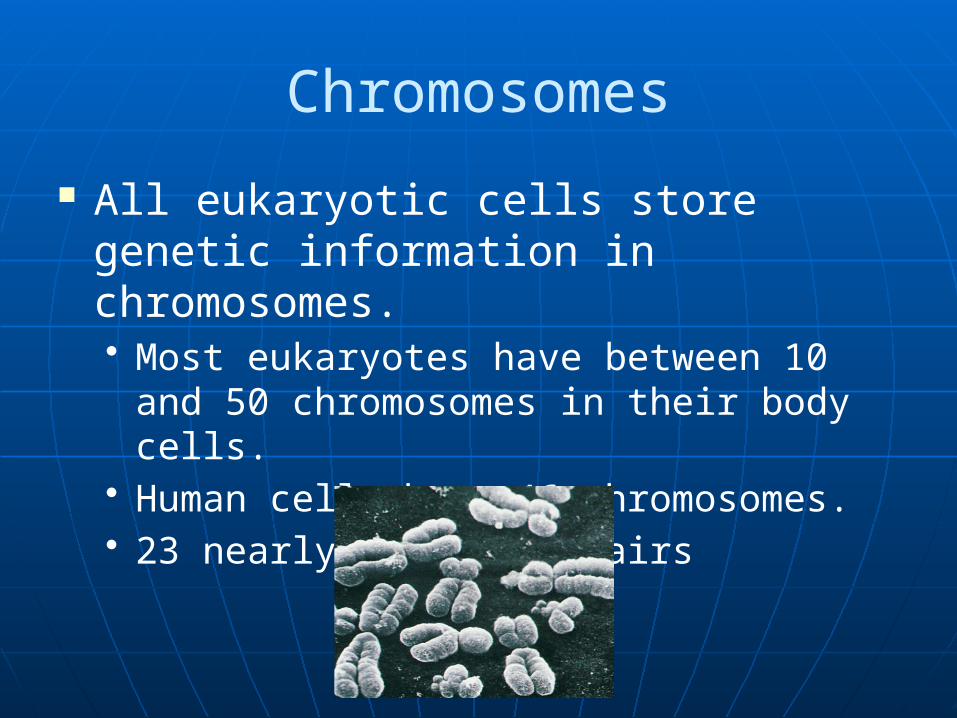
All eukaryotic cells store genetic information in chromosomes.• Most eukaryotes have between 10 and
50 chromosomes in their body cells.• Human cells have 46 chromosomes.• 23 nearly-identical pairs

Chromosomes A diploid cell has two sets of each of its chromosomes A human has 46 chromosomes (2n = 46) In a cell in which DNA synthesis has occurred all the
chromosomes are duplicated and thus each consists of two identical sister chromatids
Maternal set ofchromosomes (n = 3)
Paternal set ofchromosomes (n = 3)
2n = 6
Two sister chromatidsof one replicatedchromosome
Two nonsisterchromatids ina homologous pair
Pair of homologouschromosomes(one from each set)
Centromere

Chromosome Duplication
0.5 µm
Chromosomeduplication(including DNA synthesis)
Centromere
Separation of sister
chromatids
Sisterchromatids
Centrometers Sister chromatids
A eukaryotic cell has multiplechromosomes, one of which is
represented here. Before duplication, each chromosome
has a single DNA molecule.
Once duplicated, a chromosomeconsists of two sister chromatids
connected at the centromere. Eachchromatid contains a copy of the
DNA molecule.
Mechanical processes separate the sister chromatids into two chromosomes and distribute
them to two daughter cells.
In preparation for cell division, DNA is replicated and the chromosomes condense
Each duplicated chromosome has two sister chromatids, which separate during cell division

Because of duplication, each condensed chromosome consists of 2 identical chromatids joined by a centromere.
Each duplicated chromosome contains 2 identical DNA molecules (unless a mutation occurred), one in each chromatid:
Chromosome Duplication
Copyright © The McGraw-Hill Companies, Inc. Permission required for reproduction or display.
Two unduplicatedchromosomes
Centromere
Sisterchromatids
Sisterchromatids
Duplication
Non-sisterchromatids
Two duplicated chromosomes

6
Chromosomes, ploidy and n

7
2 Types of Cell Division
Mitosis – ordinary somatic cell division• 2 daughter cells with identical
chromosomes and genes to parents• Diploid cells – 2n chromosome
complement Meiosis – germline cell division
• Results in formation of gametes – cells with only 23 chromosomes
• Haploid cells – n chromosome complement

Phases of the Cell Cycle Interphase• G1 - primary growth • S - genome replicated• G2 - secondary growth
M - mitosis C - cytokinesis

Interphase
G1 - Cells undergo majority of growth
S - Each chromosome replicates (Synthesizes) to produce sister chromatids• Attached at centromere• Contains attachment site (kinetochore)
G2 - Chromosomes condense - Assemble machinery for division such as centrioles
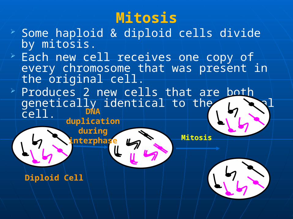
Mitosis Some haploid & diploid cells divide by
mitosis. Each new cell receives one copy of every
chromosome that was present in the original cell.
Produces 2 new cells that are both genetically identical to the original cell.
DNA duplication
during interphase Mitosis
Diploid Cell

Embryonic cell cycles The duration of the phases
varies considerably in different kinds of cells.
Early embryos may have cell cycles of 30 minutes, but there is no growth (G1 or G2) phase.
In contrast, some cells in adult animals cease division altogether (e.g., nerve cells).
Others may divide only occasionally, to replace cells that have been lost.

12
The Cell Cycle
G1
• No DNA synthesis• Lasts hours, days or years• Occurs after mitosis and before S phase
The availability of growth factors controls the animal cell cycle at a point in late G1 called the restriction point. If growth factors are not available during G1, the cells enter a quiescent stage of the cycle called G0.

13
The Cell CycleS phase
• Stage of DNA synthesis• The major variants of histones are predominantly
synthesized during S phase• Each chromosome replicates bipartite
chromosome made up of sister chromatids• End of each chromatid marked by telomeres –
specialized DNA sequences that ensure integrity of chromosomes during division
• Sister chromatids held together at centromere – region of DNA associated with kinetochore
• DNA replication happens at thousands of origins of DNA replication

14
The Cell Cycle
G2 phase• The cell is preparing for mitosis• Cell’s DNA doubled in S phase• Brief stage
G0 phase• Resting stage/quiescent stage • Neurons, RBCs arrested in G1 = G0 phase
– permanent G1 phase • Liver cells enter G0 but on damage G1 and
cell cycle continues

Figure 17-31 Molecular Biology of the Cell (© Garland Science 2008)
Centriole Replication
At a certain point in G1, the two centrioles of the pair separate.
During S phase, a daughter centriole begins to grow near the base of each mother centriole and at a right angle to it.
The elongation of the daughter centriole is usually completed by G2
16
Mitosis
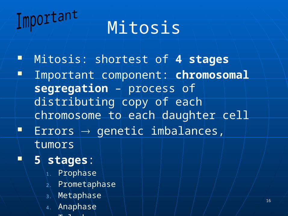
Mitosis: shortest of 4 stages Important component: chromosomal
segregation – process of distributing copy of each chromosome to each daughter cell
Errors genetic imbalances, tumors 5 stages:
1. Prophase2. Prometaphase3. Metaphase4. Anaphase5. Telophase

17
Mitosis is subdivided into prophase, prometaphase, metaphase, anaphase, and
telophase
Prophase: Condensed chromosome start to become visible, Centrosomes start moving apart, Nucleolus starts disappearing
Prometaphase: Nuclear membrane breaks down
Metaphase: Chromosomes maximally condensed and align at the metaphase plate
Anaphase: The two sister chromatids separate and move toward opposite poles
Telophase: nucleus reassembly occurs, cytokinesis occurs

18

Stages of mitosis in an animal cell

Fluorescence micrographs of chromatin, keratin, and microtubules during mitosis

21
Prophase Condensation of chromosomes Disintegration and disappearance of nucleolus Formation of mitotic spindle – microtubule
network
Prometaphase Nuclear membrane breaks up Congression occurs – chromosomes move
to point midway between spindle poles Condensation of chromosomes continue

22
Metaphase Maximum condensation of chromosomes
reached Arranged at equatorial plane of cell Balanced forces of microtubules from
opposite poles at kinetochores

23
The mitotic spindle is responsible for chromosome movements during mitosis
Spindle assembly and chromosome attachment• Minus ends anchored in
centrosome• Kinetochore: a platelike,
three layer structure made of proteins attached to CEN sequences located in the centromeres’ DNA
• Two kinetochores are located on opposite sides of a chromosome and anchor kinetochore microtubules

24
Anaphase Begins when sister
chromatids separate Sister chromatids
separate into daughter chromosomes
Daughter chromosomes move to opposite poles

25
Telophase and Cytokinesis Telophase
• Chromosomes decondense• Nuclear membrane reforms
Cytokinesis divides the cytoplasm • Cleavage begins as a slight
indentation in cell surface deepens into a cleavage furrow.
• Actin contractile ring and myosin motors drive the process
• Cytoplasm cleaves separating daughter cells

26
Meiosis One round of DNA synthesis
2 rounds of chromosomal segregation• Meiosis I – reduction division
– chromosome no. goes from diploid haploid
Meiotic crossing over or recombination also occurs here: homologous segments of DNA exchanged between nonsister chromatids of pair of homologous chromosomes
Recombination important also for chromosome segregation
• Meiosis II – like mitosis without a preceding DNA replication stage
Sister chromatids separate

27
Meiosis Meiosis can convert one diploid cell into four
haploid cells• Meiosis I: the first meiotic division• Homologous pairs join together to form a bivalent
Meiosis I produces two haploid cells that have chromosomes composed of sister chromatids• Unlike mitosis, in meiosis I, sister chromatids stay
together. • Thus, the cells at the end of meiosis I are
considered haploid because it contains only one of the two chromosomes of each bivalent
• Genetic recombination

28
Meiosis I
Prophase I: Homologous chromosomes become paired and exchange DNA• Divided into 5 stages• Formation of synaptonemal complex and crossing-over occurs
Metaphase I: Bivalents align at the spindle equator• The kinetochores of sister chromatids lie side by side and face
the same pole of the cell• Random orientation of maternal or paternal chromosomes• Chromosomes held together by chiasmata
Anaphase I: Homologous chromosomes move to opposite spindle poles
Telophase I and cytokinesis: Two haploid cells are produced

29

30
Prophase I Leptotene
• chromatin begins to condense, 2 sister chromatids so close, cannot be distinguished
Zygotene• Homologs pair along entire length – synapsis• Chromosomes held together by synaptonemal complex
Pachytene• Synapsis complete, chromosome more tightly coiled• Chromosome appear bivalent – tetrad formation• Recombination takes place
Diplotene• Synaptonemal complex disappears• Bivalents begin to separate but held together at centromeres
and ~ 50 points of crossing over – chiasmata Diakinesis – stage of max condensation

31

32
Bivalent Formation and Crossing-over

33
Anaphase I Disjunction occurs – members of each bivalent move
apart• HOMOLOGS (and usually alleles) SEPARATE!!!
Maternal and paternal chromosomes sort independently 223 combinations• Add to this the variation due to crossing over e.g. chr 1 typically
contains 3-5 segments Error-prone step: particularly nondisjunction
Metaphase I Nuclear membrane disappears Spindle forms Bivalents align at equator

Chromosome segregation in meiosis I

35
Telophase I & Cytokinesis
Telophase I – 2 chromosome sets group at opposite poles
Cytokinesis – cell divides into daughter cells enter meiotic interphase (no DNA synthesis)• Spermatogenesis: equal• Oogenesis: secondary oocyte receives
most cytoplasm, other cell 1st polar body

36
Meiosis II Meiosis II resembles a mitotic division Prophase II is very brief Metaphase II
• Kinetochores of sister chromatids now face in opposite directions
Anaphase II• Sister chromatids separate

37
Genetic Consequences of Meiosis At the end of meiosis II there are four daughter
cells, each containing a haploid set of chromosomes• Reduction of chromosome number
Segregation of alleles Random assortment of the homologues (law of
independent assortment) • Meiosis generates genetic diversity • Over 8 million different combinations of
chromosomes Additional shuffling by crossing over Nondisjunction refers to the failure of the two
members of a homologous chromosome pair to separate during anaphase• Leads to aneuploidy diseases (trisomy 21-Down
syndrome)

Solve This Question A couple in their late 30's has a baby girl with a
trisomy 21 karyotype. Genetic analysis of a polymorphic VNTR locus on chromosome 21 reveals that the baby has allelic variants A, C, D; the mother has alleles A, D; the father has alleles B, C. When did meiotic nondisjunction most likely occur and in which parent?
Answer: Nondisjunction occurred during meiosis I in the mother.
38

Comparison of meiosis and mitosis

40
Human Gametogenesis

41
Spermatogenesis Sperm formed in seminiferous tubules after sexual maturity Tubules lined with spermatogonia in different stages of
differentiation Last cell type is primary spermatocyte meiosis I 2 haploid
secondary spermatocytes meiosis II 4 spermatids sperm• Process takes 64 days• 200 million sperm produced per ejaculate and 1012 in a lifetime

42
Oogenesis Occurs only during prenatal development Oogonia in ovarian cortex originate from
primordial germ cells by about 30 mitoses By 3rd month, oogonia primary oocytes which
enter prophase INo primary oocytes develop after month 5 of
fetal life At birth, ~ 2.5 million primary oogonia, only ~
400 eventually mature At sexual maturity, individual follicles mature,
oocyte completes meiosis I one secondary oocyte (ovum)
Meiosis II begins but is arrested in metaphase until fertilization

Oogenesis - Continued If fertilization takes place, this secondary oocyte will complete 2nd
meiotic division. Most of the cytoplasm is retained by the fertilized secondary oocyte
and the 2nd polar body receives very little cytoplasm. The 2nd polar body is extruded and maturation of oocyte is completed
43

44