
1 C2 Domain Membrane Penetration by Group IVA Cytosolic ...
33
1 C2 Domain Membrane Penetration by Group IVA Cytosolic Phospholipase A 2 Induces Membrane Curvature Changes Katherine E. Ward a , James P. Ropa a , Emmanuel Adu-Gyamfi a,b , and Robert V. Stahelin a,b,c1 a Department of Chemistry and Biochemistry and b the Eck Institute for Global Health, University of Notre Dame, South Bend, IN, 46556 and c Department of Biochemistry and Molecular Biology, Indiana University School of Medicine-South Bend, South Bend, IN 46617. 1 To whom correspondence should be addressed: Indiana University School of Medicine-South Bend, 143 Raclin-Carmichael Hall, 1234 Notre Dame Avenue, South Bend, Indiana 46617; TEL: 1-574-631-5054; FAX: 1-574-631-7821; Email: [email protected]. Running Title: C2 Domain Induces Membrane Curvature Changes Abbreviations: AA, arachidonic acid; BAR, Bin-Amphiphysin-Rvs167; BCA, bicinchoninic acid; CBL, calcium binding loop; C1P, ceramide-1-phosphate; CD, circular dichroism; cPLA 2 α, group IVA cytosolic phospholipase A 2 ; EM, electron microscopy; FcR. Fc Receptor; GFP, green fluorescence protein; ITO, indium tin oxide; LUV, large unilamellar vesicle; PC, phosphatidylcholine; PCR, polymerase chain reaction; PE, phosphatidylethanolamine; POPC, 1-palmitoyl-2-oleoyl-sn-glycero-3-phosphocholine; POPE, 1-palmitoyl-2-oleoyl-sn-glycero-3- phosphoethanolamine; POPS, 1-palmitoyl-2-oleoyl-sn-glycero-3-phosphatidylserine; RU, resonance unit; SPR, surface plasmon resonance; TEM, transmission electron microscopy. by guest, on February 18, 2018 www.jlr.org Downloaded from
Transcript of 1 C2 Domain Membrane Penetration by Group IVA Cytosolic ...

1
C2 Domain Membrane Penetration by Group IVA Cytosolic Phospholipase A2 Induces Membrane Curvature Changes
Katherine E. Warda, James P. Ropaa, Emmanuel Adu-Gyamfia,b, and Robert V. Stahelina,b,c1
aDepartment of Chemistry and Biochemistry and bthe Eck Institute for Global Health, University of Notre Dame, South Bend, IN, 46556 and cDepartment of Biochemistry and Molecular Biology, Indiana University School of Medicine-South Bend, South Bend, IN 46617. 1To whom correspondence should be addressed: Indiana University School of Medicine-South Bend, 143 Raclin-Carmichael Hall, 1234 Notre Dame Avenue, South Bend, Indiana 46617; TEL: 1-574-631-5054; FAX: 1-574-631-7821; Email: [email protected].
Running Title: C2 Domain Induces Membrane Curvature Changes Abbreviations: AA, arachidonic acid; BAR, Bin-Amphiphysin-Rvs167; BCA, bicinchoninic acid; CBL, calcium binding loop; C1P, ceramide-1-phosphate; CD, circular dichroism; cPLA2α, group IVA cytosolic phospholipase A2; EM, electron microscopy; FcR. Fc Receptor; GFP, green fluorescence protein; ITO, indium tin oxide; LUV, large unilamellar vesicle; PC, phosphatidylcholine; PCR, polymerase chain reaction; PE, phosphatidylethanolamine; POPC, 1-palmitoyl-2-oleoyl-sn-glycero-3-phosphocholine; POPE, 1-palmitoyl-2-oleoyl-sn-glycero-3-phosphoethanolamine; POPS, 1-palmitoyl-2-oleoyl-sn-glycero-3-phosphatidylserine; RU, resonance unit; SPR, surface plasmon resonance; TEM, transmission electron microscopy.
by guest, on February 18, 2018
ww
w.jlr.org
Dow
nloaded from

2
Abstract Group IVA cytosolic phospholipase A2 (cPLA2α) is an 85-kDa enzyme that regulates release of
arachidonic acid (AA) from the sn-2 position of membrane phospholipids. It is well established
that cPLA2α binds zwitterionic lipids such as phosphatidylcholine in a Ca2+-dependent manner
through its N-terminal C2 domain, which regulates its translocation to cellular membranes. In
addition to its role in AA synthesis, it has been shown that cPLA2α promotes tubulation and
vesiculation of the Golgi and also regulates trafficking of endosomes. Additionally, the isolated
C2 domain of cPLA2α is able to reconstitute Fc receptor mediated phagocytosis suggesting that
C2 domain membrane binding is sufficient for phagosome formation. These reported activities
of cPLA2α and its C2 domain require changes in membrane structure but to date the ability of
the C2 domain to promote changes in membrane shape has not been reported. Here we
demonstrate that the C2 domain of cPLA2α is able to induce membrane curvature changes to
lipid vesicles, giant unilamellar vesicles, and membrane sheets. Biophysical assays combined
with mutagenesis of C2 domain residues involved in membrane penetration demonstrate
membrane insertion by the C2 domain is required for membrane deformation suggesting that
C2 domain induced membrane structural changes may be an important step in signaling
pathways mediated by cPLA2α.
Keywords: calcium, C2 domain, cytosolic phospholipase A2α, lipid binding, membrane curvature, membrane penetration.
by guest, on February 18, 2018
ww
w.jlr.org
Dow
nloaded from
3
Introduction
Group IVA cytosolic phospholipase A2 (cPLA2α) is an 85-kDa enzyme, consisting of a N-
terminal lipid-binding C2 domain (~ 120 residues) and a C-terminal catalytic or lipase domain (~
600 residues) that is separated by a flexible linker (1, 2). cPLA2α regulates arachidonic acid
release from the sn-2 positions of membrane phospholipids that is used in the synthesis of
leukotrienes and prostaglandins in response to inflammatory agonists (3). cPLA2α has also
been implicated in a number of pathological conditions including asthma (4), cancers (5),
arthritis (6), cerebral ischemia (7), and heart disease (8). The general principles governing
cPLA2α in vitro membrane binding (9, 10) and activation (11) as well as cellular translocation
(12, 13) are well established where the C2 domain binds with high affinity to zwitterionic
membranes in a Ca2+-dependent manner (9), while the catalytic domain binds to membranes
independent of Ca2+ albeit weakly (14). This functionality allows the C2 domain to act as a Ca2+
sensor in cells, which drives the cellular localization to the Golgi, ER, and nuclear membranes
(2, 12, 13).
The C2 domain (~ 120 amino acids) contains three calcium-binding loops (CBL), two of
which, CBL1 and 3 harbor hydrophobic and aromatic amino acids (See Fig. 1) that promote
binding to zwitterionic membranes (9). Ca2+ binding induces a dramatic change in electrostatic
potential lowering the desolvation penalty associated with membrane insertion (15) and
promoting the docking of the C2 domain to the membrane bilayer. This penetration into the
membrane has been shown to be significant with a depth of ~ 15 Å (16), which extends
extensively into the hydrocarbon region of the membrane. The significant membrane
penetration of cPLA2α is important for its membrane targeting to zwitterionic membranes and
also its ability to produce arachidonic acid (AA). Recently, two anionic lipids, ceramide-1-
phosphate (C1P) (17, 18) and PtdIns(4,5)P2 (19, 20) have been demonstrated to bind and
activate cPLA2α with emerging roles in the cellular translocation of the enzyme (21, 22).
by guest, on February 18, 2018
ww
w.jlr.org
Dow
nloaded from

4
Besides its role in eicosanoid biosynthesis, cPLA2α is selectively activated upon Fc
receptor (FcR)-mediated phagocytosis in macrophages where it rapidly translocates to the
nascent phagosome (23). Unexpectedly, however, it was shown that membrane binding by the
isolated C2 domain of cPLA2α was sufficient to induce phagosome formation (23) suggesting
that the C2 domain alone has membrane binding activity that regulates phagocytosis. This was
further verified with a mutation in the C2 domain, D43N, which abrogates Ca2+-binding and
could not rescue phagocytosis (23). cPLA2α also plays a role in membrane curvature
generation through regulation of aberrant Golgi vesiculation (24), Golgi tubulation (25), and
vesiculation of cholesterol-rich GPI-anchored, protein-containing endosomes (26). While it is
speculated the C2 domain may induce changes to membrane structure (23), to date, direct
evidence is lacking. These recent studies suggest that cPLA2α and its C2 domain have
membrane remodeling activity that is critical to biological signaling pathways.
Recently, a number of peripheral proteins, mainly attributed to their modular lipid binding
domains have been found to induce membrane curvature changes (27) including ENTH (28),
BAR (29), PH (30), ACCH (31), and C2 domains (32). Here, we investigate the ability of the
isolated C2 domain of cPLA2α to induce changes to lipid structure. A number of imaging assays
are employed including electron microscopy (EM) of large unilamellar vesicles (LUVs), imaging
of giant unilamellar vesicles (GUVs), and imaging of membrane sheets. In addition, biophysical
assays including monolayer penetration analysis and surface plasmon resonance (SPR) were
used to correlate membrane penetration and affinity with membrane remodeling activity. Results
provide evidence that Ca2+-dependent membrane insertion of CBL1 and 3 of the C2 domain
drive membrane curvature changes.
by guest, on February 18, 2018
ww
w.jlr.org
Dow
nloaded from

5
Materials and Methods
Materials
1-palmitoyl-2-oleoyl-sn-glycero-3-phosphocholine (POPC), 1-palmitoyl-2-oleoyl-sn-glycero-3-
phosphoethanolamine (POPE), and 1-palmitoyl-2-oleoyl-sn-glycero-3-phosphatidylserine
(POPS) were purchased from Avanti Polar Lipids, Inc. (Alabaster, AL) and used without further
purification. Octyl glucoside, (3-[3-cholamidopropyl) dimethylammonio]-1-propane-sulfonate
(CHAPS), Nunc Lab-Tek I Chambered Cover Glasses 8-well and bicinchoninic acid (BCA)
protein assay kit were from Thermo Fisher Scientific (Waltham, MA). L1 sensor chips were from
GE Healthcare (Piscataway, NJ). N-(3-(Triethylammoniumpropyl)-4-(4-
(Diethylamino)styryl)Pyridinium Dibromide (FM® 2-10) lipophilic dye was from Life Technologies
(Grand Island, NY). Restriction endonucleases and enzymes for molecular biology were
obtained from New England Biolabs (Beverly, MA).
Cloning and Protein Expression
The QuikChange site-directed mutagenesis kit (Agilent Technologies, Santa Clara, CA)
was used to introduce mutations into the pET28a vector with a His6 tag engineered into the N-
terminus of the cPLA2α C2 domain gene (9). All mutated constructs were sequenced to ensure
presence of the desired mutation. The C2 domain and respective mutations were expressed
and purified from E. Coli BL21(DE3) cells as previously described (9). Protein concentrations
were determined by the BCA method and all protein aliquots were stored in 20 mM HEPES, pH
7.4, containing 160 mM KCl,
Electron Microscopy
200 µL of 1 mg/ml POPC LUVs were prepared as previously described (32). Briefly, the lipids
were dried under nitrogen gas and resuspended in 20 mM HEPES, pH 7.4, containing 160 mM
KCl and either 100 µM CaCl2 or 100 µM EGTA. The vesicles were incubated at 37°C and
extruded through an 800 nm filter (Avanti Polar Lipids, Alabaster, AL). The respective C2
by guest, on February 18, 2018
ww
w.jlr.org
Dow
nloaded from

6
domain and mutations were incubated at a concentration of 10 µM with the POPC vesicles for
30 minutes at 25°C. Samples were then applied to a carbon-formvar-coated copper grid and
stained with 2% uranyl acetate. Liposome morphologies were then imaged at 80 kV on a FEI
80-300 D3203 electron microscope at 6300x magnification.
Giant Unilamellar Vesicle Assay
An aliquot of lipids containing POPC, POPE and POPS suspended in chloroform were prepared
in a 60:20:20 molar ratio. The suspension was dried under nitrogen gas and resuspended in
chloroform to a final concentration of 0.4 mg/mL. The lipid suspension was dried onto an indium
tin oxide (ITO) coated slide and dehydrated under a vacuum for 1 hour. The GUV apparatus
was assembled and a 350 mM D-sucrose solution was placed into the reservoir containing the
dried lipids. Another glass plate was placed on top to eliminate air from the system then a sin
wave generator was applied at 3V and 20 Hz for 5 hours at 25°C. The GUV solution was
collected and stored at 25°C until use. The GUV solution was diluted 20-fold in 20mM HEPES,
pH 7.4, containing 160 mM KCl and 10µM FM® 2-10 lipophilic dye. Samples were prepared
with either 100 µM EGTA, 10 µM CaCl2 or 500 nM CaCl2 as necessary for experimental
conditions. GUV vesiculation was assessed after a 5 min incubation with either 2 µM or 500 nM
cPLA2α-C2, 500 nM full-length cPLA2α, or 2 µM of respective mutants and was subsequently
imaged via confocal microscopy (Zeiss LSM 710) on Nunc Lab-Tek I Chambered Cover
Glasses 8-well using a 63x 1.4 NA oil objective. Three replicates for each control or sample
were quantified by counting 60-100 GUV’s per replicate. The number of vesiculated GUV’s
were quantified separately and compared to the total number of GUV’s in each replicate. The
degree of vesiculation was then expressed as a percentage, compared to the control, and
quantified using an unpaired student t-test.
Membrane Sheets
2 µL of 10 mM POPC was prepared in chloroform and spotted onto Nunc Lab Tek I 8-well
Chambered Cover Glasses then dried under a vacuum. The lipid was rehydrated with 20 mM
by guest, on February 18, 2018
ww
w.jlr.org
Dow
nloaded from

7
HEPES, pH 7.4, containing 160 mM KCl and 20 µM FM® 2-10 and allowed to rehydrate for 15
minutes. Samples were prepared with either 100 µM EGTA or 100 µM CaCl2 as necessary for
experimental conditions. The experiments contained either 2 µM cPLA2α-C2 or respective
mutants and were subsequently imaged after a 15 minute incubation with a confocal
microscope (Nikon A1R-MP with a 100x 1.4 NA oil objective).
Monolayer Penetration
Surface pressure (π) of solution in a circular Teflon trough (2 mL) was measured using a wire
probe connected to a Kibron MicroTrough X (Kibron, Inc., Helsinki) as previously described (33).
Phospholipid solution (2 – 8 µL) in hexane/ethanol (9:1 v/v) was spread onto 2 mL of subphase
to form a monolayer with a given initial surface pressure (π0). The subphase was stirred
continuously at 30 revolutions/min with a small magnetic stir bar. Following stabilization of the
surface pressure of the monolayer (~ 5 min), the protein solution (typically 10 µL) was injected
into the subphase and the change in surface pressure (Δπ) was measured as a function of time.
Generally, the Δπ value reached a maximum after 20 min. The maximal Δπ value depends
upon the protein concentration and reached saturation at [cPLA2α-C2 > 1 µg/mL] as previously
reported (34). Protein concentrations in the subphase are maintained above such values to
ensure the Δπ represents a maximal value. The Δπ versus π0 plots were constructed from
these measurements to obtain the x-intercept or critical pressure (πc) defined as the value in
which the protein penetrates up to (35).
SPR Assays
All SPR measurements were performed at 25 °C. A detailed protocol for coating the L1
sensor chip has been described elsewhere (34). Briefly, after washing the sensor chip surface,
90 µl of POPC vesicles were injected at 5 µl/min to give a response of 6500 resonance units
(RU). An uncoated flow channel was used as a control surface. Under our experimental
conditions, no binding was detected to this control surface beyond the refractive index change
for the C2 domain of cPLA2α as previously reported (18, 34). Each lipid layer was stabilized by
by guest, on February 18, 2018
ww
w.jlr.org
Dow
nloaded from

8
injecting 10 µl of 50 mM NaOH three times at 100 µl/min. SPR measurements were done at the
flow rate of 5 µl/min. 50-90 µl of protein in 10 mM HEPES, pH 7.4, containing 160 mM KCl and
50 µM Ca2+ was injected to give an association time to reach saturation of binding signal (Req)
(See Fig. 5C). The saturation responses for WT and mutations were normalized where
maximum WT saturation response was set to 1 to compare the binding capacity of WT versus
mutations. The lipid surface was regenerated using 10 µl of 50 mM NaOH. Each of the
sensorgrams was corrected for refractive index change by subtracting the control surface
response from the binding curve. A minimum of three data sets was collected for each protein
at a minimum of five different concentrations for each protein within a 10-fold range of Kd. Req
values were then plotted versus protein concentration and the Kd value was determined by a
nonlinear least-squares analysis of the binding isotherm using an equation, Req = Rmax/(1 + Kd/C)
(36). Each data set was repeated three times to calculate a standard deviation.
Circular Dichroism (CD) Spectroscopy
To ensure the WT and mutant proteins retained a stable structure, CD was utilized to assess
the secondary structure of each recombinant protein used in the study. The spectra were taken
on a JASCO 815 CD spectrometer scanned from 195-250 nm in a 1 mm quartz
spectrophotometer cell (Starna Cells Inc. Atascadero, CA) at 20 °C in 10 mM Hepes, 80 mM
KCl, pH 7.4. Each measurement was performed in triplicate and averaged to yield the
representative scans shown in Figure S1A. Molar ellipticity was defined according to the
JASCO software and was subtracted from a control buffer scan. WT and mutations displayed
overlapping spectra consistent with β-sheet structure.
Calcium Binding Assay
To measure the calcium binding capacity of WT cPLA2α-C2 and the point mutants the calcium
detector Bis-Fura-2 (Life Technologies, Carlsbad, CA) was employed according to the
manufacture’s protocol. Briefly, 2 µM protein was incubated for 30 minutes with control or 10
by guest, on February 18, 2018
ww
w.jlr.org
Dow
nloaded from

9
µM Ca2+ standard in a black fluorescent plate with a clear bottom (Costar Life Science,
Tewksbury, MA). The difference in the unknown Ca2+ concentration was determined in relation
to a standard curve by measuring the ratio of the emission at 510 nm at excitation wavelengths
of 350 nm and 380 nm, respectively. Percent binding was normalized to the average WT
binding capacity. Measurements were performed in triplicate for WT and mutations to
determine the standard deviation (Fig. S1B).
Results
Electron Microscopy of Liposome Morphology Changes Induced by the C2 domain
To date electron microscopy (EM) has been used to effectively characterize changes in
liposome morphologies induced by ENTH (28), BAR (29), ACCH (31), and C2 domains (32). To
assess the ability of the C2 domain of cPLA2α to induce changes to liposome morphologies we
used transmission EM (TEM) with negative staining to visualize liposomes before and after
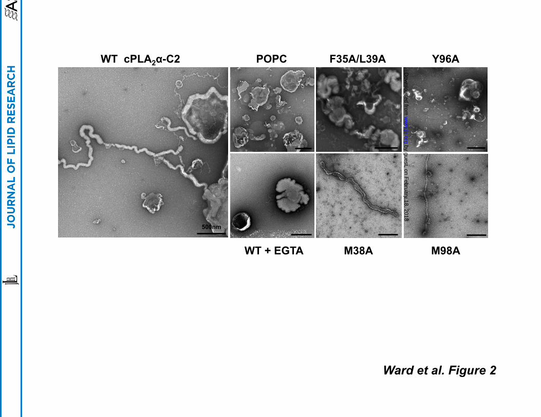
incubation with the C2 domain. As shown in Figure 2, the C2 domain induced dramatic
changes in POPC liposome structures as long tubules were extensively visualized through the
grids. Moreover, the tubulation of liposomes induced by the C2 domain was Ca2+-dependent as
experiments performed in the presence of 100 µM EGTA in place of CaCl2 did not display
detectable changes in liposome morphology (Fig. 2). ENTH, BAR and ACCH domains insert
into the hydrocarbon region of the membrane bilayer, which is a prerequisite for their ability to
induce membrane curvature changes (31, 37, 38, 39). To test if hydrophobic and aromatic
residues, which typically insert into membranes, were required for liposome morphology
changes we prepared mutations of hydrophobic and aromatic residues in calcium binding loops
1 and 3 of the C2 domain (Fig. 1). Earlier studies have demonstrated the ability of these
calcium-binding loops to penetrate deeply into membranes and monolayers where hydrophobic
and aromatic residues in these loops protrude into the hydrocarbon region of the membrane (9,
16). Indeed, F35A/L39A and Y96A displayed a drastic reduction in liposome morphology
by guest, on February 18, 2018
ww
w.jlr.org
Dow
nloaded from
10
changes and displayed a lack of long thin tubules emanating from liposomes as seen for the WT
C2 domain. M38A and M98A, which have been shown to have a lesser effect than F35A, L39A
or Y96A on cPLA2 membrane binding (9) still induced changes in liposome structure albeit to a
lesser extent than WT.
Quantification of Membrane Curvature Changes Using GUVs
GUVs have served as an effective platform for monitoring changes to membrane
structure as they can be fluorescently labeled, imaged with confocal microscopy and are more
easily quantified than EM experiments. GUVs have been effective in monitoring membrane
curvature changes for the ENTH domain (37) and viral matrix proteins (40). In addition, they are
relatively flat (mean diameter ~ 30 µM) in comparison to LUVs (mean diameter ~ 400 nm) so
they can be used to assess if proteins induce membrane curvature changes on different
membrane surfaces. GUVs composed of POPC:POPE:POPS (60:20:20) were prepared and
used to quantify membrane curvature changes for WT C2 domain and respective mutations.
All experiments were performed in triplicate and at least 60 GUVs were counted in each
experiment and assessed for membrane curvature changes in response to C2 domain binding.
As shown in Figure 3A, WT C2 domain induced vesiculation of GUVs in the presence of Ca2+,
which was not observed in the presence of 100 µM EGTA. Hydrophobic and aromatic
mutations, F35A/L39A and Y96A, which greatly reduce alterations to liposome morphology in
the EM assays significantly reduced GUV vesiculation for which their quantitative value was
similar to that of the control. M38A and M98A displayed a statistically significant reduction in
GUV vesiculation in line with the EM assays, which detected appreciable changes to liposome
structure albeit to a lesser extent than WT. To assess the ability of the C2 domain to induce
membrane curvature changes we assessed the ability of the C2 domain to induce vesiculation
in GUVs at 200 nM WT C2 domain in the presence of 500 nM CaCl2. Indeed, under these
conditions, which are closer to physiological concentrations of cytoplasmic Ca2+ the C2 domain
induced substantial GUV vesiculation (See Fig. 3C and D). Lastly, we assessed the ability of
by guest, on February 18, 2018
ww
w.jlr.org
Dow
nloaded from
11
full-length cPLA2α to induce membrane curvature changes to GUVs at 200 nM protein in the
presence of 500 nM CaCl2. The full-length enzyme not only induced GUV vesiculation but also
prompted extensive tubulation emanating from the GUVs.
C2 Domain Induces Fragmentation of Membrane Sheets
Membrane sheets labeled with fluorescent dye, which represent a relatively flat
membrane surface, have been used to image membrane curvature changes for the PH domain
of FAPP1 and 2 (30). Here we employed POPC membrane sheets labeled with FM® 2-10 dye
to assess the ability of the C2 domain to induce changes to membrane sheet structure.
Membrane sheets were imaged before and after introduction of C2 domain and mutants to
observe changes in real-time (Figure 4A). In the presence of Ca2+ the C2 domain induced rapid
fragmentation of POPC membrane sheets (Fig. 4A). The specific nature of this finding was
confirmed by adding the same volume of protein storage buffer to ensure that changes in
volume did not induce membrane swelling or membrane fragmentation. Additionally, mutations
that greatly reduced membrane structural changes in the EM or GUV assays, F35A/L39A and
Y96A, also abolished membrane fragmentation of POPC sheets (Fig. 4B). M98A displayed
similar membrane fragmentation as WT C2 but M38A did not induce detectable changes in
membrane sheet structure up to 25 min after the addition of protein. Taken together, the C2
domain is able to induce changes to membrane structure for small highly curved membranes
(LUVs) as well as less curved and relatively flat membranes (GUVs and membrane sheets).
Membrane Penetration and Lipid Binding Affinity of C2 domain and Mutations
Membrane penetration of C2 domain and mutations into POPC monolayers was
detected by injecting protein into the subphase buffer at varying initial surface pressure (π0)
values (See Fig. 5A and B). This allows determination of the critical pressure (πc), which is the
pressure up to which the protein will penetrate (x-intercept) (35). As previously reported (9, 34),
the WT C2 domain robustly penetrated a POPC monolayer with a value of ~ 36 mN/m. In
by guest, on February 18, 2018
ww
w.jlr.org
Dow
nloaded from

12
contrast, F35/L39A and Y96A, which abrogate membrane curvature changes also significantly
reduce the ability of the C2 domain to penetrate POPC monolayers with πc values of 23 and 20
mN/m, respectively. In addition, M38A and M98A, which had slightly reduced membrane-
deforming capabilities also slightly reduced membrane penetration with πc values of 30 and 31
mN/m, respectively. These results demonstrate that the C2 domain can effectively penetrate
physiological bilayers since the surface pressure of cell membranes and LUVs is estimated to
be in a range of 30-35 mN/m (41). Since monolayer penetration studies with WT and mutants
are performed at saturating amounts of protein where maximal binding of WT or mutants occurs
this signifies even at saturating conditions ofF35A/L39A and Y96A the proteins are not
significantly penetrating into the membrane while M38A and M98A have reduced penetration
compared to WT. In the absence of Ca2+ the C2 domain did not insert into the monolayer, and
the πc value of POPC monolayers was essentially undetectable as previously reported (9, 34).
Similarly, the mutation D43N in the C2 domain, which reduces Ca2+ binding and was unable to
reconstitute FcR mediated phagocytosis (23) also reduced the πc value (20 mN/m). Thus,
membrane penetration is necessary to induce membrane curvature changes as observed in the
EM, GUV, and membrane sheet assays. Likewise, phosphoinositides are necessary for the
ENTH, PH, and ACCH domains to sufficiently penetrate membranes and induce membrane
deformation (31, 39, 42).
To quantitatively assess the effect of mutations on the ability of the C2 domain to bind
POPC vesicles, we performed SPR assays (Fig. 5 C and D). A blank surface was used as a
control as it has been shown previously that the C2 domain of cPLA2α does not exhibit
nonspecific binding to the L1 chip surface (32, 34). The dissociation constants (Kds), obtained in
triplicate, demonstrate that the C2 domain associated with 21 ± 4 nM affinity to POPC vesicles
at 50 µM CaCl2 but binding was not detectable up to 5 µM in the presence of 100 µM EGTA
(data not shown). While mutations that abolish membrane-deforming activity, F35A/L39A and
by guest, on February 18, 2018
ww
w.jlr.org
Dow
nloaded from
13
Y96A, demonstrate >140- and 57-fold increases in Kd (Figure 5D) consistent with their role in
membrane penetration and in inducing alterations in membrane structure. Lastly, mutations that
slightly reduced membrane curvature changes and membrane penetration, M38A and M98A,
increase the Kd by 6.2- and 4.4-fold, respectively. In order to rule out misfolded C2 domain
mutations we used CD to determine the CD spectra of each mutant in comparison to wild type.
As shown in Fig. S1A CD spectra’s from all mutations overlapped with that of wild type C2
domain and displayed a spectra indicative of β-sheet with an energy minima at ~ 215 nm. Next,
in order to rule out changes in calcium binding for the mutants we quantified the calcium binding
ability of wild type, F35A/L39A, M38A and M98A. As shown in Fig. S1B mutations had
comparable calcium binding ability to wild type with M38A and M98A displaying slightly reduced
binding that was not statistically significant. Taken together our data indicate that membrane
affinity of the C2 domain and respective mutations correlates with the ability to penetrate
membranes and induce membrane curvature changes.
Discussion
Trafficking of membrane vesicles, endocytosis, and lipid-enveloped viral egress are a
few of the cellular pathways where major membrane curvature changes are necessary. These
are highly dynamic processes that cannot occur spontaneously as a significant energy barrier
has to be overcome to shape the lipid bilayer into a highly curved vesicle (43, 44). To overcome
this energy barrier, protein-mediated effects or lipid bilayer asymmetry can mediate curvature
changes. Protein induced changes are often facilitated by insertion of proteins into the
membrane bilayer or scaffolding of proteins on the membrane surface through oligomerization
(45). Lipid-mediated changes in bilayer structure can be attributed to lipid asymmetry mediated
by cone-shaped and inverted-cone-shaped lipids, where lipid shape can induce positive or
negative curvature changes (44).
by guest, on February 18, 2018
ww
w.jlr.org
Dow
nloaded from
14
Membrane curvature changes induced by lipid binding domains were first appreciated
with the discovery of the ENTH domain and its ability to induce changes to liposome structure in
a PI(4,5)P2-dependent manner (28). The ENTH domain deeply penetrates membranes with a
N-terminal amphipathic α-helix and also forms oligomers on the membrane (37), both of which
are necessary for effective membrane tubulation. This activity is essential to endocytosis and
clathrin coated vesicle formation, which requires substantial changes to plasma membrane
structure to form highly curved membrane vesicles (46). Subsequently, the discovery of the
BAR domains of amphiphysin (29) and endophilin (47) lead to the notion that intrinsic curvature
from the crescent moon shaped BAR domains is essential to remodeling membranes. This lead
to further investigation, which demonstrated that BAR domains form elegant scaffolds on the
membrane where mutation of residues that mediate scaffolding abrogates membrane curvature
changes (48, 49). Additionally, as with the ENTH domain some BAR domains have a N-
terminal α-helix that can penetrate into the membrane and an insert on the membrane binding
interface that inserts and is predicted to be a second amphipathic α-helix (38). The depth and
orientation of this penetration may also be important in regulating membrane curvature changes
and membrane fission (50). For instance, it was recently shown that insertion of the amphipathic
α-helix drives vesiculation and thus scission by the ENTH domain. In contrast, an antagonistic
relationship between the number and length of amphipathic helices in BAR domains was
discovered where membrane fission can be restricted by the BAR domains’ crescent shape
(50). Taken together, depth and area of insertion as well as inherent protein scaffold shape play
a critical role in the type of membrane curvature generated and whether or not membrane
fission will proceed.
PH domains (30) and C2 domains (32) have also been shown to induce membrane
curvature changes. The FAPP1 and 2 PH domains require insertion of a turret loop adjacent to
the PI(4)P binding pocket (42) to induce membrane remodeling where the inherent shape of
by guest, on February 18, 2018
ww
w.jlr.org
Dow
nloaded from
15
FAPP1 or 2 may also play a critical role (30). However, unlike the ENTH and BAR domain
elegant models of membrane scaffolding and modes of membrane curvature induction for PH
and C2 domains have not been investigated. In addition, the relationship between membrane
penetration of C2 domains and membrane curvature changes is still unknown. Recently, it was
shown that the C2B domain alone or the tandem C2AC2B domains of synaptotagmin 1, which
can induce membrane tubulation (32) as well as vesicle aggregation (51) could induce lipid
demixing of PS in POPC:POPS vesicles (52), which is thought to induce positive bilayer
curvature changes.
Here we demonstrate that the C2 domain of cPLA2α, long appreciated as a high affinity
target for zwitterionic lipids (9) with an ability to deeply penetrate the hydrocarbon core of
zwitterionic membranes, is able to induce substantial changes to membrane structure. Imaging
of liposomes with TEM, GUVs with confocal microscopy, or membrane sheets with confocal
microscopy demonstrated dramatic changes in membrane structure induced in a Ca2+-
dependent manner by the cPLA2α C2 domain. Detectable changes in membrane structure
correlated strongly with membrane penetrating ability and lipid binding affinity. As shown in
figure 6, membrane penetration of the C2 domain generated positive membrane curvature as
evidenced in the TEM and confocal assays. Positive curvature generation by the C2 domain is
consistent with the role of cPLA2α in formation of the phagosome, Golgi tubulation and Golgi
vesiculation, which occur by budding into the cytoplasm. It is also important to note the protein
concentrations of C2 domain employed in the membrane curvature assays were similar or lower
than most of the previous studies in the literature (28, 29, 32), supporting the specific nature of
our findings. For the C2 domain it seems there may be a threshold affinity and depth or extent
of penetration that is responsible for generating curvature as both M38A and M98A, which
reduced membrane penetration and affinity still induced statistically significant changes in
membrane curvature. In the sheet assay, M38A did not induce detectable membrane
by guest, on February 18, 2018
ww
w.jlr.org
Dow
nloaded from
16
fragmentation while M98A did. M38A is located in CBL1 and has lower affinity than M98A,
which is located in CBL3 (See Fig. 1). It has been shown that CBL1 penetrates more deeply into
the bilayer than CBL3 (53), which could perhaps play a role in the different observations in the
membrane sheet assays. While the origin of this discrepancy is still unknown it leaves room for
extensive biophysical studies of C2 domain parameters required for membrane curvature
generation. It also appears M38A and M98A may cause some vesiculation in the liposome
assays as visualized by EM as well as differences in the extent of vesiculation in the GUV
assays suggesting the depth of penetration and/or membrane affinity may play an important role
in the type of membrane curvature or membrane reorganization that is generated. Studying the
C2 domain’s role in the type of curvature generation in conjunction with membrane fission (50)
will be essential to better understanding the mechanism of curvature generation for this C2
domain. Penetration of hydrophobic residues by the C2 domain occurs into the hydrocarbon
layer (~ 15 Å) reminiscent of the ENTH domain (37, 39) so its possible membrane fission and
vesiculation (50) may occur, which is supported by membrane fragmentation in the membrane
sheet assays. C2 domains and PH domains may also induce different types of curvature. For
instance, in this study the C2 domain induced membrane fragmentation whereas studies with
PH domains have demonstrated extensive positive curvature induction in the form of tubules in
the membrane sheet assays (42).
Future studies will need to consider the role of C2 domain membrane binding and
penetration in inducing or contributing to membrane curvature changes in conjunction with
cPLA2α activity. The fact that the C2 domain alone is able to reconstitute FcR-mediated
phagocytosis (23) suggests the cPLA2α C2 domain possibly has a high membrane remodeling
activity that is essential to membrane reorganization. Additionally, inhibition of cPLA2α activity in
cells with the inhibitor pyrrophenone generated an allosteric block that prevented cPLA2α
translocation (23), which doesn’t allow one to account for C2 membrane binding and insertion in
by guest, on February 18, 2018
ww
w.jlr.org
Dow
nloaded from

17
assessing the generation of lysophospholipids in curvature generation (26). Nonetheless, the
prior study demonstrated the C2 domain process is Ca2+ dependent as D43N, which abrogates
calcium binding could not reconstitute phagocytosis. In conjunction with the current study this
strongly suggests membrane penetration of the C2 domain is necessary for these effects as
calcium is required for membrane penetration of the C2 domain (Fig. 1C and 5A) (9). It is also
now well established the cationic β-groove of the C2 domain binds C1P (21), which is important
for cPLA2α activity (21) and cellular translocation (54). Additionally, the role of ceramide kinase
and its product C1P are key players in phagocytosis (55, 56). Thus, its been hypothesized that
C1P has an important role in recruitment of cPLA2α in phagocytosis (57). To this end it’s
tempting to speculate that cPLA2α may be able to induce reorganization or clustering of
membranes harboring C1P.
The surface area of insertion for the C2 domain (58) is more substantial than that of the
ENTH (39, 50) and PH domains (59), at least for a monomer, however, this alone may not
account for the membrane mediated curvature changes. It was first thought the N-terminal α-
helix insertion for ENTH domains and to some degree for BAR domains was the major cause of
the membrane curvature induction, however, more recent and sophisticated studies have
demonstrated the ability of these proteins to scaffold on the membrane (48, 49). This
scaffolding is essential for both in vitro and cellular observations of membrane curvature
changes (48, 49). Future studies will need to be directed towards resolving the molecular details
of C2 oligomerization as well as the role of β-groove C1P binding in membrane curvature
changes or membrane reorganization. Additionally, the type of membrane curvature generated
by the C2 domain as well as full-length cPLA2α will require extensive analysis using a
combination of biophysical, biochemical and cellular assays. Investigating how the C2 domain
insertion and catalytic domain generation of lysophospholipids contribute to in vitro as well as
by guest, on February 18, 2018
ww
w.jlr.org
Dow
nloaded from

18
cellular membrane curvature changes should solve a number of burning questions in the fields
of membrane trafficking and phagosome formation.
The type of curvature generated by the C2 domain or full-length enzyme may also be
key to normal and aberrant physiological processes linked to cPLA2α activity. cPLA2α has been
shown to function in generation of cholesterol rich, GPI-protein containing endosomes (26),
Golgi tubulation and vesiculation (24, 25), and Golgi to PM trafficking (60). Additionally, cPLA2α
association with the Golgi has been show to regulate the function of endothelial cells (61, 62),
which serve a barrier function in the luminal surfaces of blood vessels. Proliferation of
endothelial cells has been shown to occur for formation of blood vessels in wound healing and
tumor formation while blocking cPLA2α activity through an inhibitor or siRNA blocks endothelial
cell proliferation and cell cycle entry (61). In terms of pathophysiological states cPLA2α has
been linked to diseases such as asthma (4), arthritis (6) and cancers (5). Thus, up or
downregulation of cPLA2α enzyme levels may alter the transport of vesicles from the Golgi to
the PM, modify intra-Golgi transport, or effect endothelial barrier function through the combined
generation of fatty acids and lysophospholipids and membrane penetration of the C2 domain of
cPLA2α. Additionally, because cPLA2α enzyme inhibitors act as an allosteric block that reduces
or precludes cPLA2α membrane translocation it is difficult to rule out the C2 domain mediated
effects. Thus, our data supports a model where vesiculation or tubulation of the Golgi may occur
in response to C2 domain membrane penetration of the full-length enzyme under conditions of
low cPLA2α activity. Linkage of biochemical and biophysical studies in vitro and in cells with
cellular and disease models that can tease apart the role of C2 domain penetration and cPLA2α
activity in these processes will be key to unraveling the full mechanism of membrane curvature
generation.
by guest, on February 18, 2018
ww
w.jlr.org
Dow
nloaded from

19
Acknowledgements Support for these studies were from the American Heart Association (SDG0735350N and
GRNT12080254) to R.V.S. K.E.W. is supported by an American Heart Association Predoctoral
Fellowship (AHA 11PRE7640028) and a NIH CBBI Training Fellowship (T32GM075762). E.A.G.
is supported by an Eck Institute for Global Health predoctoral training fellowship. This work was
also supported by the Indiana University School of Medicine-South Bend Imaging and Flow
Cytometry Core Facility and the Notre Dame Integrated Imaging Facility (R.V.S).
References
1. Clark, J. D., A. R. Schievella, E. A. Nalefski, and L. L. Lin. 1995. Cytosolic phospholipase A2. Journal of lipid mediators and cell signalling 12: 83-117. 2. Leslie, C. C., T. A. Gangelhoff, and M. H. Gelb. 2010. Localization and function of cytosolic phospholipase A2alpha at the Golgi. Biochimie 92: 620-626. 3. Shimizu, T., T. Ohto, and Y. Kita. 2006. Cytosolic phospholipase A2: biochemical properties and physiological roles. IUBMB life 58: 328-333. 4. Hewson, C. A., S. Patel, L. Calzetta, H. Campwala, S. Havard, E. Luscombe, P. A. Clarke, P. T. Peachell, M. G. Matera, M. Cazzola, C. Page, W. M. Abraham, C. M. Williams, J. D. Clark, W. L. Liu, N. P. Clarke, and M. Yeadon. 2012. Preclinical evaluation of an inhibitor of cytosolic phospholipase A2alpha for the treatment of asthma. The Journal of pharmacology and experimental therapeutics 340: 656-665. 5. Sundarraj, S., S. Kannan, R. Thangam, and P. Gunasekaran. 2012. Effects of the inhibition of cytosolic phospholipase A(2)alpha in non-small cell lung cancer cells. Journal of cancer research and clinical oncology 138: 827-835. 6. Tai, N., K. Kuwabara, M. Kobayashi, K. Yamada, T. Ono, K. Seno, Y. Gahara, J. Ishizaki, and Y. Hori. 2010. Cytosolic phospholipase A2 alpha inhibitor, pyrroxyphene, displays anti-arthritic and anti-bone destructive action in a murine arthritis model. Inflamm Res 59: 53-62. 7. Kishimoto, K., R. C. Li, J. Zhang, J. A. Klaus, K. K. Kibler, S. Dore, R. C. Koehler, and A. Sapirstein. 2010. Cytosolic phospholipase A2 alpha amplifies early cyclooxygenase-2 expression, oxidative stress and MAP kinase phosphorylation after cerebral ischemia in mice. Journal of neuroinflammation 7: 42. 8. Kerkela, R., M. Boucher, R. Zaka, E. Gao, D. Harris, J. Piuhola, J. Song, R. Serpi, K. C. Woulfe, J. Y. Cheung, E. O'Leary, J. V. Bonventre, and T. Force. 2011. Cytosolic phospholipase A(2)alpha protects against ischemia/reperfusion injury in the heart. Clinical and translational science 4: 236-242. 9. Bittova, L., M. Sumandea, and W. Cho. 1999. A structure-function study of the C2 domain of cytosolic phospholipase A2. Identification of essential calcium ligands and hydrophobic membrane binding residues. J Biol Chem 274: 9665-9672. 10. Corbin, J. A., J. H. Evans, K. E. Landgraf, and J. J. Falke. 2007. Mechanism of specific membrane targeting by C2 domains: localized pools of target lipids enhance Ca2+ affinity. Biochemistry 46: 4322-4336. 11. Tucker, D. E., M. Ghosh, F. Ghomashchi, R. Loper, S. Suram, B. S. John, M. Girotti, J. G. Bollinger, M. H. Gelb, and C. C. Leslie. 2009. Role of phosphorylation and basic residues in
by guest, on February 18, 2018
ww
w.jlr.org
Dow
nloaded from

20
the catalytic domain of cytosolic phospholipase A2alpha in regulating interfacial kinetics and binding and cellular function. J Biol Chem 284: 9596-9611. 12. Evans, J. H., and C. C. Leslie. 2004. The cytosolic phospholipase A2 catalytic domain modulates association and residence time at Golgi membranes. J Biol Chem 279: 6005-6016. 13. Evans, J. H., D. M. Spencer, A. Zweifach, and C. C. Leslie. 2001. Intracellular calcium signals regulating cytosolic phospholipase A2 translocation to internal membranes. J Biol Chem 276: 30150-30160. 14. Das, S., and W. Cho. 2002. Roles of catalytic domain residues in interfacial binding and activation of group IV cytosolic phospholipase A2. J Biol Chem 277: 23838-23846. 15. Murray, D., and B. Honig. 2002. Electrostatic control of the membrane targeting of C2 domains. Molecular cell 9: 145-154. 16. Frazier, A. A., M. A. Wisner, N. J. Malmberg, K. G. Victor, G. E. Fanucci, E. A. Nalefski, J. J. Falke, and D. S. Cafiso. 2002. Membrane orientation and position of the C2 domain from cPLA2 by site-directed spin labeling. Biochemistry 41: 6282-629222. 17. Pettus, B. J., A. Bielawska, P. Subramanian, D. S. Wijesinghe, M. Maceyka, C. C. Leslie, J. H. Evans, J. Freiberg, P. Roddy, Y. A. Hannun, and C. E. Chalfant. 2004. Ceramide 1-phosphate is a direct activator of cytosolic phospholipase A2. J Biol Chem 279: 11320-11326. 18. Subramanian, P., R. V. Stahelin, Z. Szulc, A. Bielawska, W. Cho, and C. E. Chalfant. 2005. Ceramide 1-phosphate acts as a positive allosteric activator of group IVA cytosolic phospholipase A2 alpha and enhances the interaction of the enzyme with phosphatidylcholine. J Biol Chem 280: 17601-17607. 19. Balsinde, J., M. A. Balboa, W. H. Li, J. Llopis, and E. A. Dennis. 2000. Cellular regulation of cytosolic group IV phospholipase A2 by phosphatidylinositol bisphosphate levels. J Immunol 164: 5398-5402. 20. Mosior, M., D. A. Six, and E. A. Dennis. 1998. Group IV cytosolic phospholipase A2 binds with high affinity and specificity to phosphatidylinositol 4,5-bisphosphate resulting in dramatic increases in activity. J Biol Chem 273: 2184-2191. 21. Stahelin, R. V., P. Subramanian, M. Vora, W. Cho, and C. E. Chalfant. 2007. Ceramide-1-phosphate binds group IVA cytosolic phospholipase a2 via a novel site in the C2 domain. J Biol Chem 282: 20467-20474. 22. Casas, J., M. A. Gijon, A. G. Vigo, M. S. Crespo, J. Balsinde, and M. A. Balboa. 2006. Phosphatidylinositol 4,5-bisphosphate anchors cytosolic group IVA phospholipase A2 to perinuclear membranes and decreases its calcium requirement for translocation in live cells. Molecular biology of the cell 17: 155-162. 23. Zizza, P., C. Iurisci, M. Bonazzi, P. Cossart, C. C. Leslie, D. Corda, and S. Mariggio. 2012. Phospholipase A2IValpha regulates phagocytosis independent of its enzymatic activity. J Biol Chem 287: 16849-16859. 24. Grimmer, S., M. Ying, S. Walchli, B. van Deurs, and K. Sandvig. 2005. Golgi vesiculation induced by cholesterol occurs by a dynamin- and cPLA2-dependent mechanism. Traffic 6: 144-156. 25. San Pietro, E., M. Capestrano, E. V. Polishchuk, A. DiPentima, A. Trucco, P. Zizza, S. Mariggio, T. Pulvirenti, M. Sallese, S. Tete, A. A. Mironov, C. C. Leslie, D. Corda, A. Luini, and R. S. Polishchuk. 2009. Group IV phospholipase A(2)alpha controls the formation of inter-cisternal continuities involved in intra-Golgi transport. PLoS biology 7: e1000194. 26. Cai, B., S. Caplan, and N. Naslavsky. 2012. cPLA2alpha and EHD1 interact and regulate the vesiculation of cholesterol-rich, GPI-anchored, protein-containing endosomes. Molecular biology of the cell 23: 1874-1888. 27. Campelo, F., H. T. McMahon, and M. M. Kozlov. 2008. The hydrophobic insertion mechanism of membrane curvature generation by proteins. Biophysical journal 95: 2325-2339. 28. Ford, M. G., I. G. Mills, B. J. Peter, Y. Vallis, G. J. Praefcke, P. R. Evans, and H. T. McMahon. 2002. Curvature of clathrin-coated pits driven by epsin. Nature 419: 361-366.
by guest, on February 18, 2018
ww
w.jlr.org
Dow
nloaded from
21
29. Peter, B. J., H. M. Kent, I. G. Mills, Y. Vallis, P. J. Butler, P. R. Evans, and H. T. McMahon. 2004. BAR domains as sensors of membrane curvature: the amphiphysin BAR structure. Science 303: 495-499. 30. Cao, X., U. Coskun, M. Rossle, S. B. Buschhorn, M. Grzybek, T. R. Dafforn, M. Lenoir, M. Overduin, and K. Simons. 2009. Golgi protein FAPP2 tubulates membranes. Proceedings of the National Academy of Sciences of the United States of America 106: 21121-21125. 31. Heller, B., E. Adu-Gyamfi, W. Smith-Kinnaman, C. Babbey, M. Vora, Y. Xue, R. Bittman, R. V. Stahelin, and C. D. Wells. 2010. Amot recognizes a juxtanuclear endocytic recycling compartment via a novel lipid binding domain. J Biol Chem 285: 12308-12320. 32. Martens, S., M. M. Kozlov, and H. T. McMahon. 2007. How synaptotagmin promotes membrane fusion. Science (New York, N.Y 316: 1205-1208. 33. Hom, R. A., M. Vora, M. Regner, O. M. Subach, W. Cho, V. V. Verkhusha, R. V. Stahelin, and T. G. Kutateladze. 2007. pH-dependent binding of the Epsin ENTH domain and the AP180 ANTH domain to PI(4,5)P2-containing bilayers. Journal of molecular biology 373: 412-423. 34. Stahelin, R. V., and W. Cho. 2001. Roles of calcium ions in the membrane binding of C2 domains. The Biochemical journal 359: 679-685. 35. Cho, W., L. Bittova, and R. V. Stahelin. 2001. Membrane binding assays for peripheral proteins. Analytical biochemistry 296: 153-161. 36. Stahelin, R. V., D. Karathanassis, D. Murray, R. L. Williams, and W. Cho. 2007. Structural and membrane binding analysis of the PX domain of Bem1p: Basis of phosphatidylinositol-4-phosphate specificity. J Biol Chem. 282, 25737-25747. 37. Yoon, Y., J. Tong, P. J. Lee, A. Albanese, N. Bhardwaj, M. Kallberg, M. A. Digman, H. Lu, E. Gratton, Y. K. Shin, and W. Cho. 2010. Molecular basis of the potent membrane-remodeling activity of the epsin 1 N-terminal homology domain. J Biol Chem 285: 531-540. 38. Gallop, J. L., C. C. Jao, H. M. Kent, P. J. Butler, P. R. Evans, R. Langen, and H. T. McMahon. 2006. Mechanism of endophilin N-BAR domain-mediated membrane curvature. The EMBO journal 25: 2898-2910. 39. Stahelin, R. V., F. Long, B. J. Peter, D. Murray, P. De Camilli, H. T. McMahon, and W. Cho. 2003. Contrasting membrane interaction mechanisms of AP180 N-terminal homology (ANTH) and epsin N-terminal homology (ENTH) domains. J Biol Chem 278: 28993-28999. 40. Shnyrova, A. V., J. Ayllon, Mikhalyov, II, E. Villar, J. Zimmerberg, and V. A. Frolov. 2007. Vesicle formation by self-assembly of membrane-bound matrix proteins into a fluidlike budding domain. The Journal of cell biology 179: 627-633. 41. Blume, A. 1979. A comparative study of the phase transitions of phospholipid bilayers and monolayers. Biochimica et biophysica acta 557: 32-44. 42. He, J., J. L. Scott, A. Heroux, S. Roy, M. Lenoir, M. Overduin, R. V. Stahelin, and T. G. Kutateladze. 2011. Molecular basis of phosphatidylinositol 4-phosphate and ARF1 GTPase recognition by the FAPP1 pleckstrin homology (PH) domain. J Biol Chem 286: 18650-18657. 43. Baumgart, T., B. R. Capraro, C. Zhu, and S. L. Das. 2011. Thermodynamics and mechanics of membrane curvature generation and sensing by proteins and lipids. Annual review of physical chemistry 62: 483-506. 44. Kooijman, E. E., V. Chupin, N. L. Fuller, M. M. Kozlov, B. de Kruijff, K. N. Burger, and P. R. Rand. 2005. Spontaneous curvature of phosphatidic acid and lysophosphatidic acid. Biochemistry 44: 2097-2102. 45. Graham, T. R., and M. M. Kozlov. 2010. Interplay of proteins and lipids in generating membrane curvature. Current opinion in cell biology 22: 430-436. 46. McMahon, H. T., and E. Boucrot. 2011. Molecular mechanism and physiological functions of clathrin-mediated endocytosis. Nature reviews 12: 517-533. 47. Gallop, J. L., P. J. Butler, and H. T. McMahon. 2005. Endophilin and CtBP/BARS are not acyl transferases in endocytosis or Golgi fission. Nature 438: 675-678.
by guest, on February 18, 2018
ww
w.jlr.org
Dow
nloaded from

22
48. Frost, A., R. Perera, A. Roux, K. Spasov, O. Destaing, E. H. Egelman, P. De Camilli, and V. M. Unger. 2008. Structural basis of membrane invagination by F-BAR domains. Cell 132: 807-817. 49. Shimada, A., H. Niwa, K. Tsujita, S. Suetsugu, K. Nitta, K. Hanawa-Suetsugu, R. Akasaka, Y. Nishino, M. Toyama, L. Chen, Z. J. Liu, B. C. Wang, M. Yamamoto, T. Terada, A. Miyazawa, A. Tanaka, S. Sugano, M. Shirouzu, K. Nagayama, T. Takenawa, and S. Yokoyama. 2007. Curved EFC/F-BAR-domain dimers are joined end to end into a filament for membrane invagination in endocytosis. Cell 129: 761-772. 50. Boucrot, E., A. Pick, G. Camdere, N. Liska, E. Evergren, H. T. McMahon, and M. M. Kozlov. 2012. Membrane fission is promoted by insertion of amphipathic helices and is restricted by crescent BAR domains. Cell 149: 124-136. 51. Arac, D., X. Chen, H. A. Khant, J. Ubach, S. J. Ludtke, M. Kikkawa, A. E. Johnson, W. Chiu, T. C. Sudhof, and J. Rizo. 2006. Close membrane-membrane proximity induced by Ca(2+)-dependent multivalent binding of synaptotagmin-1 to phospholipids. Nature structural & molecular biology 13: 209-217. 52. Lai, A. L., L. K. Tamm, J. F. Ellena, and D. S. Cafiso. 2011. Synaptotagmin 1 modulates lipid acyl chain order in lipid bilayers by demixing phosphatidylserine. J Biol Chem 286: 25291-25300. 53. Malmberg, N. J., D. R. Van Buskirk, and J. J. Falke. 2003. Membrane-docking loops of the cPLA2 C2 domain: detailed structural analysis of the protein-membrane interface via site-directed spin-labeling. Biochemistry 42: 13227-13240. 54. Lamour, N. F., P. Subramanian, D. S. Wijesinghe, R. V. Stahelin, J. V. Bonventre, and C. E. Chalfant. 2009. Ceramide 1-phosphate is required for the translocation of group IVA cytosolic phospholipase A2 and prostaglandin synthesis. J Biol Chem 284: 26897-26907. 55. Hinkovska-Galcheva, V., L. A. Boxer, A. Kindzelskii, M. Hiraoka, A. Abe, S. Goparju, S. Spiegel, H. R. Petty, and J. A. Shayman. 2005. Ceramide 1-phosphate, a mediator of phagocytosis. J Biol Chem 280: 26612-26621. 56. Hinkovska-Galcheva, V. T., L. A. Boxer, P. J. Mansfield, D. Harsh, A. Blackwood, and J. A. Shayman. 1998. The formation of ceramide-1-phosphate during neutrophil phagocytosis and its role in liposome fusion. J Biol Chem 273: 33203-33209. 57. Falke, J. J. 2012. Lipid targeting domain with dual-membrane specificity that expands the diversity of intracellular targeting reactions. Proceedings of the National Academy of Sciences of the United States of America 109: 1816-1817. 58. Malkova, S., F. Long, R. V. Stahelin, S. V. Pingali, D. Murray, W. Cho, and M. L. Schlossman. 2005. X-ray reflectivity studies of cPLA2{alpha}-C2 domains adsorbed onto Langmuir monolayers of SOPC. Biophysical journal 89:1861-1873. 59. Lenoir, M., U. Coskun, M. Grzybek, X. Cao, S. B. Buschhorn, J. James, K. Simons, and M. Overduin. 2010. Structural basis of wedging the Golgi membrane by FAPP pleckstrin homology domains. EMBO reports 11: 279-284. 60. Choukroun G. J., Marshansky, V., Gustafson, C. E., McKee, M., Hajjar, R. J., Rosenzweig, A., Brown, D., Bonventre, J. V. 2000. Cytosolic phospholipase A2 regulates Golgi structure and modulates intracellular trafficking of membrane proteins. J. Clin. Invest. 106: 983-993. 61. Herbert, S. P., Ponnambalam, S., Walker, J. H. 2005. Cytosolic phospholipase A2-α mediates endothelial cell proliferation and is inactivated by association with the Golgi apparatus. Mol. Biol. Cell 16: 3800-3809. 62. Herbert, S. P., Odell, A. F., Ponnambalam, S., Walker, J. H. 2007. The confluence-dependent interaction of cytosolic phospholipase A2-α with annexin A1 regulates endothelial cell prostaglandin E2 generation. J. Biol. Chem. 282: 34468-34478.
by guest, on February 18, 2018
ww
w.jlr.org
Dow
nloaded from

23
Figure Legends
Fig. 1 Structural depiction of the C2 domain of cPLA2α. A. The C2 domain (PDB ID 1CJY) is
shown in grey in surface transparency mode to depict hydrophobic amino acids in calcium
binding loops 1 and 3 (red). The two Ca2+ ions bound to the C2 domain are shown in yellow. B.
A close up view of the calcium binding and membrane penetration regions of the C2 domain of
cPLA2α. Amino acids mutated in this study to assess membrane penetration and membrane
curvature are shown in red (Phe35, Met38, and Leu39 in CBL1 and Tyr96 and Met98 in CBL3). Ca2+
ions crystalized with the protein are shown in yellow. C. The C2 domain has been shown to
deeply penetrate zwitterionic membranes. Here the C2 domain is shown with the depth of
penetration and orientation previously resolved by EPR (16) deeply penetrating hydrophobic
and aromatic residues are shown in red (Phe35, Met38, and Leu39 in CBL1 and Tyr96 and Met98 in
CBL3) and 2 Ca2+ ions in yellow. The domain was docked to the membrane according to
previous biophysical studies, which provided molecular insight into the depth and orientation of
the C2 domain binding to membranes (16). The protein shown is docked to a POPC
membrane, which displays the importance of Phe35, Met38, and Leu39 in CBL1 and Tyr96 and
Met98 in CBL3 in penetrating the lipid bilayer.
Fig. 2 The C2 domain induces membrane tubulation of POPC LUVs. Transmission electron
microscopy was used to assess the ability of the C2 domain and mutants to induce changes to
liposome morphology. All measurements were done with 10 µM protein in 20mM HEPES, pH
7.4, containing 160 mM KCl, and either 100 µM CaCl2 or 100 µM EGTA . WT C2 induced
extensive tubulation of POPC liposomes in a Ca2+-dependent manner. However, mutations
F35A/L39A and Y96A greatly reduced changes in liposome morphology while M38A and M98A
by guest, on February 18, 2018
ww
w.jlr.org
Dow
nloaded from

24
induced formation of tubules from liposomes but to a lesser extent than WT. Incubation of the
C2 domain in 100 µM EGTA in place of CaCl2 with POPC liposomes did not induce appreciable
changes in liposome morphology. Scale bars = 500 nm.
Fig. 3 GUV assay demonstrates the C2 domain’s ability to induce membrane budding. A.
Experiments with POPC:POPE:POPS (60:20:20) GUVs were performed in triplicate with a
minimum of 60 GUVs assessed per measurement to provide a quantitative representation of
membrane curvature changes shown in B. B. Quantitative representation of GUV vesiculation
induced by C2 domain and respective mutants. WT induced significant vesiculation of GUVs in
the presence of 10 µM CaCl2 compared to control experiments performed in 100 µM EGTA.
M38A and M98A did display some induction of GUV vesiculation and membrane reorganization
but to a significantly lesser extent than WT. F35A/L39A and Y96A did not appreciably induce
GUV vesiculation when compared to the control. C. WT C2 domain and full-length cPLA2α
were assessed at 200 nM protein concentration for their ability to induce membrane curvature
changes in the presence of 500 nM CaCl2 to GUVs containing POPC:POPE:POPS (60:20:20).
WT C2 domain induced substantial vesiculation while full-length cPLA2α induced vesiculation
and long tubule formation from GUVs. D. Quantification of vesiculation in control versus WT C2
experiments shown in C. The p value for each protein was determined in comparison to the
control in C and D (ns = not significant, * p < 0.001, ** p < 0.0001) using an unpaired student t-
test. Scale bars = 5 µm.
Fig. 4 C2 domain induces lipid fragmentation of membrane sheets. POPC membrane sheets
were used to test the ability of the C2 domain to induce membrane fragmentation to relatively
flat membrane surfaces. Membranes were hydrated then incubated with 2 µM WT or mutant
C2 domain for 15 minutes. A. WT C2 domain induced extensive membrane fragmentation from
by guest, on February 18, 2018
ww
w.jlr.org
Dow
nloaded from

25
membrane sheets, which was not observed in control experiments with buffer alone. B.
Hydrophobic and aromatic mutations reduced C2 domain membrane fragmentation. Only M98A
displayed detectable membrane fragmentation compared to WT, F35A/L39A, M38A, and Y96A.
All membrane sheets were imaged before and after protein incubation. All scale bars = 25 µm.
Fig. 5 Mutations that reduce or abrogate membrane deformation reduce or abolish membrane
penetration and membrane affinity. A. Insertion of the wild type C2 domain in the presence of
Ca2+ (filled circles) or EGTA (open circles) into a POPC monolayer monitored as a function of
π0. Insertion of F35A/L39A (filled squares), M38A (filled triangles), or D43N (filled diamonds)
was also monitored in the presence of Ca2+. B. Insertion of the wild type C2 domain (filled
circles), Y96A (filled squares) or M98A (filled triangles). All measurements performed in the
presence of Ca2+ C. The normalized saturation response (Req) from WT cPLA2α-C2 (filled
circles), M38A (filled triangles) or Y96A (filled squares) binding at each respective protein
concentration was plotted versus [C2] to fit with a nonlinear least squares analysis of the binding
isotherm (Req = Rmax/(1 + Kd/C) to determine the Kd. D. Kd values for WT and respective
mutations binding to POPC vesicles. The binding experiments were completed from
independent experiments in triplicate and are listed with their respective Kd ± standard deviation.
Fig. 6 Membrane penetration by the C2 domain of cPLA2α is sufficient to induce membrane
curvature changes. The hydrophobic residues essential in penetrating the membrane are also
key for membrane curvature generation. The deep ~ 15 Å penetration of these hydrophobic
and aliphatic residues as well as a significant area of insertion (~ 2110 Å2 (82)) are sufficient to
reduce the energetic barrier to bend the membrane as deletion of one of these key residues
abolishes this effect as shown for F35A/L39A and Y96A mutants. Although the overall
mechanism is currently unknown, our data suggests that membrane penetration of the C2
by guest, on February 18, 2018
ww
w.jlr.org
Dow
nloaded from
26
domain is vital for membrane bending, tubulation, vesiculation and fragmentation depending on
the initial curvature of the membrane.
by guest, on February 18, 2018
ww
w.jlr.org
Dow
nloaded from

M38A
F35A L39A
M98A
Y96A
B A
C
CBL1 CBL3
Ward et al. Figure 1
by guest, on February 18, 2018
ww
w.jlr.org
Dow
nloaded from
POPC
WT + EGTA
Y96A F35A/L39A
M38A M98A
WT cPLA2α-C2
Ward et al. Figure 2
500nm
by guest, on February 18, 2018
ww
w.jlr.org
Dow
nloaded from

0
1
2
3
4
5
6
7
8
Vesi
cula
ted
GU
Vs/ C
ontr
ol
WT
+ 10µM CaCl2
100µM EGTA F35A/L39A M38A M98A Y96A
Ward et al. Figure 3
A
B
ns
*
**
5µm
Control WT Full Length
0
1
2
3
4
5
Control 200nM WT
Vesi
cula
ted
GU
Vs/
Con
trol
*
C
D
by guest, on February 18, 2018
ww
w.jlr.org
Dow
nloaded from

Control +
Buf
fer
+ cPLA2 α-C2
Ward et al. Figure 4A
A
Scale bar = 25 µm
25µm
by guest, on February 18, 2018
ww
w.jlr.org
Dow
nloaded from

F35A/L39A
+ W
T
M38A Y96A M98A
Ward et al. Figure 4B
post
hyd
ratio
n
B
25µm
by guest, on February 18, 2018
ww
w.jlr.org
Dow
nloaded from

Ward et al. Figure 5
A B
C D Kd measurements for the C2 domain of cPLA2α. Measurements were determined with SPR at 50 µM Ca2+ in 10 mM HEPES, pH 7.4 containing 0.16 M KCl buffer.
POPC Protein Kd (M) Fold Increase in Kda
WT (2.1 ± 0.4) x 10-8 ---- F35/L39A > 3 x 10⁻⁶ > 140
M38A (1.3 ± 0.2) x 10-7 6.2 Y96A (1.2 ± 0.3) x 10-6 57 M98A (9.5 ± 0.7) x 10-8 4.4
aFold increase in Kd relative to the binding of WT C2 to POPC vesicles.
by guest, on February 18, 2018
ww
w.jlr.org
Dow
nloaded from

Ward et al. Figure 6
by guest, on February 18, 2018
ww
w.jlr.org
Dow
nloaded from


![Hydrogen Peroxide-Mediated Cytosolic Acidification Is a ...€¦ · [CANCER RESEARCH 64, 7867–7878, November 1, 2004] Hydrogen Peroxide-Mediated Cytosolic Acidification Is a Signal](https://static.fdocuments.net/doc/165x107/5eaae4556521d7256f027b39/hydrogen-peroxide-mediated-cytosolic-acidification-is-a-cancer-research-64.jpg)


![Cytosolic [Ca]](https://static.fdocuments.net/doc/165x107/56814e3f550346895dbbac79/cytosolic-ca.jpg)

![Glutaredoxin GRXS17 Associates with the Cytosolic … · Glutaredoxin GRXS17 Associates with the Cytosolic Iron-Sulfur Cluster Assembly Pathway1[OPEN] Sabrina Iñigo2, Astrid Nagels](https://static.fdocuments.net/doc/165x107/5b9f750709d3f267388b4bda/glutaredoxin-grxs17-associates-with-the-cytosolic-glutaredoxin-grxs17-associates.jpg)

