Brain stem & Cerebellum. The brain Telencephalon Diencephalon Cerebellum Brain stem.

BRAINA JOURNAL OF NEUROLOGY
Evidence of a non-progressive course of alternatinghemiplegia of childhood: study of a large cohort ofchildren and adultsEleni Panagiotakaki,1 Giuseppe Gobbi,2 Brian Neville,3 Friedrich Ebinger,4 Jaume Campistol,5
Sona Nevsımalova,6 Laura Laan,7 Paul Casaer,8 Georg Spiel,9 Melania Giannotta,2 Carmen Fons,5
Miriam Ninan,3 Guenter Sange,9 Tsveta Schyns,10 Rosaria Vavassori,10 Dominique Poncelin,10
The ENRAH Consortium10,* and Alexis Arzimanoglou1,11
1 Institute for Children and Adolescents with Epilepsy (IDEE), Hopital Femme Mere Enfant, University Hospitals of Lyon (HCL), France
2 Child Neurology Unit, Department of Neuroscience, Maggiore Hospital, Bologna, Italy
3 Institute of Child Health, UCL, London, UK
4 Department of Child Neurology, Heidelberg University Hospital, Germany
5 Department of Neurology, Sant Joan de Deu Hospital, Barcelona, Spain
6 Department of Neurology, Charles University, 1st Faculty of Medicine and Teaching Hospital, Prague, Czech Republic
7 Department of Neurology, K5Q, Leiden University Medical Centre, Leiden, The Netherlands
8 Department of Paediatrics University Hospital Gasthuisberg, Leuven, Belgium
9 Department of Neurology and Psychiatry of Children and Adolescents, General Hospital, Klagenfurt, Austria
10 The ENRAH Consortium
11 Inserm U821 and TIGER Research Group, Lyon, France
*The members of the The ENRAH Consortium are listed in the Appendix 1.
Correspondence to: Pr. Alexis Arzimanoglou,
Head of the Institute for Children and Adolescents with Epilepsy (IDEE),
Hopital Femme Mere Enfant,
Hospices Civils de Lyon,
59 Boulevard Pinel, 69677,
LYON, France
E-mail: [email protected]
Alternating hemiplegia of childhood is a neurological disorder characterized by episodes of hemiplegia, various non-epileptic
paroxysmal events and global neurological impairment. Characterization of the evolution and outcome into adulthood has not
been sufficiently investigated. The goal of this study was to elucidate the natural history of alternating hemiplegia within a large
cohort of 157 patients, as part of the European Network for Research on Alternating Hemiplegia project. A questionnaire was
formulated to determine the severity of both paroxysmal and global neurological impairment and address progression of the
disorder by allocating data to specific age epochs up to and over 24 years of age. Patients in early age groups were consistently
present in subsequent later age groups and for each patient, data were collected for each corresponding age epoch. The study
was based on predominantly retrospective and, for a period of 2 years, prospective data. At inclusion, patients were aged from
9 months to 52 years. The median age at diagnosis was 20 months. All patients experienced hemiplegic attacks; 86.5% reported
episodes of bilateral weakness, 88% dystonic attacks, 53% epileptic seizures, 72% developed chorea and/or dystonia
and 92% mental retardation. When data over the course of the illness were examined for the whole cohort, the severity of
symptoms did not appear to change, with the exception of abnormal ocular movements and hypotonia that regressed, but did
doi:10.1093/brain/awq295 Brain 2010: Page 1 of 13 | 1
Received January 6, 2010. Revised August 31, 2010. Accepted September 5, 2010.
� The Author (2010). Published by Oxford University Press on behalf of the Guarantors of Brain. All rights reserved.
For Permissions, please email: [email protected]
Brain Advance Access published October 24, 2010 by guest on January 21, 2011
brain.oxfordjournals.orgD
ownloaded from

not disappear into adulthood (from 86 to 36% and 76 to 36%, respectively). No statistically significant correlation between a
history of severe paroxysmal hemiplegic/dystonic episodes and a worse neurological outcome was identified. Seven patients
died, some of whom experienced severe plegic attacks or epileptic seizures at the time of death. History of severe plegic/
dystonic attacks was not found to be an aggravating factor for deceased patients. Our results provide evidence that the natural
history of alternating hemiplegia is highly variable and unpredictable for individual patients. However, we did not find evidence
to support a steadily progressive and degenerative course of the disorder when patients were analysed as a group. For a minority
of patients, a risk of sudden death was associated with more severe neurological impairment. The European Network for
Research on Alternating Hemiplegia Registry, validated by our study, includes all major neurological signs and symptoms of
alternating hemiplegia and may thus be used as a precedent for the progressive inclusion and follow-up of patients as well as a
reference for genetic studies and treatment trials.
Keywords: alternating hemiplegia; AHC; evolution; adulthood; sudden death
Abbreviations: AHC = alternating hemiplegia of childhood; ENRAH = European Network for Research on Alternating Hemiplegia
IntroductionAlternating hemiplegia of childhood (AHC) is a disorder character-
ized by transient episodes of alternating hemiplegia/hemiparesis
and often tetraplegia as well as other paroxysmal manifestations
(dystonic attacks, paroxysmal nystagmus, episodes of autonomic
disturbances and epileptic seizures) starting in the first 18 months
of life (Verret and Steele, 1971; Aicardi, 1987). Between attacks,
patients have an abnormal neurological examination with signs of
ataxia, involuntary abnormal movements (such as athetosis or
chorea) and the majority develop mental retardation (Bourgeois
et al., 1993). The condition was initially described in 1971
(Verret and Steele, 1971) and other authors have since added to
the description (Dittrich et al., 1979; Krageloh and Aicardi, 1980).
The disappearance of paroxysmal symptoms with, and immedi-
ately following sleep was reported for the first time in 1987
(Aicardi, 1987). Several authors suggested that similar patho-
physiological mechanisms with hemiplegic migraine could be impli-
cated in AHC (Golden and French, 1975; Hosking et al., 1978;
Hockaday, 1979). However, fixed neurological deficits are unusual
in hemiplegic migraine and the overall clinical picture and evolu-
tion are very different (Verret and Steele, 1971; Hockaday, 1979).
Approximately half of patients present with epilepsy and seizures
may sometimes occur simultaneously with plegic/dystonic attacks
(Neville and Ninan, 2007). The seizures are predominantly partial
but may manifest as status epilepticus requiring urgent medical
attention (Aicardi et al., 1995b).
Onset of the disease usually occurs before the age of 6 months
(sometimes in the neonatal period) with repeated tonic/dystonic
attacks and paroxysmal (often monocular) nystagmus, with hemi-
plegic and bilateral events appearing later (Aicardi et al., 1995a;
Sweney et al., 2009). During hemiplegic events a varying intensity
of paralysis may occur from one moment to another and can
sometimes alternate between sides during the same episode,
often mixed with dystonia. Consciousness is preserved during
episodes that can last from a few minutes to several days.
Bilateral plegic attacks, monocular nystagmus and other abnormal
ocular movements like paroxysmal strabismus (Bursztyn et al.,
2000) are considered to be highly characteristic of the disease
(Aicardi, 1987).
Due to the rare nature of the disorder, estimated at about one
in a million, previous reports have not sufficiently investigated
outcome of AHC in adulthood. The present study describes the
data obtained from the largest cohort of AHC patients to date; a
European web-based registry that was created within the ENRAH
project (European Network for Research on Alternating
Hemiplegia; http://www.enrah.net). The aim was to collect,
review and evaluate the clinical aspects of almost all known
cases of the disease in the participating countries, with emphasis
on the natural history and long-term outcome of the disease.
Materials and methods
The ENRAH projectThe ENRAH is a non-profit European organization, initiated through
a project funded by the European Commission within the sixth
Framework Programme (scheduled between April 2005 and June
2007). The ENRAH was established by patient organizations and
health workers with the goal to promote knowledge, communication
and research concerning AHC.
To assess the clinical presentation of AHC with age, a questionnaire
was designed to gather information on the clinical presentation of
AHC at specific ages from a total of 157 patients. The questionnaire
was structured as a multiple choice to include specific questions that
were repeated for each age group (Appendix 1 in online
Supplementary material). Any further information was written on
space provided, as required. The questionnaire was developed through
comprehensive discussions within the ENRAH group and validated by
the treating child neurologists (E.P., G.G., B.N., F.E., J.C., S.N., L.L.,
P.C., G.S. and A.A.).
Data collectionData were collected retrospectively for the period preceding the onset
of the project and prospectively between April 2005 and June 2007
from paediatric neurology university centres from nine European coun-
tries (see below). Data collection was undertaken by 12 delegated
participating clinicians (one or two per reference centre) with the
informed consent of the patient and/or his/her legal representative,
after approval from independent national ethics committees, in
2 | Brain 2010: Page 2 of 13 E. Panagiotakaki et al.
by guest on January 21, 2011brain.oxfordjournals.org
Dow
nloaded from

accordance with European and national legislation and regulations.
The delegated clinicians completed the questionnaire either after
direct contact with patients and/or after medical records revision and
using additional information provided by the treating physician
(paediatrician, paediatric neurologist, neurologist or other practitioner)
or family. For each of the patients, data were collected for every
corresponding age epoch. The final visit (prospective) corresponded
to the age of the patient at inclusion in this study. An average evalu-
ation over a period of years was obtained after reviewing subsequent
clinical visits and completing information by interviewing family
and physicians. National parent associations assisted in the collection
of data and guided physicians to contact patients. To ensure a
consistent standard of collected information throughout the study,
regular meetings were held between the participating clinicians.
Following initial data collection, one member of the ENRAH
Validation Committee (A.A., B.N. or G.G.) reviewed data from a
particular centre (data were not reviewed from a centre by a residing
member). Any queries raised by a Committee member were discussed
with the delegated clinician, who provided any further information
sometimes through discussion with the previously treating physician
or family members. Patient data were ultimately scrutinized again
by the Validation Committee who together reviewed the final
157 included cases. Information of these 157 patients was subse-
quently transferred in an electronic form to the European Registry.
This electronic database is available on a secure Internet site
(HC Forum� Internet platform) and access to the registry is secured
by the use of a smart card (an integrated circuit card requiring a
password and card reader). These data are accessible only to project
participants or other persons with a research interest, following
approval by the Steering Committee of ENRAH.
The participating paediatric neurology university centres included:
(i) Department of Neurology and Psychiatry of Children and
Adolescents, General Hospital, Klagenfurt, Austria; (ii) Department
of Paediatrics University Hospital Gasthuisberg, Leuven, Belgium;
(iii) Department of Neurology, Charles University, First Faculty
of Medicine and Teaching Hospital, Prague, Czech Republic;
(iv) Department of Paediatric Neurology, Robert Debre Hospital,
APHP, Paris, France; (v) Institute for Children and Adolescents with
Epilepsy (IDEE), Women and Children’s Hospital, University Hospitals
of Lyon (HCL), France; (vi) Department of Child Neurology, University
Hospital of Heidelberg, Germany; (vii) Child Neurology Unit,
Department of Neuroscience, Maggiore Hospital Bologna, Italy;
(viii) Department of Neurology, K5Q, Leiden University Medical
Centre, Leiden, The Netherlands; (ix) Department of Neurology,
Hospital Sant Joan de Deu, Barcelona, Spain; and (x) the
Neurosciences Unit, the UCL Institute of Child Health, London, UK.
Patient inclusionA total of 157 patients with AHC (87 females and 70 males) were
included in this study. At inclusion patients were aged between
9 months and 52 years; and included two pairs of siblings, of which
one were monozygotic female twins.
Some of the subjects may have been included in previously
published studies, particularly 20 out of 54 patients of French origin
who were included in the series of Sweney et al. (2009). However,
all of the information regarding patients included in our study was
collected and reviewed independently of all prior studies. Particularly
for the 20 patients of French origin, data on last visit(s) was collected
prospectively by the same investigator (E.P.) in collaboration with the
treating paediatric neurologist and the family. Seven diagnostic criteria
for AHC were used for inclusion (Table 1).
The ENRAH validation Committee agreed that, given the lack of an
AHC biological marker, the criteria 1 (onset 518 months), 2 (bouts of
alternating hemiplegia), 3 (bilateral hemiplegia) and 7 (the fact that
the symptoms present could not be attributable to another disorder)
are mandatory for considering a patient as having a ‘typical’ AHC
form. However, for the few patients that evolution allowed confirm-
ation of AHC diagnosis at inclusion into our study, the diagnosis of a
typical form was accepted even if the exact age at onset (criterion 1)
or the presence of episodes of bilateral hemiplegia could not be
retraced with certainty from the patient file (especially because of
the retrospective nature of a majority of data with information some-
times going back decades). Cases with abnormal laboratory findings
were included (e.g. an abnormal cerebral imaging), if clinical presen-
tation was typical of AHC and such a finding, when taken alone, could
not explain the clinical picture (criterion 7).
On their own, none of the AHC diagnostic criteria can be
considered as pathognomonic. In clinical practice, it is the co-existence
of some of the signs and symptoms, and for some cases later evolu-
tion, that allow diagnosis. Consequently, for our study, we considered
that the presence of highly suggestive symptoms, such as abnormal
eye movements in very young patients, and/or the inhibitory effect of
sleep, reinforced the diagnosis of ‘typical’ AHC.
Information collectedThe questionnaire is given in Appendix 1 in online Supplementary
material. General information included a detailed family history to
investigate the presence of neurological disease, migraine, epilepsy
and other paroxysmal disorders in the family. Medical history was
subsequently collated to include pregnancy and delivery details,
growth and other somatic complaints, as well as the presence of
any comorbidities. Characteristics of paroxysmal events were detailed,
as were the presence of premonitory signs or aura and any beneficial
effects of sleep (disappearance of paroxysmal symptoms). During
the course of the disease, epileptic episodes were distinguished from
usual paroxysmal events (plegic, dystonic attacks) on the basis of
clearly different clinical presentation and by the presence of an ictal
electroencephalogram or abnormal interictal electroencephalographic
tracings, which contrasted with previous normal or non-specific
tracings.
We searched for the presence of neurological signs between parox-
ysmal events, such as abnormal movements (chorea, dystonia, myo-
clonus and tremor), abnormalities associated with independent walking
Table 1 Diagnostic criteria used for patient inclusion
1 Onset of paroxysmal events before 18 months of age.
2 Repeated bouts of hemiplegia involving right and left side of thebody in some attacks.
3 Episodes of bilateral hemiplegia or quadriplegia starting either asgeneralization of a hemiplegic episode or bilateral from thestart.
4 Other paroxysmal disturbances including tonic/dystonic attacks,nystagmus, strabismus, dyspnoea and other autonomicphenomena occurring during hemiplegic bouts or in isolation.
5 Immediate disappearance of all symptoms upon sleep, withprobable recurrence of long-lasting bouts 10–20 min afterawakening.
6 Evidence of developmental delay, mental retardation, neurologicabnormalities, choreoathetosis and dystonia or ataxia.
7 Not attributable to other disorders.
Evolution of alternating hemiplegia Brain 2010: Page 3 of 13 | 3
by guest on January 21, 2011brain.oxfordjournals.org
Dow
nloaded from
and gross and fine motor skills, mental retardation and behavioural
and communication problems. The questionnaire also addressed
school attendance and social integration in adulthood.
To assess disease severity with age, detailed information concerning
paroxysmal and non-paroxysmal features was collected for different
age epochs: 0–2, 2–6, 6–12, 12–18, 18–24 and424 years. Laboratory
tests, imaging, neurophysiological and other investigations were also
included and classified according to age group, together with informa-
tion of different treatments (relative efficacy) and trigger events and
alleviating factors of the paroxysmal events.
Data analysisTo assess the severity of paroxysmal hemiplegic/dystonic events and
other more general disabling effects a ‘paroxysmal disability index’ and
a ‘non-paroxysmal disability index’ were developed, respectively, on
the basis of the most characteristic signs and symptoms of the
disorder.
To determine if the severity of attacks was related to a poor prognosis,
we firstly searched for a correlation between the values of paroxysmal
and non-paroxysmal disability indices for each age group using
the Pearson correlation coefficient (Microsoft Office Excel 2003).
Secondly, in order to identify a cumulative effect of severe attacks
with time, and for patients with a sufficiently long follow-up period
(12–18 years), we compared the mean value of paroxysmal disability
indices from all age epochs investigated for each patient with their
final non-paroxysmal disability index at the end of the follow-up.
For the purpose of statistical evaluation, the effect of age on
paroxysmal and non-paroxysmal indices was calculated initially using
data from all patients and subsequently excluding atypical cases and
also two cases with chromosomal abnormalities and one case with a
cerebral structural abnormality (polymicrogyria).
To determine if the severity of attacks or if the severity of global
disability was related to sudden death, the non-parametric Mann–
Whitney test (Wilcoxon test) was used to compare the values of parox-
ysmal disability indices, non-paroxysmal disability indices and the final
non-paroxysmal indices between the groups of deceased and
non-deceased patients.
Determination of ‘paroxysmal disabilityindex’The ‘paroxysmal disability index’ was based on the three major vari-
ables that determine the extent of plegic and dystonic attacks: (i) severity;
(ii) frequency and (iii) duration. It is defined as the sum of scores allocated
to the three variables, divided by the total number of available variables.
The three variables were determined for both plegic and tonic/dystonic
attacks (maximum of six variables/patient) and were scored as follows:
(i) severity, number of extremities involved (one limb = 1 point, more
than one limb = 2 points, both sides or 4 limbs = 3 points); (ii) frequency
(51 attack/year = 1 point, monthly attacks = 2 points, weekly = 3 points,
daily = 4 points); and (iii) duration (51 h = 1 point, 1–6 h = 2 points,
6–12 h = 3 points, 12–24 h = 4 points,424 h = 5 points).
Determination of ‘non-paroxysmaldisability index’The severity of global neurological impairment is described as
the ‘non-paroxysmal disability index’, calculated for every patient
in different age groups and at the end of the follow-up period.
The non-paroxysmal disability index was defined as the sum of
scores allocated to a possible seven variables, divided by the number of
available variables: (i) the ability to walk independently (independent
walking = 0 points, walking with help = 1 point, not possible = 2 points);
(ii) behavioural disorder (no = 0 points, yes = 1 point); (iii) communication
disorder (no = 0 points, yes = 1 point); (iv) gross motor abnormalities;
(v) fine motor abnormalities; (vi) movement disorders (chorea, dystonia,
myoclonus, tremor and complex movement disorders); and (vii) mental
retardation [variables (iv–vii) were quantified as follows: none = 0 points,
mild = 1 point, moderate = 2 points, severe = 3 points].
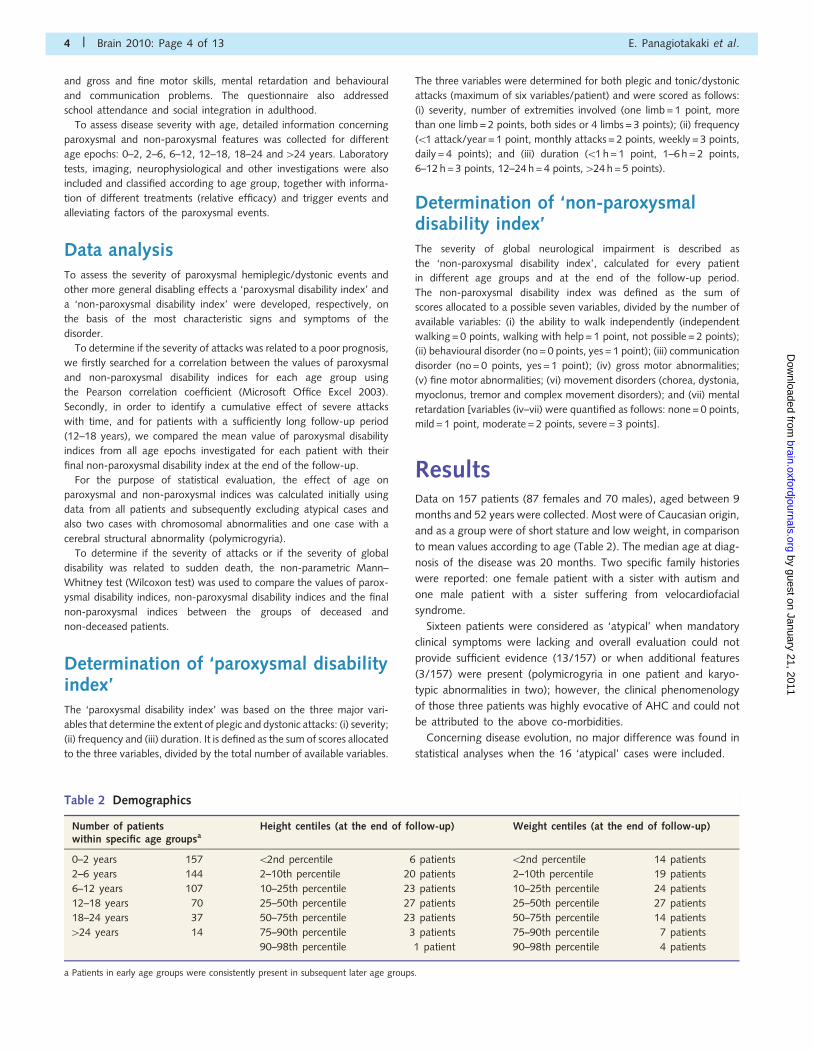
ResultsData on 157 patients (87 females and 70 males), aged between 9
months and 52 years were collected. Most were of Caucasian origin,
and as a group were of short stature and low weight, in comparison
to mean values according to age (Table 2). The median age at diag-
nosis of the disease was 20 months. Two specific family histories
were reported: one female patient with a sister with autism and
one male patient with a sister suffering from velocardiofacial
syndrome.
Sixteen patients were considered as ‘atypical’ when mandatory
clinical symptoms were lacking and overall evaluation could not
provide sufficient evidence (13/157) or when additional features
(3/157) were present (polymicrogyria in one patient and karyo-
typic abnormalities in two); however, the clinical phenomenology
of those three patients was highly evocative of AHC and could not
be attributed to the above co-morbidities.
Concerning disease evolution, no major difference was found in
statistical analyses when the 16 ‘atypical’ cases were included.
Table 2 Demographics
Number of patientswithin specific age groupsa
Height centiles (at the end of follow-up) Weight centiles (at the end of follow-up)
0–2 years 157 52nd percentile 6 patients 52nd percentile 14 patients
2–6 years 144 2–10th percentile 20 patients 2–10th percentile 19 patients
6–12 years 107 10–25th percentile 23 patients 10–25th percentile 24 patients
12–18 years 70 25–50th percentile 27 patients 25–50th percentile 27 patients
18–24 years 37 50–75th percentile 23 patients 50–75th percentile 14 patients
424 years 14 75–90th percentile 3 patients 75–90th percentile 7 patients
90–98th percentile 1 patient 90–98th percentile 4 patients
a Patients in early age groups were consistently present in subsequent later age groups.
4 | Brain 2010: Page 4 of 13 E. Panagiotakaki et al.
by guest on January 21, 2011brain.oxfordjournals.org
Dow
nloaded from

Overall clinical presentation ofalternating hemiplegia of childhood
Semiology of paroxysmal events
The median age at first attack was 3.5 months (between the first
day of life and four years) and the first hemiplegic events occurred
at a median age of 7 months (between the first month and
eight years). Attacks of hemiplegia were the first sign of the
disease in only 30% of patients (n = 47), for the remainder, hemi-
plegia followed other paroxysms with a median delay of 5 months.
Neurological episodes during the neonatal period were noted in
12.5% of patients (n = 20) and varied from unusual behaviour
and presumed seizures to clearly identified early onset dystonic,
autonomic or ocular attacks. Five children experienced their
first episode on the first day of life, manifested mainly by ocular
movements and dystonic attacks (usually limbs stiffening with a
vibratory tremor, but sometimes torticollis or opisthotonus) or an
episode of marked hypotonia.
All patients experienced hemiplegic attacks (criterion 2). The
majority of patients (152/157) also presented with other parox-
ysmal phenomena (criterion 4). Hemiplegic attacks usually
involved one upper and an ipsilateral lower limb, but could involve
either only one limb, or limbs on contralateral sides. The paralysis
was usually characterized by a flaccid weakness without pyramidal
signs. During attacks the face was usually spared, however, facial
hemiparesis did occur with mouth deviation and slurred speech.
Two children described an inability to speak only when hemipar-
esis was on the right side and continued to speak normally when
paralyzed on the left. Eighty-eight percent (138/157) of patients
also experienced tonic/dystonic attacks involving one limb or two
or more limbs on ipsilateral or contralateral sides of the body and
occurring alone or mixed with hemiplegic episodes. Dystonic at-
tacks could also involve the tongue causing difficulties in breathing
and swallowing. Some children appeared cyanotic and experi-
enced a feeling of suffocation during dystonic attacks; these epi-
sodes were reported to be particularly stressful.
During bilateral plegic attacks (86.5% of patients), children
were unable to swallow and experienced excessive salivation and
drooling. Those with language already acquired were unable to
speak during these episodes and most patients were unable to
eat and at best could only accept crushed foods. During most of
these attacks, patients were conscious, but some were somnolent
and appeared lethargic. During bilateral attacks, autonomic dis-
turbances including cardiorespiratory problems were very pro-
nounced and comprised bradycardia, stridor, bronchospasm,
hyperventilation, noisy breathing, gasping for breath or apnoeic
spells. In some cases, intestinal motility was accelerated.
Autonomic phenomena (during bilateral, other attacks or alone)
were present in 65% of patients (102/157) and also included a
reddening or pallor of the face, fever, tachycardia or bradycardia
and mydriasis. Apnoeic spells sometimes required a monitoring
device at home, and in very severe cases, intubation and mech-
anical ventilation were performed.
Paroxysmal abnormal ocular movements were present in 91%
of patients (143/157) and included unilateral or bilateral and hori-
zontal, vertical or rotatory nystagmus, as well as tonic upgaze or
tonic lateral deviation of the eyes. Ocular movements sometimes
involved convergent strabismus or loss of convergence. In one
child, one eye was directed upwards and the other downwards
during a paroxysmal episode. Eye movements were predominantly
homolateral to the side of paralysis (if they presented concomi-
tantly with attacks of hemiplegia).
Headaches were reported by 58% (53/91) of patients, in whom
the presence or absence of headache could be ascertained; exclu-
sion was made of patients younger than 6 years old (50 patients)
and of those with mental retardation or communication problems
who were unable to answer definitively (16 patients). Headache
was either in the form of classic isolated migraine (16%; 15/91) or
in the form of migraine or headache with hemiplegia. Localized
pain in affected limbs during attacks was sometimes severe to the
point that, in one child, regular morphine administration was
required.
Epileptic seizures were experienced by 53% of patients (83/157),
at least once in their lifetime. Epileptic seizures tended to be mostly
focal or focal with secondary generalization (in 45–67%, according
to age), although three out of the four patients over 24 years of
age who had epilepsy, presented with generalized seizures.
Overall, seizures were not very frequent and occurred one to
six times a year for 52–75% of patients taking antiepileptic medica-
tion (percentages varied according to age group). Epilepsy of
patients with AHC in our study was therefore relatively well
controlled with medication. Five children presented unexplained
episodes of explosive, violent laughter accompanied by limb move-
ments, terror and ocular movements or mydriasis. In most of these
children these episodes were interpreted as epileptic seizures and in
one patient they decreased after vagus nerve stimulation
implantation.
Premonitory signs or aura before paroxysmal episodes were
reported by 41% of patients (64/157). Some of these patients
exhibited a different behavioural pattern and appeared irritable.
Others reported a sensation of pinpricks or discomfort of the
hands or feet that later spread to adjacent parts of the body in
an ascending or descending manner, with a progression of
paralysis. One child described a sore throat before attacks,
concomitant with a strange sensation in the hand.
The inhibitory effect of sleep on attacks was reported for
the majority of patients (83%; 130/157). For 28 patients this
information could not be confirmed with certainty. This was
defined as loss of abnormal posture, the ability to move within
the limit of what could be observed and uniquely normal motor
function for a short period of 10–20 min on awakening. Relaxation
in a calm environment (reassurance, music, massage and averting
attention) was reported to have a beneficial effect on attacks. The
number of attacks increased in adolescent/adult female patients
during menstruation.
Long-term neurological impairmentAt walking age (150/157 patients older than 2 years), gait was
considered to be unsteady in 84% (126/150) of patients. Of the
total number of 157 patients, 113 (72%) had long-term chorea
and/or dystonia, 117 (75%) hypotonia and 144 (92%) develop-
mental delay or mental retardation: 29 (18%) mild, 78 (50%)
Evolution of alternating hemiplegia Brain 2010: Page 5 of 13 | 5
by guest on January 21, 2011brain.oxfordjournals.org
Dow
nloaded from

moderate, 37 (23.5%) severe. Nine (6%) exhibited no retardation
and for four (2.5%) there was insufficient information. The impact
of paroxysmal events on school attendance and out-of-home
activities was severe with only 34 patients attending for more
than 90% of the expected time, 56 patients for 50–90% of
time and 13 patients for 550% of time. For the remaining,
13 patients were of preschool age, eight were not following
school due to very severe handicap, and no information was avail-
able for 33 patients. During adulthood, from a total of 37 adult
patients, only one patient was working independently in employ-
ment, 13 patients were working in an assisted environment,
20 patients had never worked and for three patients there was
no available information.
Evolution of clinical presentation ofalternating hemiplegia of childhood inpatientsData during the first 2 years was available for all 157 patients.
Further data corresponding to 2–6 years were available for 144
patients, 6–12 years for 107 patients, 12–18 years for 70 patients,
18–24 years for 37 patients and 424 years for 14 patients (Table
2). Clinical presentation at different age groups was analysed for
all 157 patients and was similar to that of the subset of 14 pa-
tients, each with a follow-up period of 424 years (Figs 1 and 2).
Paroxysmal eventsDuring the initial report (0–2 years) attacks of hemiplegia occurred
in at least 93% (146/157) of patients. Prevalence of plegic attacks
moderately decreased with age and by adulthood. Tonic attacks
occurred initially in 74% (116/157) of patients and their preva-
lence remained relatively stable. During adulthood all patients con-
tinued to experience either plegic and tonic or only plegic attacks
and only one patient experienced only tonic attacks. Episodes
of abnormal ocular movements, characteristic of the disease in
infancy and early childhood were present in 86% (135/157) of
patients at 0–2 years, but were decreased to about a third of
patients in the older age groups (18–24 and 424 years). Events
of autonomic dysfunction were present in 49% (77/157) of
patients at 0–2 years and their prevalence remained constant.
The prevalence of epilepsy was relatively stable and varied
between approximately one-third to less than half of patients in
different age groups, with a peak frequency in adolescence.
Global neurological impairmentIn this cohort all 157 patients presented neurological abnormalities
in addition to paroxysmal events (they all fulfilled criterion 6). At
0–2 years, 84% (132/157) of patients experienced gross motor
delay and gross motor skill abnormalities that remained constant
with time and present in at least three-quarters of adult patients.
Figure 1 Paroxysmal features of AHC at different ages in (A) all patients and (B) a subset of 14 patients each with at least 24 years
follow-up period.
6 | Brain 2010: Page 6 of 13 E. Panagiotakaki et al.
by guest on January 21, 2011brain.oxfordjournals.org
Dow
nloaded from

Only 27% (41/157) of patients were able to walk without help
at the age of 2 years. Likewise, 79% (124/157) of patients experi-
enced fine motor delay at an early age. Fine motor skill abnorm-
alities increased with age and were present in the vast majority of
adult patients. This was also the case with chorea and dystonia.
Hypotonia was present early in 76% (119/157) of patients, ap-
peared to decrease with time and was present in only one-third of
adult patients.
Effect of time on the paroxysmal andnon-paroxysmal disability indicesThe paroxysmal disability index was calculated for all patients at
all time periods and the median values and standard deviations
are presented in Fig. 3A. The results demonstrate that there was
no significant difference in paroxysmal events with time, and the
extent of plegic and tonic attacks remained fairly constant
throughout the different age groups. There was no difference
when analysis was performed after exclusion of the 16 atypical
patients.
When the paroxysmal disability index was plotted against time
for each patient individually, with a follow-up period of at least
18 years, significant variability of severity of paroxysmal attacks
was observed between patients (Fig. 4A).
The effect of the non-paroxysmal disability index with time was
analysed for all patients at all time periods (Fig. 3B). Similar to that
of the paroxysmal disability index, it was not possible to identify a
pattern of improvement or regression with time, although a high
degree of individual variability was noted when the data of indi-
vidual patients with a follow-up period of at least 18 years were
plotted with time (Fig. 4B). There was no difference when analysis
was performed after exclusion of the 16 atypical patients. Among
the 37 older patients with a follow-up period of at least 18 years,
three could be considered to follow a consistently progressive
course.
Individually, a fluctuating course, characterized by alternating
periods of more and less severe attacks and global disability was
therefore a frequent feature, making any specific patterns of
progression difficult to identify. Furthermore, no correlation was
identified between the values of paroxysmal and non-paroxysmal
disability indices for the different age groups (r =�0.07, �0.18,
0.01, 0.01, �0.15 and �0.42 for each age group in ascending
order and after exclusion of the 16 aforementioned cases,
r =�0.09, �0.2, �0.06, �0.006, �0.17 and �0.4). Also, no
correlation was identified when the mean value of paroxysmal
disability indices throughout lifetime (for older patients with a
follow-up of at least 12–18 years), was compared with the final
non-paroxysmal disability index at the end of the follow-up period
Figure 2 Non-paroxysmal features of AHC at different ages in (A) all patients and (B) a subset of 14 patients each with at least 24 years
follow-up period.
Evolution of alternating hemiplegia Brain 2010: Page 7 of 13 | 7
by guest on January 21, 2011brain.oxfordjournals.org
Dow
nloaded from

(r =�0.06, and after exclusion of the 16 atypical cases
r =�0.014).
Patient deathsSeven patients in this cohort died, one of whom died during the
prospective period. One female patient died at the age of 25 years
from cardiorespiratory failure. A 12-year-old male patient died
after a very severe and prolonged state of quadriplegia and a
second 12-year-old male died during an epileptic seizure compli-
cated by cardiorespiratory arrest (the latter child was previously
hospitalized in an intensive care unit for severe respiratory prob-
lems during bilateral attacks). Two female patients died at the age
of 2 years following a very severe attack characterized as status
epilepticus, although some months before frequent plegic attacks,
with or without epileptic seizures, were experienced. The
28-year-old patient who died during the prospective part of the
study did so during a very prolonged plegic attack. During the few
months that preceded his death he was experiencing predomin-
antly bilateral and prolonged (424 h) attacks. The last patient died
at the age of 3.5 years. No information was available about
the circumstances of her death. Her principle paroxysmal events
were bilateral plegic attacks which occurred weekly, lasting from
12–24 h, and were associated with autonomic dysfunction and
were unresponsive to treatment. She had no epileptic seizures.
To identify disease characteristics associated with a risk of death,
we compared the mean paroxysmal disability index between the
groups of deceased and non-deceased patients. No significant
difference was observed (P = 0.6). However, when either the
mean non-paroxysmal disability index or final non-paroxysmal dis-
ability index was compared between the groups of deceased and
non-deceased patients, both were significantly higher in the group
of deceased patients (P = 0.05, 0.01, respectively; Fig. 5).
Laboratory investigationsThe registry included the results of extensive investigations
performed at various stages of the disorder, however, none were
characteristic or informative and as such, are not reported in detail.
Magnetic resonance imaging abnormalities such as non-specific
cerebral atrophy, discrete vermian atrophy, hippocampal sclerosis,
cortical dysplasia and syringomyelia were reported in isolated
cases. One patient presented extensive bilateral polymicrogyria
with typical AHC clinical presentation. Characteristic hemiplegic
events, provoked by excitement during clinical examination with
no electroencephalographic modification, were reported (E.P.,
A.A.). The patient was included in the cohort on the basis that
the clinical picture was not explained by cerebral imaging.
Two patients presented chromosomal abnormalities; one female
demonstrated a 47, XXX karyotype and one male a translocation
(t11,22) (q23;11.2), thus far unreported in patients with AHC (the
mother and grandmother of the boy had the same translocation
and were in good health). Both patients had a clinically typical
presentation of AHC and were also included in the cohort.
Screening of the ATP1A2 gene was performed in 20 patients,
the CACNA1A gene in nine patients and the SCN1A gene in three
patients, however no mutations were identified for any of the
genes. Furthermore, no mutations associated with myoclonic epi-
lepsy associated with ragged red fibres (MERRF) in three patients,
or mitochondrial myopathy, encephalopathy, lactic acidosis and
stroke (MELAS) in six patients, were identified.
TreatmentThe study was not specifically designed to evaluate treatment
strategies and outcome. Patients received a number of different
treatments simultaneously, furthermore, the natural progression of
the disease is such that periods of ‘ups’ and ‘downs’, even without
changes due to treatment, are common. We can therefore only
report on the general impressions of treatment, as expressed by
the patients themselves, their families or the physician.
Nearly all patients were given flunarizine as a prophylactic agent
of non-epileptic attacks. Flunarizine was considered to be partially
effective in 76% (108/141) of patients. Antiepileptic drugs, alone
or in combination, were administered in nearly all patients for
prophylaxis of paroxysmal non-epileptic (hemiplegic, dystonic at-
tacks, etc.) as well as epileptic episodes. None of the drugs used
had a prominent effect on non-paroxysmal events. Occasionally
some reduction in frequency or severity of the paroxysmal events
was reported. Benzodiazepines (and particularly diazepam) or
some antiepileptic drugs were often used to reduce the duration
of dystonic events, occasionally with success. Agents that induce
sleep, such as melatonin, chloral hydrate and niaprazine, were also
used during acute episodes. A minority of patients also tried other
agents like neuroleptics, beta-blockers, L-dopa, antihistamines,
Figure 3 Median disability indices of all patients as a function
of time. (A) Median values of paroxysmal disability index.
(B) Median values of non-paroxysmal disability index. Bars
depict standard deviation (1 or 2 SD).
8 | Brain 2010: Page 8 of 13 E. Panagiotakaki et al.
by guest on January 21, 2011brain.oxfordjournals.org
Dow
nloaded from

selective serotonin reuptake inhibitors and others, without clinically
meaningful effect.
Two children were misdiagnosed with opsoclonus-myoclonus
syndrome at an early stage and were treated with corticosteroids
and immunoglobulins. For one child there was no improvement,
however a marked improvement was observed for the other child,
especially with steroid treatment.
DiscussionIn this predominantly retrospective study, we investigated the clin-
ical features of AHC in a cohort of 157 patients, which included
37 adults. Patients were from nine different European countries,
within the ENRAH project, and to our knowledge this is the most
extensive cohort of patients with AHC reported. Our results firstly
emphasize the significant variability of the disease course between
individuals, and secondly, indicate no general pattern of
progression, supporting the notion that AHC is a non-progressive
or non-degenerative disorder.
General description of the disease is consistent with former studies.
We identified an age of onset of between 0 and 50 months
(median 3.5 months) in agreement with previous reports
(Bourgeois et al., 1995; Sakuragawa, 1995; Mikati et al.,
2000). Hemiplegic attacks were the first sign in only 30% of
cases (similar to the series of Mikati et al., 2000). Plegic attacks
(hemiplegic or bilateral plegic events) occurred in all patients and
were the main paroxysmal event of the disease. The second
most common phenomena were abnormal ocular movements
(91%) and tonic attacks (88%). Some authors (Bourgeois
et al., 1993; Sweney et al., 2009) observed a similar high inci-
dence for both the latter phenomena, although lower incidences
of 65 and 60%, respectively, have also been observed (Mikati
et al., 2000). The prevalence of epileptic seizures varied between
28 and 47% depending on the age group (Fig. 1) and 53% of
patients experienced epileptic seizures at least once in their lives,
Figure 4 Paroxysmal (A) and non-paroxysmal (B) disability index values as a function of time for individual patients with available clinical
information up to adulthood (at least 18 years of age, n = 37).
Evolution of alternating hemiplegia Brain 2010: Page 9 of 13 | 9
by guest on January 21, 2011brain.oxfordjournals.org
Dow
nloaded from

in agreement with previous studies (Sakuragawa, 1995), although
other authors have described a lower prevalence of epilepsy
(Mikati et al., 2000; Sweney et al., 2009). Mental retardation was
present in at least 92% of patients (of which 50% were moderately
retarded) confirming previous reports (Bourgeois et al., 1995;
Sakuragawa, 1995; Mikati et al., 2000; Sweney et al., 2009).
Evolution of paroxysmal eventsIt has been reported that paroxysmal nystagmus disappears in
all cases before the age of 10 years (Bourgeois et al., 1995) and
earlier reports indicated that tonic attacks tend to decrease in fre-
quency and severity or even disappear with time after 5–7 years
(Bourgeois et al., 1993). Other authors have reported similar
results (Aicardi, 1987; Siemes and Cordes, 1993; Silver and
Andermann, 1993; Mikati et al., 2000). The data of patients
reported here indicate that the occurrence of abnormal ocular move-
ments diminished in adulthood, but that these movements did not
completely disappear in all patients. Occurrence of tonic attacks
seemed to remain constant when analysis was performed for
the whole patient group, however, there was significant individual
variability. The same was true for hemiplegic phenomena.
Evolution of global neurologicalimpairmentNeurological disability is present in all patients with AHC and a
normal neurological examination should make one reconsider the
diagnosis of AHC. For patients described here, gross motor
abnormalities were reported early (Fig. 2) and were constantly
present when the different age epochs were examined. Early in
life, motor milestones were achieved with a variable delay. The
majority of patients had an ataxic gait and they rarely acquired
independent walking before 2 years of age. The prevalence of
gross motor skill abnormalities was constant and was still present
in at least three-quarters of patients in adult life.
Abnormalities of fine motor skills were present in the vast
majority of patients and increased (or became more evident)
with age, as previously reported (Bourgeois et al., 1993).
Abnormal movements like chorea and dystonia were present
with frequencies similar to those in previous reports (Bourgeois
et al., 1993, 1995; Sakuragawa, 1995; Mikati et al., 2000). The
majority of patients (91.5%) presented some kind of abnormal
movements (chorea, dystonia, myoclonus or tremor) in various
combinations and degrees. Hypotonia generally improved with
time and was present in only a third of adult patients.
Patients often had difficulties in acquiring speech and presented
with attention deficit disorder or other behavioural problems as
well as neuropsychological deficits that have previously been
reported (Shafer et al., 2005).
When the non-paroxysmal index was calculated on the basis of
behavioural and communicative disability and mental retardation
alone, in the absence of all motor disabilities; i.e. the ‘cognitive
severity index’, no significant difference was identified with this
index with time. However, an increase in behavioural troubles was
observed for a subset of patients.
Patient deathsPrevious studies have reported unexplained sudden death of
patients with AHC, however, due to the rarity of such cases,
events have not been described in any detail. In our study,
4% (7/157) of patients died suddenly, usually associated with
very severe attacks or seizures. For at least two cases, sudden
death was associated with a severe and prolonged plegic episode
and three other patients died during very prolonged episodes
combining plegic phenomena with what was characterized as
status epilepticus. During these episodes, patients presented a
cardiorespiratory failure and died as a result of a cardiac arrest.
Whereas the severity of plegic/dystonic attacks was not
increased in the group of deceased patients, the mean and final
non-paroxysmal disability indices were significantly greater in
this group. Moreover, the association of particularly severe
plegic attacks or seizures at the moment of death suggests that
autonomic disturbances are a precipitating factor of sudden death,
reminiscent of sudden death associated with epilepsy. Accordingly,
this would suggest the presence of factors, as yet unknown, that
may influence susceptibility of sudden death; acquired or genetic
factors that determine a worse neurological outcome and a
shorter life span in some patients, independent of the burden of
paroxysmal events.
Overall outcomeOur results suggest, in agreement with previous reports (Bourgeois
et al., 1993; Aicardi et al., 1995b; Mikati et al., 2000), that neuro-
logical signs of the disease become more apparent after the first
years of life and overall, remain more or less fixed. This is in
contrast to other reports (Nevsımalova et al., 1994; Nishiki
et al., 1994; Gordon, 1995) that suggest a progressive neurologic-
al deficit. As shown in Figs 3 and 4, for the majority of patients
there were no identifiable groups that differ in the severity of
attacks or progression of general disability (improvement or
regression). A decrease in variation was observed into adulthood
(patients tended to be more ‘similar’ with age), although this dif-
ference may merely reflect the lower patient numbers. However, a
significant degree of individual variability was observed and each
patient exhibited his/her own disease course (Fig. 4).
Figure 5 Comparisons of mean paroxysmal and
non-paroxysmal index values and final non-paroxysmal index
values between deceased and non-deceased patients.
10 | Brain 2010: Page 10 of 13 E. Panagiotakaki et al.
by guest on January 21, 2011brain.oxfordjournals.org
Dow
nloaded from

Our results confirm that paroxysmal phenomena persist into
adulthood. However, it remained unclear as to whether there
was an eventual correlation between the severity of paroxysmal
events and the evolution of neurological disability. Amelioration
by different drugs can offer symptomatic improvement without
necessarily altering the evolution in terms of final disability
score, supporting the concept that the severity of paroxysmal
events may not be directly related to an eventual severe outcome.
In this study, we were unable to identify a correlation between
paroxysmal and non-paroxysmal disability indices, further sup-
porting the lack of evidence for a cause-and-effect relationship
between the paroxysmal attacks and eventual overall neurological
disability. The existence of more severe attacks and greater
disability in some patients could simply be the expression of a
more severe form of the disease due to differences in genetic
and/or environmental factors.
In previous studies (Aicardi et al., 1995a), as is the case for
some patients in the present study, parents reported a loss of
skills following very severe and prolonged, particularly bilateral,
plegic episodes. In general, this loss of skills (in our study and
previous studies) is not permanent, but recovery can last many
months. However, patients usually lack a proper evaluation of
skills, before and after recovery, and information is mainly based
on the family’s estimation of a child’s neurological state.
Moreover, in the present cohort some families paradoxically also
reported the opposite situation, that is, an overall improvement
and acquisition of new skills after a severe episode.
As is the case with other reports, recognition of early disease
progression (in the first 2 years) was not possible since early time
periods were based purely on a retrospective description, which
were, furthermore, pooled from patients of different ages. A
longitudinal study would be required to further overcome the
limitations encountered in this study.
Aetiological factorsAt present there is no biological marker specific for the disease.
As a result patients generally undergo extensive investigations
such as imaging, neurophysiological studies, biochemical and
genetic tests, which are usually inconclusive. Some of the patients
reported here had non-specific structural abnormalities based
on magnetic resonance imaging scanning and an extensive bilat-
eral polymicrogyria was identified in only one case. Previous
reports (Sakuragawa, 1992; Saito et al., 1998) have also
mentioned structural abnormalities of unknown significance, such
as cerebellar atrophy in four of 23 Japanese patients. Genetic
screening for mutations in genes that encode for ion channel
transporters (ATP1A2, CACNA1A, SCN1A) was negative, incon-
sistent with previous reports that identified one mutation in
the ATP1A2 gene (Kanavakis et al., 2003; Bassi et al., 2004;
Swoboda et al., 2004), but in agreement with other studies
that did not identify mutations in the genes ATP1A2, CACNA1A
and SLC1A3 associated with familial hemiplegic migraine and
episodic ataxia (Haan et al., 2000; Kors et al., 2004; De Vries
et al., 2006).
TreatmentA number of different drug treatments for AHC have been shown
to be relatively unsuccessful in the past, with the exception of
flunarizine (Casaer and Azou, 1984; Casaer, 1987; Silver and
Andermann, 1993) with which open trials indicated a positive
response for at least 75% of patients studied (Sakuragawa,
1995; Mikati et al., 2000). We have obtained similar results
with flunarizine, demonstrating at least partial efficacy in �76%
of patients. Topiramate has recently been proposed as an alterna-
tive to flunarizine (Di Rosa et al., 2006; Jiang et al., 2006). In this
cohort, the drug was reported to be only moderately efficacious
in six out of 20 patients. Drug efficacy is difficult to evaluate as
the disease often exhibits a ‘roller-coaster’ course, with periods of
aggravation and improvement. Moreover, the majority of patients
often receive multiple drugs and it is difficult to attribute a positive
result to any particular medication. There is a need for prospective
double-blinded studies, although this may be limited by small
patient numbers.
ConclusionWhen all patients were examined collectively, the reported severity
of clinical presentation and neurological disability remained constant
with age, with the exception of abnormal ocular movements and
hypotonia that appeared to regress, but not disappear, into adult-
hood. When analysed individually, however, the reported clinical
presentation was sometimes highly variable with age.
For 7/157 patients, sudden death occurred, sometimes asso-
ciated with severe plegic attacks and epileptic seizures. Patients
who suffered sudden death, however, did not generally experi-
ence more severe plegic/dystonic attacks than other patients, but
severity of global neurological impairment was significantly higher
in the group of deceased patients. We speculate that increased
autonomic dysfunction is a precipitating factor for sudden death.
In summary, on the basis of the largest cohort of patients with
AHC to date, we present data suggesting that the natural history
of the disorder is variable in severity and rather unpredictable.
However, when patients were analysed as a group, we did not
find strong evidence suggesting a steady progressive and degen-
erative course. Although much attention was given to accuracy of
data collection, definitive conclusions are nonetheless subject to
the partly retrospective nature of the study. A prospective
European–US study, although very difficult to perform, may be
needed to further explore the overall evolution of the disorder.
AcknowledgementsSpecial thanks to the following doctors for giving access to
their patients’ files: France: Aicardi J, Arbues AS, Bednarek N,
Billette T, Bourgeois M, Chaigne D, Chaunu MP, Chiron C,
Cournelle AM, De StMartin A, Desguerres I, DesPortes V, Dulac O,
Dusser A, Echenne B, Evrard P, Goutieres F, Lazaro L, Motte J,
Moutard ML, Nassogne MC, Neau JP, Pedespan JM, Penniello
Valette MJ, Perelman S, Ponsot G, Pouplard F, Quillerou D,
Evolution of alternating hemiplegia Brain 2010: Page 11 of 13 | 11
by guest on January 21, 2011brain.oxfordjournals.org
Dow
nloaded from

Rivier F, Roelens P, Vallee L, Vanhulle C; Italy: Santucci M,
Tedesi B, Pantaleoni C, Zucca C, Pelliccia A, Piccinelli, Cardona F,
Manfredi M, Galeone D, Laverda A, Dalla Bernardina B,
Gambardella A, Vigevano F, Cotrufo R, Antonelli C, Granata T,
Perini A, Di Rosa G, Gaggero R, Veneselli E, Giacanelli M;
Germany, Austria: Altmann B, Brand J, Fischer M, Gildberg K,
Hallman V, Hantschel P, Heidelmann M Holthausen H, Kippert,
Korinthenberg R, Krageloh-Mann I, Kutschke G, Lauffer H,
Laun J, Niemann G, Niethammer A, Opp J, Porsch P,
Reinhardt W.Schmitt B, Schulze F, Skirl G, Steinlin M, Sterken
W Stizmann FC, Strehl H, Traue C, Weidner B; UK: Philip S,
Short D, Childs AM, Jayawanth S, Das K, Hanna M,
Wimalaratna S, Coldwell S, Cottrell A, Webb D, Gale A,
Damman D; Spain: Campos J, Eiris J, Mora MD, Narbona J,
Nieto M, Roche C, Arrabal L, Rufo M, Velazquez R, Blanco B,
Calvo MJ, Garcia Penas JJ; Czech Republic: Havlova M,
Tauberova A, Prihodova I, Kemlink D; The Netherlands:
van Nieuwenhuizen O, Coo R, Stroink H, Renier WO, Coo R,
De Coo IFM, Arts WF, Eekhof JAH. Special thanks to Mrs Tsveta
Schyns for initiating and coordinating the ENRAH for SMEs
project; to Mrs Rosaria Vavassori for the management of the
registry, to the AHC family associations from France, Italy,
Spain, Germany, UK and The Netherlands; and to the patients
and their families who participated in the Registry and supported
the project. Special thanks to Mr Oliver Gubbay for reviewing
English.
FundingThis work was supported by grant (LSSM-CT-2005-516513
ENRAH for SMEs) of the European Commission Research
Programme FP6 from April 2005 throughout June 2007.
Supplementary materialSupplementary material is available at Brain online.
ReferencesAicardi J. Alternating hemiplegia of childhood. International Pediatrics
1987; 2: 115–9.
Aicardi J, Bourgeois M, Fusco L, Vigevano F, Silver K, Andermann F.
Alternating hemiplegia of childhood: an overview. In: Andermann F,
Aicardi J, Vigevano F, editors. Alternating hemiplegia of childhood.
New York: Raven Press; 1995a. p. 207–12.Aicardi J, Bourgeois M, Goutieres F. Alternating hemiplegia of childhood:
clinical findings and diagnostic criteria. In: Andermann F, Aicardi J,
Vigevano F, editors. Alternating hemiplegia of childhood. New York:
Raven Press; 1995b. p. 3–18.
Bassi MT, Bresolin N, Tonelli A, Nazos K, Crippa F, Baschirotto C, et al. A
novel mutation in the ATP1A2 gene causes alternating hemiplegia of
childhood. J Med Genet 2004; 41: 621–8.
Bourgeois M, Aicardi J, Goutieres F. Alternating hemiplegia of childhood.
J Pediatr 1993; 122: 673–9.
Bourgeois M, Nevsımalova S, Aicardi J, Andermann F. Alternating hemi-
plegia of childhood: long-term outcome. In: Andermann F, Aicardi J,
Vigevano F, editors. Alternating hemiplegia of childhood. New York:
Raven Press; 1995. p. 49–54.
Bursztyn J, Mikaeloff Y, Kaminska A, Plouin P, Soufflet C, Dulac O, et al.
Alternating hemiplegia of childhood and oculomotor anomalies. J Fr
Ophtalmol 2000; 23: 161–4.
Casaer P. Flunarizine in alternating hemiplegia in childhood: an interna-
tional study in 12 children. Neuropediatrics 1987; 18: 191–5.Casaer P, Azou M. Flunarizine in alternating hemiplegia in childhood.
Lancet 1984; 2: 579.De Vries B, Haan J, Stam AH, Vanmolkot KR, Stroink H, Laan LA, et al.
Alternating hemiplegia of childhood: no mutations in the glutamate
transporter EAAT1. Neuropediatrics 2006; 37: 302–4.Di Rosa G, Spano M, Pustorino G, Ferrari MD, Stam AH, Sgro DL, et al.
Alternating hemiplegia of childhood successfully treated with topira-
mate: 18 months of follow-up. Neurology 2006; 66: 146.Dittrich J, Havlova M, Nevsımalova S. Paroxysmal hemipareses in
childhood. Dev Med Child Neurol 1979; 21: 800–7.
Golden GS, French JH. Basilar artery migraine in young children.
Pediatrics 1975; 56: 722–6.
Gordon N. Alternating hemiplegia of childhood. Dev Med Child Neurol
1995; 37: 464–8.
Haan J, Kors EE, Terwindt GM, Vermeulen FL, Vergouwe MN, van den
Maagdenberg AM, et al. Alternating hemiplegia of childhood: no
mutations in the familial hemiplegic migraine CACNA1A gene.
Cephalalgia 2000; 20: 696–700.
Hockaday JM. Basilar migraine in childhood. Dev Med Child Neurol
1979; 21: 455–63.
Hosking GP, Cavanagh NP, Wilson J. Alternating hemiplegia: compli-
cated migraine of infancy. Arch Dis Child 1978; 53: 656–9.
Jiang W, Chi Z, Ma L, Du B, Shang W, Guo H, et al. Topiramate: a new
agent for patients with alternating hemiplegia of childhood.
Neuropediatrics 2006; 37: 229–33.
Kanavakis E, Xaidara A, Papathanasiou-Klontza D, Papadimitriou A,
Velentza S, Youroukos S. Alternating hemiplegia of childhood: a
syndrome inherited with an autosomal dominant trait. Dev Med
Child Neurol 2003; 45: 833–6.
Kors EE, Vanmolkot KR, Haan J, Kheradmand Kia S, Stroink H, Laan LA,
et al. Alternating hemiplegia of childhood: no mutations in the second
familial hemiplegic migraine gene ATP1A2. Neuropediatrics 2004; 35:
293–6.
Krageloh I, Aicardi J. Alternating hemiplegia in infants: report of five
cases. Dev Med Child Neurol 1980; 22: 784–91.
Mikati MA, Kramer U, Zupanc ML, Shanahan RJ. Alternating hemiplegia
of childhood: clinical manifestations and long-term outcome. Pediatr
Neurol 2000; 23: 134–41.
Neville BG, Ninan M. The treatment and management of
alternating hemiplegia of childhood. Dev Med Child Neurol 2007;
49: 777–80.
Nevsımalova S, Dittrich J, Havlova M, Tauberova A, Korsova B, Hajek M,
et al. Alternating hemiplegia in childhood: a cross-sectional study.
Brain Dev 1994; 16: 189–94.
Nishiki T, Takeuchi Y, Yamazoe I, Yoshioka H, Sawada T.
Patient with unusual alternating hemiplegia. Pediatr Neurol 1994;
10: 153–6.
Saito Y, Sakuragawa N, Sasaki M, Sugai K, Hashimoto T. A case of
alternating hemiplegia of childhood with cerebellar atrophy. Pediatr
Neurol 1998; 19: 65–8.
Sakuragawa N. Alternating hemiplegia in childhood: 23 cases in Japan.
Brain Dev 1992; 14: 283–8.Sakuragawa N. Clinical findings in 23 Japanese patients with alternating
hemiplegia of childhood. In: Andermann F, Aicardi J, Vigevano F,
editors. Alternating hemiplegia of childhood. New York: Raven Press;
1995. p. 43–7.
Shafer ME, Mayfield JW, McDonald F. Alternating hemiplegia of child-
hood: a study of neuropsychological functioning. Appl Neuropsychol
2005; 12: 49–56.
12 | Brain 2010: Page 12 of 13 E. Panagiotakaki et al.
by guest on January 21, 2011brain.oxfordjournals.org
Dow
nloaded from

Siemes H, Cordes M. Single-photon emission tomography investigations
of alternating hemiplegia of childhood. Dev Med Child Neurol 1993;
35: 346–50.
Silver K, Andermann F. Alternating hemiplegia of childhood: a study of
10 patients and results of flunarizine treatment. Neurology 1993; 43:
36–41.
Sweney MT, Silver K, Gerard-Blanluet M, Pedespan JM, Renault F,
Arzimanoglou A, et al. Alternating hemiplegia of childhood: early
characteristics and evolution of a neurodevelopmental syndrome.
Pediatrics 2009; 123(3): e534–41.
Swoboda KJ, Kanavakis E, Xaidara A, Johnson JE, Leppert MF,
Schlesinger-Massart MB, et al. Alternating hemiplegia of childhood
or familial hemiplegic migraine?: a novel ATP1A2 mutation. Ann
Neurol 2004; 55: 884–7.
Verret S, Steele JC. Alternating hemiplegia of childhood: a report of eight
patients with complicated migraine beginning in infancy. Pediatrics
1971; 47: 675–80.
Appendix 1The ENRAH Consortium Arzimanoglou Alexis (France), Campistol
Jaume (Spain), Casaer Paul (Belgium), Ebinger Friedrich
(Germany), Fons Carmen (Spain), Giannotta Melania (Italy),
Gobbi Giuseppe (Italy), Kemlink David (Czech Republic), Laan
Laura (The Netherlands), Neville Brian (UK), Nevsımalova Sona
(Czech Republic), Ninan Miriam (UK), Oechsler Claudia,
Panagiotakaki Eleni (France), Spiel Georg (Austria), Bassi Maria
Teresa (Italy), Casari Giorgio (Italy), Crawley Francis (Belgium),
Martinelli Boneschi Filippo (Italy), Nicole Sophie (France),
Poncelin Dominique (France), Sange Guenter (Austria), Schyns
Tsveta (Austria), van den Maagdenberg Arn (The Netherlands),
Vavassori Rosaria (Italy), Zucca Claudio (Italy).
Evolution of alternating hemiplegia Brain 2010: Page 13 of 13 | 13
by guest on January 21, 2011brain.oxfordjournals.org
Dow
nloaded from

![[All] - Home - Springer978-3-540-69677...Albeverio, S., Merlini, D., and Tartini, R., Una breve introduzione a diffusioni su insiemi [raitali e ad alcuni essempi di sistemi dinamici](https://static.fdocuments.net/doc/165x107/5ae9baee7f8b9ad73f8c380d/all-home-springer-978-3-540-69677albeverio-s-merlini-d-and-tartini.jpg)