1 Biochemistryof...
97
Biochemistry of connective tissue - extracellular matrix Lenka Fialová Institute of Medical Biochemistry and Laboratory Diagnostics, 1st Faculty of Medicine, Charles University, Prague 1
Transcript of 1 Biochemistryof...
Biochemistry ofconnective tissue
- extracellular matrix
Lenka FialováInstitute of Medical Biochemistry andLaboratory Diagnostics, 1st Faculty ofMedicine, Charles University, Prague
1
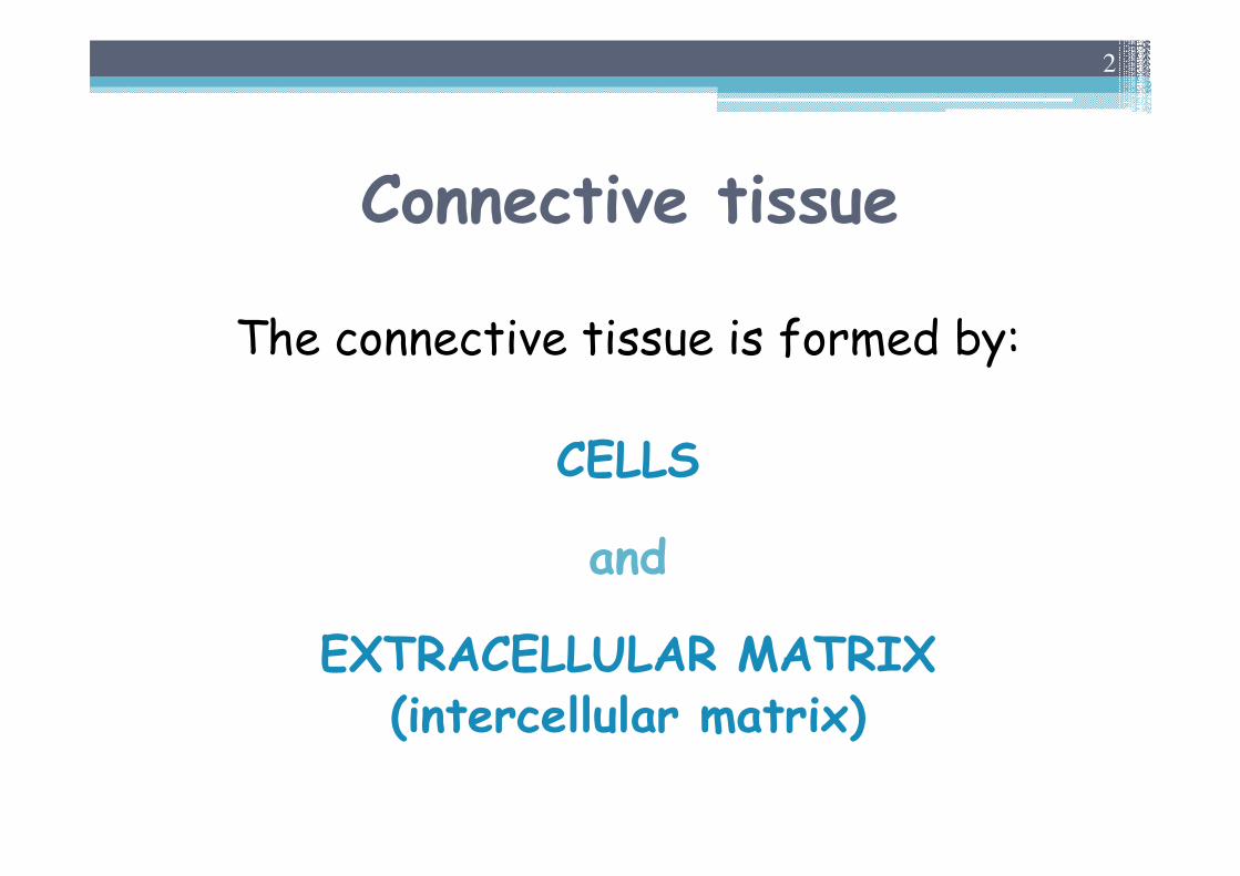
Connective tissue
The connective tissue is formed by:
CELLS
and
EXTRACELLULAR MATRIX (intercellular matrix)
2

Extracellular matrix
Function
▫ stabilisation of tissue structure ▫ regulation cell behavior� survival, development, migration, proliferation▫ membrane filtration barrier (glomerules)▫ exchange of different metabolites, ions and water▫ reparation function▫ immune processes▫ participation in inflammation
3
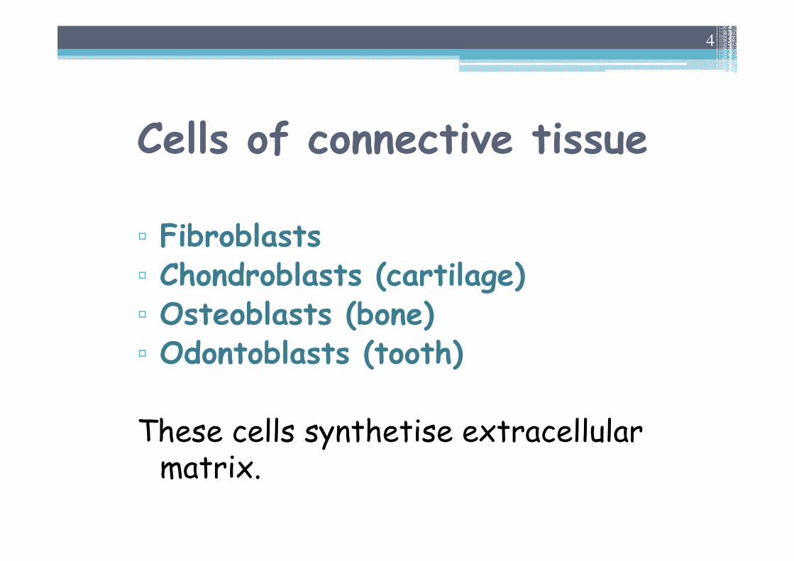
Cells of connective tissue
▫ Fibroblasts▫ Chondroblasts (cartilage)▫ Osteoblasts (bone)▫ Odontoblasts (tooth)
These cells synthetise extracellularmatrix.
4
Extracellular matrix
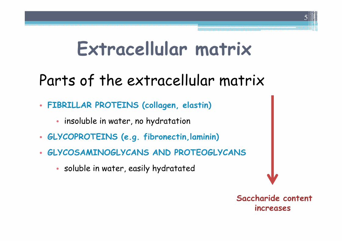
Parts of the extracellular matrix
• FIBRILLAR PROTEINS (collagen, elastin)
• insoluble in water, no hydratation
• GLYCOPROTEINS (e.g. fibronectin,laminin)
• GLYCOSAMINOGLYCANS AND PROTEOGLYCANS
• soluble in water, easily hydratated
5
Saccharide contentincreases
Extracellular matrix
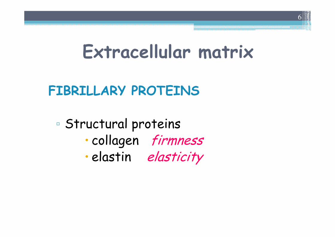
FIBRILLARY PROTEINS
▫ Structural proteins� collagen firmness� elastin elasticity
6
COLLAGENS
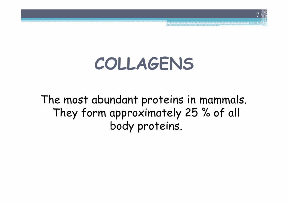
The most abundant proteins in mammals. They form approximately 25 % of all
body proteins.
7
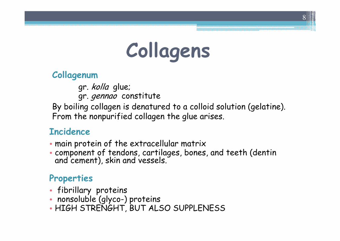
CollagensCollagenum
gr. kolla glue;gr. gennao constitute
By boiling collagen is denatured to a colloid solution (gelatine). From the nonpurified collagen the glue arises.
Incidence• main protein of the extracellular matrix• component of tendons, cartilages, bones, and teeth (dentin and cement), skin and vessels.
Properties• fibrillary proteins• nonsoluble (glyco-) proteins• HIGH STRENGHT, BUT ALSO SUPPLENESS
8
Structure of collagen
Collagens
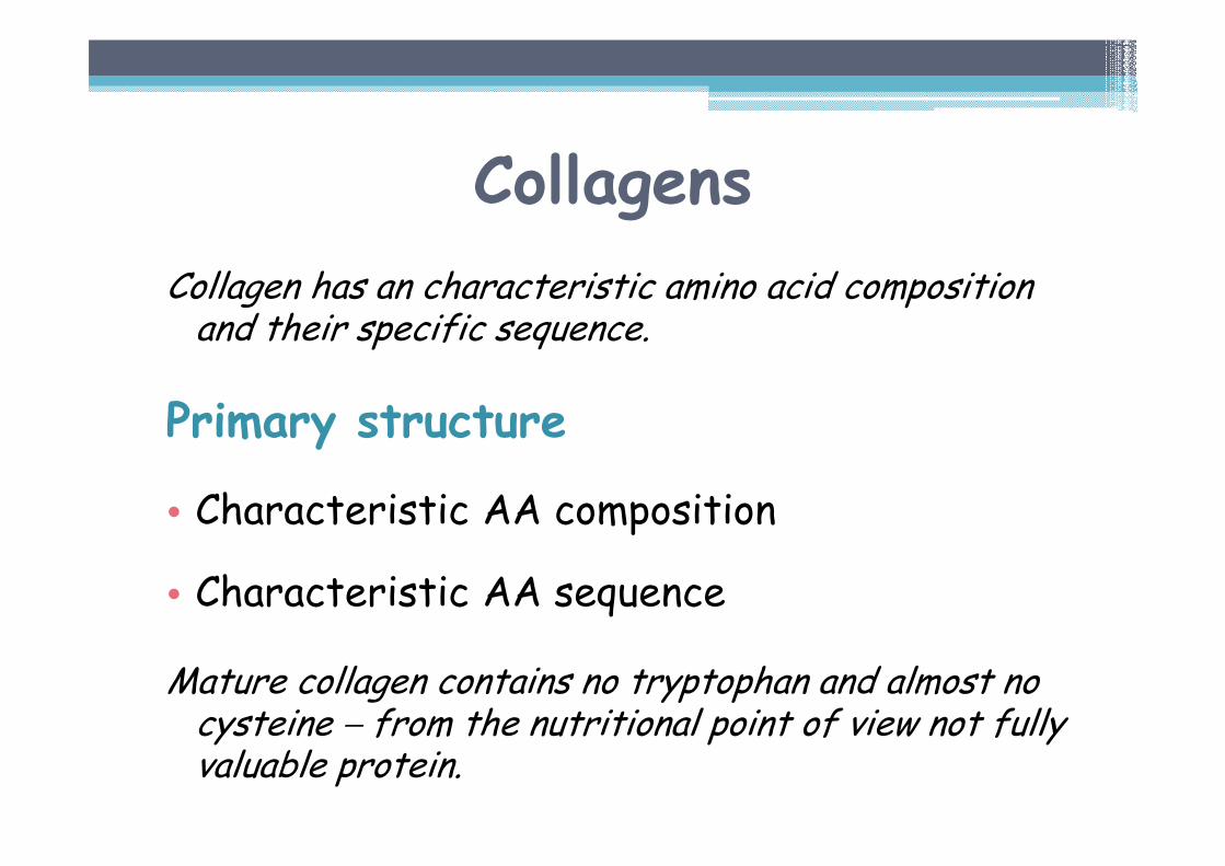
Collagen has an characteristic amino acid composition and their specific sequence.
Primary structure
• Characteristic AA composition
• Characteristic AA sequence
Mature collagen contains no tryptophan and almost no cysteine − from the nutritional point of view not fully valuable protein.
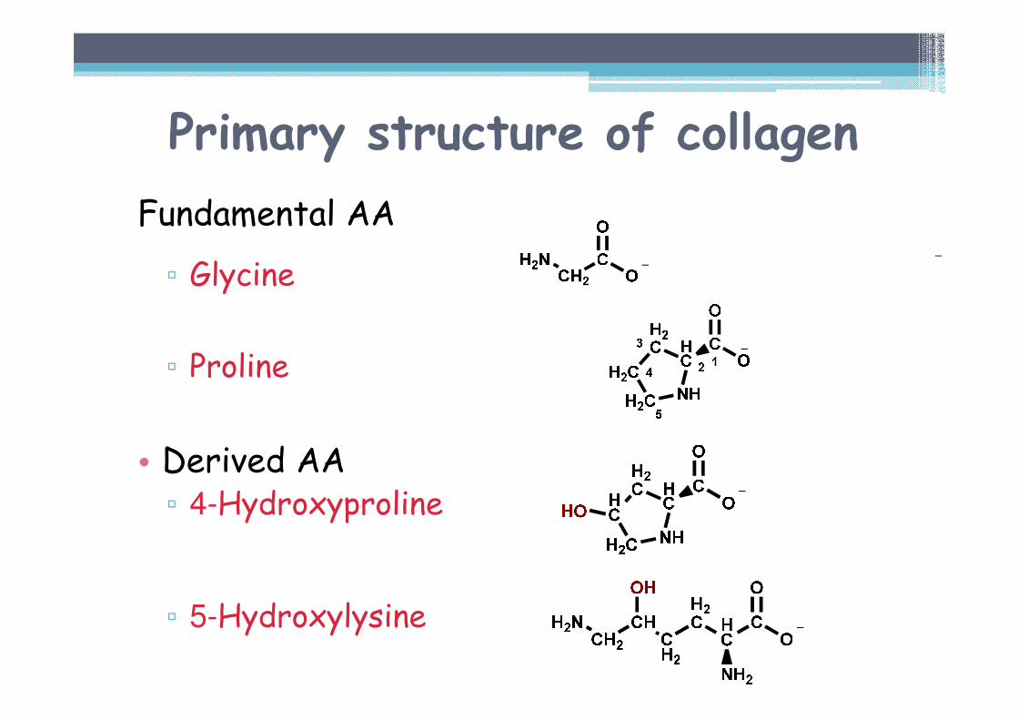
Primary structure of collagen
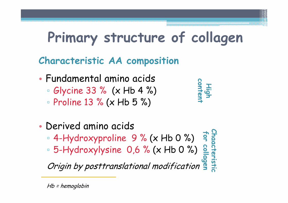
Characteristic AA composition
• Fundamental amino acids▫ Glycine 33 % (x Hb 4 %)▫ Proline 13 % (x Hb 5 %)
• Derived amino acids▫ 4-Hydroxyproline 9 % (x Hb 0 %)▫ 5-Hydroxylysine 0,6 % (x Hb 0 %)
Origin by posttranslational modification
Hb = hemoglobin
Chaacte
risticfor
collagen
High
content
Primary structure of collagen
Fundamental AA
▫ Glycine
▫ Proline
• Derived AA▫ 4-Hydroxyproline
▫ 5-Hydroxylysine
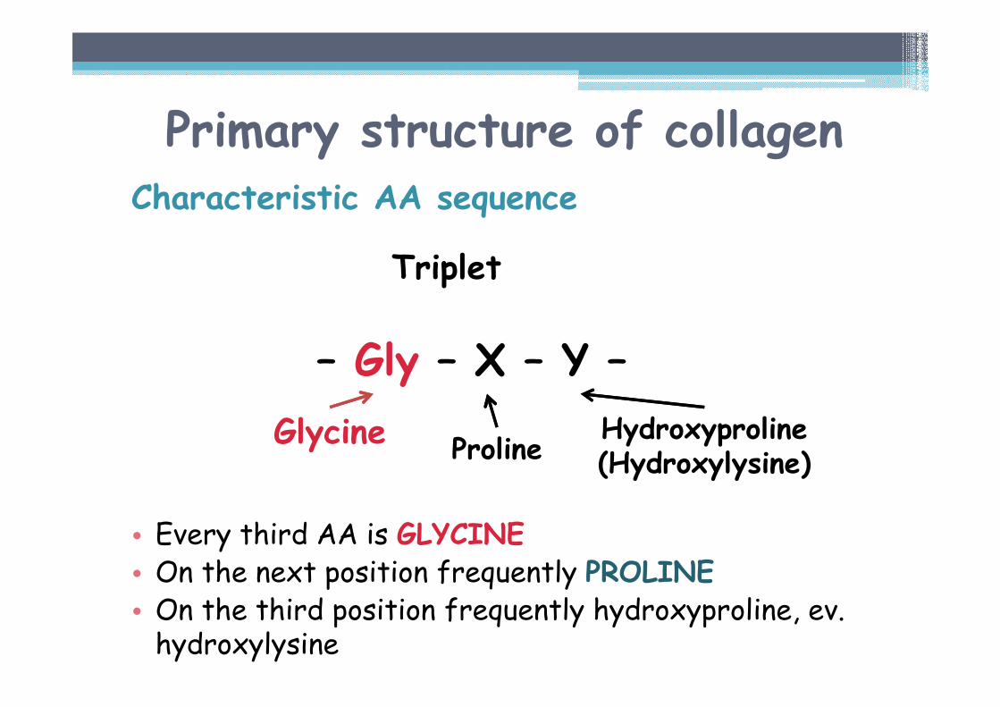
Primary structure of collagenCharacteristic AA sequence
Triplet
– Gly – X – Y –
• Every third AA is GLYCINE• On the next position frequently PROLINE• On the third position frequently hydroxyproline, ev. hydroxylysine
ProlineHydroxyproline(Hydroxylysine)
Glycine
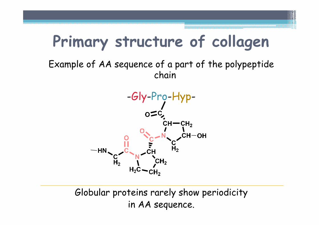
Primary structure of collagenExample of AA sequence of a part of the polypeptide
chain
-Gly-Pro-Hyp-
Globular proteins rarely show periodicityin AA sequence.
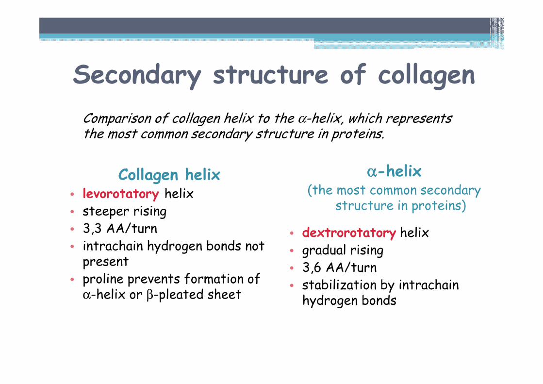
Secondary structure of collagen
Collagen helix• levorotatory helix• steeper rising • 3,3 AA/turn• intrachain hydrogen bonds not present
• proline prevents formation of α-helix or β-pleated sheet
αααα-helix(the most common secondary
structure in proteins)
• dextrorotatory helix• gradual rising • 3,6 AA/turn• stabilization by intrachainhydrogen bonds
Comparison of collagen helix to the α-helix, which represents the most common secondary structure in proteins.
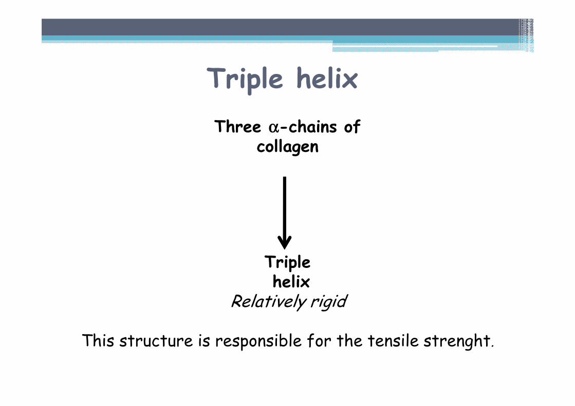
Triple helix
Three αααα-chains ofcollagen
Triplehelix
Relatively rigid
This structure is responsible for the tensile strenght.
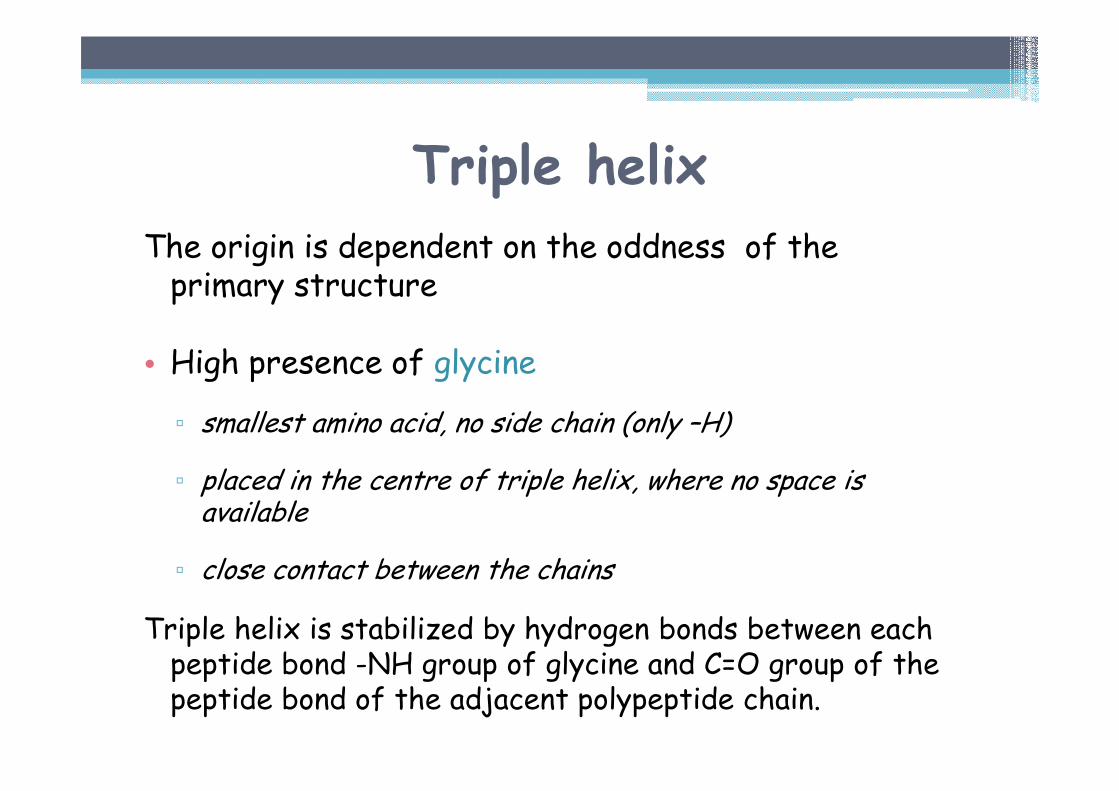
Triple helix
The origin is dependent on the oddness of the primary structure
• High presence of glycine
▫ smallest amino acid, no side chain (only –H)
▫ placed in the centre of triple helix, where no space is available
▫ close contact between the chains
Triple helix is stabilized by hydrogen bonds between each peptide bond -NH group of glycine and C=O group of the peptide bond of the adjacent polypeptide chain.
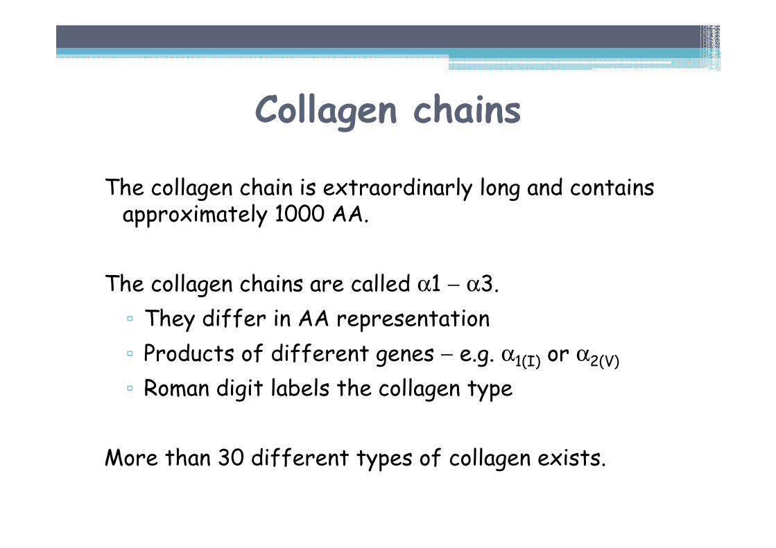
Collagen chains
The collagen chain is extraordinarly long and contains approximately 1000 AA.
The collagen chains are called α1 − α3.
▫ They differ in AA representation
▫ Products of different genes − e.g. α1(I) or α2(V)
▫ Roman digit labels the collagen type
More than 30 different types of collagen exists.
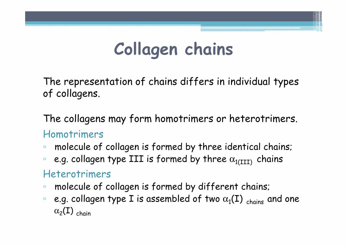
The representation of chains differs in individual types of collagens.
The collagens may form homotrimers or heterotrimers.
Homotrimers▫ molecule of collagen is formed by three identical chains;▫ e.g. collagen type III is formed by three α1(III) chains
Heterotrimers▫ molecule of collagen is formed by different chains;▫ e.g. collagen type I is assembled of two α1(I) chains and one
α2(I) chain
Collagen chains
Collagen synthesis
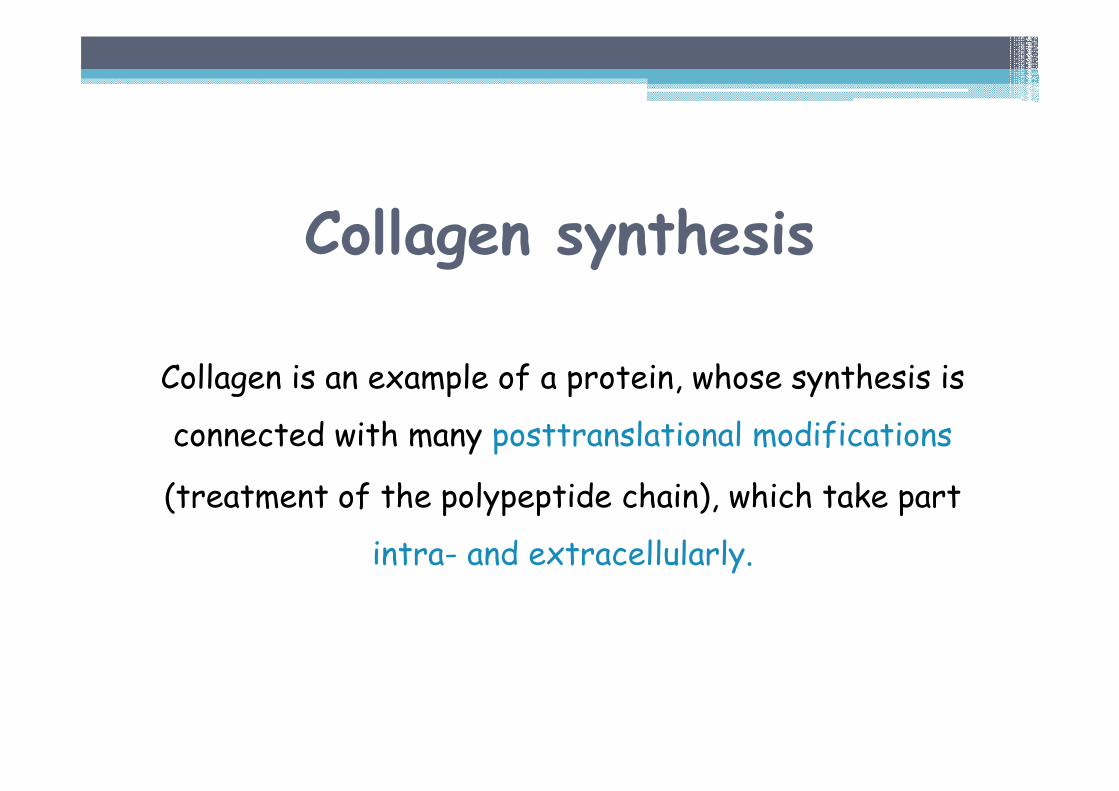
Collagen is an example of a protein, whose synthesis is
connected with many posttranslational modifications
(treatment of the polypeptide chain), which take part
intra- and extracellularly.
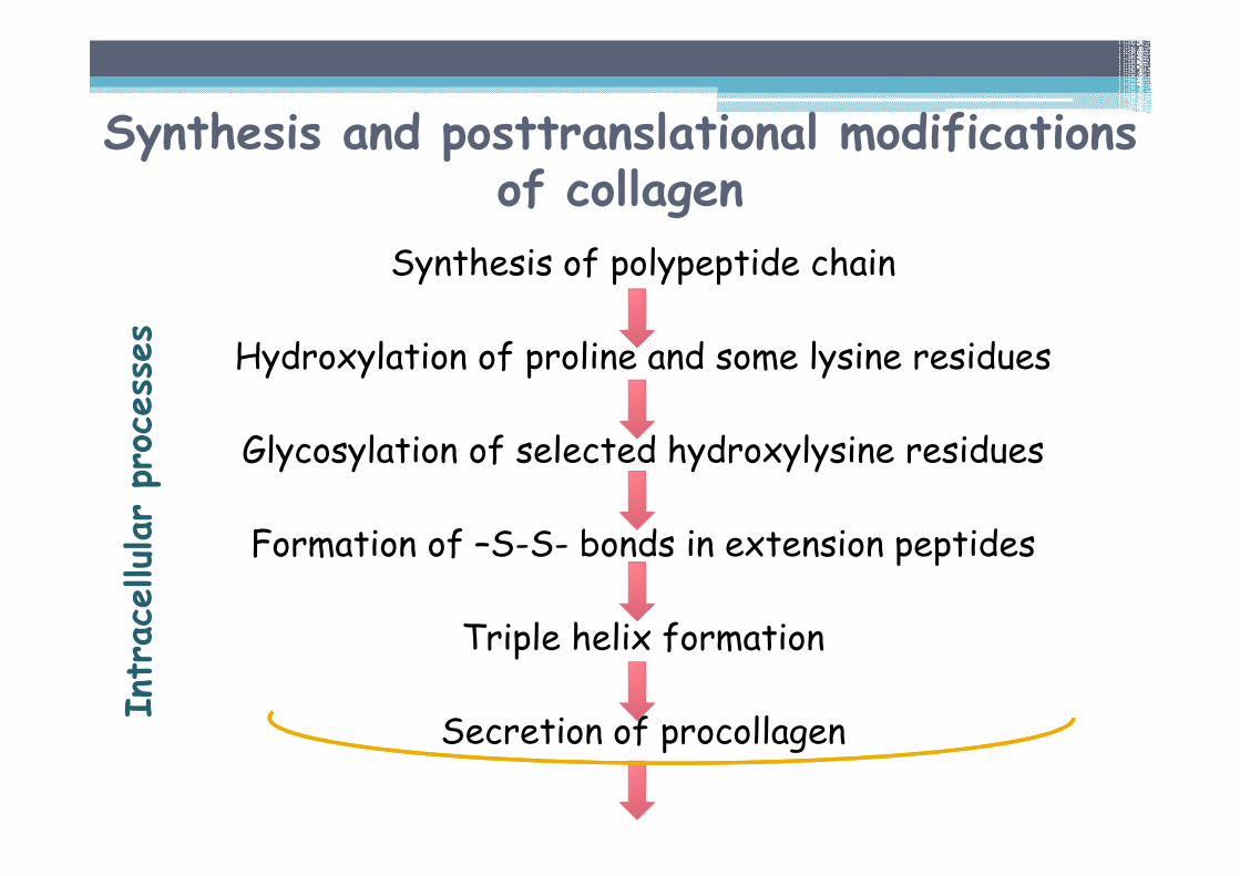
Synthesis and posttranslational modifications of collagen
Synthesis of polypeptide chain
Hydroxylation of proline and some lysine residues
Glycosylation of selected hydroxylysine residues
Formation of –S-S- bonds in extension peptides
Triple helix formation
Secretion of procollagen
Intracellularprocesses
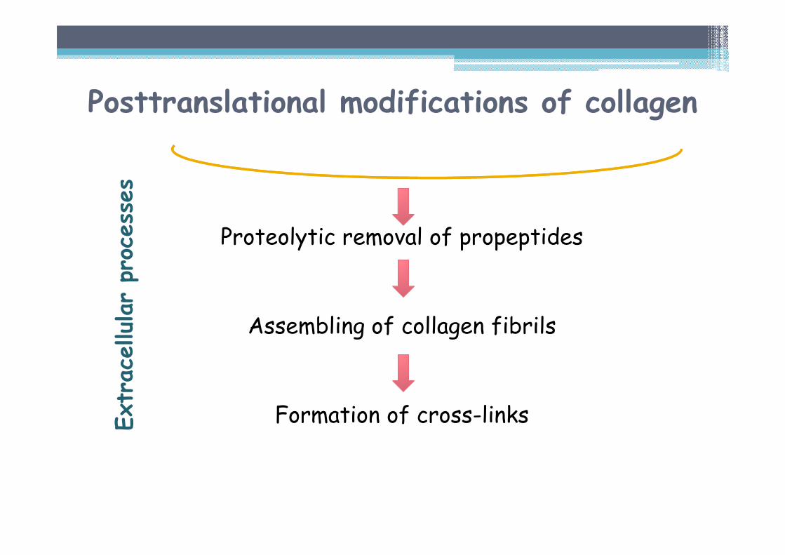
Proteolytic removal of propeptides
Assembling of collagen fibrils
Formation of cross-linksExtracellularprocesses
Posttranslational modifications of collagen
Posttranslational modifications in the course of collagen synthesis
INTRACELLULAR PROCESSES
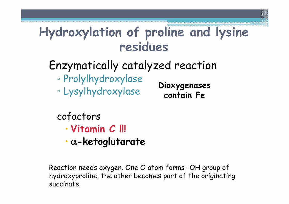
Hydroxylation of proline and lysineresidues
Enzymatically catalyzed reaction▫ Prolylhydroxylase▫ Lysylhydroxylase
cofactors� Vitamin C !!!� αααα-ketoglutarate
Reaction needs oxygen. One O atom forms -OH group of hydroxyproline, the other becomes part of the originating succinate.
Dioxygenasescontain Fe
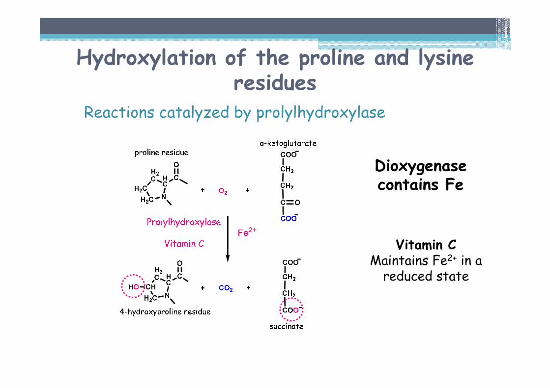
Hydroxylation of the proline and lysine residues
Reactions catalyzed by prolylhydroxylase
Vitamin CMaintains Fe2+ in a reduced state
Dioxygenasecontains Fe
Hydroxylation of proline and lysine
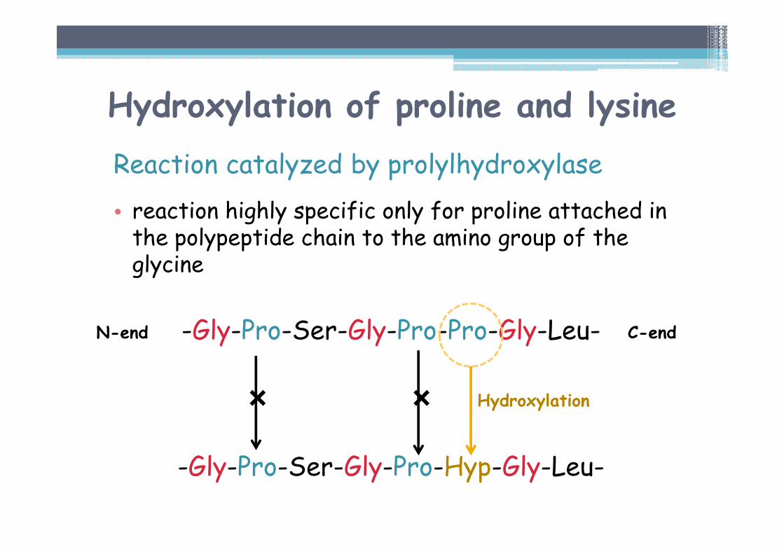
Reaction catalyzed by prolylhydroxylase
• reaction highly specific only for proline attached in the polypeptide chain to the amino group of the glycine
-Gly-Pro-Ser-Gly-Pro-Pro-Gly-Leu-
� �
-Gly-Pro-Ser-Gly-Pro-Hyp-Gly-Leu-
N-end C-end
Hydroxylation
Hydroxylation of proline and lysine
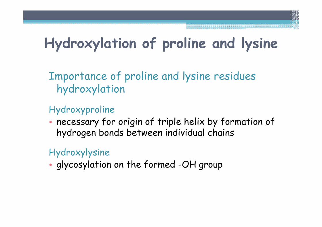
Importance of proline and lysine residues hydroxylation
Hydroxyproline• necessary for origin of triple helix by formation of hydrogen bonds between individual chains
Hydroxylysine• glycosylation on the formed -OH group
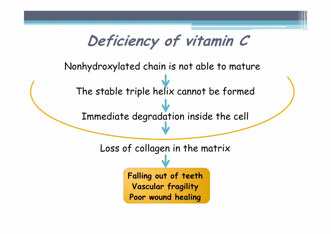
Nonhydroxylated chain is not able to mature
The stable triple helix cannot be formed
Immediate degradation inside the cell
Loss of collagen in the matrix
Falling out of teethVascular fragility Poor wound healing
Deficiency of vitamin C
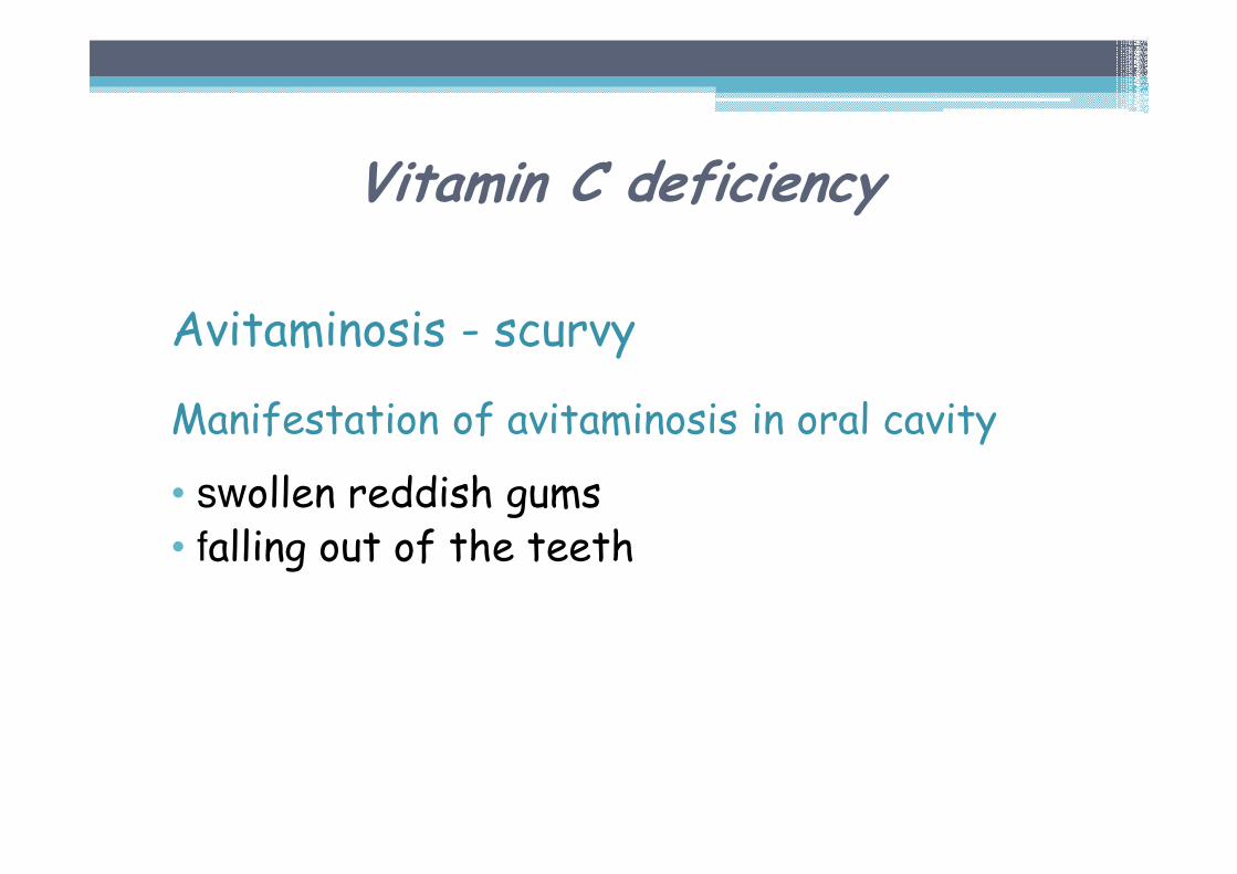
Vitamin C deficiency
Avitaminosis - scurvy
Manifestation of avitaminosis in oral cavity
• swollen reddish gums• falling out of the teeth
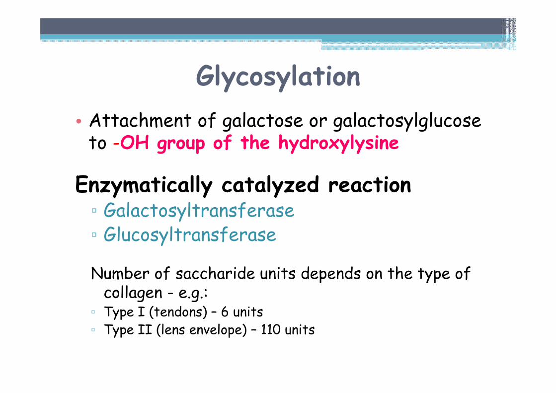
Glycosylation
• Attachment of galactose or galactosylglucoseto -OH group of the hydroxylysine
Enzymatically catalyzed reaction▫ Galactosyltransferase▫ Glucosyltransferase
Number of saccharide units depends on the type of collagen - e.g.: ▫ Type I (tendons) – 6 units▫ Type II (lens envelope) – 110 units
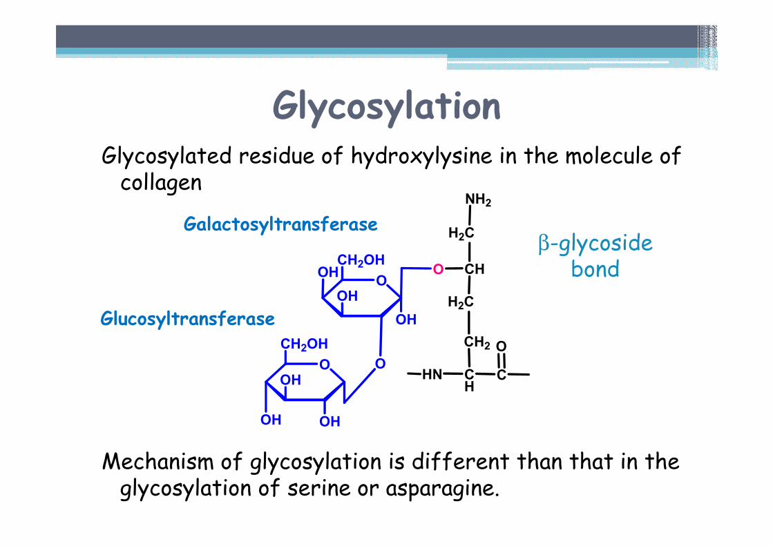
Glycosylated residue of hydroxylysine in the molecule of collagen
Mechanism of glycosylation is different than that in the glycosylation of serine or asparagine.
O
OH
OHOH
CH2OH
CHN CH
CH2
H2C
CH
H2C
NH2
O
OOHO
OH
O
CH2OH
OH
Glycosylation
Galactosyltransferase
Glucosyltransferase
β-glycosidebond
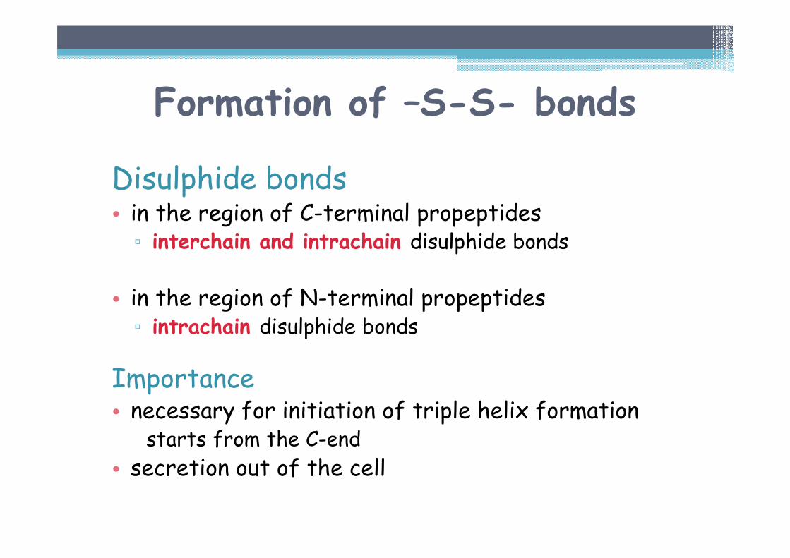
Formation of –S-S- bonds
Disulphide bonds• in the region of C-terminal propeptides▫ interchain and intrachain disulphide bonds
• in the region of N-terminal propeptides▫ intrachain disulphide bonds
Importance• necessary for initiation of triple helix formation
starts from the C-end
• secretion out of the cell
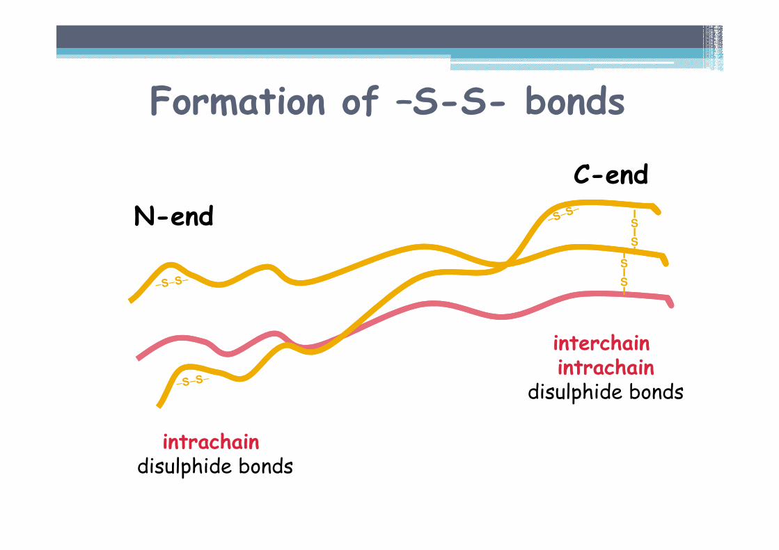
Formation of –S-S- bonds
ISISII
SISI
C-end
N-end
interchainintrachain
disulphide bonds
intrachaindisulphide bonds
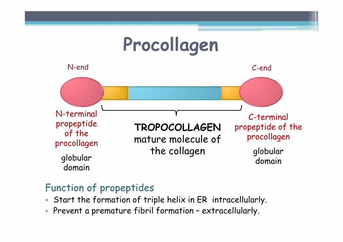
Procollagen
Function of propeptides• Start the formation of triple helix in ER intracellularly.• Prevent a premature fibril formation – extracellularly.
N-terminalpropeptideof the
procollagen
globulardomain
C-terminalpropeptide of the
procollagen
globulardomain
TROPOCOLLAGENmature molecule of
the collagen
N-end C-end
Posttranslational modificationsin the process of collagen synthesis
EXTRACELLULAR PROCESSES
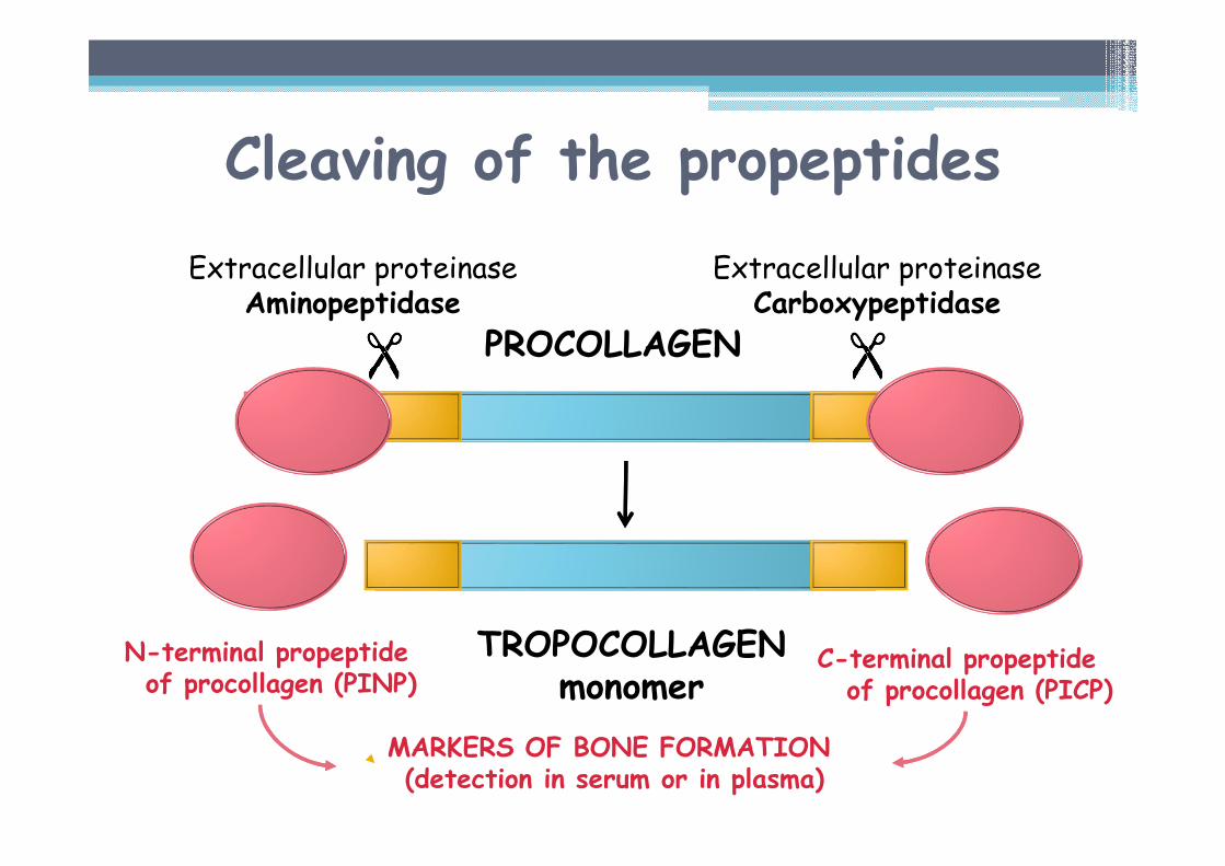
N-terminal propeptideof procollagen (PINP)
MARKERS OF BONE FORMATION (detection in serum or in plasma)
Cleaving of the propeptides
C-terminal propeptideof procollagen (PICP)
Extracellular proteinaseAminopeptidase
Extracellular proteinaseCarboxypeptidase�� �� �� ��
PROCOLLAGEN
TROPOCOLLAGENmonomer
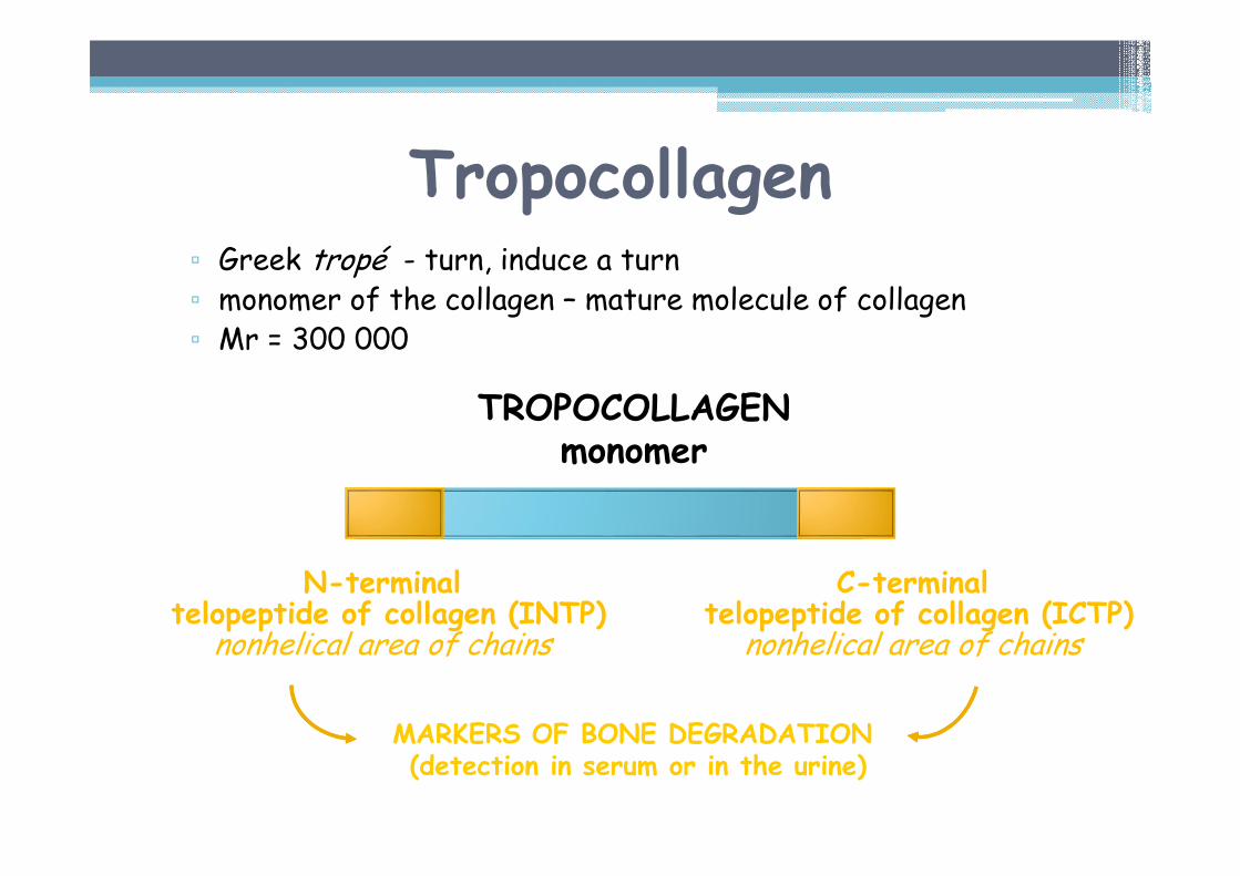
Tropocollagen▫ Greek tropé - turn, induce a turn▫ monomer of the collagen – mature molecule of collagen▫ Mr = 300 000
N-terminaltelopeptide of collagen (INTP)
nonhelical area of chains
TROPOCOLLAGENmonomer
C-terminaltelopeptide of collagen (ICTP)
nonhelical area of chains
MARKERS OF BONE DEGRADATION (detection in serum or in the urine)
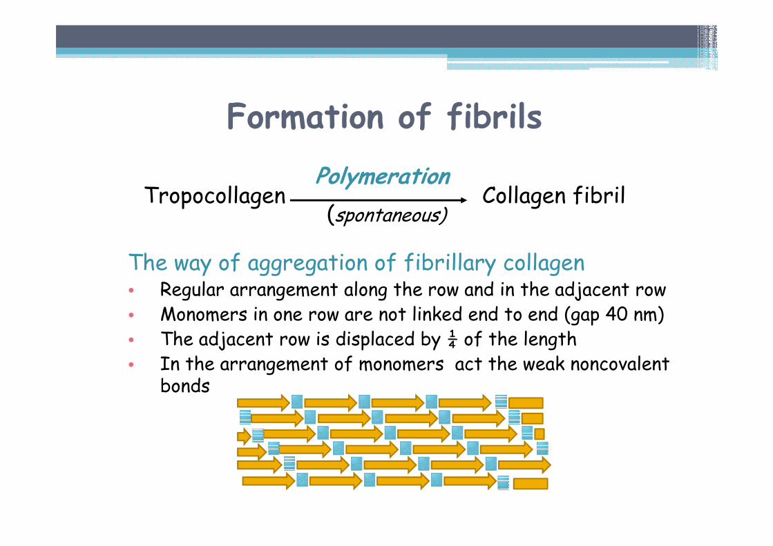
The way of aggregation of fibrillary collagen• Regular arrangement along the row and in the adjacent row• Monomers in one row are not linked end to end (gap 40 nm)• The adjacent row is displaced by ¼ of the length• In the arrangement of monomers act the weak noncovalent
bonds
Formation of fibrils
PolymerationTropocollagen Collagen fibril
(spontaneous)
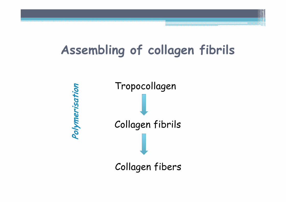
Tropocollagen
Collagen fibrils
Polymerisation
Collagen fibers
Assembling of collagen fibrils
Formation of cross-links
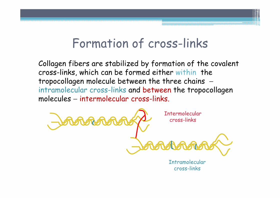
Collagen fibers are stabilized by formation of the covalent cross-links, which can be formed either within the tropocollagen molecule between the three chains −intramolecular cross-links and between the tropocollagenmolecules − intermolecular cross-links.
Intermolecularcross-links
Intramolecularcross-links
Formation of cross-links
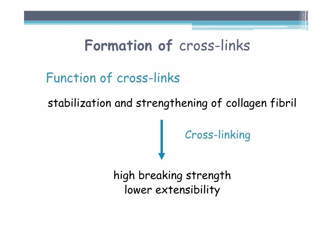
Function of cross-links
stabilization and strengthening of collagen fibril
high breaking strengthlower extensibility
Cross-linking
Formation of cross-links
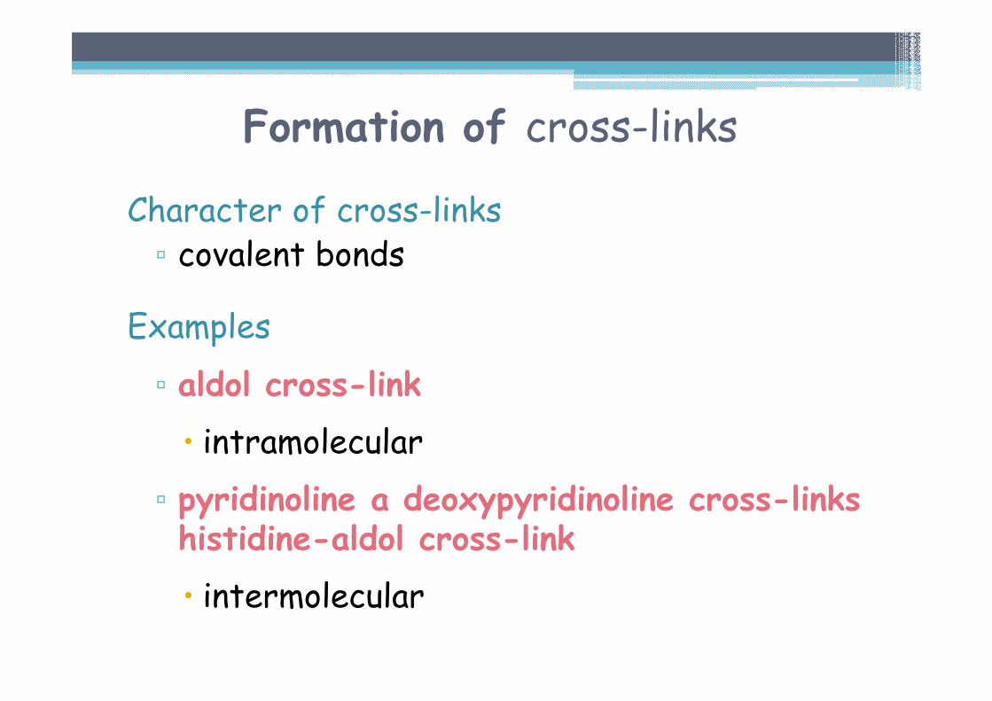
Character of cross-links▫ covalent bonds
Examples
▫ aldol cross-link
� intramolecular
▫ pyridinoline a deoxypyridinoline cross-links histidine-aldol cross-link
� intermolecular
Aldol cross-link
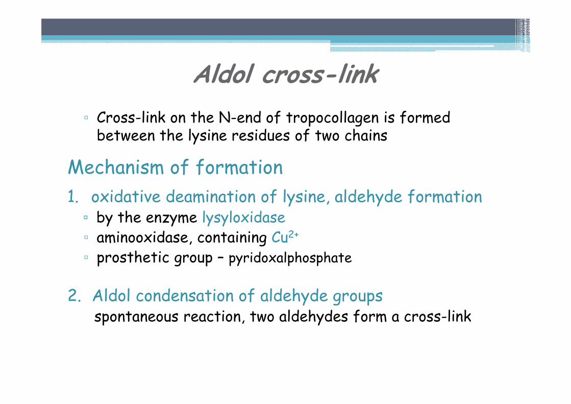
▫ Cross-link on the N-end of tropocollagen is formed between the lysine residues of two chains
Mechanism of formation
1. oxidative deamination of lysine, aldehyde formation▫ by the enzyme lysyloxidase▫ aminooxidase, containing Cu2+
▫ prosthetic group – pyridoxalphosphate
2. Aldol condensation of aldehyde groupsspontaneous reaction, two aldehydes form a cross-link
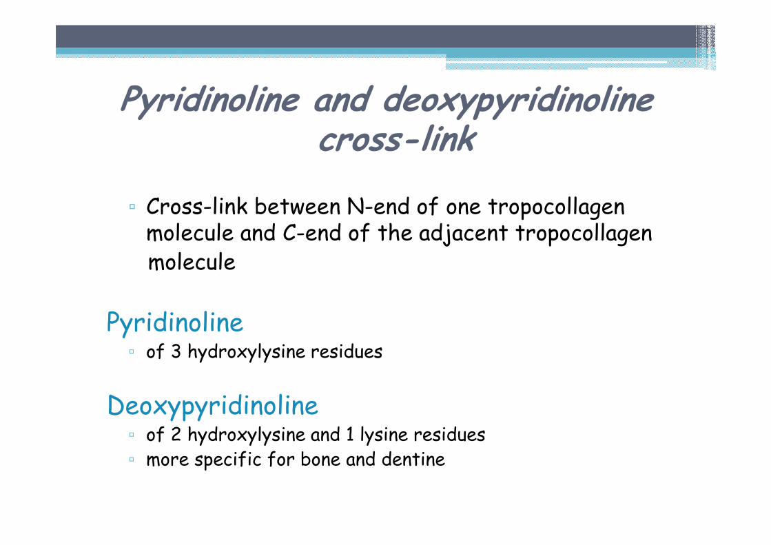
Pyridinoline and deoxypyridinolinecross-link
▫ Cross-link between N-end of one tropocollagenmolecule and C-end of the adjacent tropocollagenmolecule
Pyridinoline▫ of 3 hydroxylysine residues
Deoxypyridinoline▫ of 2 hydroxylysine and 1 lysine residues ▫ more specific for bone and dentine
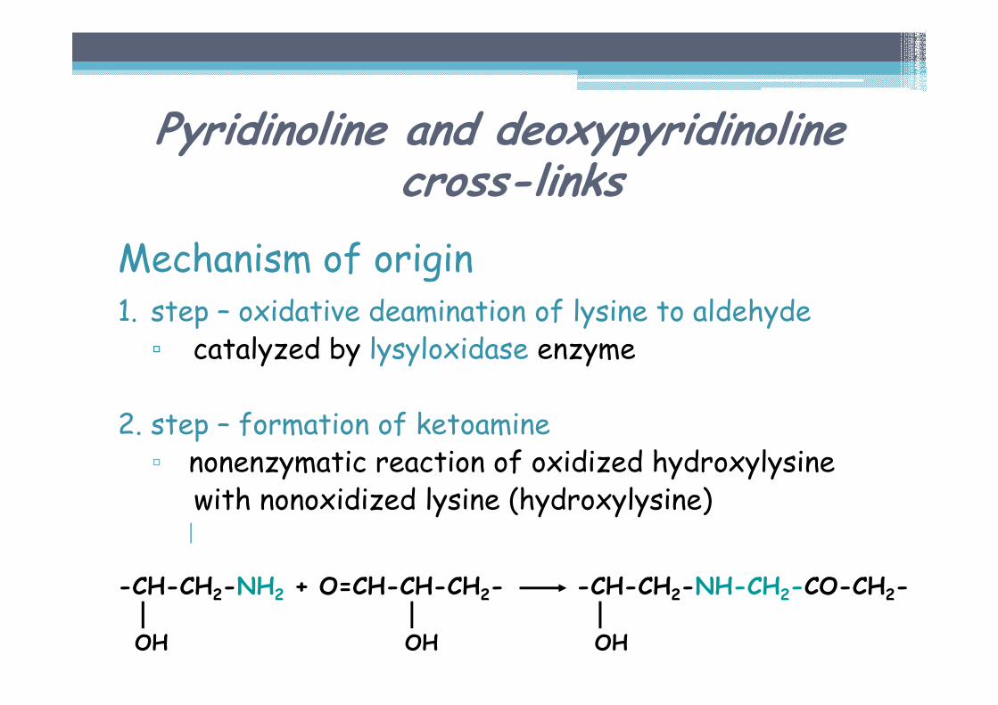
Mechanism of origin1. step – oxidative deamination of lysine to aldehyde▫ catalyzed by lysyloxidase enzyme
2. step – formation of ketoamine▫ nonenzymatic reaction of oxidized hydroxylysine
with nonoxidized lysine (hydroxylysine)|
OH OH OH
Pyridinoline and deoxypyridinolinecross-links
-CH-CH2-NH2 + O=CH-CH-CH2- -CH-CH2-NH-CH2-CO-CH2-
Pyridinoline a deoxypyridinolinecross-links
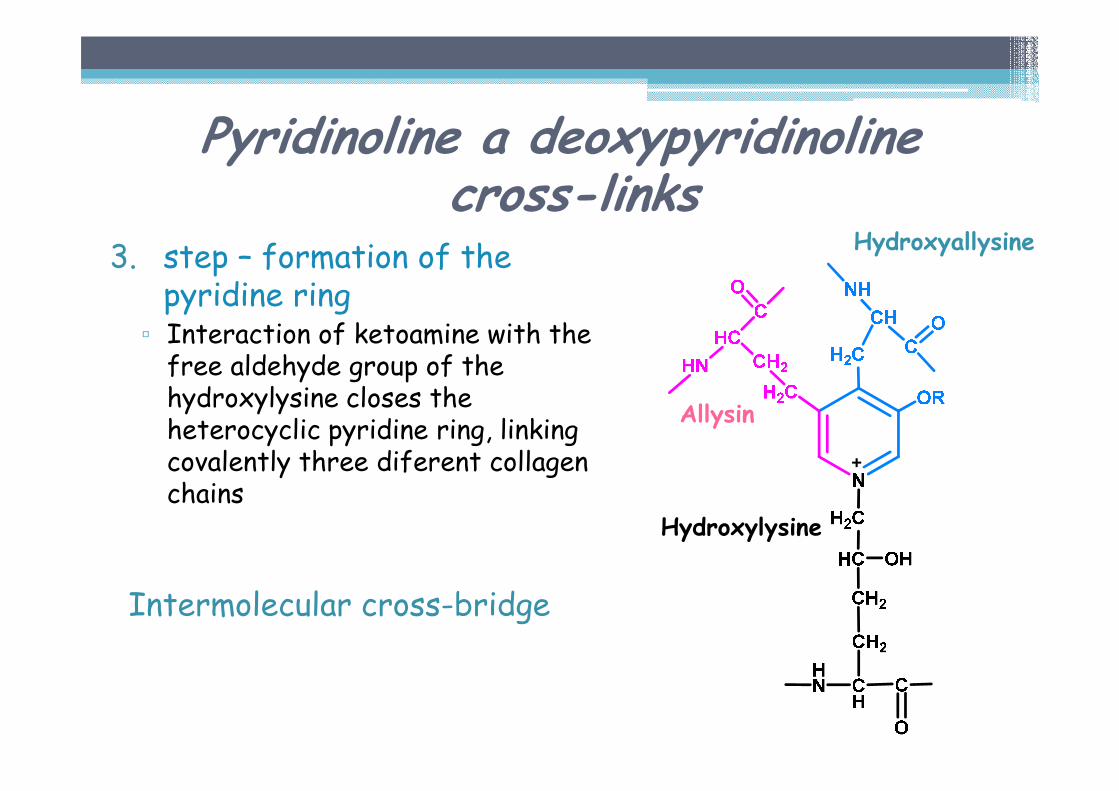
3. step – formation of the pyridine ring▫ Interaction of ketoamine with the free aldehyde group of the hydroxylysine closes the heterocyclic pyridine ring, linking covalently three diferent collagen chains
Intermolecular cross-bridge
Allysin
Hydroxyallysine
Hydroxylysine
Pyridinoline a deoxypyridinolinecross-links
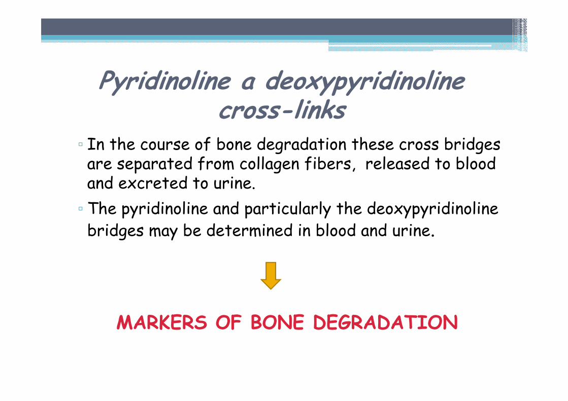
▫ In the course of bone degradation these cross bridges are separated from collagen fibers, released to blood and excreted to urine.
▫ The pyridinoline and particularly the deoxypyridinolinebridges may be determined in blood and urine.
MARKERS OF BONE DEGRADATION
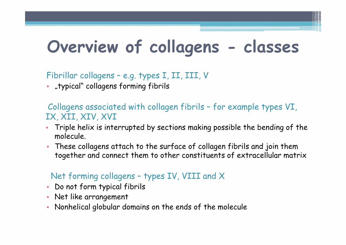
Fibrillar collagens – e.g. types I, II, III, V • „typical“ collagens forming fibrils
Collagens associated with collagen fibrils – for example types VI, IX, XII, XIV, XVI• Triple helix is interrupted by sections making possible the bending of the molecule.
• These collagens attach to the surface of collagen fibrils and join them together and connect them to other constituents of extracellular matrix
Net forming collagens – types IV, VIII and X• Do not form typical fibrils• Net like arrangement• Nonhelical globular domains on the ends of the molecule
Overview of collagens - classes
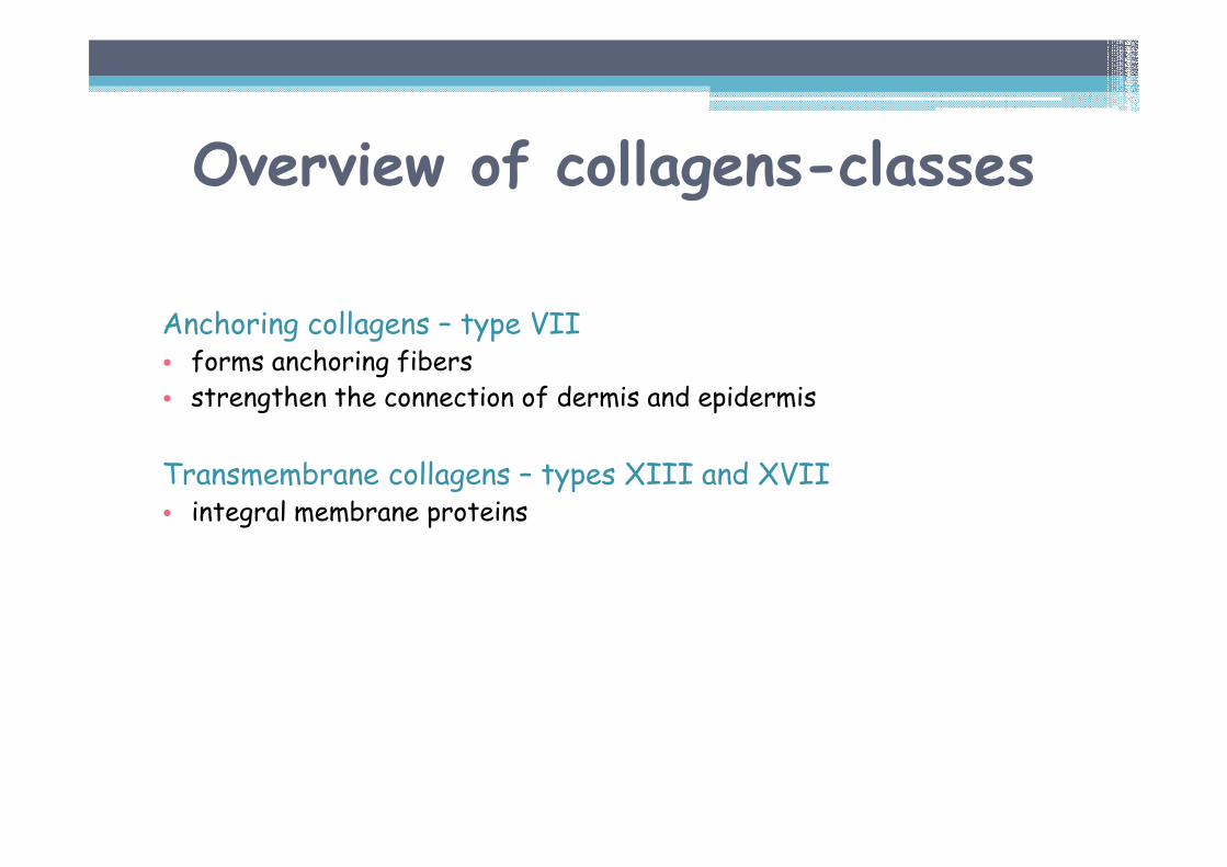
Anchoring collagens – type VII• forms anchoring fibers• strengthen the connection of dermis and epidermis
Transmembrane collagens – types XIII and XVII• integral membrane proteins
Overview of collagens-classes
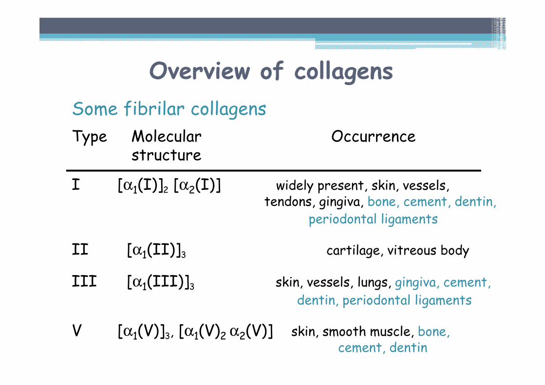
Overview of collagens
Some fibrilar collagens
Type Molecular Occurrencestructure
I [α1(I)]2 [α2(I)] widely present, skin, vessels, tendons, gingiva, bone, cement, dentin,
periodontal ligaments
II [α1(II)]3 cartilage, vitreous body
III [α1(III)]3 skin, vessels, lungs, gingiva, cement,dentin, periodontal ligaments
V [α1(V)]3, [α1(V)2 α2(V)] skin, smooth muscle, bone, cement, dentin
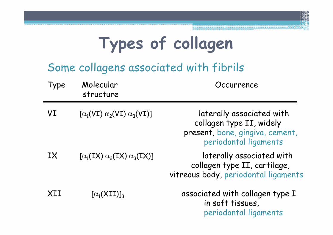
Types of collagen
Some collagens associated with fibrils
Type Molecular Occurrencestructure
VI [α1(VI) α2(VI) α3(VI)] laterally associated withcollagen type II, widely
present, bone, gingiva, cement, periodontal ligaments
IX [α1(IX) α2(IX) α3(IX)] laterally associated with collagen type II, cartilage,
vitreous body, periodontal ligaments
XII [α1(XII)]3 associated with collagen type I in soft tissues,periodontal ligaments
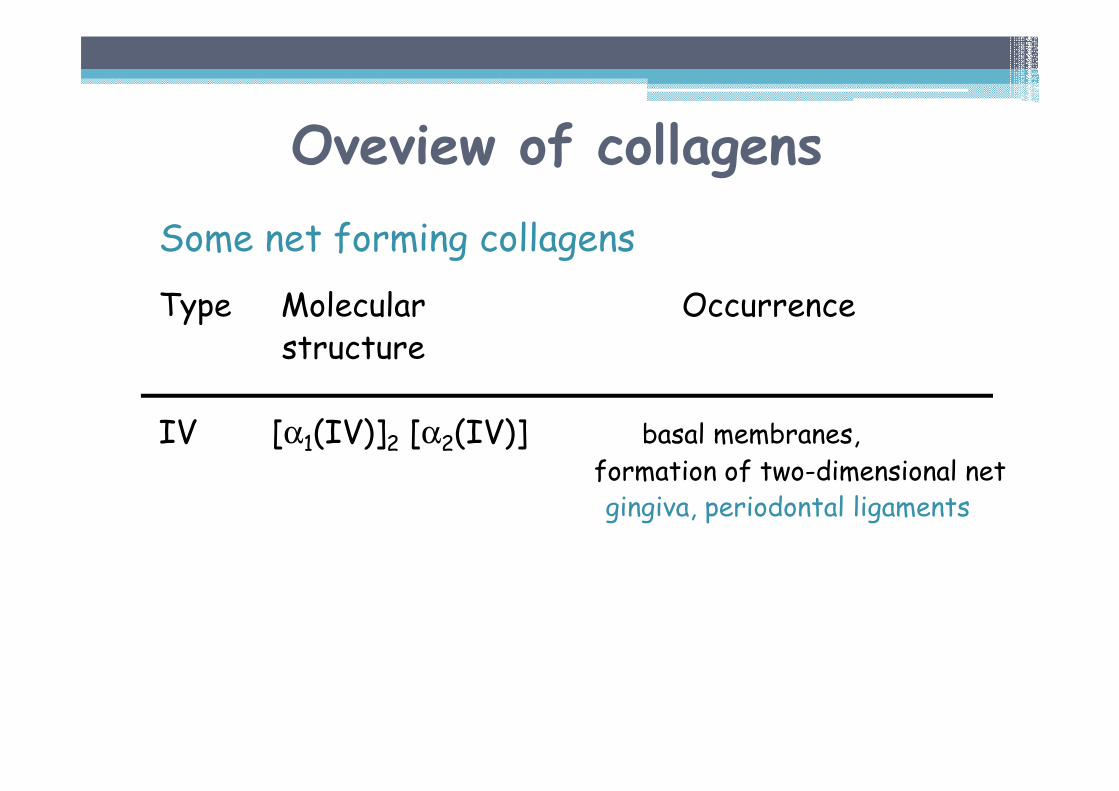
Oveview of collagens
Some net forming collagens
Type Molecular Occurrencestructure
IV [α1(IV)]2 [α2(IV)] basal membranes,formation of two-dimensional netgingiva, periodontal ligaments
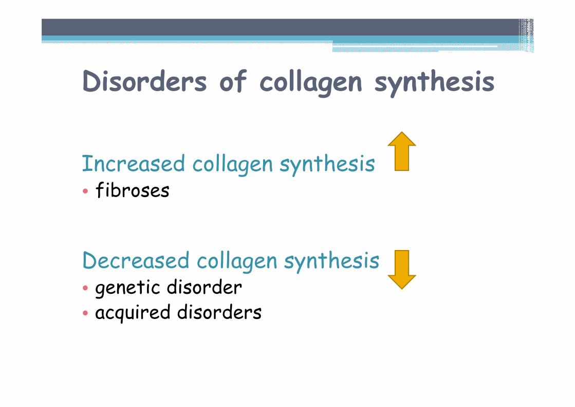
Disorders of collagen synthesis
Increased collagen synthesis • fibroses
Decreased collagen synthesis • genetic disorder• acquired disorders
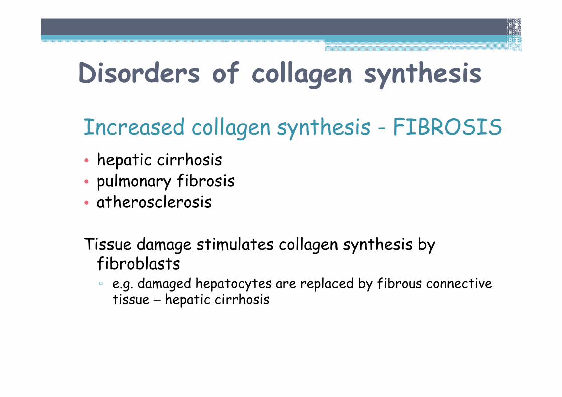
Disorders of collagen synthesis
Increased collagen synthesis - FIBROSIS
• hepatic cirrhosis • pulmonary fibrosis• atherosclerosis
Tissue damage stimulates collagen synthesis by fibroblasts▫ e.g. damaged hepatocytes are replaced by fibrous connective tissue − hepatic cirrhosis
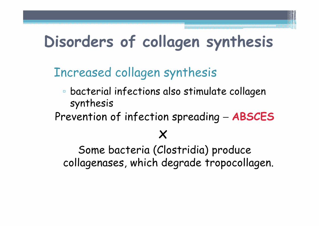
Disorders of collagen synthesis
Increased collagen synthesis
▫ bacterial infections also stimulate collagen synthesis
Prevention of infection spreading − ABSCES
xSome bacteria (Clostridia) produce
collagenases, which degrade tropocollagen.
Disorders of collagen synthesis
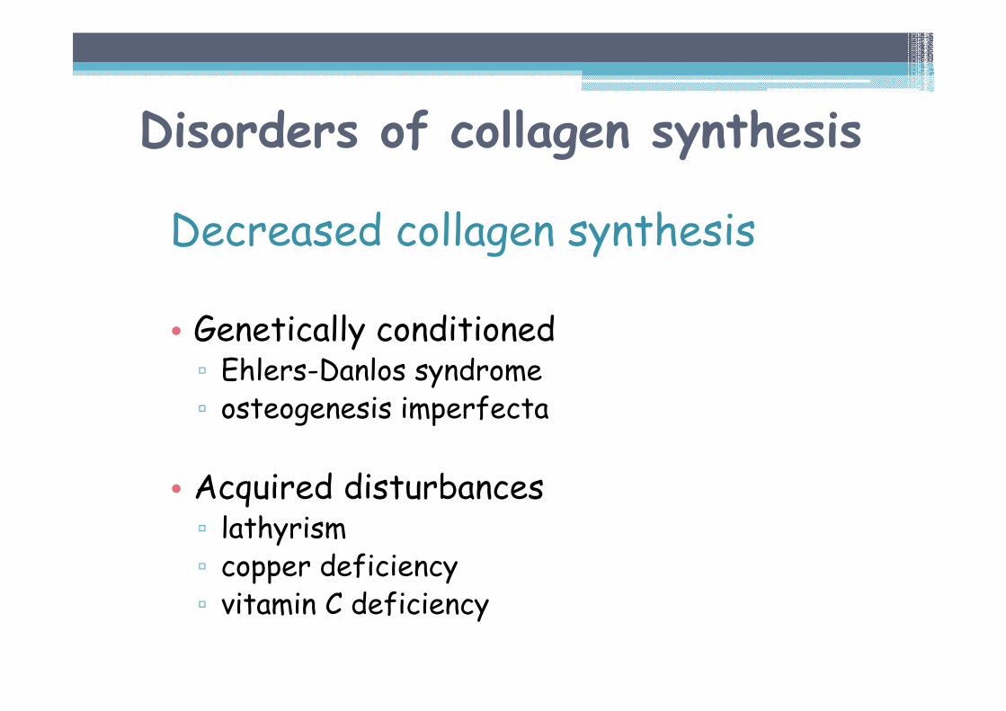
Decreased collagen synthesis
• Genetically conditioned▫ Ehlers-Danlos syndrome▫ osteogenesis imperfecta
• Acquired disturbances▫ lathyrism▫ copper deficiency ▫ vitamin C deficiency
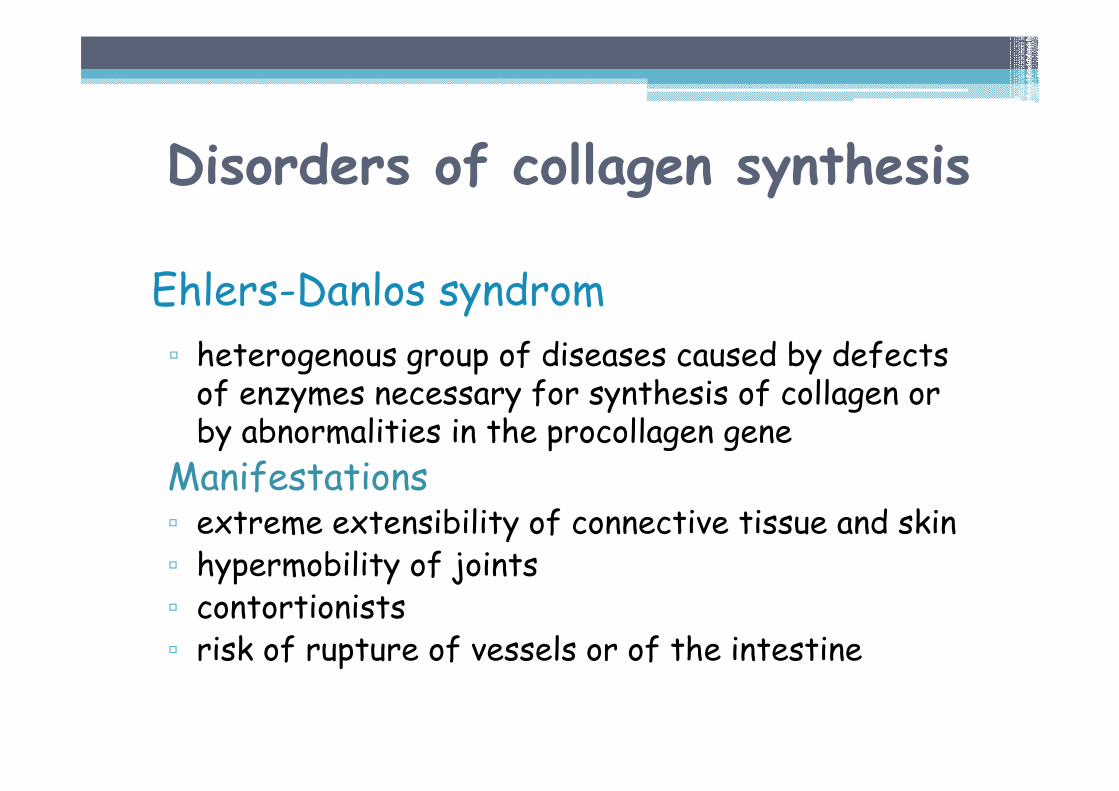
Ehlers-Danlos syndrom
▫ heterogenous group of diseases caused by defects of enzymes necessary for synthesis of collagen or by abnormalities in the procollagen gene
Manifestations▫ extreme extensibility of connective tissue and skin▫ hypermobility of joints▫ contortionists▫ risk of rupture of vessels or of the intestine
Disorders of collagen synthesis
Disorders of collagen synthesis
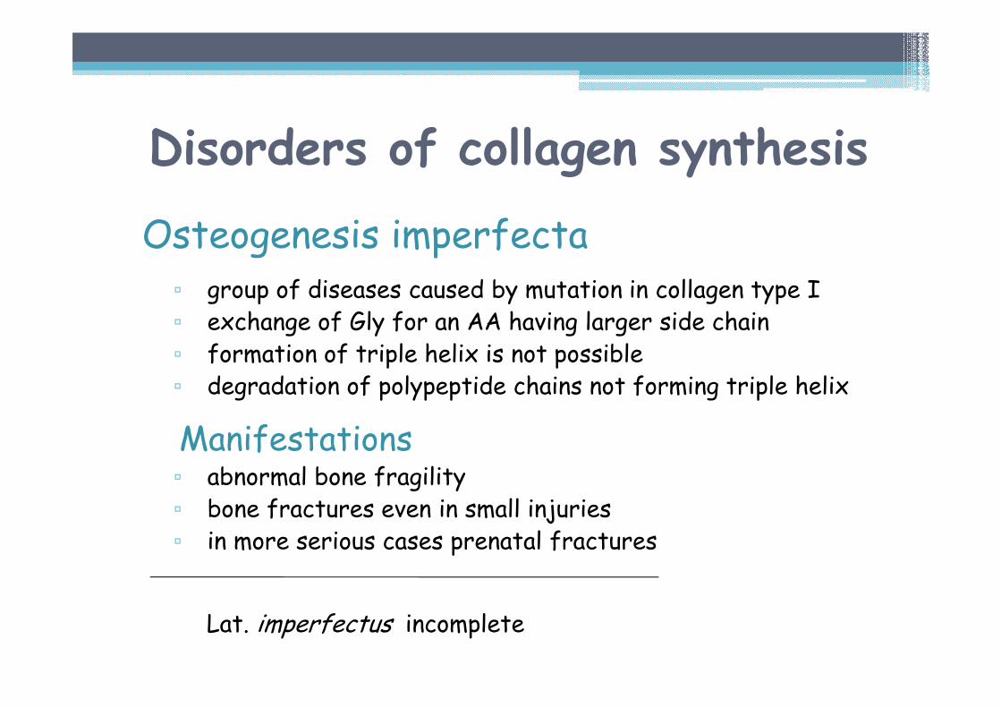
Osteogenesis imperfecta▫ group of diseases caused by mutation in collagen type I▫ exchange of Gly for an AA having larger side chain▫ formation of triple helix is not possible▫ degradation of polypeptide chains not forming triple helix
Manifestations▫ abnormal bone fragility▫ bone fractures even in small injuries▫ in more serious cases prenatal fractures
Lat. imperfectus incomplete

Dentinogenesis imperfecta
▫ group of diseases caused by mutation in α1(I)▫ associated with osteogenesis imperfecta
Manifestations▫ thin enamel ▫ discolouring of teeth (yellow, brown, grey) ▫ opalescence of the teeth▫ lower mechanical resistance of the teeth
Disorders of collagen synthesis
Disorders of collagen synthesis
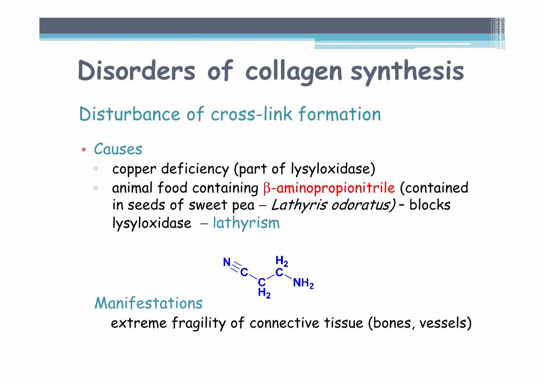
Disturbance of cross-link formation
• Causes▫ copper deficiency (part of lysyloxidase)▫ animal food containing β-aminopropionitrile (contained
in seeds of sweet pea − Lathyris odoratus) – blocks lysyloxidase − lathyrism
Manifestations extreme fragility of connective tissue (bones, vessels)
ELASTIN
Elastin is the main protein of elastic fibers, providing elasticity to the tissues.
61
Elastin
Occurrence
• in arteries, particularly in aorta • in skin, tendons and loose connective tissue (relatively low content) • in lungs
Synthesis takes place in early development or after tissue damage
Half-time is approximately 70 years (lower content in elderly people).
Elastin
Properties
EXTENSIBILITY AND CONTRACTILITY
▫ resembles the rubber▫ after extension elastin is able to return to original size and original form▫ tensile strength is lower than in collagen▫ hydrophobic, practically insoluble in aqueous solutions
Primary structure of elastin
Occurrence of amino acids▫ 1/3 glycine▫ high content of nonpolar AA (Ala, Val, Leu, Ileu)▫ low hydroxyproline▫ no hydroxylysine − elastin is not glycosylated
Sequence of amino acids▫ typical triplet as in collagen is not present
Alternation of short hydrophobic and hydrophilic sections.Hydrophilic sections, which represent a minority part, are rich in lysine, which takes part in forming of cross - links.
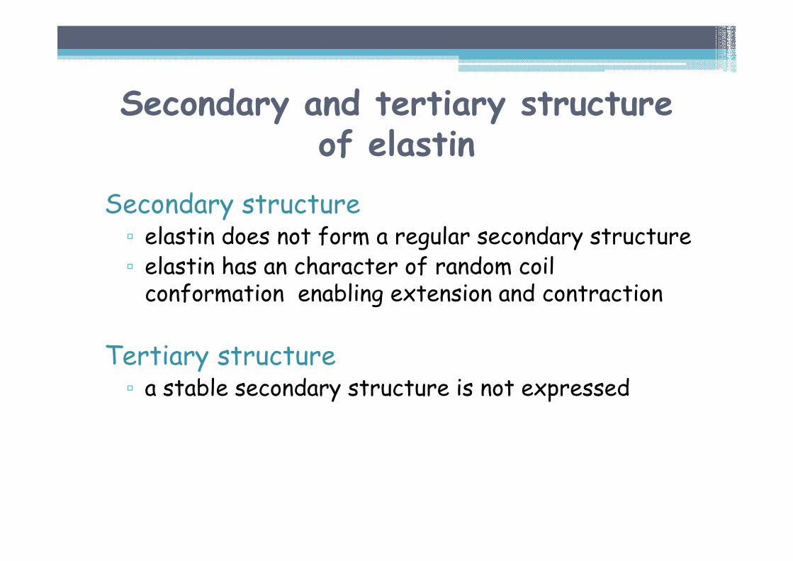
Secondary and tertiary structureof elastin
Secondary structure▫ elastin does not form a regular secondary structure▫ elastin has an character of random coil conformation enabling extension and contraction
Tertiary structure▫ a stable secondary structure is not expressed
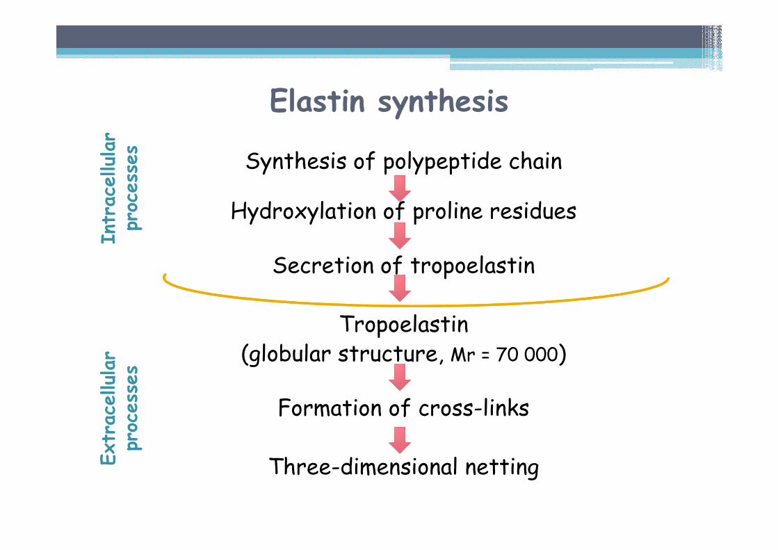
Elastin synthesis
Synthesis of polypeptide chain
Hydroxylation of proline residues
Secretion of tropoelastin
Tropoelastin(globular structure, Mr = 70 000)
Formation of cross-links
Three-dimensional netting
Intracellular
processes
Extracellular
processes
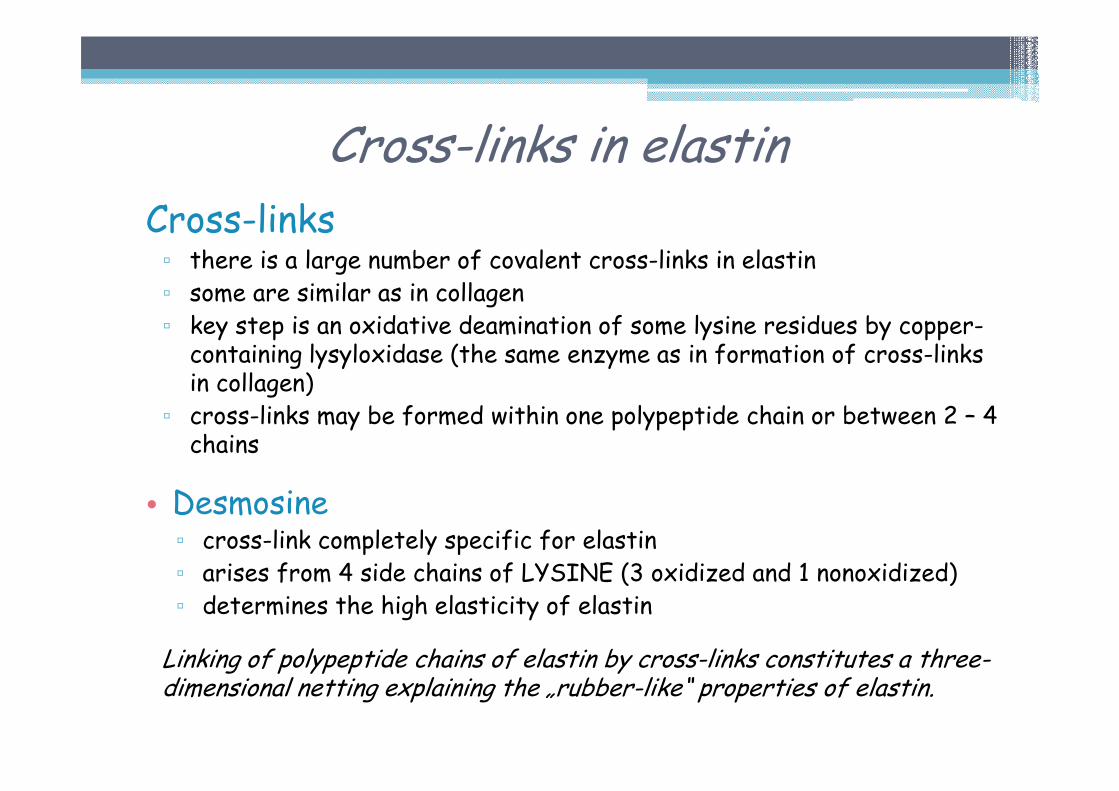
Cross-links in elastinCross-links▫ there is a large number of covalent cross-links in elastin▫ some are similar as in collagen▫ key step is an oxidative deamination of some lysine residues by copper-containing lysyloxidase (the same enzyme as in formation of cross-links in collagen)
▫ cross-links may be formed within one polypeptide chain or between 2 – 4 chains
• Desmosine▫ cross-link completely specific for elastin▫ arises from 4 side chains of LYSINE (3 oxidized and 1 nonoxidized)▫ determines the high elasticity of elastin
Linking of polypeptide chains of elastin by cross-links constitutes a three-dimensional netting explaining the „rubber-like“ properties of elastin.
GLYCOSAMINOGLYCANS
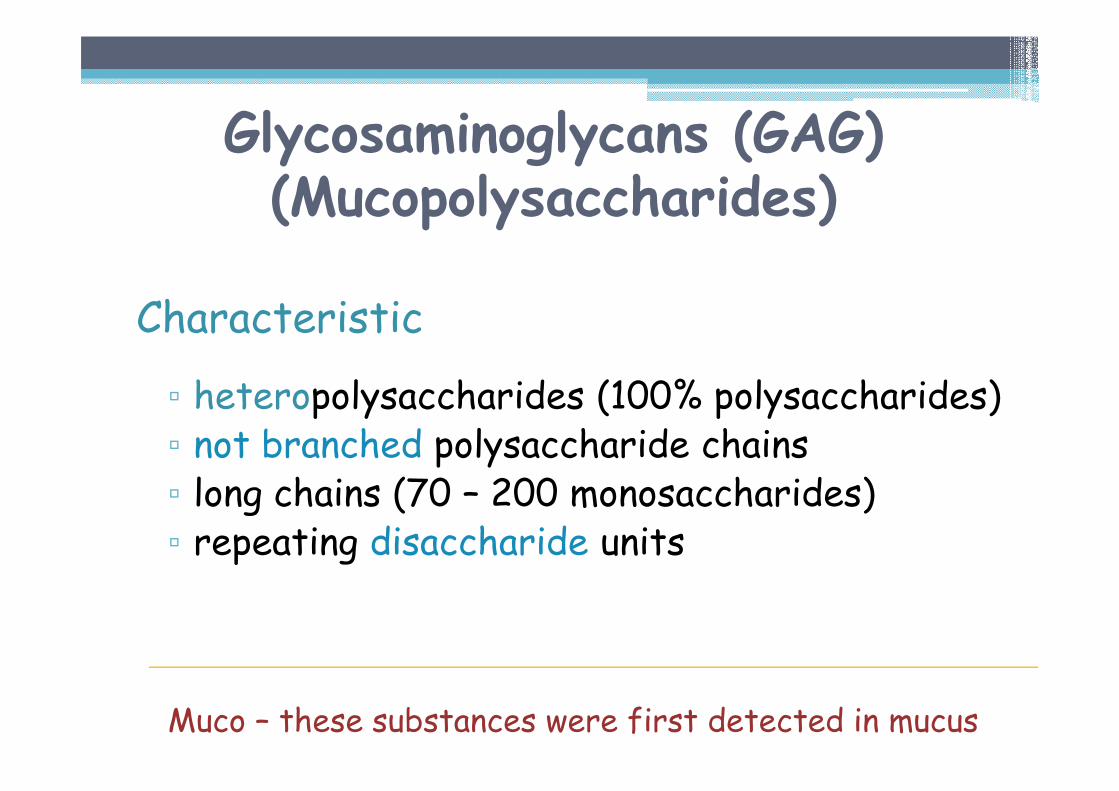
Glycosaminoglycans (GAG)(Mucopolysaccharides)
Characteristic
▫ heteropolysaccharides (100% polysaccharides)▫ not branched polysaccharide chains ▫ long chains (70 – 200 monosaccharides)▫ repeating disaccharide units
Muco – these substances were first detected in mucus
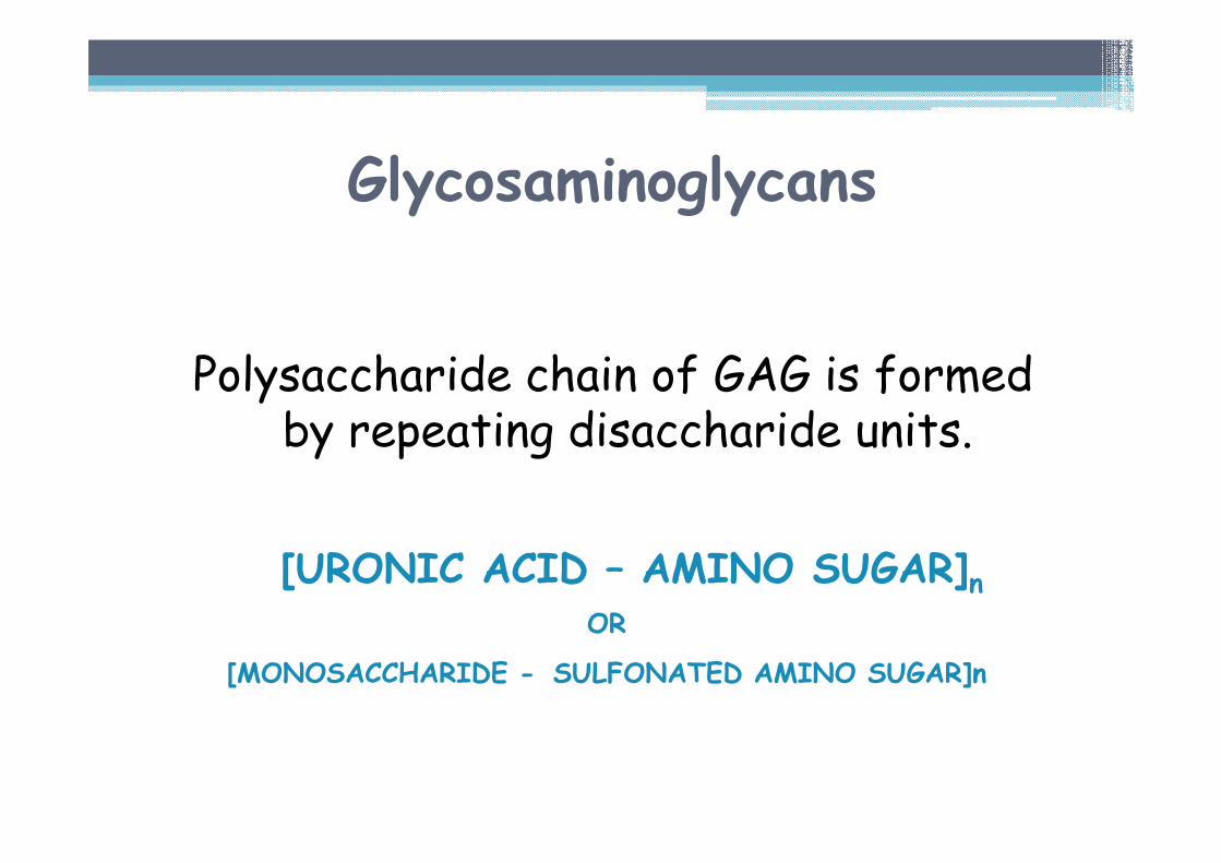
Glycosaminoglycans
Polysaccharide chain of GAG is formed by repeating disaccharide units.
[URONIC ACID – AMINO SUGAR]nOR
[MONOSACCHARIDE - SULFONATED AMINO SUGAR]n
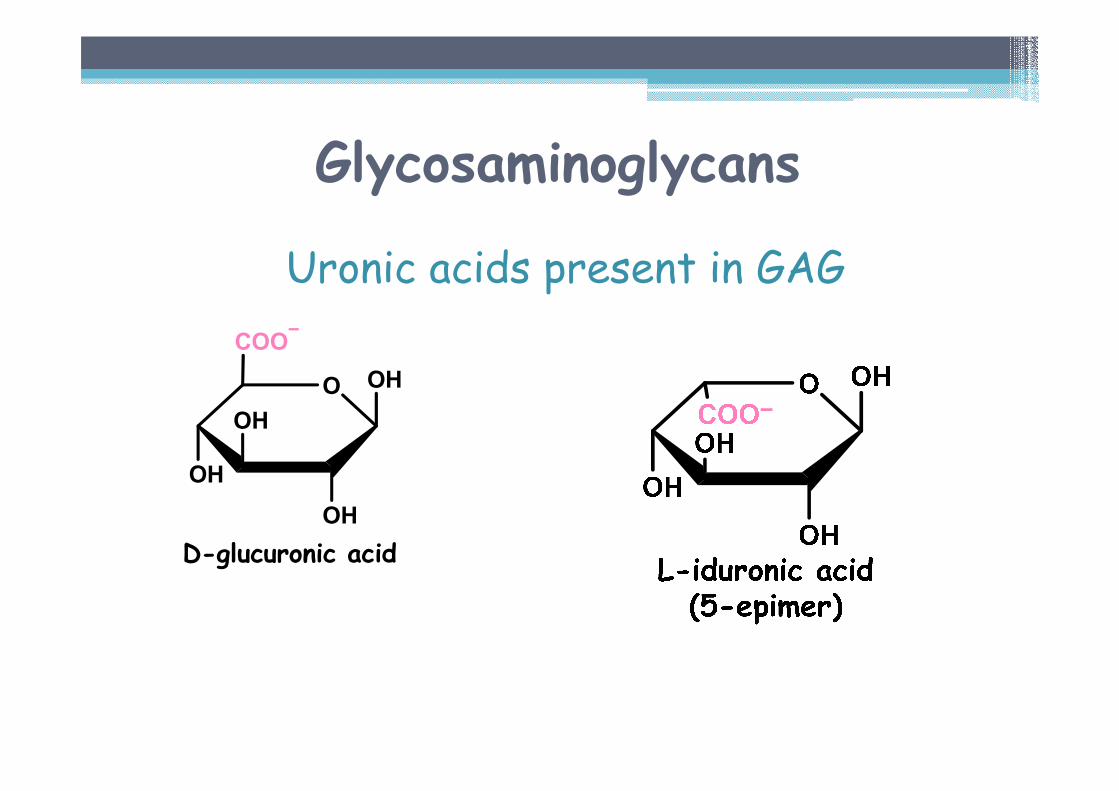
Glycosaminoglycans
Uronic acids present in GAG
O OH
OH
OH
OH
COO
D-glucuronic acid
Glycosaminoglycans
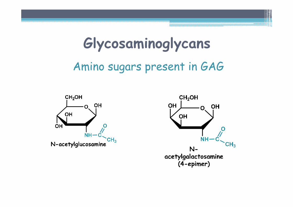
Amino sugars present in GAG
Glycosaminoglycans
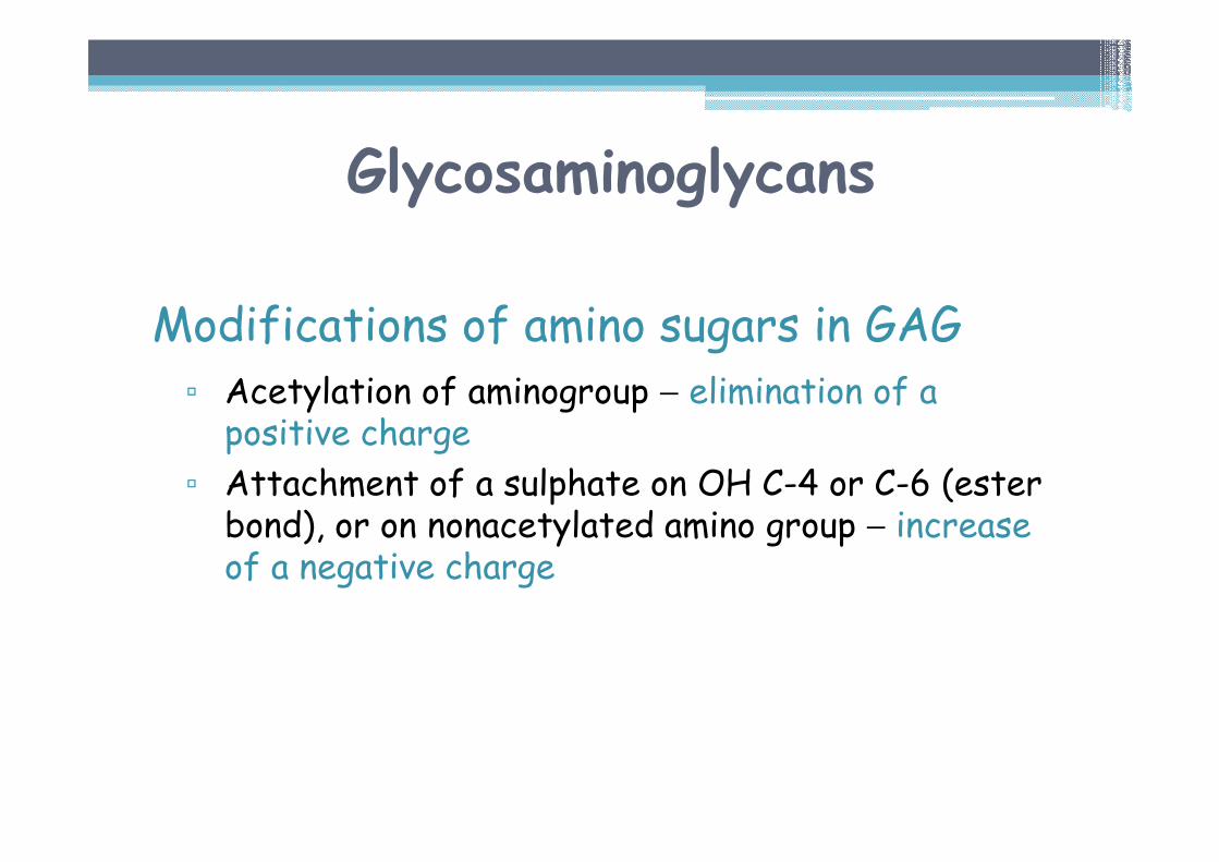
Modifications of amino sugars in GAG▫ Acetylation of aminogroup − elimination of a positive charge
▫ Attachment of a sulphate on OH C-4 or C-6 (ester bond), or on nonacetylated amino group − increase of a negative charge
Glycosaminoglycans
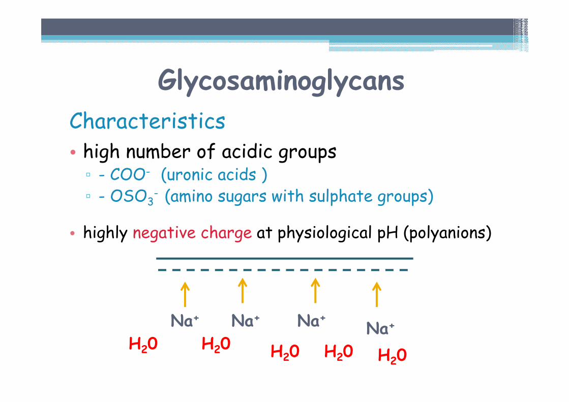
Characteristics• high number of acidic groups▫ - COO- (uronic acids )▫ - OSO3
- (amino sugars with sulphate groups)
• highly negative charge at physiological pH (polyanions)
Na+ Na+ Na+ Na+H20 H20 H20
H20 H20
Glycosaminoglycans
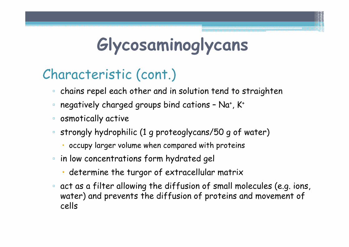
Characteristic (cont.)▫ chains repel each other and in solution tend to straighten
▫ negatively charged groups bind cations – Na+, K+
▫ osmotically active
▫ strongly hydrophilic (1 g proteoglycans/50 g of water)
� occupy larger volume when compared with proteins
▫ in low concentrations form hydrated gel
� determine the turgor of extracellular matrix
▫ act as a filter allowing the diffusion of small molecules (e.g. ions, water) and prevents the diffusion of proteins and movement of cells
Glycosaminoglycans
Types of glycosaminoglycans
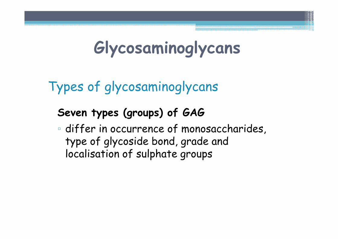
Seven types (groups) of GAG
▫ differ in occurrence of monosaccharides, type of glycoside bond, grade and localisation of sulphate groups
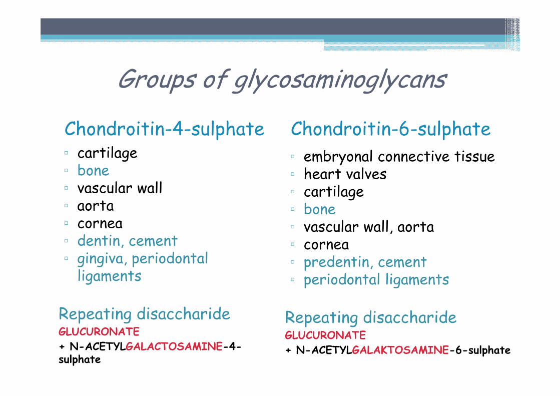
Groups of glycosaminoglycans
Chondroitin-4-sulphate▫ cartilage▫ bone▫ vascular wall▫ aorta▫ cornea▫ dentin, cement▫ gingiva, periodontal ligaments
Repeating disaccharideGLUCURONATE
+ N-ACETYLGALACTOSAMINE-4-sulphate
Chondroitin-6-sulphate▫ embryonal connective tissue▫ heart valves▫ cartilage▫ bone▫ vascular wall, aorta▫ cornea▫ predentin, cement▫ periodontal ligaments
Repeating disaccharideGLUCURONATE
+ N-ACETYLGALAKTOSAMINE-6-sulphate
Groups of glycosaminoglycans
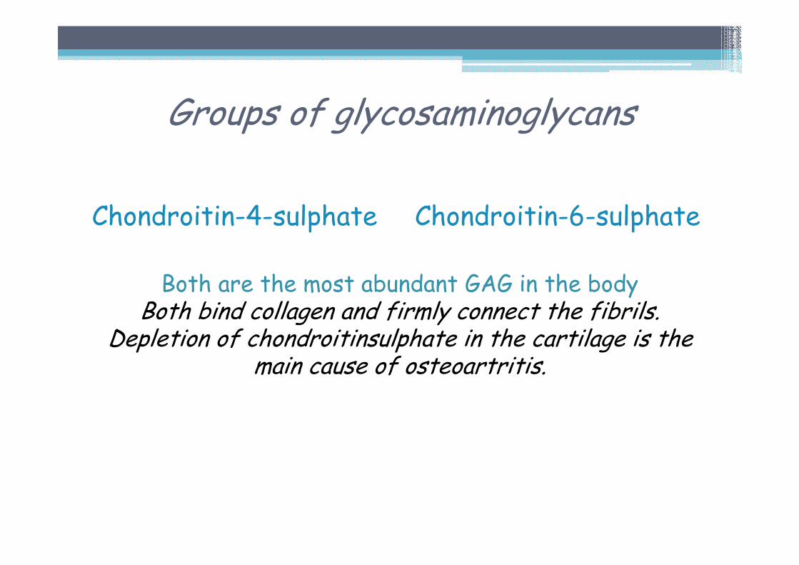
Chondroitin-4-sulphate Chondroitin-6-sulphate
Both are the most abundant GAG in the bodyBoth bind collagen and firmly connect the fibrils.
Depletion of chondroitinsulphate in the cartilage is the main cause of osteoartritis.
Groups of glycosaminoglycans
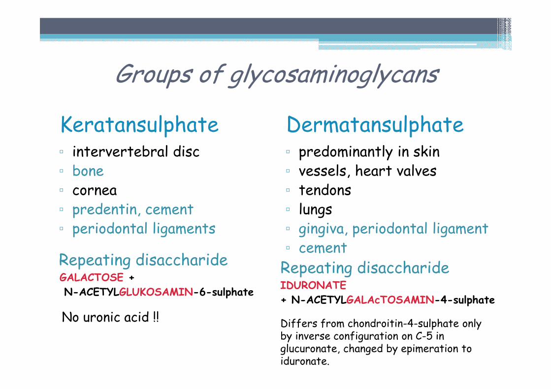
Keratansulphate▫ intervertebral disc▫ bone▫ cornea▫ predentin, cement▫ periodontal ligaments
Repeating disaccharideGALACTOSE +
N-ACETYLGLUKOSAMIN-6-sulphate
No uronic acid !!
Dermatansulphate▫ predominantly in skin▫ vessels, heart valves▫ tendons▫ lungs▫ gingiva, periodontal ligament▫ cement
Repeating disaccharideIDURONATE
+ N-ACETYLGALAcTOSAMIN-4-sulphate
Differs from chondroitin-4-sulphate only by inverse configuration on C-5 in glucuronate, changed by epimeration to iduronate.
Groups of glycosaminoglycans
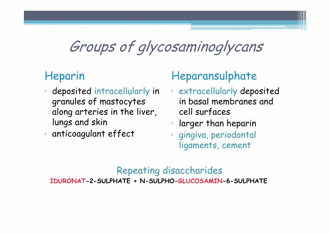
Heparin▫ deposited intracellularly in granules of mastocytesalong arteries in the liver, lungs and skin▫ anticoagulant effect
Heparansulphate▫ extracellularly deposited in basal membranes and cell surfaces▫ larger than heparin ▫ gingiva, periodontal ligaments, cement
Repeating disaccharidesIDURONAT-2-SULPHATE + N-SULPHO-GLUCOSAMIN-6-SULPHATE
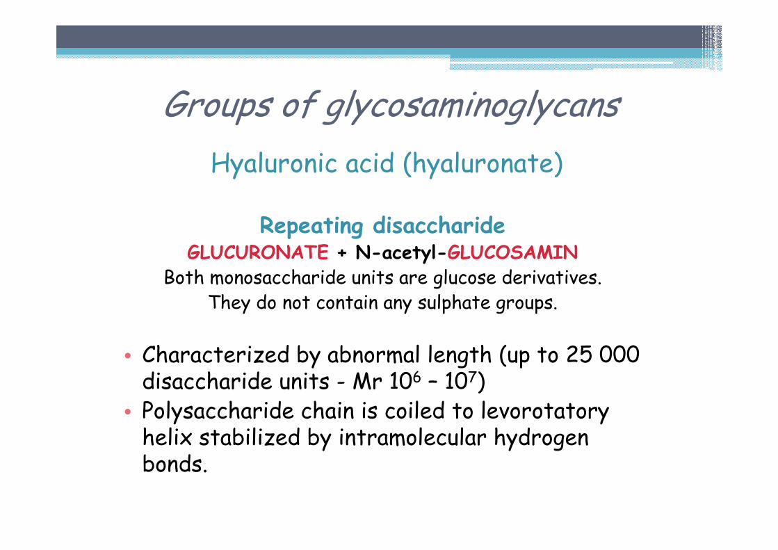
Hyaluronic acid (hyaluronate)
Repeating disaccharideGLUCURONATE + N-acetyl-GLUCOSAMIN
Both monosaccharide units are glucose derivatives.They do not contain any sulphate groups.
• Characterized by abnormal length (up to 25 000 disaccharide units - Mr 106 – 107)• Polysaccharide chain is coiled to levorotatory helix stabilized by intramolecular hydrogen bonds.
Groups of glycosaminoglycans
Hyaluronic acid (hyaluronate)
Occurrence▫ proteoglycan aggregates▫ vitreous body▫ synovial fluid (lubricating function)▫ umbilical cord▫ production increases during wound healing▫ gingiva, periodontal ligaments▫ cement
Groups of glycosaminoglycans
Groups of glycosaminoglycans
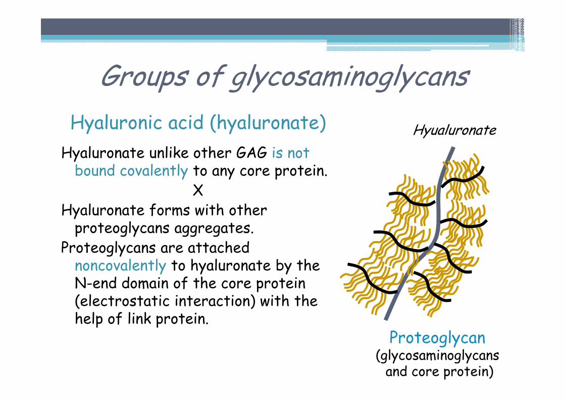
Hyaluronic acid (hyaluronate)
Hyaluronate unlike other GAG is not bound covalently to any core protein.
XHyaluronate forms with other proteoglycans aggregates.
Proteoglycans are attached noncovalently to hyaluronate by the N-end domain of the core protein (electrostatic interaction) with the help of link protein.
Proteoglycan(glycosaminoglycansand core protein)
Hyualuronate
Glycosaminoglycans
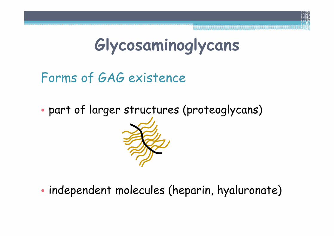
Forms of GAG existence
• part of larger structures (proteoglycans)
• independent molecules (heparin, hyaluronate)
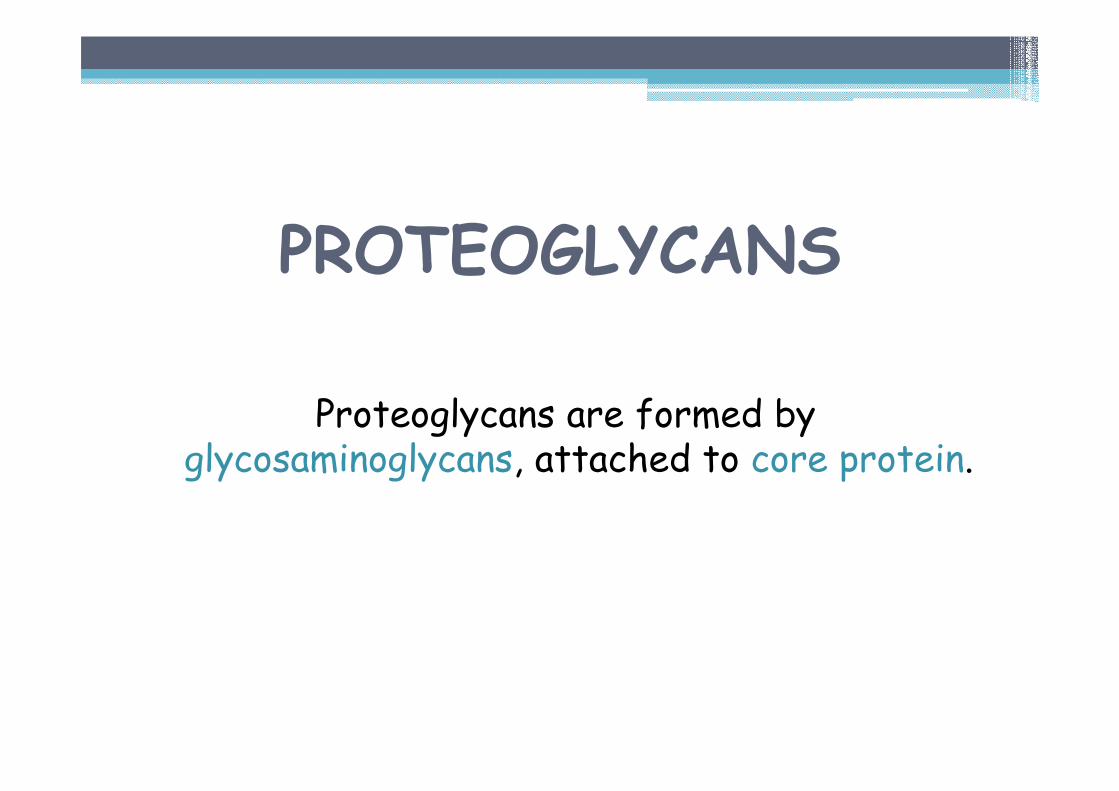
PROTEOGLYCANS
Proteoglycans are formed by glycosaminoglycans, attached to core protein.
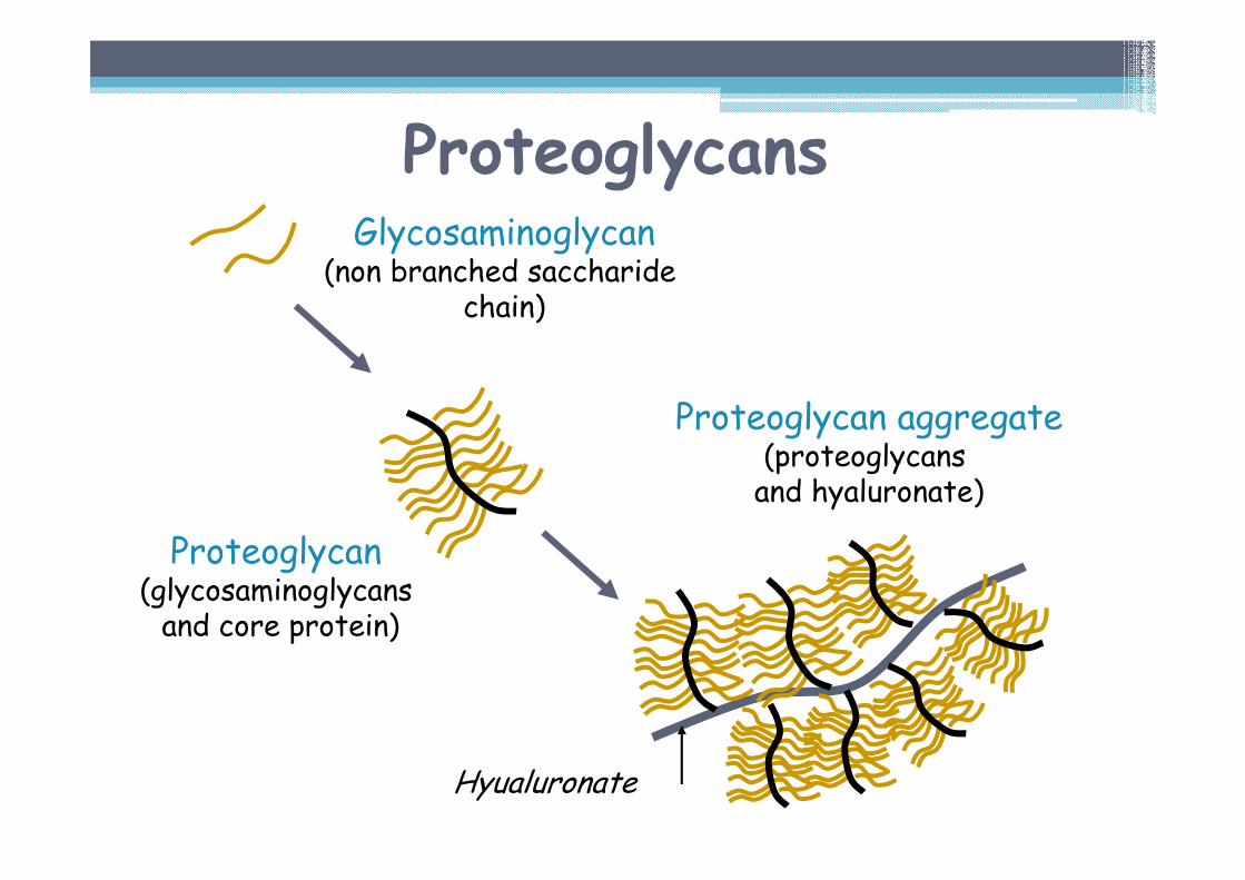
ProteoglycansGlycosaminoglycan
(non branched saccharidechain)
Proteoglycan(glycosaminoglycansand core protein)
Proteoglycan aggregate(proteoglycans and hyaluronate)
Hyualuronate
Proteoglycans
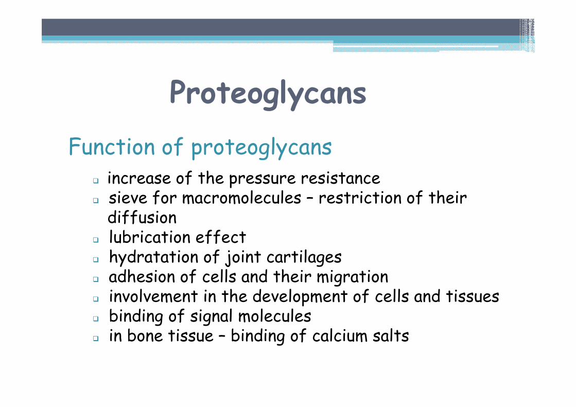
Function of proteoglycans� increase of the pressure resistance� sieve for macromolecules – restriction of their diffusion
� lubrication effect� hydratation of joint cartilages � adhesion of cells and their migration� involvement in the development of cells and tissues� binding of signal molecules� in bone tissue – binding of calcium salts
Proteoglycans
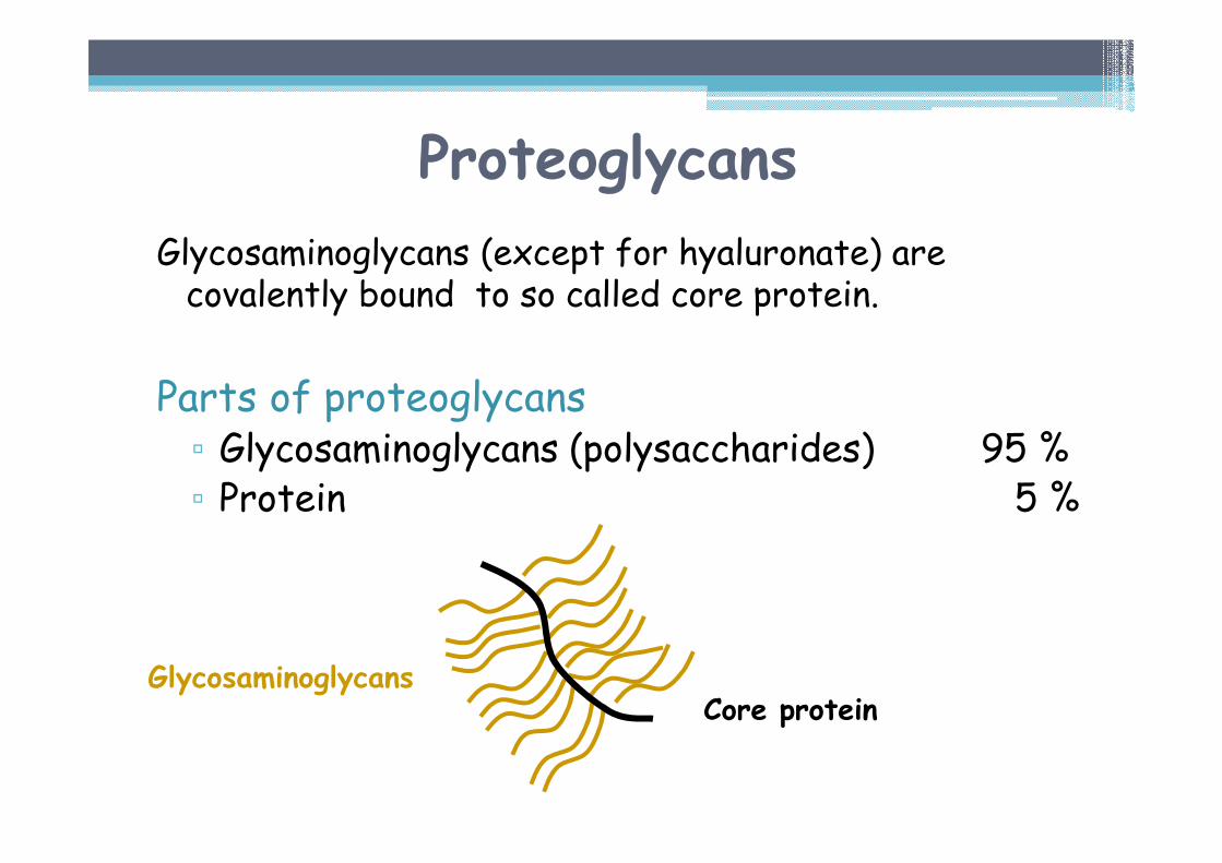
Glycosaminoglycans (except for hyaluronate) are covalently bound to so called core protein.
Parts of proteoglycans▫ Glycosaminoglycans (polysaccharides) 95 %▫ Protein 5 %
Core proteinGlycosaminoglycans
Proteoglycans
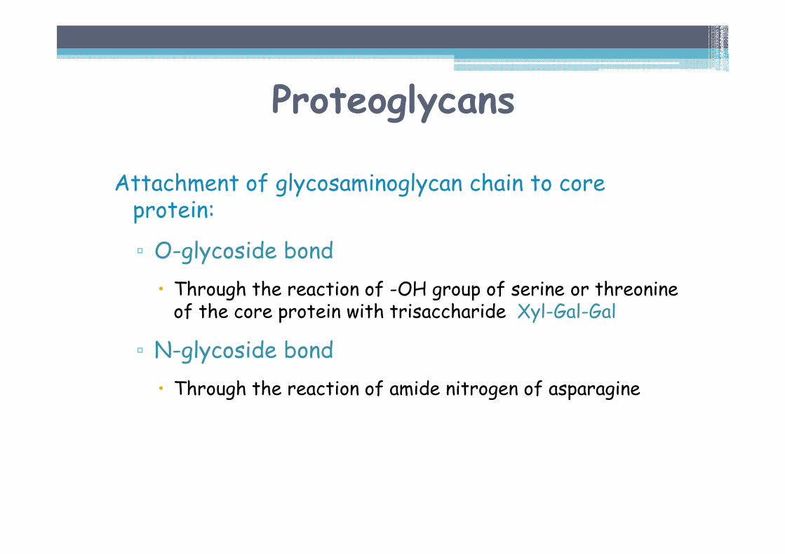
Attachment of glycosaminoglycan chain to core protein:
▫ O-glycoside bond
� Through the reaction of -OH group of serine or threonineof the core protein with trisaccharide Xyl-Gal-Gal
▫ N-glycoside bond
� Through the reaction of amide nitrogen of asparagine

Proteoglycans
• Proteoglycans are characterized by structural diversity:
▫ different core proteins
▫ different GAG chains
▫ different length of GAG chains
• Proteoglycans differ also in localisation:
▫ proteoglycans attached to basal membrane
▫ interstitial proteoglycans
Function
forms proteoglycanaggregates with hyaluronate
• cartilage, gingiva
• binds to collagen• belongs to a group of small proteoglycan rich in leucine
• gingiva
• present in basal membrane• long core protein• forms a barrier limiting penetration of macromolecules through the basal membrane
Selected proteoglycans
Proteoglycan
Versican
Aggrecan
Decorin
Perlecan
Typ GAG
chondroitinsulphatedermatansulphate
chondroitinsulphatekeratansulphate
chondroitinsulphatedermatansulphate
heparansulphate
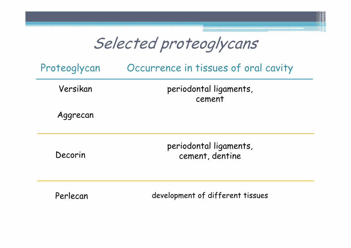
Selected proteoglycans
Proteoglycan
Versikan
Aggrecan
Decorin
Perlecan
Occurrence in tissues of oral cavity
periodontal ligaments,cement
periodontal ligaments,cement, dentine
development of different tissues
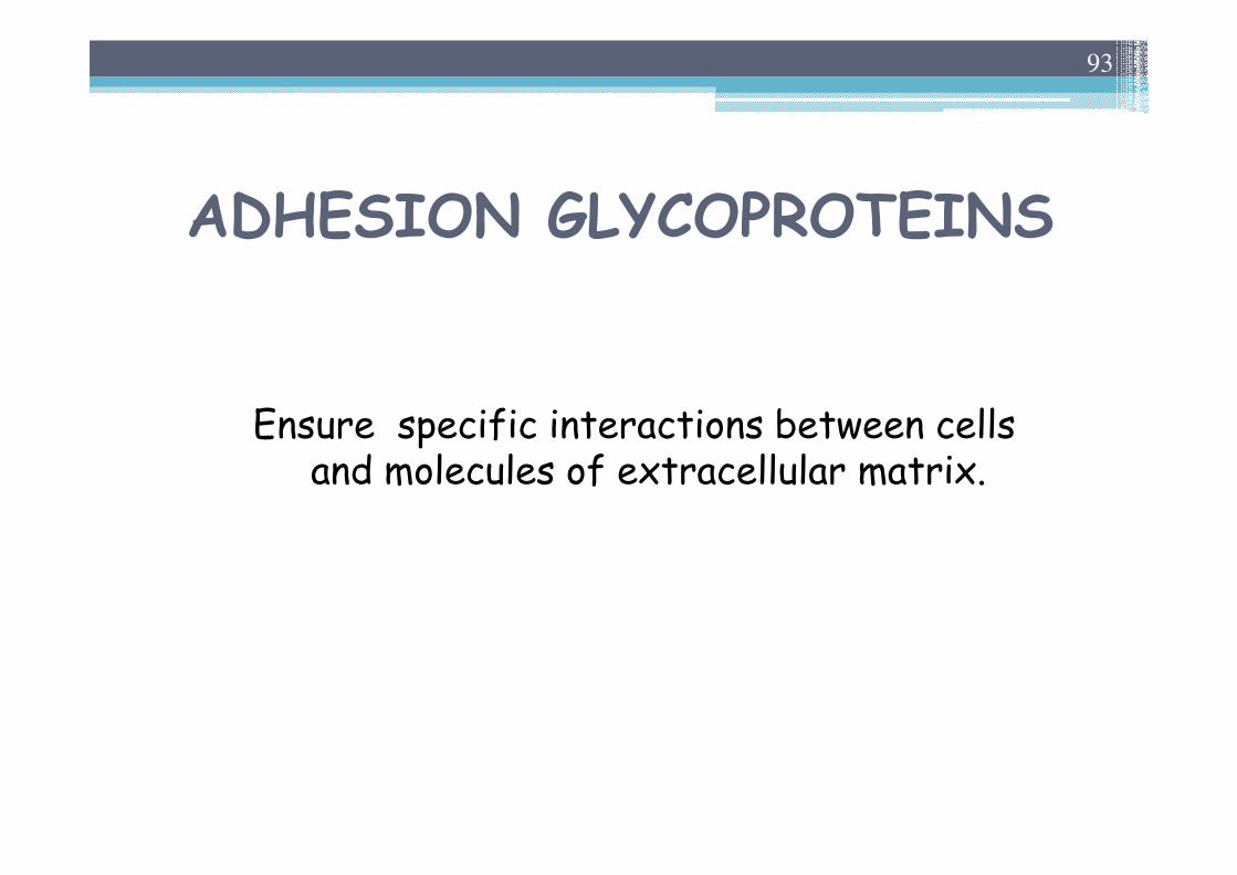
ADHESION GLYCOPROTEINS
Ensure specific interactions between cells and molecules of extracellular matrix.
93
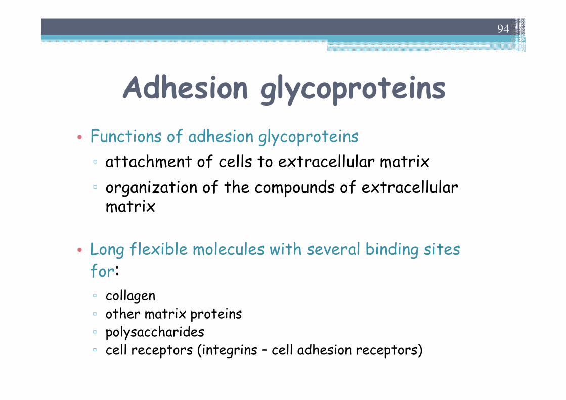
Adhesion glycoproteins
• Functions of adhesion glycoproteins
▫ attachment of cells to extracellular matrix
▫ organization of the compounds of extracellular matrix
• Long flexible molecules with several binding sitesfor:▫ collagen▫ other matrix proteins▫ polysaccharides▫ cell receptors (integrins – cell adhesion receptors)
94
Adhesion glycoproteins
• Selected representatives of adhesionglycoproteins
▫ fibronectin▫ laminin▫ osteonectin▫ chondronectin
95
Adhesion glycoproteins
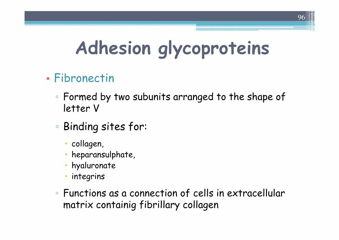
• Fibronectin
▫ Formed by two subunits arranged to the shape of letter V
▫ Binding sites for:
� collagen, � heparansulphate, � hyaluronate� integrins
▫ Functions as a connection of cells in extracellular matrix containig fibrillary collagen
96
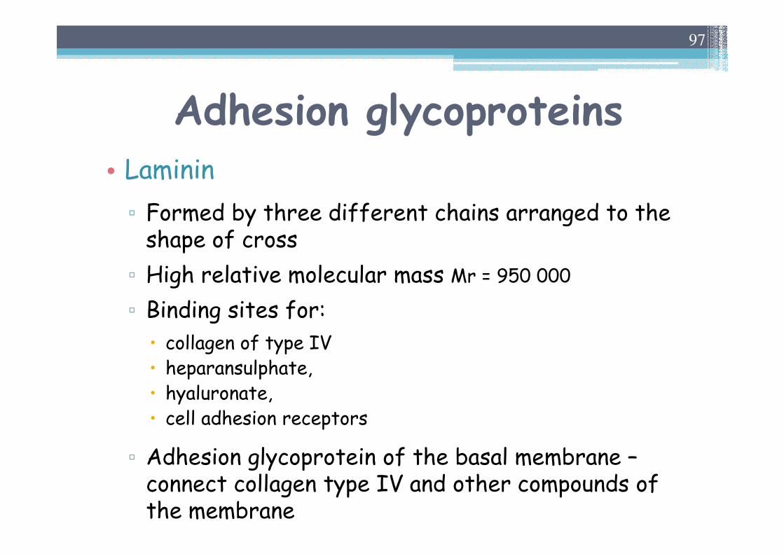
Adhesion glycoproteins• Laminin
▫ Formed by three different chains arranged to the shape of cross
▫ High relative molecular mass Mr = 950 000
▫ Binding sites for:
� collagen of type IV� heparansulphate, � hyaluronate,� cell adhesion receptors
▫ Adhesion glycoprotein of the basal membrane –connect collagen type IV and other compounds of the membrane
97