1. Benign disorders of WBCs By/ Mr. Waqqas Elaas; M.Sc; MLT 2.
30
1
-
Upload
john-garrett -
Category
Documents
-
view
216 -
download
0
Transcript of 1. Benign disorders of WBCs By/ Mr. Waqqas Elaas; M.Sc; MLT 2.
1
Benign disorders of WBCs
By/Mr. Waqqas Elaas;
M.Sc; MLT
2
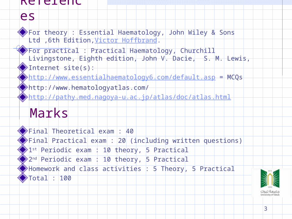
References
For theory : Essential Haematology, John Wiley & Sons Ltd ,6th Edition,Victor Hoffbrand.For practical : Practical Haematology, Churchill Livingstone, Eighth edition, John V. Dacie, S. M. Lewis, Internet site(s):http://www.essentialhaematology6.com/default.asp = MCQshttp://www.hematologyatlas.com/http://pathy.med.nagoya-u.ac.jp/atlas/doc/atlas.html
Final Theoretical exam : 40Final Practical exam : 20 (including written questions)1st Periodic exam : 10 theory, 5 Practical 2nd Periodic exam : 10 theory, 5 PracticalHomework and class activities : 5 Theory, 5 PracticalTotal : 100
Marks
3
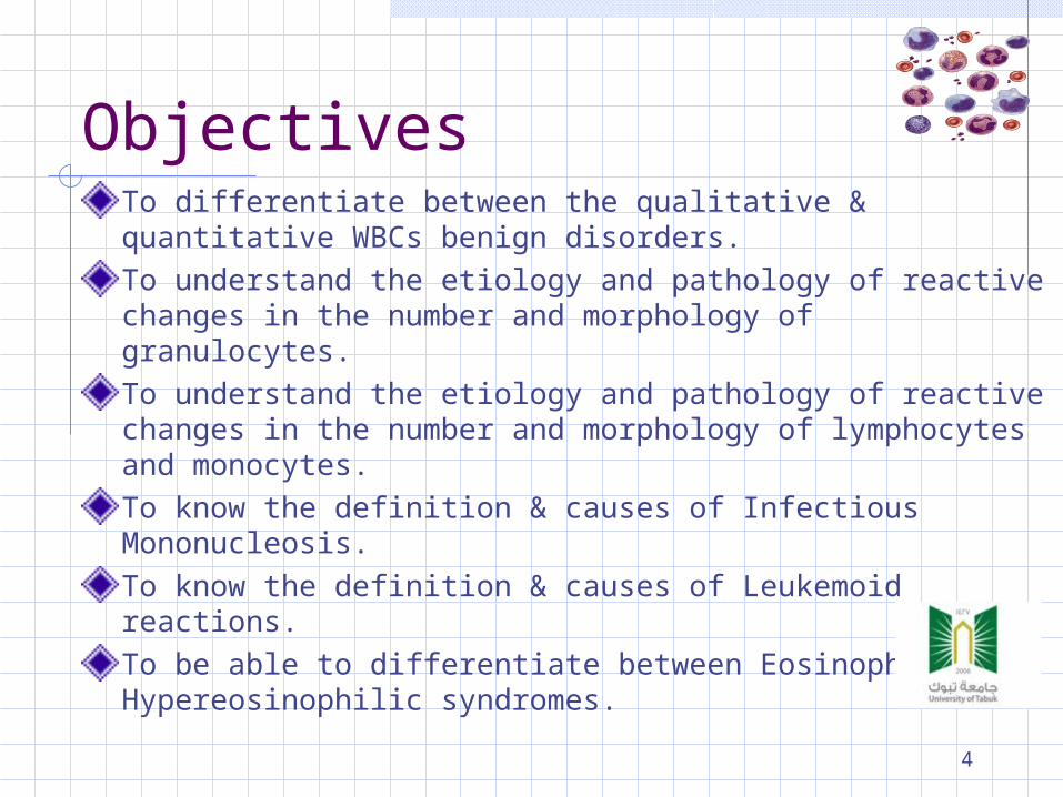
ObjectivesTo differentiate between the qualitative & quantitative WBCs benign disorders.To understand the etiology and pathology of reactive changes in the number and morphology of granulocytes.To understand the etiology and pathology of reactive changes in the number and morphology of lymphocytes and monocytes.To know the definition & causes of Infectious Mononucleosis.To know the definition & causes of Leukemoid reactions.To be able to differentiate between Eosinophilia & Hypereosinophilic syndromes.
4
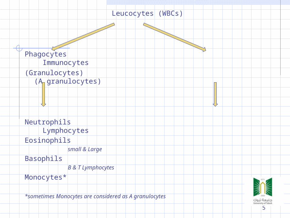
Leucocytes (WBCs)
Phagocytes Immunocytes(Granulocytes) (A granulocytes)
Neutrophils LymphocytesEosinophils small & Large
Basophils B & T Lymphocytes
Monocytes*
*sometimes Monocytes are considered as A granulocytes
5

Normal leucocytes morphology
6
LEUCOCYTES BENIGN DISORDERS
Quantitative Change in number
Terminology Cytosis / philia
Increase in number Cytopenia/penia
Decrease in number
Qualitative Morphologic changes Functional changes
7
LEUCOCYTES BENIGN DISORDERS Quantitative changes
Relative & Absolute values
To make an accurate assessment, consider both relative and absolute values. For example a relative value of 70% neutrophils may seem within normal limits; however, if the total WBC is 20,000, the absolute value (70% of 20,000) would be an abnormally high count of 14,000.
8
LEUCOCYTOSIS
Definition
Raised TWBC above 11.0 x 10⁹/L in adults, due
to elevation of any of a single lineage. Note: elevation of the minor cell populations
can occur without a rise in the total white cell count.
Normal reference range (adults) 4.5 -- 11.0 x 10⁹/L
9
LEUCOPENIA
DefinitionTWBC lower than 4.5 x 10⁹/L in adults Leucopenia may affect one or more lineages
and it is possible to be severely neutropenic or lymphopenic without a reduction in total white cell count.
10
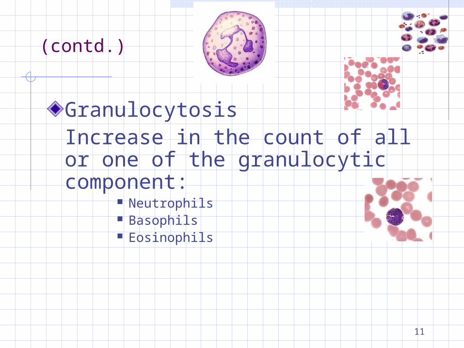
(contd.)
GranulocytosisIncrease in the count of all or one of the granulocytic component:
Neutrophils Basophils Eosinophils
11
NEUTROPHILIA
Definition Increase in the number of neutrophils and / or its
precursors In adults count >7.5 x 10⁹/L but the counts are
age dependent Increase may results from alteration in the
normal steady state of Production Transit Migration Destruction
12
NEUTROPHILIA (contd.)
Causes of Neutrophilia Infection
Bacterial Inflammatory conditions
Autoimmune disorders Gout
Neoplasia Metabolic conditions
Uraemia Acidosis Haemorhage
Corticosteroids Marrow infiltration/fibrosis Myeloproliferative disorders
13
Excessive reactive leucocytosis. Applied to chronic Neutrophilia with marked leucocytosis
(>20 x 109/L) The usual feature is the shift to the left of myeloid cells Causes include Infections Marrow infiltration Systemic disease (e.g.: Acute liver failure)
(Left shift : indicates that the neutrophils present in the blood are at a slightly earlier stage of maturation than usual. The Band and the stages before. This is often seen in acute infections).
(Right shift : an increase in the percentage of multilobed neutrophils).
Leukemoid reactions
14
NEUTROPENIA
Neutropenia is an absolute reduction in the number of circulating neutrophils
Mild (1- 1.5 x 10⁹/L) Moderate (0.5 – 1 x 10⁹/L) Severe (<0.5 x 10⁹/L)
Symptoms are rare with the neutrophil count above 1 x 10⁹/L
Bacterial infections are the commonest. Fungal, viral and parasitic infection are
relatively uncommon.
15
(NEUTROPENIA) contd.
Causes of Neutropenia Racial Congenital Marrow aplasia Marrow infiltration Megaloblastic anemia Acute infections
Typhoid, Miliary TB, viral hepatitis Drugs Irradiation exposure Immune disorders
HIV SLE Neonatal isoimmune and autoimmune neutropenia
Hyperslplenism
16
(EOSINOPHILIA)
Increase in the eosinophil count must prompt for further investigation (>0.6 x 10⁹/L)The causes of eosinophilia can be considered under following headings
Allergy Atopic, drug sensitivity and pulmonary eosinophilia
Infection Parasites, recovery from infections
Malignancy Hodgkin’s disease, NHL and myeloproliferative
disorders Drugs Skin disorders Gastrointestinal disorders Hypereosinophilic syndrome
17
(EOSINOPHILIA) Contd.
Hypereosinophilic syndrome Criteria of diagnosis
Peripheral blood eosinophil >1.5 x 10⁹/L Persistence of counts more than 6 months End organ damage Absence of any obvious cause for eosinophilia
Organ most commonly involved Heart Lung Skin Neurological
18
(MONOCYTOSIS)
Absolute monocyte count is age dependentCount rarely exceeds >1.0 x 10⁹/LHave no marrow reservesCauses of monocytosis can be grouped as Infections
Chronic infection (TB, typhoid fever, infective endocarditis)
Recovery from acute infection Malignant disease
MDS, AML, HD, NHL Connective tissue disorders
Ulcerative colitis, Sarcoidosis, Crohn’s disease Post splenectomy
19
(BASOPHILIA)
Basophils are least common of the granulocytesReference range for adult is 0 – 0.2 x 10⁹/LMost commonly associated with hypersensitivity reactions to drugs or foodInflammatory conditions e.g RA, ulcerative colitis are also sometime associated with basophiliaMyeloproliferative disordersChronic myeloid leukemia
20
(LYMPHOCYTOSIS)
The blood contain only few percent of total body lymphocytesThe most consistent variation is seen with ageAlteration of lymphocyte counts can result from
The redistribution of lymphocytes Absolute increase of lymphocyte number Loss of lymphocytes Combination of these
21
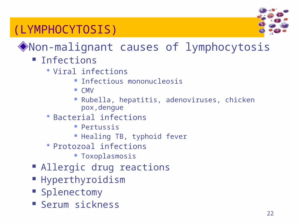
(LYMPHOCYTOSIS)
Non-malignant causes of lymphocytosis Infections
Viral infections Infectious mononucleosis CMV Rubella, hepatitis, adenoviruses, chicken pox,dengue
Bacterial infections Pertussis Healing TB, typhoid fever
Protozoal infections Toxoplasmosis
Allergic drug reactions Hyperthyroidism Splenectomy Serum sickness
22
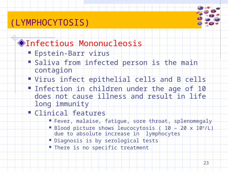
(LYMPHOCYTOSIS)
Infectious Mononucleosis Epstein-Barr virus Saliva from infected person is the main contagion Virus infect epithelial cells and B cells Infection in children under the age of 10 does not
cause illness and result in life long immunity Clinical features
Fever, malaise, fatigue, sore throat, splenomegaly Blood picture shows leucocytosis ( 10 – 20 x 109/L) due to
absolute increase in lymphocytes Diagnosis is by serological tests There is no specific treatment
23
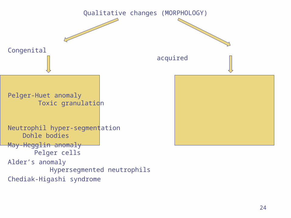
Qualitative changes (MORPHOLOGY)
Congenital acquired
Pelger-Huet anomaly Toxic granulation
Neutrophil hyper-segmentation Dohle bodies May-Hegglin anomaly Pelger cellsAlder’s anomaly Hypersegmented
neutrophils Chediak-Higashi syndrome
24
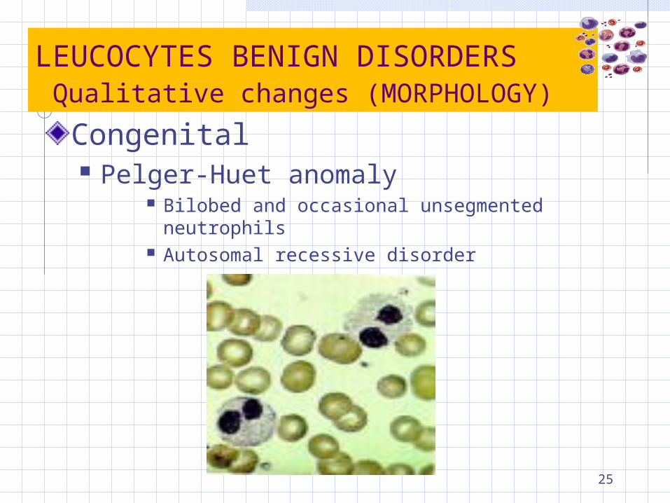
LEUCOCYTES BENIGN DISORDERS Qualitative changes (MORPHOLOGY)
Congenital Pelger-Huet anomaly
Bilobed and occasional unsegmented neutrophils Autosomal recessive disorder
25
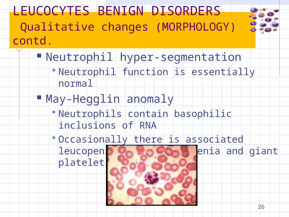
LEUCOCYTES BENIGN DISORDERS Qualitative changes (MORPHOLOGY) contd.
Neutrophil hyper-segmentation Neutrophil function is essentially normal
May-Hegglin anomaly Neutrophils contain basophilic inclusions of
RNA Occasionally there is associated leucopenia,
Thrombocytopenia and giant platelet are frequent
26
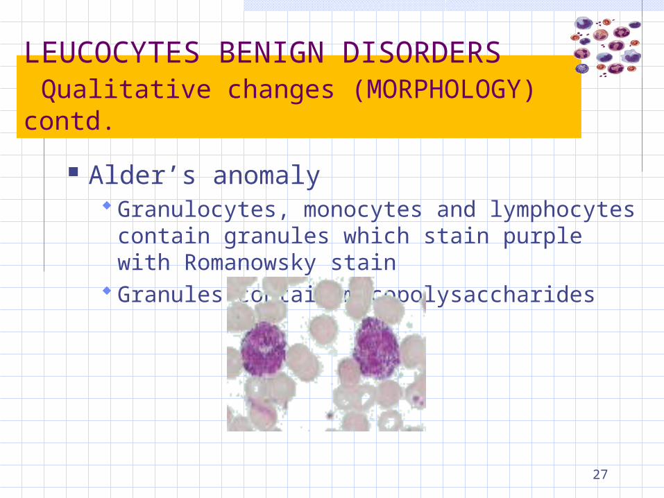
LEUCOCYTES BENIGN DISORDERS Qualitative changes (MORPHOLOGY) contd.
Alder’s anomaly Granulocytes, monocytes and lymphocytes
contain granules which stain purple with Romanowsky stain
Granules contain mucopolysaccharides
27
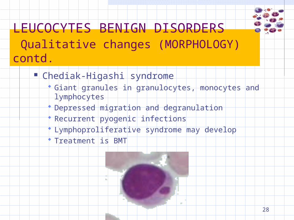
LEUCOCYTES BENIGN DISORDERS Qualitative changes (MORPHOLOGY) contd.
Chediak-Higashi syndrome Giant granules in granulocytes, monocytes and
lymphocytes Depressed migration and degranulation Recurrent pyogenic infections Lymphoproliferative syndrome may develop Treatment is BMT
28
LEUCOCYTES BENIGN DISORDERS Qualitative changes (MORPHOLOGY) contd.
Acquired Toxic granulation Dohle bodies Pelger cells Hypersegmented neutrophils
29
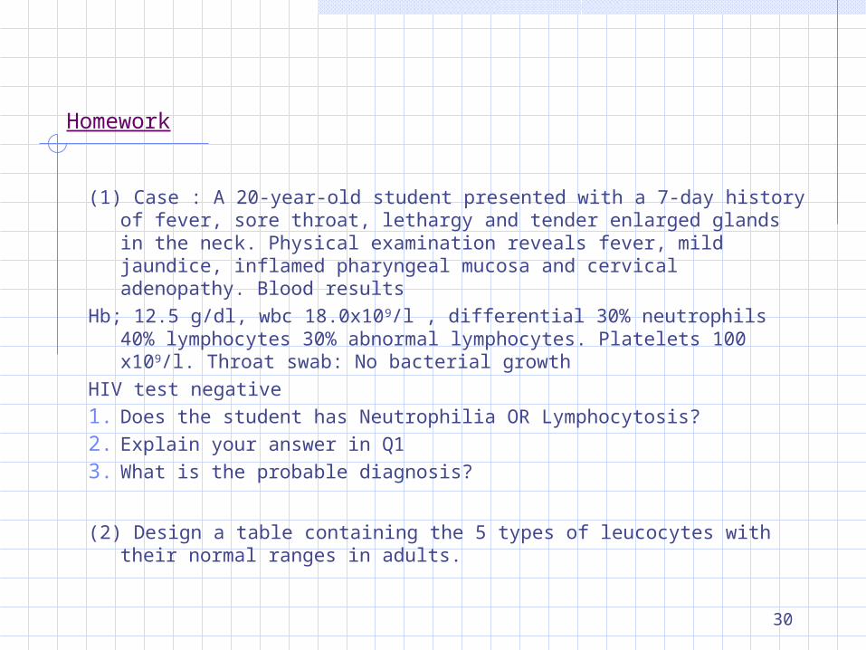
Homework
(1) Case : A 20-year-old student presented with a 7-day history of fever, sore throat, lethargy and tender enlarged glands in the neck. Physical examination reveals fever, mild jaundice, inflamed pharyngeal mucosa and cervical adenopathy. Blood results
Hb; 12.5 g/dl, wbc 18.0x10⁹/l , differential 30% neutrophils 40% lymphocytes 30% abnormal lymphocytes. Platelets 100 x109/l. Throat swab: No bacterial growth
HIV test negative1. Does the student has Neutrophilia OR Lymphocytosis?2. Explain your answer in Q13. What is the probable diagnosis?
(2) Design a table containing the 5 types of leucocytes with their normal ranges in adults.
30