1 Azin Nowrouzi, PhD TUMS. 2 1.Optical properties of amino acids –S–Stereoisomerism in -amino...
69
1 Azin Nowrouzi, PhD TUMS
-
Upload
dennis-underwood -
Category
Documents
-
view
224 -
download
2
Transcript of 1 Azin Nowrouzi, PhD TUMS. 2 1.Optical properties of amino acids –S–Stereoisomerism in -amino...

1
Azin Nowrouzi, PhD
TUMS

2
1. Optical properties of amino acids– Stereoisomerism in -amino acids, D and L isomers
2. Ionization of amino acids– Titration curves– Isoelectric pH (pI)
3. Chemical reactions of the carboxyl group– Include all the reactions of weak acids
• Salt production with bases• Esters with alcohols• Decarboxylation• Amides with amines
4. Chemical reactions of the amino group• Amides with carboxilic acids• Deamination• Reaction with ninhydrin• Reaction with dinitrofluorobenzene (DNP-aa)• Reaction with phenyl isothiocyanate
5. Separation and analysis of amino acids
Physical and chemical properties of amino acids

3
Examples of decarboxylation
• Formation of Serotonin from tryptophan.
• Formation of -aminobutyric acid (GABA) from glutamate.
• Formation of neurotransmitters, such as norepinephrine, dopamine, histamine in the nervous system.

4
Deamination• Deamination takes place in the liver.
• It is the process by which amino acids are broken down.
• The amino group is removed from the amino acid and converted to ammonia.
• The rest of the amino acid is made up of mostly carbon
and hydrogen, and is recycled or oxidized for energy.
• Ammonia is toxic to the human system, and enzymes convert it to urea or uric acid in the urea cycle.
• Urea and uric acid can safely diffuse into the blood and then be excreted in urine.
5
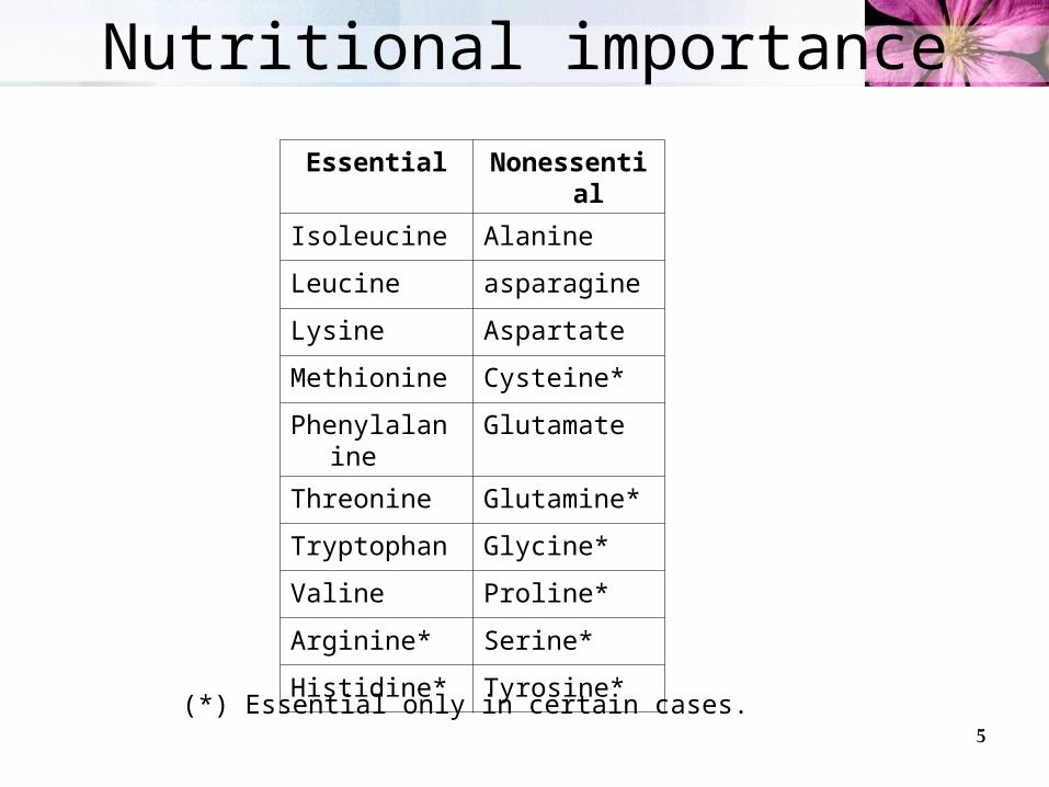
Nutritional importance
Essential Nonessential
Isoleucine Alanine
Leucine asparagine
Lysine Aspartate
Methionine Cysteine*
Phenylalanine Glutamate
Threonine Glutamine*
Tryptophan Glycine*
Valine Proline*
Arginine* Serine*
Histidine* Tyrosine*
(*) Essential only in certain cases.

6
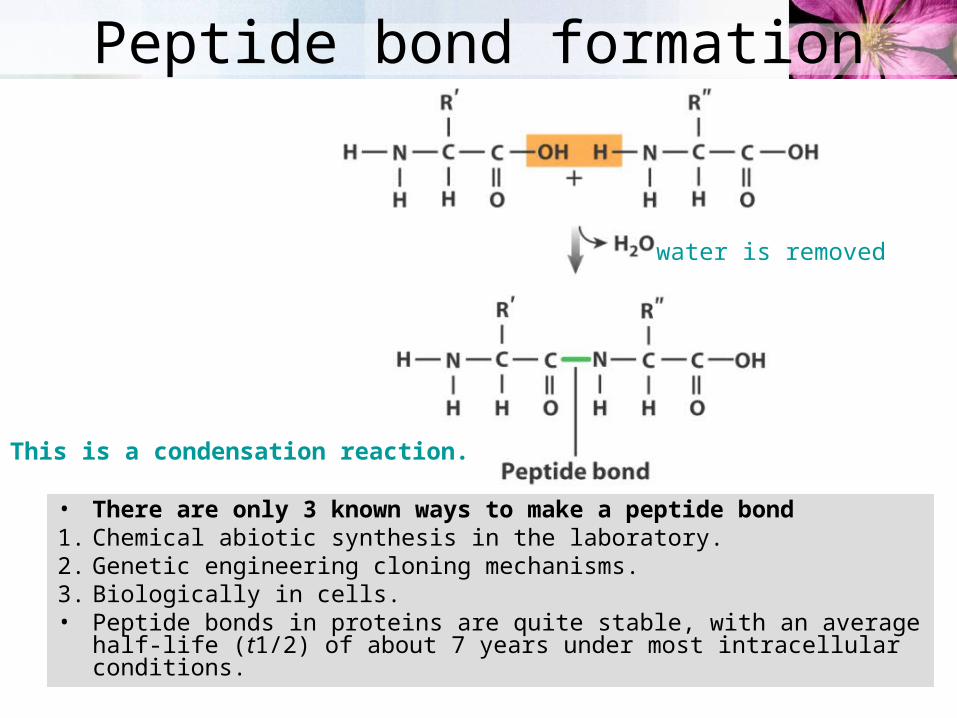
3- Chemical reactions of the carboxyland the amino groups (Amide or peptide bond formation)
• Polymers of amino acids.• Two amino acids join covalently through a peptide bond.• Another name for peptide bond is amide bond or linkage.
7
water is removed
Peptide bond formation
• There are only 3 known ways to make a peptide bond1. Chemical abiotic synthesis in the laboratory.2. Genetic engineering cloning mechanisms.3. Biologically in cells.• Peptide bonds in proteins are quite stable, with an average half-life
(t1/2) of about 7 years under most intracellular conditions.
This is a condensation reaction.

8
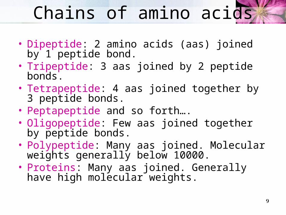
Peptides are chains of amino acids
9
Chains of amino acids
• Dipeptide: 2 amino acids (aas) joined by 1 peptide bond.
• Tripeptide: 3 aas joined by 2 peptide bonds.• Tetrapeptide: 4 aas joined together by 3 peptide
bonds.• Peptapeptide and so forth…. • Oligopeptide: Few aas joined together by
peptide bonds.• Polypeptide: Many aas joined. Molecular weights
generally below 10000.• Proteins: Many aas joined. Generally have high
molecular weights.

10
Nomenclature
• The pentapeptide serylglycyltyrosylalanylleucine, or Ser–Gly–Tyr–Ala–Leu.
• Peptides are named beginning with the amino terminal residue, which by convention is placed at the left.
• The peptide bonds are shaded in yellow; the R groups are in red.

11
Ionization behavior of peptides
carboxyl-terminal (C-terminal) residue
amino-terminal (N-terminal) residue
Like free amino acids, peptides have characteristic titration curves and a characteristic isoelectric pH (pI) at which they do not move in an electric field.
Alanylglutamylglycyllysine

12
Physical and chemical properties of amino acids
1. Optical properties of amino acids– Stereoisomerism in -amino acids
2. Ionization of amino acids– Titration curves– Isoelectric pH (pI)
3. Chemical reactions of the carboxyl group– Include all the reactions of weak acids.
• Salt production with bases• Esters with alcohols• Decarboxylation• Amides with amines
4. Chemical reactions of the amino group• Amides with carboxilic acids• Reaction with ninhydrin (detection and measurement of concentration).• Reaction with dinitrofluorobenzene (DNP-aa) (detection of first amino
acid in a chain and others that have side chains with amino groups)• Reaction with phenyl isothiocyanate (protein sequencing).
5. Separation and analysis of amino acids
13
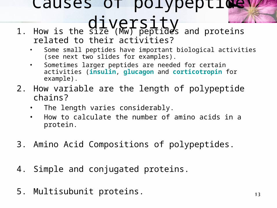
Causes of polypeptide diversity 1. How is the size (Mw) peptides and proteins related to
their activities? • Some small peptides have important biological activities (see next
two slides for examples).• Sometimes larger peptides are needed for certain activities (insulin,
glucagon and corticotropin for example).
2. How variable are the length of polypeptide chains?• The length varies considerably.• How to calculate the number of amino acids in a protein.
3. Amino Acid Compositions of polypeptides.
4. Simple and conjugated proteins.
5. Multisubunit proteins.

14
Some peptides with important biological activities
Peptide hormones:
Oxytocin- a nonapeptide involved in parturition and lactation.
Vasopressin-maintains water balance Structurally similar, but
different functions Contain disulfide bridges
covalent linkages intramolecular cross-links.
Enkephalins Either of two penta-peptides
with opiate & analgesic activity (involved in pain control).
Occur naturally in brain & have marked affinity for opiate receptors.
• Aspartame low calorie sweetener L-aspartyl-L-phenylalanine
methyl ester.
15
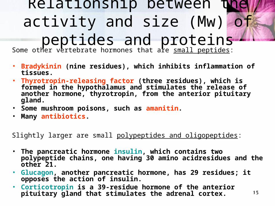
Relationship between the activity and size (Mw) of peptides and proteins
Some other vertebrate hormones that are small peptides:
• Bradykinin (nine residues), which inhibits inflammation of tissues.
• Thyrotropin-releasing factor (three residues), which is formed in the hypothalamus and stimulates the release of another hormone, thyrotropin, from the anterior pituitary gland.
• Some mushroom poisons, such as amanitin.• Many antibiotics.
Slightly larger are small polypeptides and oligopeptides: • The pancreatic hormone insulin, which contains two polypeptide
chains, one having 30 amino acidresidues and the other 21.• Glucagon, another pancreatic hormone, has 29 residues; it
opposes the action of insulin. • Corticotropin is a 39-residue hormone of the anterior pituitary
gland that stimulates the adrenal cortex.
16
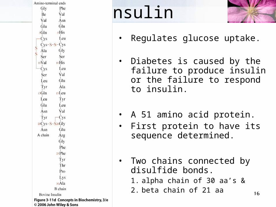
Insulin• Regulates glucose uptake.
• Diabetes is caused by the failure to produce insulin or the failure to respond to insulin.
• A 51 amino acid protein.• First protein to have its
sequence determined.
• Two chains connected by disulfide bonds.1. alpha chain of 30 aa’s & 2. beta chain of 21 aa

17
Length of polypeptide chains• Lengths vary considerably.

18
How to calculate the number of amino acids in a protein
• We can calculate the approximate number of amino acid residues in a simple protein containing no other chemical constituents by dividing its molecular weight by 110.
• Although the average molecular weight of the 20 common amino acids
is about 138, the smaller amino acids predominate in most proteins.
• If we take into account the proportions in which the various amino acids occur in proteins, the average molecular weight of protein amino acids is nearer to 128.
• Because a molecule of water (Mr 18) is removed to create each peptide bond, the average molecular weight of an amino acid residue in a protein is about 128 -18 = 110.

19
Polypeptides have characteristicamino acid compositions
• The 20 common amino acids almost never occur in equal amounts in a protein.
• Some amino acids may
occur only once or not at all in a given type of protein; others may occur in large numbers.
20
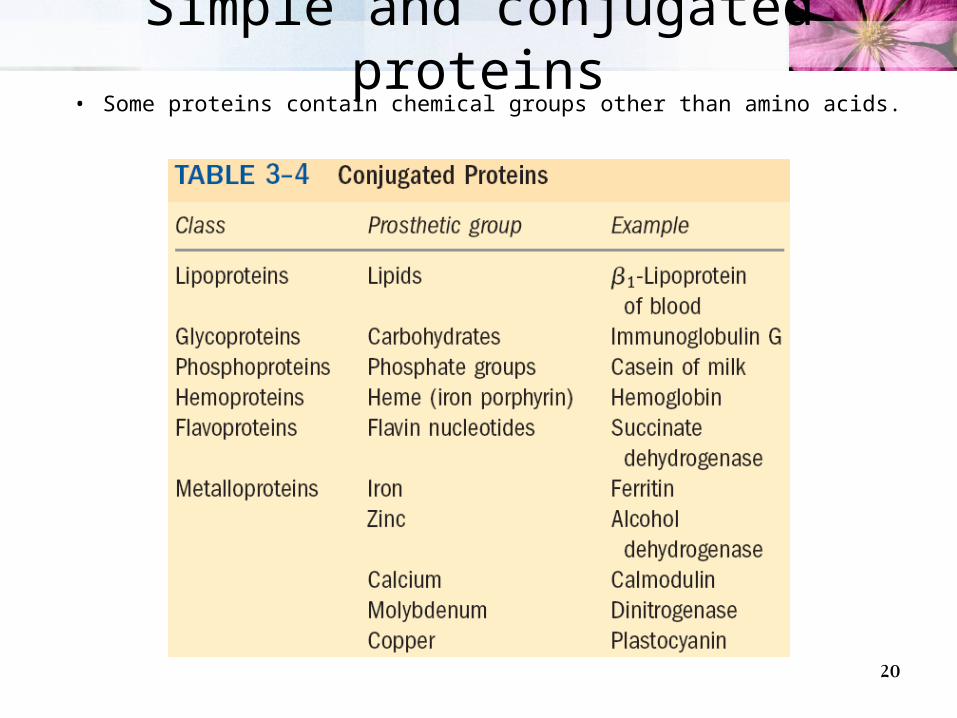
Simple and conjugated proteins• Some proteins contain chemical groups other than amino acids.

21
Multisubunit proteins
• When two or more polypeptides are associated noncovalently.– Hemoglobin, for example, has four polypeptide
subunits: two identical chains and two identical chains, all four held together by noncovalent interactions.
• A few proteins contain two or more polypeptide chains linked covalently. – For example, the two polypeptide chains of insulin are
linked by disulfide bonds. In such cases, the individual polypeptides are not considered subunits but are commonly referred to simply as chains.

22
Levels of structure in proteins

23
Levels of Protein StructureLevels of Protein Structure
Primary structure
Secondary structures
Tertiary structure
Quaternary structure
Amino acid sequence
Rigidity of the peptide bond
-carbons of adjacent amino acids are separated by three covalent bondsC-C-N-C
• Tetrahedral angles:
N-C bond is labeled (phi).
C-C bond is labeled (psi).
• Repetitive structures-Helix-Sheet
•parallel•antiparallel
• Non-repetitive structures-turn

24
Primary structure• A description of all
covalent bonds (mainly peptide and disulfide bonds) linking amino acid residues in a polypeptide chain.
• The linear sequence of amino acids within a peptide
• Written from NC, – either in three-letter code,– or, more often, in one-letter
code. • Example: Glu-Gly-Ala-Lys or
EGAK

25
Insulin’s primary structure
26
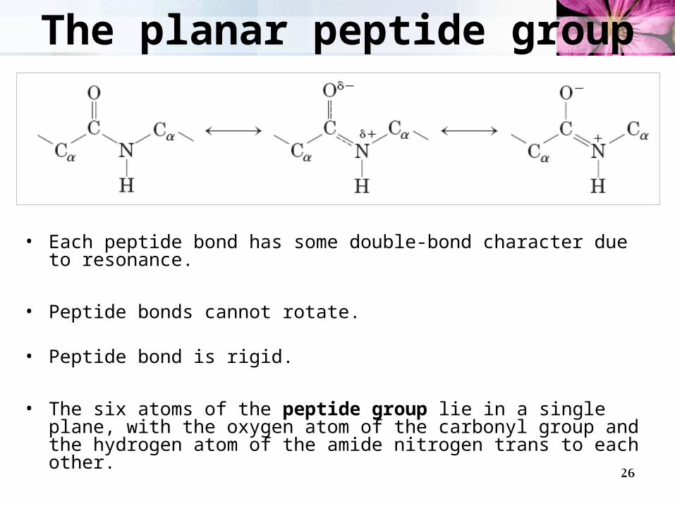
The planar peptide group
• Each peptide bond has some double-bond character due to resonance.
• Peptide bonds cannot rotate.
• Peptide bond is rigid.
• The six atoms of the peptide group lie in a single plane, with the oxygen atom of the carbonyl group and the hydrogen atom of the amide nitrogen trans to each other.

27
• The carbons of adjacent amino acid residues are separated by three covalent bonds, arranged as C-C-N-C.
• The N-C and C-C bonds can rotate, with bond angles designated and , respectively.
• The peptide C-N bond is not free to rotate.
• Other single bonds in the backbone may also be rotationally hindered, depending on the size and charge of the R groups.

28
Steric hindrance
• By convention, both and are defined as 0 when the two peptide bonds flanking that carbon are in the same plane.
• In a protein, this conformation is prohibited by steric overlap between an -carbonyl oxygen and an -amino hydrogen atom.
• To illustrate the bonds between atoms, the balls representing each atom are smaller than the van der Waals radii for
• this scale. 1 Å 0.1 nm.

29
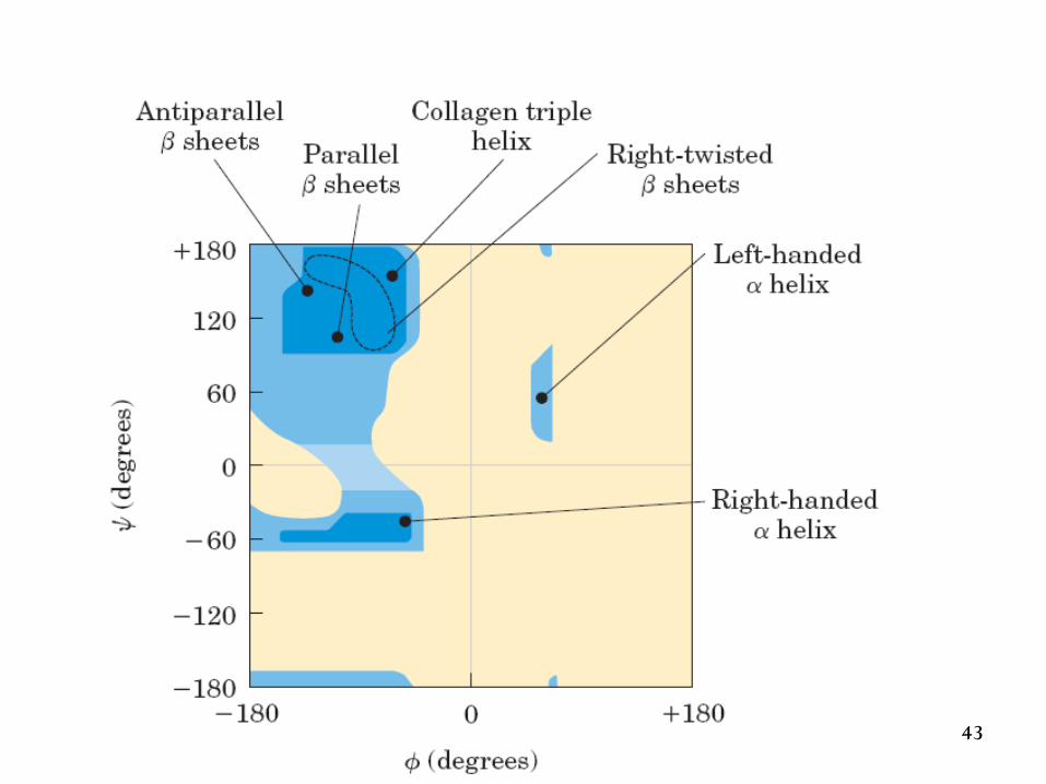
Ramachandran plot for L-Ala residues
• The areas shaded dark blue reflect conformations that involve no steric overlap and thus are fully allowed.
• Medium blue indicates conformations allowed at the extreme limits for unfavorable atomic contacts.
• The lightest blue area reflects conformations that are permissible if a little flexibility is allowed in the bond angles.

30
Human PCNA

31
Four models of the helix

32
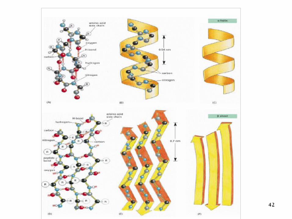
Helix
• H-bonds are inside the chain

33
Description of -helix
• The polypeptide backbone is tightly wound around an imaginary axis drawn longitudinally through the middle of the helix.
• The R groups of the amino acid residues protrude outward from the helical backbone.
• The amino acid residues in an -helix have conformations with = -40 to -50 and = -60.
• Each helical turn includes 3.6 amino acid residues.
• About one-fourth of all amino acid residues in polypeptides are found in -helices.
34
Stabilization of -helix• The structure is stabilized by a hydrogen bond between
the hydrogen atom attached to the electronegative nitrogen atom of a peptide linkage (amino acid n) and the electronegative carbonyl oxygen atom of the fourth amino acid (amino acid n+4) on the amino-terminal side of that peptide bond.
• Each successive turn of the -helix is held to adjacent turns by three to four hydrogen bonds.
• All the hydrogen bonds combined give the entire helical structure considerable stability.
• Naturally occurring L-amino acids can form either right- or left-handed helices, but extended left-handed helices have not been observed in proteins.

35
Factors affecting stability1. Electrostatic repulsion (or attraction) between successive amino acid residues
with charged R groups.
2. Bulkiness of adjacent R groups.
3. The interactions between R groups spaced three (or four) residues apart.
4. Presence of Pro or Gly residues.– Proline is only rarely found within an helix
• In proline, the nitrogen atom is part of a rigid ring and rotation about the N-C bond is not possible. Thus, a Pro residue introduces a destabilizing kink in an helix.
• In addition, the nitrogen atom of a Pro residue in peptide linkage has no substituent hydrogen to participate in hydrogen bonds with other residues.
– Glycine occurs infrequently in helices for a different reason• It has more conformational flexibility than the other amino acid residues. • Polymers of glycine tend to take up coiled structures quite different from an -helix.
5. Interaction between amino acid residues at the ends of the helical segment and the electric dipole inherent to the helix.

36
Helix dipole
• A net dipole extends along the helix that increases with helix length.
• Negatively charged amino acids
are often found near the amino terminus of the helical segment, where they have a stabilizing interaction with the positive charge of the helix dipole.
• A positively charged amino acid at the aminoterminal end is destabilizing.

37
Fully extended chains can form -sheets

38
Types of -sheets

39
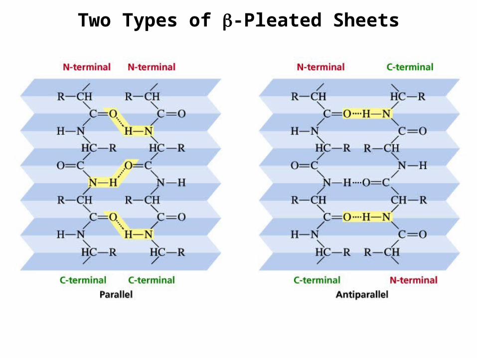
- pleated sheet
• H-bonds are between chains.
= 140° = -120°.
40
Two Types of -Pleated Sheets

41
The conformation of polypeptide chains
42
43

44

45
Non-repetitive secondary structures
1. Turns
2. Connections
3. Loops
4. Coils or random coils
• These are well ordered but non repeating configurations.

46
turns
• type I turns occur more than twice as frequently as type II. • Type II turns always have Gly as the third residue.

47
• Amino acids that are far apart in the polypeptide sequence and that reside in different types of secondary structure may interact within the completely folded structure of a protein.
• The location of bends (including turns) in the polypeptide chain and the direction and angle of these bends are determined by the number and location of specific bend-producing residues, such as Pro, Thr, Ser, and Gly.
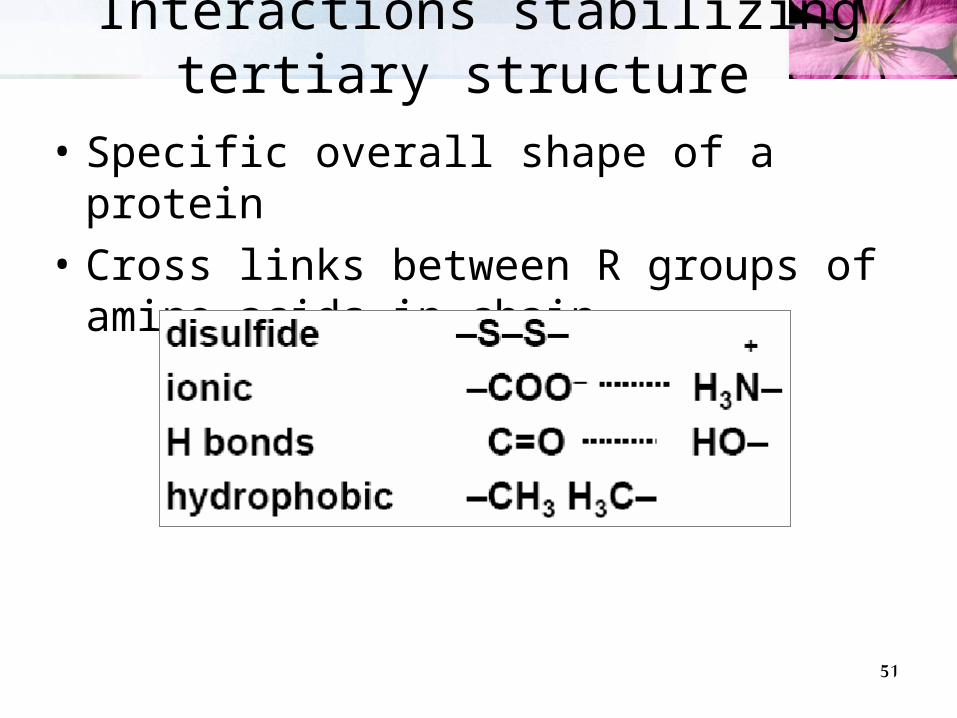
• Interacting segments of polypeptide chains are held in their characteristic tertiary positions by different kinds of weak bonding interactions (and sometimes by covalent bonds such as disulfide cross-links) between the segments.
Leptin
Tertiary structure

48
Stabilization of tertiary Structure• overall three-
dimensional arrangement of all atoms in a protein is referred to as the protein’s tertiary structure.
• Stabilized primarily through weak bonds.

49
Folding of a polypeptide chain

50
Three-dimensional structures of some small proteins
• PDB; www.rcsb.org/pdb
MyoglobinPDB ID 1MBO
LysozymePDB ID 3LYMCytochrome c
PDB ID 1CCR
RibonucleasePDB ID 3RN3
51
Interactions stabilizing tertiary structure
• Specific overall shape of a protein
• Cross links between R groups of amino acids in chain

52
Domains
• When molecular weight is larger than 20000.• The ratio of surface area to volume is small.• A protein with multiple domains may appear to
have a distinct globular lobe for each domain.
53
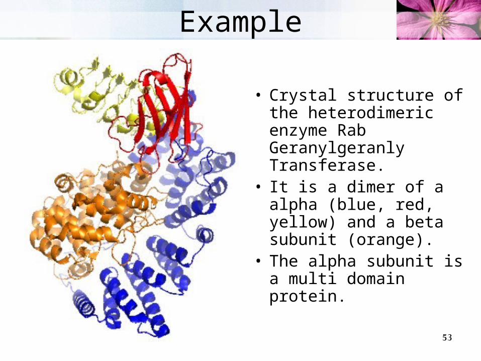
Example
• Crystal structure of the heterodimeric enzyme Rab Geranylgeranly Transferase.
• It is a dimer of a alpha (blue, red, yellow) and a beta subunit (orange).
• The alpha subunit is a multi domain protein.
54
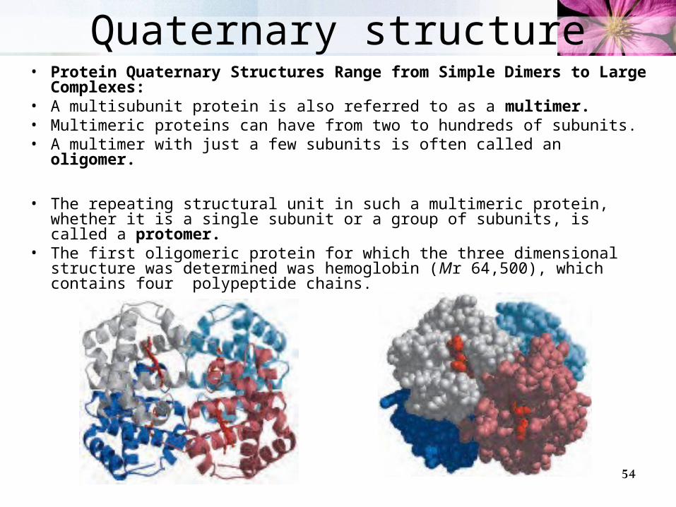
Quaternary structure• Protein Quaternary Structures Range from Simple Dimers to
Large Complexes:• A multisubunit protein is also referred to as a multimer.• Multimeric proteins can have from two to hundreds of subunits. • A multimer with just a few subunits is often called an oligomer.
• The repeating structural unit in such a multimeric protein, whether it is a single subunit or a group of subunits, is called a protomer.
• The first oligomeric protein for which the three dimensional structure was determined was hemoglobin (Mr 64,500), which contains four polypeptide chains.
55
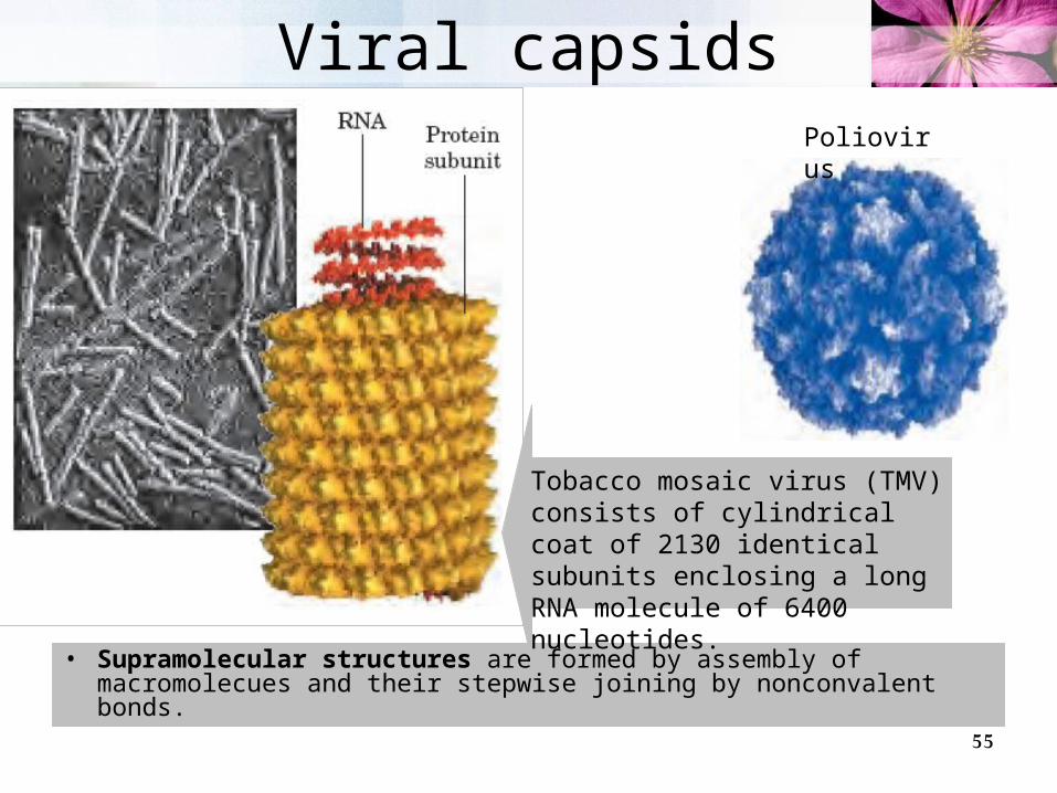
Viral capsids
• Supramolecular structures are formed by assembly of macromolecues and their stepwise joining by nonconvalent bonds.
Poliovirus
Tobacco mosaic virus (TMV) consists of cylindrical coat of 2130 identical subunits enclosing a long RNA molecule of 6400 nucleotides.

56

57
Denaturation and Renaturation
• A loss of three-dimensional structure sufficient to cause loss of function is called denaturation.
• Denaturing agents include:1. Heat2. pH3. Certain miscible organic solvents such as alcohol
or acetone.4. Certain solutes such as urea and guanidine
hydrochloride.5. Detergents, such as sodium dodecyl sulfate (SDS).
• Denaturation of some proteins is reversible.– This process is called renaturation

58
Renaturation of unfolded, denatured ribonuclease
• Urea is used to denature ribonuclease, and mercaptoethanol (HOCH2CH2SH) to reduce and thus cleave the disulfide bonds to yield eight Cys residues.
• Renaturation involves
reestablishment of the correct disulfide cross-links.
59
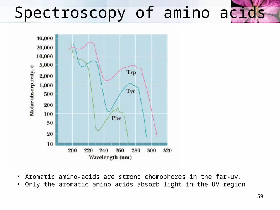
Spectroscopy of amino acids
• Aromatic amino-acids are strong chomophores in the far-uv.• Only the aromatic amino acids absorb light in the UV region

60
Absorbance can be measured by UV-spectrophotometer

61
Ninhydrin-detection of amino acids• Complete hydrolysis for 24 hr at 110 oC in 6 M HCl.• Amino acids can be detected on the chromatogram by using
ninhydrin. A solution of ninhydrin is sprayed onto the paper and heated. The amino acids show up as purple spots (proline appears yellow).

62
Paper chromatograms
2D chromatogram eluting with a different solvent mixture in each direction.

63
Electrophoresis• Electrophoresis is a technique that uses the net charge of peptides
(amino acids) as a basis for separation. A potential difference is applied across a solid material (e.g. paper
for amino acid analysis) permeated by an electrolyte.
Anions migrate to the anode and cations to the cathode. The rate of diffusion is related to the size and net charge. Small highly charged proteins migrate more quickly.
64
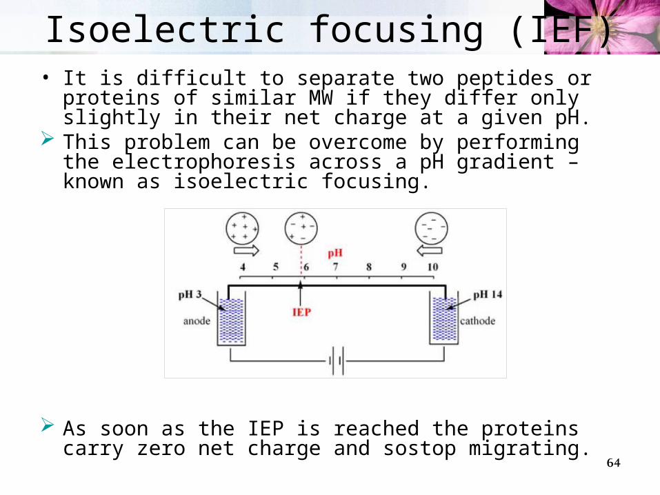
Isoelectric focusing (IEF)• It is difficult to separate two peptides or proteins of
similar MW if they differ only slightly in their net charge at a given pH.
This problem can be overcome by performing the electrophoresis across a pH gradient – known as isoelectric focusing.
As soon as the IEP is reached the proteins carry zero net charge and sostop migrating.

65
Isoelectric focusing (IEF)• This technique separates
proteins according to their isoelectric points.

66
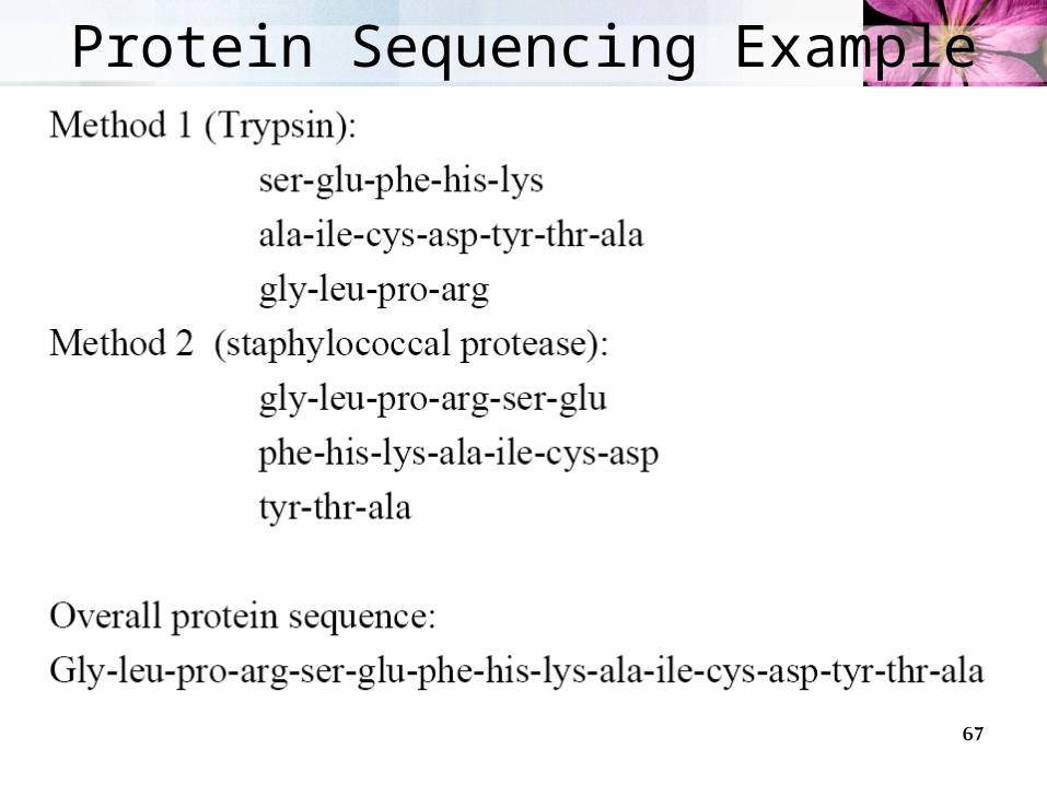
1. Proteins larger than 50 aa are first hydrolyzed into shorter peptides.
2. Chemical or enzymatic methods hydrolyze proteins at specific sites.
3. Peptides are separated by chromatography
4. Peptides generated by 2 or more cleavage methods are each sequenced separately.
5. Sequences of individual peptides are overlapped together to deduce the entire protein sequence
Specific cleavage of polypeptides
67
Protein Sequencing Example

68
Protein structureProtein structure1. 3d structure of a protein isdetermined byIts amino acid sequence.
2. The function of a protein depends on its structure.
3. Isolated proteins exist in one or a small number of stable structural forms.
4. Specific structuresof proteins are stabilizedby weak non covalent interactions.
5. Common structural patterns can help us organize our understanding of protein architecture.
69
Types of proteins• In considering these higher levels of structure, it is useful
to classify proteins into two major groups: • Fibrous proteins, having polypeptide chains arranged in
long strands or sheets.– Fibrous proteins usually consist largely of a single type of
secondary structure.– Provide support, shape, and external protection to vertebrates -Keratin– Collagen
• Globular proteins, having polypeptide chains folded into a spherical or globular shape.– Globular proteins often contain several types of secondary
structure– Most enzymes and regulatory proteins are globular proteins– Myoglobin