1 Active nuclear import and cytoplasmic retention of Activation ...
32
1 Active nuclear import and cytoplasmic retention of Activation Induced Deaminase Anne-Marie Patenaude 1 , Alexandre Orthwein 1,2 , Yi Hu 1,3 , Vanina A Campo 1 , Bodil Kavli 3 , Alejandro Buschiazzo 4,5 and Javier M Di Noia 1,2,6 1 Institut de Recherches Cliniques de Montréal, 110 Av des Pins Ouest, Montréal, QC, H2W 1R7, Canada. 2 Department of Microbiology and Immunology, Université de Montréal, Montréal, QC, Canada. 3 Department of Cancer Research and Molecular Medicine, Norwegian University of Science and Technology, Erling Skjalgssons gt. 1, DMF, 7006 Trondheim, Norway. 4 Institut Pasteur de Montevideo, Unidad de Cristalografia de Proteinas, Montevideo, Uruguay. Mataojo 2020, Montevideo 11400, Uruguay. 5 Institut Pasteur, Département de Biologie Structurale & Chimie, Paris, France. 6 Department of Medicine, University of Montreal, Montreal, QC, Canada. Correspondence should be addressed to J.M.D email: [email protected] Tel: 1-514-987-5642, Fax: 1-514-987-5645.
-
Upload
trinhxuyen -
Category
Documents
-
view
224 -
download
0
Transcript of 1 Active nuclear import and cytoplasmic retention of Activation ...
1
Active nuclear import and cytoplasmic retention of Activation Induced Deaminase
Anne-Marie Patenaude1, Alexandre Orthwein1,2, Yi Hu1,3, Vanina A Campo1, Bodil Kavli3, Alejandro
Buschiazzo4,5 and Javier M Di Noia1,2,6
1Institut de Recherches Cliniques de Montréal, 110 Av des Pins Ouest, Montréal, QC, H2W 1R7,
Canada.
2Department of Microbiology and Immunology, Université de Montréal, Montréal, QC, Canada.
3Department of Cancer Research and Molecular Medicine, Norwegian University of Science and
Technology, Erling Skjalgssons gt. 1, DMF, 7006 Trondheim, Norway.
4Institut Pasteur de Montevideo, Unidad de Cristalografia de Proteinas, Montevideo, Uruguay. Mataojo
2020, Montevideo 11400, Uruguay.
5Institut Pasteur, Département de Biologie Structurale & Chimie, Paris, France.
6Department of Medicine, University of Montreal, Montreal, QC, Canada.
Correspondence should be addressed to J.M.D email: [email protected] Tel: 1-514-987-5642,
Fax: 1-514-987-5645.

2
Abstract
The enzyme Activation Induced Deaminase (AID) triggers antibody diversification in B-cells by
catalyzing deamination and consequently mutation of immunoglobulin genes. To minimize off-target
deamination, AID is restrained by several regulatory mechanisms including nuclear exclusion, thought
to be mediated exclusively by active nuclear export. Here we identify two other mechanisms involved
in controlling AID subcellular localization. AID is unable to passively diffuse into the nucleus, despite
its small size, its nuclear entry requiring active import mediated by a conformational nuclear
localization sequence (NLS). We also identify a determinant for AID cytoplasmic retention in its C-
terminus, which hampers diffusion to the nucleus, competes with nuclear import and is critical for
maintaining the predominantly cytoplasmic localization of AID in steady-state conditions. Blocking
nuclear import alters the balance between these processes in favor of cytoplasmic retention, resulting in
reduced isotype class switching.

3
INTRODUCTION
Activation Induced Deaminase (AID) is the enzyme responsible for the diversification of rearranged
antibody genes in activated B cells1,2. AID acts as a mutator by deaminating deoxycytidine in single
stranded DNA thereby changing the base, cytosine, into a uracil (reviewed in3). Through further
processing by DNA repair enzymes that recognize uracil in DNA, this single biochemical activity
triggers different genetic modifications that are critical for a proper antibody response (reviewed in3,4).
Thus, AID initiates somatic hypermutation (SHM) and Ig gene conversion at the immunoglobulin (Ig)
variable regions allowing for affinity maturation of the antibody response1,2. AID also initiates class
switch recombination (CSR), which exchanges the exons encoding the Fc region of the antibody from
the default IgM to another isotype1,2. All the known components in these pathways except for AID are
ubiquitous DNA repair enzymes (reviewed in3-5). Indeed, AID is able to trigger SHM and CSR in non-
B cell models6,7 and since such mutagenic and recombinogenic enzyme is potentially dangerous, it
needs to be tightly regulated. This is highlighted by the cancer predisposition phenotype observed in
transgenic mice overexpressing AID7, by the finding that ectopic SHM can occur in proto-oncogenes
and tumor suppressor genes8-10 and by the involvement of AID in oncogenic chromosomal
translocations11,12. AID is normally induced in germinal center B cells13 but in order to ensure that the
genetic modifications it can cause are largely restricted to the Ig loci, there are multiple points of post-
transcriptional regulation such as regulation of mRNA stability and translation14-16, subcellular
localization17,18, protein stability19 and modification by phosphorylation20-22.
The subcellular localization of AID is especially interesting. The initially puzzling observation that an
AID-GFP fusion was exclusively cytoplasmic23 could be later explained by the fact that AID is a
nuclear-cytoplasmic shuttling protein. The mechanism by which AID is transported out of the nucleus
is known; a Leucine-rich nuclear export signal (NES) at the C-terminus of AID is recognized by the
exportin CRM117,18,24. However, the mechanism by which AID enters the nucleus has not been studied
in any detail and the observations available are inconsistent. While one report proposes a classical
bipartite nuclear localization signal (NLS) at the N-terminus of AID17, others failed to find evidence for
such a signal, suggesting that AID may simply diffuse into the nucleus18,24. We have investigated the
mechanism by which human AID enters the nucleus to address this controversy. We find that, despite
its small size, AID is actually unable to passively diffuse into the nucleus, requiring instead active
import. In addition, we describe a novel cytoplasmic retention mechanism for AID, which contributes
to the observed nuclear exclusion in steady state, thus restraining its function.

4
RESULTS
AID is actively imported into the nucleus
The expression of AID-GFP in several cell types resulted in exclusive cytoplasmic localization in
steady state, with this changing to nuclear localization after inhibition of CRM1-mediated nuclear
export by leptomycin B, as previously reported17,18,24. We indeed observed that AID-GFP accumulated
into the nucleus after leptomycin B treatment in the majority of transiently transfected 293T (80% after
4 h, n = 206) and HeLa (96% after 1 h, n = 53) cells and in stably expressing Ramos B cells (93% after
1 h, n = 91). Moreover, AID-GFP accumulated in the nucleus with different kinetics depending on the
cell line (Fig. 1 a). These observations are not consistent with the previous proposal that AID
subcellular localization is determined exclusively by active nuclear export and nuclear entry occurring
by passive diffusion18,24. In that case AID-GFP would be expected to follow the mass action law and
reach a more homogeneous distribution between nucleus and cytoplasm after leptomycin B treatment.
Rather, additional mechanisms seem to control AID localization.
Since active translocation across nuclear pores requires energy25,26, nuclear proteins that are not
retained should diffuse out of the nucleus when ATP is depleted27. We depleted ATP from HeLa cells
by glucose deprivation and assessed the localization of AID L198S-GFP, a constitutively nuclear AID
variant carrying a point mutation inactivating the NES18, and NLSSV40-GFP. The latter was used as a
positive control since it is small enough to diffuse through the nuclear pores and its nuclear localization
is mediated only by the NLS. ATP depletion led to redistribution of NLSSV40-GFP throughout the cell
in >90% of the cells and ~40% of the cells transfected with AID L198S-GFP (Fig. 1 b). Importantly,
this redistribution was reversible as both proteins accumulated back into the nucleus upon transferring
the cells to complete medium (Fig. 1 b). The differential sensitivity of AID L198S-GFP to this
treatment compared to NLSSV40-GFP may suggest nuclear retention of the former, which could be due
to a larger protein size or binding to DNA28 or to nuclear factors29, as well as a previously proposed
retention by DNA damage24. We did not pursue this observation further. Nevertheless, the fact that
energy was needed to maintain AID nuclear localization in a substantial proportion of cells supports the
existence of active nuclear import.
The ~50 kDa AID-GFP is at the upper limit of the nuclear pore cut-off29,30. We confirmed that AID
could mediate active nuclear import by increasing the size of the fusion protein so as to preclude
diffusion. Since protein shape may strongly influence the ability to diffuse30, we used APOBEC2 (A2)
as control. Given the homology with A2 and APOBEC3G31-33, the predicted three-dimensional

5
structure of AID safely allows us to postulate that the monomers of A2 (25.7 kDa) and AID (23.9 kDa)
will have a similar general folding, and therefore shape (Supplementary Fig. 1). Several controls
confirmed that A2-GFP has no information for nuclear import or export and therefore its throughout
the cell localization is achieved by passive diffusion (Fig. 1 c and Supplementary Fig. 2). Thus,
increasing the size of the A2-GFP to ~75 kDa by adding β-Lactamase (β-Lac) or to ~175 kDa using β-
Galactosidase (β-Gal) resulted in exclusive cytoplasmic localization, even after leptomycin B
treatment. Instead, AID-β-Lac-GFP accumulated in the nucleus upon leptomycin B treatment and the
export deficient mutants AID L198S-β-Lac-GFP and AID 181-β-Lac-GFP (that lacks the last 17-
residues of AID) were constitutively nuclear (Fig. 1 c). Moreover, AID 181 was also able to change the
subcellular localization of β-Gal-GFP (~150 kDa) from cytoplasmic to nuclear, just as the bona fide
nuclear protein UNG234 did (Fig. 1 d). We conclude that AID nuclear import is active.
Most of AID protein is necessary to mediate nuclear import
The existence of an NLS in AID is controversial17,18,24. We systematically investigated whether AID
contained a sequence that could be sufficient to act as an NLS. To this end, we generated a collection
of nested C-terminal deletions of AID fused to the N-terminus of GFP (Fig. 2 a). Most of these fusion
proteins were distributed throughout cells. Only AID 181 and 187 fully transported GFP into the
nucleus of 293T, Hela and Ramos cells (Fig. 2 a, b and c and Supplementary Fig. 3). To further
examine whether the N-terminal domain of AID contained an NLS without being obscured by the
effects of diffusion, we increased the size of the fusion proteins by including β-Gal. Both, AID 40-β-
Gal-GFP and AID 54-β-Gal-GFP showed only weak nuclear signals while the bipartite NLS from
nucleoplasmin and AID 181 efficiently transported β-Gal-GFP into the nucleus of 293T and Hela cells
(Fig. 2 d and not shown).
To define the regions of AID that are relevant for nuclear import by a different approach, we used a set
of five chimeric proteins (AID-A2 #1 to #5), in which different regions of AID were replaced by the
homologous A2 regions35, with a C-terminal GFP tag (Fig. 2 e and Supplementary Fig. 4). AID-A2 #1,
in which amino acids 19–57 of AID were replaced with residues 60–96 of A2, remained exclusively
cytoplasmic even after nuclear export establishing this N-terminal domain as critical for nuclear import.
AID-A2 #2 localized throughout the cells after leptomycin B treatment with AID-A2 #3 and #4
showing similar but milder defects for nuclear accumulation in HeLa cells (Fig. 2 e), which were more
evident in 293T cells (Supplementary Fig. 4). Since AID-A2 #2, #3 and #4 were catalytically inactive
(Supplementary Fig. 4), a structural defect could explain the effect seen on import. AID-A2 #5 had the

6
C-termini interchanged; replacing AID positions 154–198 (thus deleting the NES) with the last 31
amino acids of A2 (which has no detectable NES). Despite this exchange, AID-A2 #5 was not nuclear
(as it would be expected if it had a linear NLS) but distributed throughout the cell. AID-A2 #5-β-Gal-
GFP was cytoplasmic (Supplementary Fig. 4) confirming loss of active import. Importantly, AID-A2
#5 was active (Supplementary Fig. 4) ruling out a major folding alteration as a cause to prevent import.
Rather, it is consistent with the results from the C-terminal truncations indicating that a minimal length
of AID (>160 residues) is required to allow efficient active nuclear import and suggesting the need for
structural integrity.
AID has a conformational positively charged NLS
Since AID is likely to oligomerize (see Discussion), we explored the possible influence of AID
quaternary structure on nuclear import. AID dimerization through the β2 strand has been proposed
based on the structure of A233. We tested the effect of perturbing the predicted AID β2, residues 40-53
in our model, on oligomerization by introducing one (F46A), two (F46A/Y48A)33 or four
(F46A/Y48A/R50G/N51A, named AID FYRN) mutations. The ability of each of these mutants to
interact with wt AID was monitored by comparing their efficiency in coimmunoprecipitating with
AID-Flag. While AID F46A-GFP and AID F46A/Y48A-GFP coimmunoprecipitated similarly to AID-
GFP in this assay, AID FYRN-GFP did so much less efficiently or not at all depending on the
experiment (Fig. 3 a and not shown). When AID FYRN-Flag was used as bait AID FYRN-GFP or
AID-GFP failed to coimmunoprecipitate (Fig. 3 a). Failure to oligomerize correlated well with
defective nuclear import. While AID F46A-GFP and AID F46A/Y48A-GFP exhibited normal
shuttling, AID FYRN-GFP was distributed throughout cells after leptomycin B treatment (Fig. 3 b and
Supplementary Fig. 5), suggesting defective nuclear import. Indeed, AID FYRN with a truncation of
the last 17 amino acids was unable to transport β-Gal into the nucleus while the equivalent unmutated
AID 181-β-Gal-GFP did (Fig. 3 b). However, AID FYRN showed a shorter half-life than AID and it
was inactive (Supplementary Fig. 5) so a structural defect cannot be excluded as causing the import
deficiency. In any case, the failure of AID FYRN to accumulate in the nucleus could not be explained
by the diminished stability of the protein since it was still excluded from the nucleus in steady state (i.e.
export was active) and could be complemented for nuclear accumulation by the addition of NLSSV40
(Supplementary Fig. 5).
While investigating the effect of mutations in the putative oligomerization interfaces of AID33 we
found three other Arg residues that were important for nuclear import in addition to Arg50 in FYRN.

7
Mutations R19A, R24W and R112D caused import defects while mutations at Tyr114/Phe115,
Glu117/Asp118 did not (Supplementary Fig. 5). Therefore, we examined in more detail the positively
charged N-terminal domain of AID by using chimeras in which this domain was substituted in
segments of 3–5 residues with the corresponding A2 positions35. Only replacements 19–22, 34–36 or
50–54, all involving basic residues, caused mislocalization of AID after nuclear export inhibition (Fig.
3 c). The combination of two of these replacements in AID-A2 19–22/34–36 and AID-A2 34–36/50–
54, showed a more drastic effect than any single one resulting in persistent cytoplasmic localization
after leptomycin B treatment (Fig. 3 d). Confirming the importance of these determinants for import,
untagged AID-A2 #1 or AID-A2 34–36 remained largely cytoplasmic after leptomycin B (Fig. 3 e).
Here again the effect of replacing residues 34–36 was less drastic than replacing 19–57 in #1. Thus, at
least 4 determinants involving basic residues collaborate in mediating efficient nuclear import of AID.
The fact that AID requires several non-consecutive determinants, as well as a substantial length of the
protein, for efficient nuclear import strongly suggests a conformational NLS. In fact, when the residues
affecting AID import were displayed on the 3D model it became apparent that they were all in close
proximity within the same surface area except for residues 34-36, which would however be included in
the same surface in a putative AID dimer (Fig. 4 a). A number of the basic residues are exposed in this
model suggesting a positively charged, classical NLS (Fig. 4 b). This kind of NLS is recognized by
members of the importin-α family of adaptors29,36. Indeed, AID-Flag was pulled down from extracts of
Ramos cells by GST-importins-α1, α3 and α5 (Fig. 4 c). We obtained identical results with AID-GFP
(not shown). In agreement with our localization results AID-GFP and the constitutively nuclear AID
181-GFP were pulled down by GST-importin-α3 while A2-GFP control or the import-deficient AID
mutants AID FYRN and AID with an N-terminal truncation were not (Fig. 4 d). Altogether, our results
are consistent with a conformational classical NLS determined by the protein folding and, possibly,
oligomerization.
Hindered diffusion of AID from the cytoplasm
The observation that several import-deficient AID-GFP variants (AID-A2 19–22, 19–22/34–36 and 34–
36/50–54 and AID R19A) and even untagged AID-A2 #1 remained mainly cytoplasmic after nuclear
export inhibition, suggested that AID was unable to freely diffuse into the nucleus. We confirmed that
AID-A2 #1-GFP remained exclusively cytoplasmic after leptomycin B treatment also in B cells while
the similarly sized and shaped A2-GFP was localized throughout the cells (Fig. 5 a). The finding that
N-terminal tagging of unmutated AID apparently prevented its nuclear translocation reinforced the
possibility of cytoplasmic retention. Thus, GFP-AID and even Flag-AID (~25kDa) failed to enter into
8
the nucleus of B and non-B cells after leptomycin B treatment (Fig. 5 b and Supplementary Fig. 6). N-
terminal fusions do not seem to affect AID oligomerization35 (Supplementary Fig. 6) or catalytic
activity28,33,35. Rather, the position of the N-terminus respect to the import determinants suggests that
they might be masking the NLS (Fig. 4 a). Regardless of the explanation, the possibility that the
persistent cytoplasmic localization of GFP-AID or AID-A2 #1-GFP simply reflects reduced active
nuclear import is very unlikely since both remained cytoplasmic up to 6 h post leptomycin B treatment
(Fig. 5 c and Supplementary Fig. 6). By this time, they should have been able to at least passively
diffuse into the nucleus given their size and as demonstrated by the fact that of AID L198S-GFP
diffused out of the nucleus within 3 h in the energy depletion experiments. Finally, we took advantage
of the general inhibition of importin-mediated nuclear import produced by oxidative stress37,38 to
further test the cytoplasmic retention of unmutated AID after leptomycin B treatment. Indeed, AID-
GFP failed to enter the nucleus in response to leptomycin B in Hela cells that had been pretreated with
hydrogen peroxide although the distribution of A2-GFP was not affected (Fig. 5 d). Even untagged
AID, which is well below the nuclear pore cut-off, remained cytoplasmic in these conditions (Fig. 5 e).
Altogether, our results strongly suggest that the diffusion of AID from the cytoplasm is hindered,
indicating the existence of a retention mechanism.
A C-terminal cytoplasmic retention determinant
To identify regions of AID mediating cytoplasmic retention we used again AID-A2 chimeras but N-
terminally GFP tagged so as to block import. Replacing any region of AID contributing to mediate
cytoplasmic retention should, at least by passive diffusion, allow some nuclear localization after
leptomycin B treatment. Only GFP-AID-A2 #5 was able to access the nucleus (Fig. 6 a) suggesting that
AID residues 158 to 198 were necessary for cytoplasmic retention. Indeed, AID-A2 #1-GFP, which is
cytoplasmic in the presence of leptomycin B, was able to diffuse into the nucleus when the last 17
amino acids of AID were deleted (Fig. 6 b). The differential effect between treating AID-A2 #1-GFP
with leptomycin B (see Fig. 5 c) and deleting the AID C-terminal domain suggests that this region is
involved in cytoplasmic retention, apart from its role in nuclear export. This is reinforced by the
observation that several nested N-terminal truncations of AID were able to confer preferentially
cytoplasmic localization to GFP in leptomycin B treated 293T and Hela cells, including a fragment
only encompassing residues 164-198 of AID (Fig. 6 c and Supplementary Fig. 7). The retention was
not as strong as for full-length constructs, suggesting additional, perhaps structural, requirements.
We further used AID-A2 #1-GFP in an effort to find point mutations that could distinguish cytoplasmic
retention from nuclear export. Affecting export but not retention would be expected to result in nuclear

9
exclusion of AID-A2 #1-GFP both in steady state and after leptomycin B. Mutating the NES residue
L198S, produced such a phenotype (Fig. 6 d). The exclusively nuclear localization and loss of CRM1
binding of AID L198S-GFP (Fig. 6 d an e) indicated that L198S was largely causing export deficiency.
Thus, the still preferentially cytoplasmic localization of AID-A2 #1 L198S-GFP suggests that export
and retention can be functionally separated. Conversely, affecting retention but not export should result
in nuclear exclusion of AID-A2 #1-GFP in steady state but allow diffusion after leptomycin B. We
tested several mutations within the C-terminal domain of AID including at Tyr184, which could
modulate retention through phosphorylation20,21. Most variants remained cytoplasmic in the context of
AID-A2 #1-GFP and shuttled normally in the context of AID-GFP, except for those affecting residues
Asp187 and Asp188 (Supplementary Table 1). In Hela cells, AID-A2 #1 D188A-GFP was able to
diffuse into the nucleus only after leptomycin B treatment and AID-GFP bearing D187A or D188A
were no longer excluded from the nucleus in steady state (Fig. 6 d). AID D188A-GFP was still largely
cytoplasmic in 293T cells, in line with the differences observed between these two cell lines, but the
double mutation D187A/D188A caused consistent loss of nuclear exclusion in HeLa and 293T cells
(Fig. 6 d and Supplementary Fig. 7), suggesting that both contribute to retention. In addition, two
further evidences support that mutations in Asp187 and As188 are preferentially affecting retention
rather than export. First, AID D188A and AID D187A/D188A coimmunoprecipiated with CRM1 as
efficiently as AID, in stark contrast with AID L198S, which did not (Fig. 6 e). Second, AID D188A-
GFP accumulated faster than AID-GFP in the nucleus of leptomycin B-treated 293T cells, as it would
be expected for a variant with reduced retention (Fig. 6 f and Supplementary Fig. 7).
Functional impact of altering AID localization balance
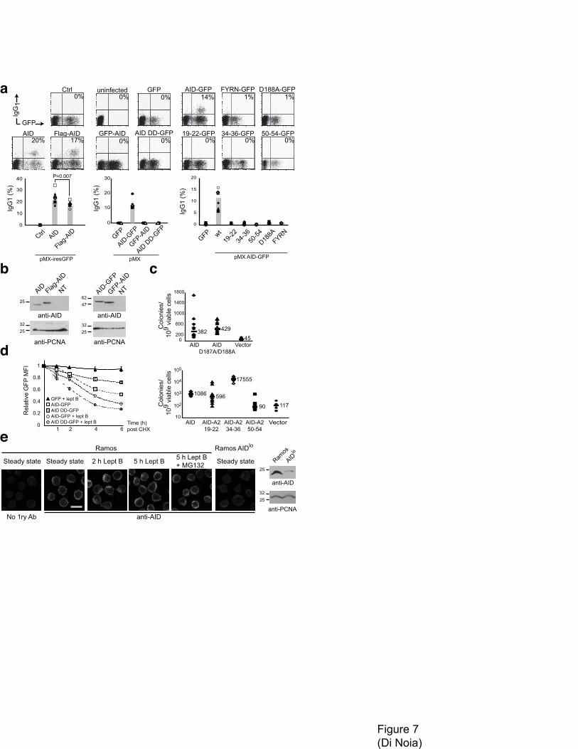
The efficiency of nuclear import and cytoplasmic retention would be predicted to influence AID
biology. We performed CSR assays on Aicda-/- mouse B cells complemented by retroviral delivery
with AID variants showing compromised nuclear import. Flag-AID was able to rescue isotype
switching to IgG1 but less efficiently than untagged AID (Fig. 4 a). This reduction is more pronounced
when the >2-fold higher protein expression of Flag-AID than AID is factored in (Fig. 7 b). On the other
hand, while AID-GFP restored isotype switching, GFP-AID was completely unable to do so despite
similar expression levels (Fig. 7 a and b). Thus, the effect of N-terminal tags on import seems
proportional to their size, in keeping with the proposed masking of the NLS and implying that
decreased nuclear import can limit CSR. We tested other AID variants with mutated import and
cytoplasmic retention determinants by this assay. AID FYRN and AID-A2 50–54 were catalytically
inactive (Fig. 7 c) and could not rescue CSR. AID with single point mutations at Arg50 or Asn51 have

10
been found to be CSR-proficient39 but our constructs bear additional mutations, which most likely
explains the difference. On the other hand, AID-A2 19–22 and 34–36 as well as AID D187A/D188A
could not complement CSR despite being catalytically active (Fig. 7 a and c). The phenotype of AID-
A2 19–22 and 34–36 may suggest that nuclear import is necessary for CSR, just as nuclear export
seems to be40. However, it cannot be excluded that these chimeras have lost binding affinity to relevant
AID partners35. Mutations at Asp187 and Asp188 may affect the C-terminal domain of AID that is
required for CSR41,42.
AID is rapidly degraded in the nucleus19, so altering the balance between the mechanisms determining
its subcellular localization should influence its half-life. If cytoplasmic retention were important in
determining the subcellular localization of AID in steady state, reduced retention of AID should lead to
an increased proportion of nuclear protein, resulting in shorter protein half-life. The degradation
kinetics of AID D187A/D188A-GFP in DT40 B cells after cycloheximide treatment show that this is
the case (Fig. 7 d). Since AID D187A/D188A-GFP has similar catalytic activity to AID-GFP, structural
problems are unlikely to cause the destabilization. Importantly, after leptomycin B treatment the
degradation of AID D187A/D188A-GFP was still more rapid than for AID-GFP (Fig. 7 d), as faster
nuclear import would predict.
Finally, to examine the importance of cytoplasmic retention we determined the subcellular localization
of endogenous AID in Ramos B cells in steady state and after leptomycin B by immunofluorescence.
We could not detect any major increase in nuclear AID signal after incubation with leptomycin B (Fig.
7 e). This observation further indicates that nuclear export is not the only mechanism excluding AID
from the nucleus and suggests the relevance of cytoplasmic retention for the compartmentalization of
AID.
11
DISCUSSION
The subcellular localization of AID determines how much AID becomes in contact with the genome,
probably a crucial parameter in balancing antibody diversification and off-target
mutations/translocations. Nuclear export is one mechanism regulating AID localization17,18,24. We now
provide strong evidence of two additional mechanisms influencing human AID subcellular distribution:
active nuclear import and cytoplasmic retention.
Active nuclear import of AID
We demonstrate herein that AID is actively imported into the nucleus. Passive diffusion of AID into
the nucleus was previously proposed based on consideration on AID size and the inability of AID fused
to the C-terminus of a large protein to enter the nucleus18,24. The observation that fusions to the N-
terminus of AID largely block its nuclear import, can now explain this difference. It has also been
proposed that AID residues 1-26 contain a classical bipartite NLS17. However, the minimal AID
fragment mediating nuclear accumulation encompasses from the N-terminus to somewhere between
residues 160 and 181, pointing to the importance of protein conformation. The N-terminal domain
contributes at least three of the four non-contiguous, positively charged determinants that we propose
conform the AID NLS. Binding to importin-α adaptors, which mediate the nuclear import of proteins
with classical basic NLS29,30,36, is in line with this. Without the 3D structure of an AID-importin
complex it is difficult to assign which residues make direct contact and which play a structural role in
displaying those residues. Hydrophobic residues W20, V18 and Y13, previously identified as necessary
for AID nuclear accumulation43, may be examples of the latter kind. NLSs often overlap with nucleic
acid binding domains44. Indeed, Arg24 and Arg112 might be involved in DNA binding by comparison
with APOBEC3G31. However, three import-deficient but catalytically active AID-A2 chimeras (#5,
19–22 and 34–36) provide separation of function between active import and nucleic acid binding.
Similarly to AID, the N-terminal domain of APOBEC1 is necessary but not sufficient for nuclear
import45,46 suggesting that APOBEC1 might also have a conformational NLS.
The stoichiometry and architecture of the biologically relevant AID molecule is unknown but many
indirect evidences suggest that it will have quaternary structure33,35,42,47,48 as all cytidine deaminases
including the APOBECs do33,49,50. Predicting AID dimerization through β2 (ref. 33) produces a
thermodynamically robust model, which is supported by the compromised oligomerization of AID
FYRN. Since AID FYRN is also compromised for nuclear import it suggests a role for AID
dimerization in conforming the NLS, which is also consistent with the location of the import
12
determinants in our AID dimer model (Fig. 4 a). However, AID FYRN is unstable and inactive.
Although this would not be unexpected if a dimer was the minimal functional unit of AID33, we cannot
rule out other structural defects impinging on the localization of AID FYRN. Solving this would need
elucidating the structure of AID by more sophisticated techniques than coimmunoprecipitation,
exceeding the focus of this work. In any case, AID FYRN still supports the conformational NLS and
the importance of the tertiary structure for nuclear import.
Cytoplasmic retention of AID
We show in a number of ways that AID is unable to diffuse into the nucleus when both active import
and export are inhibited, strongly suggesting the existence of cytoplasmic retention. The C-terminal
domain of AID, which we propose to be a flexible helix, seems necessary for cytoplasmic retention.
This domain also bears the NES17,18,24, complicating the mapping of cytoplasmic retention
determinants, which may partially overlap with it. However, comparison of mutations L198S and
D187A/D188A provides good evidence that export and retention are distinct and can be separated.
Structural integrity of AID may also be important as suggested by the less strong retention of some N-
terminal truncations of AID and by the apparently reduced retention of AID-FYRN, which unlike AID
is not able to outcompete a heterologous NLS (Supplementary Fig. 5). Coincidently, a structural
determinant mediates cytoplasmic retention of APOBEC3G49,51.
Regarding the identity of a putative AID cytoplasmic anchor, the observation that 3XFlag-AID was
resistant to digitonin washing from the cytoplasm, led others to propose retention of AID mediated by
binding to tubulin52. We did not detect any change in AID-GFP localization after treatment with
nocodazole (not shown) but other cytoskeleton molecules might be involved and further research is
needed to settle this issue. Other candidates to mediate AID cytoplasmic retention may be the 14-3-3
proteins given that AID is phosphorylated29. The analysis of partners in the uncharacterized AID high
molecular weight cytoplasmic complexes (K. Xue and M. Neuberger personal communication); or
comparisons between HeLa and 293T cells, which show different kinetics for AID nuclear import, may
help to identify relevant cytoplasmic retention and/or nuclear import AID partners.
Functional implications
Our findings demonstrate that AID compartmentalization is not determined just by nuclear export, as
the available evidences indicated so far18,24. We show that AID passive diffusion from the cytoplasm
into the nucleus is hindered and therefore AID needs to be actively imported. Both, cytoplasmic
retention and nuclear import influence the biology of AID. Hindering import results in cytoplasmic

13
retention and decreased CSR. AID variants with reduced cytoplasmic retention, are no longer excluded
from the nucleus in steady state resulting in shorter protein half-life. Thus, nuclear export seems
insufficient to outweigh AID nuclear import. The observation that leptomycin B treatment of Ramos
cells results in little AID nuclear translocation supports this scenario, although this should be confirmed
in non-transformed B cells. The difference with AID-GFP, which accumulates in the nucleus of
Ramos, could be explained by a combination of overexpression and the C-terminal tag weakening
retention, just as an N-terminal tag affects import. AID is vastly confined to the cytoplasm of germinal
center B cells48,53-55. Only a very small proportion of the B cells in rabbit appendix48 and human
tonsils53, and hardly any cell in unsynchronized populations overexpressing AID-GFP, seem to contain
predominantly nuclear AID. In the light of our results it seems unlikely that such accumulation might
be cell cycle regulated or has much physiological significance. We would rather suggest a model in
which a small proportion of nuclear AID exists at any given time, resulting from the competition
between active import, nuclear export and cytoplasmic retention. This steady state level of nuclear AID
might be enough for efficient antibody gene diversification as suggested by the CSR levels elicited by
apparently exclusively cytoplasmic AID variants, like AID V18S/R19V43 or Flag-AID. Perturbing any
of the mechanisms determining AID subcellular localization will affect this equilibrium and impact on
the efficiency and specificity of antibody diversification.
14
Acknowledgments
We thank Drs S Conticello and M Neuberger (MRC Laboratory of Molecular Biology, Cambridge,
UK), I Macara (U of Virginia School of Medicine, VA, USA), M Malim (King's College London
School of Medicine, London, UK), N Navaratnam (Imperial College London, London, UK), A Martin
(U of Toronto, ON, Canada), Swaminathan (U of Florida, FL, USA), J Archambault, E Cohen, T
Möröy and Y Guindon (IRCM, Montréal, Canada) for providing reagents, Dr T Honjo (Kyoto
University School of Medicine, Japan) for the AID-deficient mice through Dr A Lamarre (I Armand-
Frappier, Laval, QC, Canada). We thank E-L Thivierge and C Toulouse for expert animal care and
Dominic Filion for help with confocal microscopy. We are grateful to H Krokan and R Harris for
discussions and to S Conticello, C Buscaglia, D Muñoz and JF Côté for critically reading the
manuscript. This work was supported by the Canadian Institutes of Health Research (MOP 84543) and
a Canada Research Chair (to JDN). AO was supported by a fellowship from the CIHR Cancer Training
Program at the IRCM. VC was supported in part by a Michel Saucier fellowship from the Louis-
Pasteur Canadian Fund through the University of Montreal.
Author contributions
AMP, AO, YH, VAC, AB and JMD performed research. All authors analyzed data and discussed
results. JMD designed research and wrote the paper.
15
Figure Legends
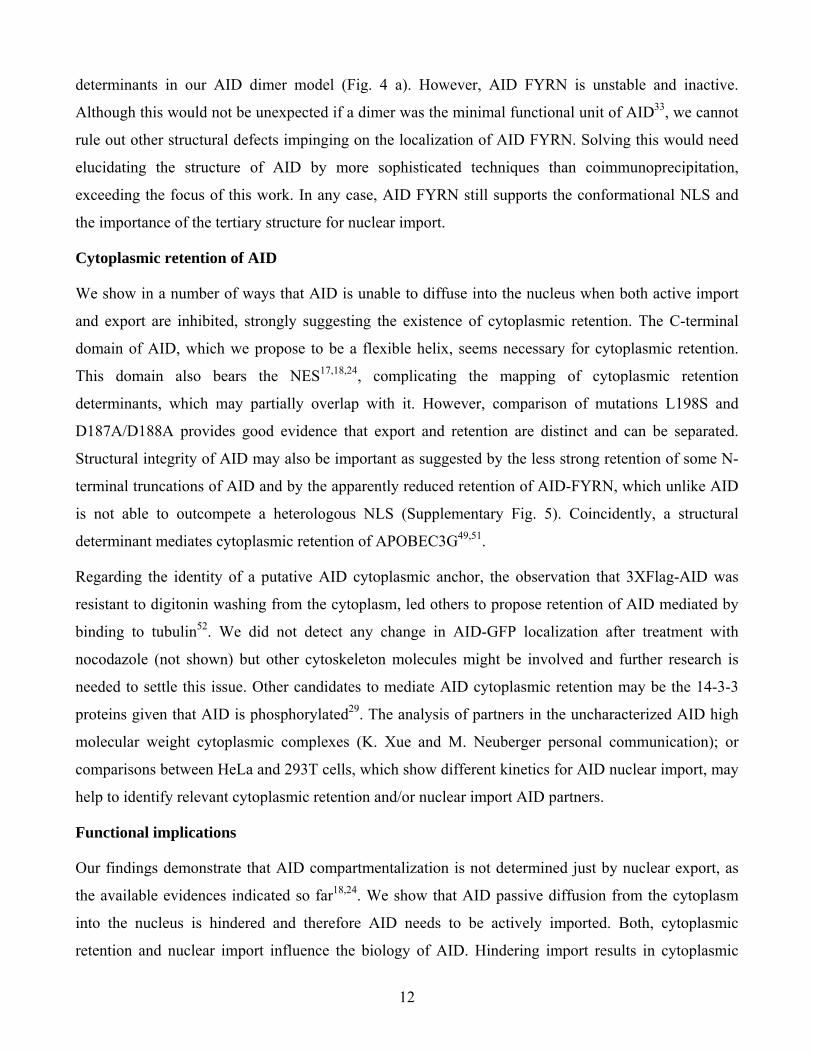
Figure 1 AID nuclear import is an active process. (a) Representative confocal images showing the
kinetics of AID-GFP nuclear accumulation in stably transfected Ramos and transiently transfected
HeLa and 293T cells. Time in h after addition of leptomycin B is indicated on the left. Propidium
iodide (PI) staining of nuclei is included for Ramos cells. (b) Energy depletion experiments in HeLa
cells transiently expressing NLSSV40-GFP or the constitutively nuclear AID L198S-GFP.
Representative confocal images are shown for untreated cells (Ctrl), energy-depleted cells (ΔGlc), or
energy-depleted cells reincubated in complete medium (ΔGlc+Glc). The average proportion of cells +
s.e.m. with clear cytoplasmic localization is plotted for each condition as determined from 2
independent experiments (58-100 cells scored). Student t-test p values are shown. (d, e) –
Representative confocal images of 293T cells transiently expressing the indicated fusions proteins. PI
was used to stain the nuclei. β-Lac, β-Lactamase; β-Gal, β-Galactosidase; UNG2, nuclear isoform of
mouse uracil DNA-glycosylase. The punctuated nature of the signal for the β-Gal fusion proteins in the
nucleus was consistently observed in all cells for both constructs. All images are at 630X
magnification. Bar, 10 µm.
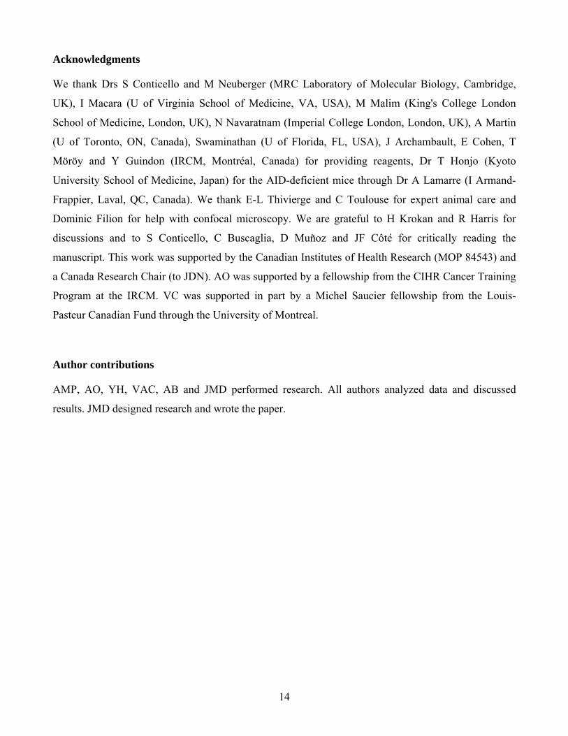
Figure 2 Most of the AID protein is required to mediate efficient nuclear import (a) Schematic
representation of C-terminally GFP-tagged AID truncations (named after the last AID residue included)
and their subcellular localization in transiently transfected 293T cells. Cells were scored from confocal
images (representative pictures shown in supplementary figure 3) and classified according to the
predominant (i.e. observed in >85% of the cells) subcellular localization (Loc) as cytoplasmic (C),
throughout the cell (N+C) or nuclear (N). (b) Representative confocal images of HeLa cells in steady
state transiently expressing the indicated constructs described in a. The localization of GFP control and
full length AID after leptomycin B treatment are shown for comparison. All transfections were
performed in parallel and images acquired using the same settings. (c) Representative confocal images
of Ramos cells in steady state transiently expressing the indicated constructs described in a. Nuclear
staining with propidium iodide (PI) is shown. (d) Representative confocal images of 293T cells in
steady state transiently expressing the indicated constructs with a C-terminal GFP tag. NLSNP, bipartite
nuclear localization signal from nucleoplasmin; β-Gal, β-Galactosidase. (e) Schematic representation
of AID-A2 chimeric proteins. Full length AID and A2 are drawn to scale and aligned. In each AID-A2
chimera (#1 to #5) the indicated AID amino acid range was replaced with the homologous region from
16
A2. NES, nuclear export signal; K/R-rich, AID N-terminal domain rich in Lys and Arg. Representative
images of HeLa cells transiently expressing the indicated C-terminally GFP tagged proteins in steady
state and after leptomycin B treatment (+ Lept B) are shown. All images are at 630X magnification.
Bars, 10 µm.
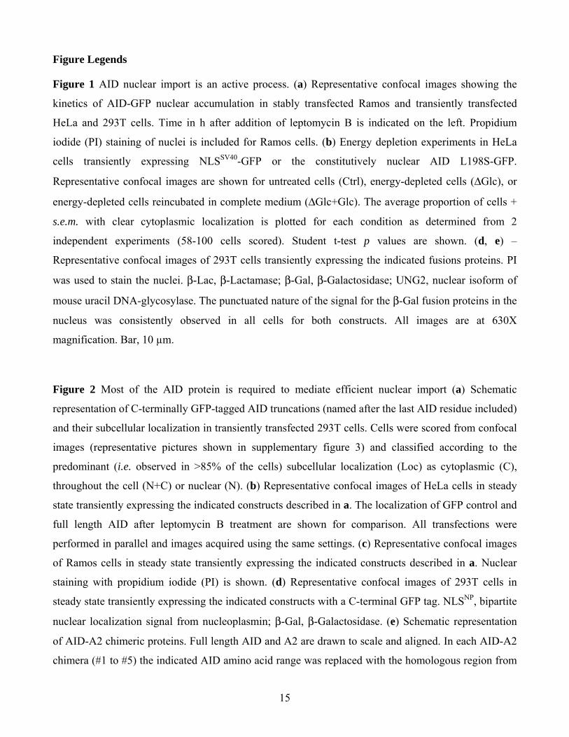
Figure 3 Several non-contiguous, positively charged residues are critical for AID nuclear import. (a)
AID oligomerization assay. The physical association between C-terminally Flag- and GFP-tagged
versions of the indicated proteins was monitored by detecting the GFP-tagged proteins in western blots
of anti-Flag immunoprecipitates from transiently cotransfected 293T cell extracts. The filters were
subsequently probed with anti-Flag to confirm similar immunoprecipitation of the bait. Aliquots
representing 5 % of the extracts were probed to control for expression levels of the GFP-tagged
proteins. One representative of three experiments done is shown. (b) Representative confocal images of
293T cells transiently expressing the indicated fusion proteins. β-Gal, β-galactosidase. Leptomycin B
was added where indicated. (c) Replacements of 3-5 consecutive residues in the N-terminal region of
AID with the corresponding residues in A2. Identities between AID and APOBEC2 are indicated in
bold on the AID sequence. Representative confocal pictures of HeLa cells expressing each AID-A2-
GFP protein are shown in the steady state or after leptomycin B treatment. (d) Representative confocal
images of HeLa cells transiently expressing AID-A2 chimeras in which the two indicated residue
stretches of AID were simultaneously replaced. (e) Representative confocal pictures of HeLa cells
transiently expressing untagged AID and AID-A2 #1 and 34-36. AID was detected by
immunofluorescence with anti-AID followed by biotinylated anti-rat and anti-biotin FITC in steady
state or after 4 h of leptomycin B treatment.. In c-d the proportion of cells with largely cytoplasmic
(black), homogeneous throughout the cell (grey) or nuclear (white) GFP signal was scored from >6
random fields from two independent experiments and the proportions indicated in the stacked bars plots
below each corresponding picture. Images at 630X except panel d at 400X magnification. Bars, 10 µm.
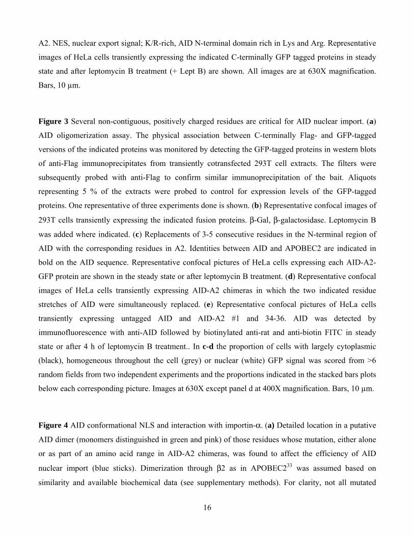
Figure 4 AID conformational NLS and interaction with importin-α. (a) Detailed location in a putative
AID dimer (monomers distinguished in green and pink) of those residues whose mutation, either alone
or as part of an amino acid range in AID-A2 chimeras, was found to affect the efficiency of AID
nuclear import (blue sticks). Dimerization through β2 as in APOBEC233 was assumed based on
similarity and available biochemical data (see supplementary methods). For clarity, not all mutated

17
residues are labeled. Asterisks indicate residues belonging to the second monomer. Tyr13, Val18 and
Trp20, identified elsewhere43, are also included. (b) Predicted solvent accessible surface of the putative
AID dimmer with those residues identified in a shown in shades of blue. Positively charged residues
within the import determinants are highlighted in bright blue to show the bias for exposed basic amino
acids. (c) Glutathione-sepharose beads loaded with the indicated purified GST-fusion protein were used
in pull down assays on cell extracts from Ramos B cells stably expressing AID-Flag. The material
eluted from the beads was analyzed by western blot using anti-AID. Ponceau-S staining of the
transferred proteins is shown as an indication of GST-protein precipitation. (d) Pull down of the
indicated GFP-tagged proteins from transiently transfected 293T cell extracts using purified GST of
GST-importin-α3 immobilized on glutathione-agarose. The material eluted from the beads and a 2%
aliquot of the input lysates were analyzed by western blot using anti-GFP. Ponceau staining of the
transferred proteins is shown as an estimation of similar GST-protein precipitation. Note that AIDΔN26
and AID 181 were appreciably degraded in the extracts (AID 181 was not visible in the extracts but it
was after concentration by the pull down).
Figure 5 AID cannot passively diffuse into the nucleus. (a) Representative confocal images of Ramos
B cells transiently expressing AID-A2 #1-GFP or APOBEC2-GFP after leptomycin B treatment. Both
proteins show the same distribution in steady state (not shown). (b) Representative confocal images of
Ramos B cells stably expressing GFP-AID (top) and DT40 B cells stably expressing Flag-AID
(bottom) in steady state of after leptomycin B treatment. Flag was detected by immunofluorescence
using mouse anti-Flag followed by anti-mouse AlexaFluor488. Nuclei are shown by PI staining. (c)
Stacked bars plot comparing the subcellular localization of AID-GFP, GFP-AID and AID-A2 #1-GFP
in Hela cells at different time points after addition of leptomycin B. The number (n) of cells indicated
above each bar was scored from multiple fields and the proportion of cells at each time point showing
largely cytoplasmic (black), homogeneous (grey) or nuclear (white) GFP signal determined.
Representative pictures are shown in supplementary figure 6. (d) Representative confocal pictures of
Hela cells transiently expressing AID-GFP or A2-GFP pretreated or not for 1 h with H2O2 before
treating for 2 h with leptomycin B where indicated. Subcellular localization was scored and plotted as
in c from three independent experiments. (e) Representative pictures of a similar experiment to that in
d but using untagged AID, which was detected by immunofluorescence as in figure 3. Subcellular
localization was scored and plotted as in c from two independent experiments. All images are at 630X
except d and e at 400X magnification. Bars, 10 µm.

18
Figure 6 The C-terminal domain of AID contains a cytoplasmic retention determinant. (a)
Representative confocal images of HeLa cells transiently expressing the indicated AID-A2 chimeras
(schematized in Fig. 2) with N-terminal GFP tag after leptomycin B treatment. (b) Representative
confocal images of HeLa cells transiently expressing AID-A2 #1-GFP in the presence of leptomycin B
or carrying a deletion of the last 17 residues of AID. (c) Representative confocal images of HeLa cells
treated with leptomycin B, transiently expressing GFP, A2 70-224-GFP (APOBEC2 with a deletion of
the first 69 residues) and AID 55-198-GFP (AID with a deletion of the first 54 residues). These
truncations were designed so that the resulting fragments are structurally equivalent. Nuclei are shown
by propidium iodide staining (PI). Experiments including a whole set of AID N-terminal truncations in
Hela and 293T cells is shown in supplementary figure 7. (d) Representative confocal images of HeLa
cells transiently expressing C-terminally GFP-tagged constructs of AID or AID-A2 #1 with the
indicated point mutations. Cells were treated with leptomycin B where indicated. In panels a, b, c and
d the subcellular localization of the constructs within the cells was scored as predominantly
cytoplasmic (black), homogenous throughout the cell (grey) or largely nuclear (white) from the
indicated number of cells (n) and the proportions plotted in the stacked bars below each corresponding
picture. All images are at 630X magnification. Bars, 10 µm. (e) 293T cells overexpressing untagged
full-length human CRM1 were cotransfected with the indicated AID-Flag variants. AID was
immunoprecipitated by anti-flag beads and the presence of CRM1 in the precipitate analyzed by
western blot. The IP filters were subsequently probed with anti-flag HRP to confirm precipitation of the
bait. One of two experiments yielding identical results is shown. (f) Line plot depicting the import
kinetics of AID-GFP vs AID D188A-GFP in transiently transfected 293T cells. The proportion of cells
with exclusively nuclear GFP signal was scored at each time point after the addition of leptomycin B to
the cultures. Two independent experiments are plotted, which only had partially overlapping time
points. For t = 0, 1 and 2 h the average and s.e.m of both experiments is plotted, the other points
represent one experiment only. Representative pictures are shown in supplementary figure 7.
Figure 7 Altering the balance between AID subcellular localization mechanisms has functional
consequences. (a) B cells from AID-deficient mice were infected with retroviruses encoding untagged
AID or Flag-AID in pMX-ires-GFP or GFP control, GFP-AID or the indicated AID-GFP variants in
pMX. The proportion of infected (GFP positive) cells that have switched to IgG1 is indicated in the top
right quadrant of representative flow cytometry profiles and summarized in the bar graphs. Different

19
symbols indicate cells coming from individual mice. Filled and empty symbols indicated independent
experiments. (b) Western blots comparing the expression of the indicated proteins. PCNA was used as
loading control. (c) E coli mutation assay to monitor the catalytic activity of AID-GFP and the
indicated derived mutants. Bottom plot is in log scale. Combined data from two independent
experiments are plotted for each construct. (d) DT40 B cells stably expressing GFP control, AID-GFP
or AID D187A/D88A-GFP (AID DD-GFP) were pretreated with cycloheximide for 1 h and the GFP
signal monitored over time by flow cytometry in the presence or absence of leptomycin B. For each
construct and condition, the mean fluorescent intensity (MFI) of the GFP signal at each time point was
normalized to the MFI at t = 0. The averages ± s.d.. of three experiments are plotted. The two curves
done in the presence of leptomycin B were analyzed by t-test and found to be significant with p<0.05 at
t = 1 and p<0.001 at all other time points. (e) Representative confocal images of untransfected Ramos
B cells treated or not with leptomycin B for 2 or 5 h (and where indicated with 10 µM MG132
proteasome inhibitor) stained with anti-AID followed by biotinylated anti-Rat and anti-biotin FITC.
Staining in which primary (1ry) anti-AID was omitted or using a Ramos clone with low AID
expression (AIDlo)56 were used as specificity controls. Western blot analysis comparing the expression
of AID in Ramos and Ramos AIDlo are shown on the right.

20
METHODS
DNA constructs. N-terminally GFP-tagged human AID, APOBEC2 and the AID-APOBEC2 chimeras
in pEGFP-C3 were a gift of Dr S. Conticello35. We generated C-terminally GFP-tagged versions by
PCR amplification of the open reading frames using oligonucleotides OJ60 and OJ166 for AID, OJ196
and OJ197 for APOBEC2 and OJ60-OJ166 for all AID-A2 except #5, for which OJ60-OJ197 were
used (see supplementary methods for all for oligonucleotide sequences). The 5’ oligonucleotides
introduced a BamHI site and a CAA triplet, a “cold” translation initiation context to reduce expression
levels, just before the ATG. The 3’ oligonucleotides eliminated the stop codon and introduced an
EcoRI site at the end of the coding regions. All fragments were cloned into BglII/EcoRI-digested
pEGFP-N3. The AID protein truncations used were generated by PCR amplification using the indicated
oligonucleotides (supplementary methods) and identically cloned into pEGFP-N3. We introduced point
mutations by the quickchange method using the oligonucleotides listed in supplementary methods. We
cloned the open reading frames of β-Lactamase, amplified from pUC18 using OJ213 and OJ214, and
β-Galactosidase, amplified from pIND/lacZ (Invitrogen) using OJ172 and OJ174, as EcoRI-BamHI
fragments into pEGFP-N3. To construct C-terminally flag-tagged versions of some of the proteins,
EGFP was excised from pEGFP-N3 using EcoRI and NotI and replaced by a synthetic Flag/HA-tag
(oligonucleotides OJ215 and OJ216). The nuclear localization signals from the SV40 large T antigen
and from nucleoplasmin were constructed by annealing oligonucleotides OJ190/OJ191 and
OJ192/OJ193, respectively. We used pCDNA3.1 (invitrogen) to express untagged AID and AID
mutants. Construct names throughout the manuscript indicate the actual order of the fragments in the
fusion proteins.
Cell methods and microscopy. Transfections were done as indicated in supplementary methods. We
inhibited nuclear export by using leptomycin B (LC labs, Woburn, MA) at 50 ng ml-1 for 4 h for 293T
cells, 50 ng ml-1 for 2 h for Hela cells and 10 ng ml-1 for 2 h for Ramos and DT40 B cells, unless
indicated differently. We inhibited nuclear import by pretreating the cells for 1 h with freshly diluted
10 mM H2O2 before any further treatment or analysis. Adherent cells were grown on coverslips and B
cell lines were allowed to attach to poly-L-Lysine-coated coverslips for 20 min at 37˚C in PBS. For
immunofluorescence the fixed cells were permeabilized in 0.25% (v/v) Triton X-100, blocked
overnight and stained in PBS 5% (w/v) BSA using mouse MAb anti-Flag M2 (1:100 Sigma-Aldrich)
followed by anti-mouse AlexaFluor 488 of 546 (1:1000 Invitrogen) or rat MAb anti-AID (1:100 EK2
5G9, Cell Signaling) followed by biotinylated goat anti-Rat antibodies (1:100 Abcam) detected with
anti-biotin FITC (1:50 Milteny Biotec). For energy depletion experiments we plated transfected cells in
21
triplicate. We subsequently incubated two of the wells in glucose-free DMEM (Wisent Inc)
supplemented with 10% (v/v) dialyzed FCS, 6 mM 2-deoxy-D-glucose (Sigma-Aldrich), to inhibit
glycolysis, and 10 mM sodium azide (Bioshop), to inhibit mitochondrial respiration, for 3 h. After
microscopic examination we fixed one of the wells and incubated the remaining well for further 3 h in
complete DMEM before fixing and processing along with the untreated control. For microscopy we
fixed cells in 3.7% (w/v) p-formaldehyde for 15 min, washed 3 times in PBS and stained 15 min in
PBS 0.5% (v/v) Triton-X100, 200 µg ml-1 RNase A, 10 µg ml-1 propidium iodide; washed in PBS and
mounted using FluorSafe (Calbiochem). Detailed confocal image acquisition and scoring is explained
in supplementary methods.
Coimmunprecipitation, 293T cells cotransfected with GFP and Flag-tagged versions of the indicated
proteins were homogenized in Lysis Buffer [20 mM Tris pH 7.8, 137 mM NaCl, 10 % (v/v) Glycerol,
2mM EDTA, 1 % (v/v) Triton X-100, Complete (Roche) protease inhibitors] 48 h post-transfection.
Lysates were incubated with 20 µl of anti-Flag®M2 affinity gel (Sigma-Aldrich) for 3 h at 4˚C and
eluted with 50 µL of 3x Flag peptide (Sigma-Aldrich) following the manufacturer protocol. Eluates
were analyzed by western blot with anti-eGFP and 1:5000 anti-Flag-HRP (Sigma-Aldrich) or 1:1000
anti-CRM1 (Santa Cruz Biotechnology). Full-length human CRM1 cloned into pCDNA3 was a gift
from Dr S Swaminathan. GST-fusion protein pull downs were done on cell extracts using loaded
glutathione-agarose beads as detailed in supplementary methods. Full-length human importin-α3
cloned in pGEX4T1 was a gift of Dr I. Macara, a similar construct with human importin-α5 was a gift
of Dr M. Malim. Full-length human importin-α1 was subcloned into pGEX-4T1 as an EcoRI/XhoI
fragment from pJG4-5-importin-α145, a gift of Dr N. Navaratnam. Western blots were developed using
SuperSignal® West Pico Chemiluminiscent substrate (Thermo Scientific).
AID assays. We assayed catalytic activity of AID by a mutation assay as described57 using the ung-
deficient E coli strain BW310 expressing AID-GFP and various mutants subcloned as NheI-NotI
fragments into a modified pTrc99a. We assayed isotype class switching by retrovirally deliverying AID
variants into in vitro activated B cells, obtained from Aicda-/- mice. The IRCM Animal Ethics
Committee approved all animal handling. Purification of resting B cells from splenic lymphocytes by
CD43-depletion, retroviral infection and analysis of isotype class switching to IgG1 has been
described58. AID-GFP variants and GFP-AID were subcloned as EcoRI-NotI fragments into the pMXs
retroviral vector. Untagged human AID and Flag-AID were subcloned as BamHI-NotI fragments into
pMXs-ires-GFP59. AID stability monitoring is explained in supplementary methods.

22
Structure modeling. See supplementary methods.
Satistics. Where indicated, two experimental data sets were compared using paired, one-tailed,
Student’s t-Test.

23
References
1. Muramatsu, M. et al. Class switch recombination and hypermutation require activation-induced cytidine deaminase (AID), a potential RNA editing enzyme. Cell 102, 553-63 (2000).
2. Revy, P. et al. Activation-induced cytidine deaminase (AID) deficiency causes the autosomal recessive form of the Hyper-IgM syndrome (HIGM2). Cell 102, 565-75 (2000).
3. Peled, J.U. et al. The Biochemistry of Somatic Hypermutation. Annu Rev Immunol (2007). 4. Di Noia, J.M. & Neuberger, M.S. Molecular mechanisms of antibody somatic hypermutation.
Annu Rev Biochem 76, 1-22 (2007). 5. Chaudhuri, J. et al. Evolution of the immunoglobulin heavy chain class switch recombination
mechanism. Adv Immunol 94, 157-214 (2007). 6. Martin, A. & Scharff, M.D. Somatic hypermutation of the AID transgene in B and non-B cells.
Proc Natl Acad Sci USA 99, 12304-8 (2002). 7. Okazaki, I.M. et al. Constitutive expression of AID leads to tumorigenesis. J Exp Med 197,
1173-81 (2003). 8. Liu, M. et al. Two levels of protection for the B cell genome during somatic hypermutation.
Nature 451, 841-5 (2008). 9. Pasqualucci, L. et al. BCL-6 mutations in normal germinal center B cells: evidence of somatic
hypermutation acting outside Ig loci. Proc Natl Acad Sci U S A 95, 11816-21 (1998). 10. Shen, H.M., Peters, A., Baron, B., Zhu, X. & Storb, U. Mutation of BCL-6 gene in normal B
cells by the process of somatic hypermutation of Ig genes. Science 280, 1750-2 (1998). 11. Dorsett, Y. et al. A role for AID in chromosome translocations between c-myc and the IgH
variable region. J Exp Med 204, 2225-32 (2007). 12. Ramiro, A.R. et al. AID is required for c-myc/IgH chromosome translocations in vivo. Cell 118,
431-8 (2004). 13. Crouch, E.E. et al. Regulation of AID expression in the immune response. J Exp Med 204,
1145-56 (2007). 14. de Yébenes, V.G. et al. miR-181b negatively regulates activation-induced cytidine deaminase in
B cells. J Exp Med (2008). 15. Dorsett, Y. et al. MicroRNA-155 suppresses activation-induced cytidine deaminase-mediated
Myc-Igh translocation. Immunity 28, 630-8 (2008). 16. Teng, G. et al. MicroRNA-155 is a negative regulator of activation-induced cytidine deaminase.
Immunity 28, 621-9 (2008). 17. Ito, S. et al. Activation-induced cytidine deaminase shuttles between nucleus and cytoplasm like
apolipoprotein B mRNA editing catalytic polypeptide 1. Proc Natl Acad Sci USA 101, 1975-80 (2004).
18. McBride, K.M., Barreto, V.M., Ramiro, A.R., Stavropoulos, P. & Nussenzweig, M.C. Somatic hypermutation is limited by CRM1-dependent nuclear export of activation-induced deaminase. J Exp Med 199, 1235-44 (2004).
19. Aoufouchi, S. et al. Proteasomal degradation restricts the nuclear lifespan of AID. J Exp Med 205, 1357-68 (2008).
20. Basu, U. et al. The AID antibody diversification enzyme is regulated by protein kinase A phosphorylation. Nature 438, 508-11 (2005).
21. McBride, K.M. et al. Regulation of hypermutation by activation-induced cytidine deaminase phosphorylation. Proc Natl Acad Sci U S A 103, 8798-803 (2006).
22. Pasqualucci, L., Kitaura, Y., Gu, H. & Dalla-Favera, R. PKA-mediated phosphorylation regulates the function of activation-induced deaminase (AID) in B cells. Proc Natl Acad Sci USA 103, 395-400 (2006).

24
23. Rada, C., Jarvis, J.M. & Milstein, C. AID-GFP chimeric protein increases hypermutation of Ig genes with no evidence of nuclear localization. Proc Natl Acad Sci USA 99, 7003-8 (2002).
24. Brar, S.S., Watson, M. & Diaz, M. Activation-induced cytosine deaminase (AID) is actively exported out of the nucleus but retained by the induction of DNA breaks. J Biol Chem 279, 26395-401 (2004).
25. Richardson, W.D., Mills, A.D., Dilworth, S.M., Laskey, R.A. & Dingwall, C. Nuclear protein migration involves two steps: rapid binding at the nuclear envelope followed by slower translocation through nuclear pores. Cell 52, 655-64 (1988).
26. Newmeyer, D.D. & Forbes, D.J. Nuclear import can be separated into distinct steps in vitro: nuclear pore binding and translocation. Cell 52, 641-53 (1988).
27. Guiochon-Mantel, A. et al. Nucleocytoplasmic shuttling of the progesterone receptor. EMBO J 10, 3851-9 (1991).
28. Larijani, M. et al. AID associates with single-stranded DNA with high affinity and a long complex half-life in a sequence-independent manner. Mol Cell Biol 27, 20-30 (2007).
29. Macara, I.G. Transport into and out of the nucleus. Microbiol Mol Biol Rev 65, 570-94, table of contents (2001).
30. Görlich, D. & Kutay, U. Transport between the cell nucleus and the cytoplasm. Annu Rev Cell Dev Biol 15, 607-60 (1999).
31. Chen, K. et al. Structure of the DNA deaminase domain of the HIV-1 restriction factor APOBEC3G. Nature 452, 116-9 (2008).
32. Holden, L.G. et al. Crystal structure of the anti-viral APOBEC3G catalytic domain and functional implications. Nature 456, 121-4 (2008).
33. Prochnow, C., Bransteitter, R., Klein, M.G., Goodman, M.F. & al., e. The APOBEC-2 crystal structure and functional implications for the deaminase AID. Nature 445, 447-51 (2006).
34. Nilsen, H. et al. Analysis of uracil-DNA glycosylases from the murine Ung gene reveals differential expression in tissues and in embryonic development and a subcellular sorting pattern that differs from the human homologues. Nucleic Acids Res 28, 2277-85 (2000).
35. Conticello, S.G. et al. Interaction between antibody-diversification enzyme AID and spliceosome-associated factor CTNNBL1. Mol Cell 31, 474-84 (2008).
36. Lange, A. et al. Classical nuclear localization signals: definition, function, and interaction with importin alpha. J Biol Chem 282, 5101-5 (2007).
37. Miyamoto, Y. et al. Cellular stresses induce the nuclear accumulation of importin alpha and cause a conventional nuclear import block. J Cell Biol 165, 617-23 (2004).
38. Kodiha, M., Chu, A., Matusiewicz, N. & Stochaj, U. Multiple mechanisms promote the inhibition of classical nuclear import upon exposure to severe oxidative stress. Cell Death Differ 11, 862-74 (2004).
39. Shivarov, V., Shinkura, R. & Honjo, T. Dissociation of in vitro DNA deamination activity and physiological functions of AID mutants. Proc Natl Acad Sci USA 105, 15866-71 (2008).
40. Doi, T. et al. The C-terminal region of activation-induced cytidine deaminase is responsible for a recombination function other than DNA cleavage in class switch recombination. Proc Natl Acad Sci USA (2009).
41. Barreto, V., Reina-San-Martin, B., Ramiro, A.R., McBride, K.M. & Nussenzweig, M.C. C-terminal deletion of AID uncouples class switch recombination from somatic hypermutation and gene conversion. Mol Cell 12, 501-8 (2003).
42. Ta, V. et al. AID mutant analyses indicate requirement for class-switch-specific cofactors. Nat Immunol 4, 843-8 (2003).
43. Shinkura, R. et al. Separate domains of AID are required for somatic hypermutation and class-switch recombination. Nat Immunol 5, 707-12 (2004).
25
44. LaCasse, E.C. & Lefebvre, Y.A. Nuclear localization signals overlap DNA- or RNA-binding domains in nucleic acid-binding proteins. Nucleic Acids Res 23, 1647-56 (1995).
45. Chester, A. et al. The apolipoprotein B mRNA editing complex performs a multifunctional cycle and suppresses nonsense-mediated decay. EMBO J 22, 3971-82 (2003).
46. Yang, Y. & Smith, H.C. Multiple protein domains determine the cell type-specific nuclear distribution of the catalytic subunit required for apolipoprotein B mRNA editing. Proc Natl Acad Sci USA 94, 13075-80 (1997).
47. Dickerson, S.K., Market, E., Besmer, E. & Papavasiliou, F.N. AID mediates hypermutation by deaminating single stranded DNA. J Exp Med 197, 1291-6 (2003).
48. Yang, G. et al. Activation-induced deaminase cloning, localization, and protein extraction from young VH-mutant rabbit appendix. Proc Natl Acad Sci USA 102, 17083-8 (2005).
49. Bennett, R.P., Presnyak, V., Wedekind, J.E. & Smith, H.C. Nuclear Exclusion of the HIV-1 Host Defense Factor APOBEC3G Requires a Novel Cytoplasmic Retention Signal and Is Not Dependent on RNA Binding. J Biol Chem 283, 7320-7 (2008).
50. Lau, P.P., Zhu, H.J., Baldini, A., Charnsangavej, C. & Chan, L. Dimeric structure of a human apolipoprotein B mRNA editing protein and cloning and chromosomal localization of its gene. Proc Natl Acad Sci USA 91, 8522-6 (1994).
51. Stenglein, M.D., Matsuo, H. & Harris, R.S. Two regions within the amino-terminal half of APOBEC3G cooperate to determine cytoplasmic localization. J Virol 82, 9591-9 (2008).
52. Wu, X., Geraldes, P., Platt, J.L. & Cascalho, M. The double-edged sword of activation-induced cytidine deaminase. in J Immunol Vol. 174 934-41 (2005).
53. Cattoretti, G. et al. Nuclear and cytoplasmic AID in extrafollicular and germinal center B cells. Blood 107, 3967-75 (2006).
54. Greiner, A. et al. Differential expression of activation-induced cytidine deaminase (AID) in nodular lymphocyte-predominant and classical Hodgkin lymphoma. J Pathol 205, 541-7 (2005).
55. Pasqualucci, L. et al. Expression of the AID protein in normal and neoplastic B cells. Blood 104, 3318-25 (2004).
56. Zhang, W. et al. Clonal instability of V region hypermutation in the Ramos Burkitt's lymphoma cell line. Int Immunol 13, 1175-84 (2001).
57. Petersen-Mahrt, S.K., Harris, R.S. & Neuberger, M.S. AID mutates E. coli suggesting a DNA deamination mechanism for antibody diversification. Nature 418, 99-103 (2002).
58. Di Noia, J.M. et al. Dependence of antibody gene diversification on uracil excision. J Exp Med 204, 3209-19 (2007).
59. Kitamura, T. et al. Retrovirus-mediated gene transfer and expression cloning: powerful tools in functional genomics. Exp Hematol 31, 1007-14 (2003).

b
a
dβ-Gal-GFP (~150 kDa)
UNG2-β-Gal-GFP
AID 181-β-Gal-GFP
PIGFP
Figure 1(Di Noia)
AID–GFPPI
Ramos
t pos
t Lep
t B (
h)
HeLa 293T
0.5
1
2
4
0
Cyt
opla
smic
sig
nal (
% o
f cel
ls)
ΔGlc+Glc
ΔGlc
Ctrl
AID L198S-GFP
AID L198S-GFP
NLSSV40-GFPNLSSV40-GFP
0
20
40
60
80
100
cAID-
β-Lac-GFP(~75 kDa)A2-β-Lac-GFPA20-GFP
AID L198S-β-Lac-GFP
GFP-β-Lac-GFPAID 181-β-Lac-GFP
+Le
ptB
0
20
40
60
80
100
ΔGlc
+GlcΔG
lcCtrl
P=0.002P=0.004

NES
HxE
K/R rich
PCxxC
AID 30AID 40AID 54AID 85AID 103AID 142AID 160AID 181AID 187AID L198S
N+CC
Loc
N+CN+CN+CN+CN+CN+C
NNN
AID
GFP
AID +Lept B
AID 40 AID 187AID 160GFP
a
b
c
d
Figure 2(Di Noia)
AID
PI
GFP
AID 40 AID 142 AID 160 AID 187
e
#1 #2 #3 #4 #5
AID
AID-A2
A2
NESK/R rich
+ L
ept B
A2 #1 #2 #3 #4 #5
19-57 59-84 88-116 118-150 154-198GFP
AID
GFP PI

c
d
e
Figure 3(Di Noia)
+ L
ept B
+ Lept B
19–22
AID-A2
25–28 39–42 43–46 50–54 55–57
...KNVRWAKGRRETYLCYVVKRRDSATSFSLDFGYLRNKNG-CHVE...
EYSSA2
25 39 43 50 5519
NKTF EAQ GGQVQASR EDEHAAAHA
34
AID
34–36
Lysa
tes
IP a
nti-F
lagAnti-
GFP
Anti-GFP
AID FYRN-FlagAnti-Flag
AID-G
FP
GFPAID
FYRN-G
FP
A2-GFP
8362
32
47
25
47
25
WB
AID-Flag
AID-G
FP
AIDF46
A-GFP
AIDF46
A/Y48
A-GFP
AID F
YRN-GFP
A2-GFP
6247
47
25
aAID-GFP
AID FYRN-GFP
b
Ctrl FYRN
+ Lept B
+ Lept B+ Lept B
AID-A234–36/50–54
+ Lept B
n = 50 n = 20 n = 44 n = 25 n = 19 n = 19n = 26
n = 22
AID-A2 34–36
n = 12
AID-A2 #1
n = 16
AID
n = 25
n = 36 n = 34
n = 21
n = 9
C
N+C
N
AID-A219–22/34–36

Arg19Arg24
Arg50
Lys52Arg35*
Arg36*
Arg34*Arg112
Tyr13Trp20
Lys22
Figure 4(Di Noia)
c d
GST
3225
GSTIn
put
Ponceau
anti-Flag
anti-GFP
GST
Ponceau
8362
32
47
25
AIDAID
6247
AID F
YRN
AID 1
81
A2
62
32
47
25
Lysates
a
b6287
32
47
25

c
+ H2O2
+ H2O2
d
e
a
b
Figure 5(Di Noia)
AID-GFP
t post lept B (h)0
n =
1 2 4 6 0 1 2 4 6 0 1 2 4 6
23 14 28 28 32 17 6 16 27 30 12 6 15 28 28GFP-AID #1-GFP
+ LeptB (4 h)
+ L
eptB
+ L
eptB
GFP-AID
AID-A2 #1-GFP + Lept B A2-GFP + Lept BA2-GFPAID-GFP
Untagged AID
Flag-AID
PI
PI
α-FLAG
GFP PI
GFP
0
25
50
75
100
C
Cells(%)
N+CN
n = 17
n = 57 n = 53 n = 6
n = 20n = 49n = 63
n = 17
n = 16 n = 18

a
c
d
+ L
ept B
+ L
ept B
+ L
ept B
Figure 6(Di Noia)
b
f
e
175
25
175
vecto
r
wt
AID-Flag/HA
D187A
/D18
8A
L196
S/L19
8S
D188A
L198
S
IP anti-Flag
Lysates
Flag
CRM1
CRM183
WB
Time post lept B (min)
Cel
ls w
ith n
ucle
ar-o
nly
AID
-GF
P (
%)
D188Awt25
50
75
100
0 6030 120 180 240
GFP-AID-A2 (+ Lept B)
#1 #2 #3 #4 #5
n = 28n = 50 n = 41 n = 16 n = 14
C
N+C
N
AID-A2 #1-GFP
steady state + Lept B steady state + Lept B
AID-GFP
n = 15
n = 26
n = 17
n = 22
n = 30
n = 20
n = 28
n = 19
n = 26
n = 37
n = 22
n = 12 n = 15
n = 16
n = 24
L198S
D187A/D188A
D187A
D188A
GFP
PI
A2 70-224-GFP
PI
AID 55-198-GFP
PI
AID-A2 #1-GFPAID-A2 #1ΔC17-GFP
n = 48n = 74
+ LeptB
GFP
IgG
1
GFP0%
GFP-AID0%
AID20%
Ctrl0%
uninfected 0%
Flag-AID17%
IgG
1 (%
)
GFP
AID-G
FP
AID D
D-GFP
pMX
GFP-AID
0
10
20
30
IgG
1 (%
)
Ctrl
pMX-iresGFP
AID
Flag-A
ID0
10
20
30
40 P=0.007
AID DD-GFP0%
a
e
cb
Figure 7(Di Noia)
10
103
104
105
102
AID AID-A219-22
AID-A234-36
AID-A250-54
Vector
1086 596
17555
11790Col
onie
s/10
9 vi
able
cel
lsC
olon
ies/
109
viab
le c
ells
0200
600
1000
1400
1800
AID AIDD187A/D188A
Vector
382 429
45
0
5
10
15
20 Ig
G1
(%)
wt
19-2
2
34-3
6
50-5
4
D188A
FYRNGFP
pMX AID-GFP
34-36-GFP
FYRN-GFP1%
0%
D188A-GFP1%
50-54-GFP0%
19-22-GFP
AID-GFP
0%
14%
No 1ry Ab anti-AID
Ramos Ramos AIDlo
AIDlo
Ramos
d
0
0.2
0.4 GFP + lept BAID-GFPAID DD-GFPAID-GFP + lept BAID DD-GFP + lept B
0.6
0.8
1
1 2Time (h) post CHX4 6
Rel
ativ
e G
FP
MF
I
2 h Lept B 5 h Lept B5 h Lept B+ MG132
Steady state Steady stateSteady state
anti-AID
anti-PCNA
32
25
25
AID-G
FP
GFP-A
ID
NT
anti-AID
anti-PCNA
32
25
62
47
AID Flag-A
ID
NT
anti-AID
anti-PCNA
32
25
25