
1 ˜, &’, *’+ 2,*-˜’,% ’,2!# 0’+’*˜/...
7
405 Introduction Exposure to prolonged disuse or microgravity produces a va- riety of effects on skeletal muscle, including fiber atrophy, a re- duction in maximal force, and reduced endurance 1 . For example, given the major alterations that can ensue even after just several days of exposure to microgravity, human spaceflight currently requires astronauts to participate in daily exercise countermea- sures to help offset the effects of weightlessness 2 . Although a recent report suggests that high intensity exercise combined with optimal nutrition may mitigate bone and muscle loss 3 , novel ap- proaches to reduce negative effects of spaceflight or prolonged bed rest/immobilization on musculoskeletal health, including drug therapies, are being sought 4,5 . Diagnostic tools for the assessment of muscle and bone loss due to disuse or weightlessness are, however, quite limited. Standard methods used to evaluate these changes such as dual- energy X-ray absorptiometry (DXA) and quantitative com- puted tomography 6,7 are expensive and inconvenient for regular clinical use. Moreover, they are not feasible in space- flight, given the size, weight, and power requirements of the equipment. Ultrasound is being studied for muscle loss assess- ment in spaceflight, but quantifying the measurements requires substantial procedural modification 8 . Simple force-testing dy- J Musculoskelet Neuronal Interact 2013; 13(4):405-411 Spaceflight and hind limb unloading induce similar changes in electrical impedance characteristics of mouse gastrocnemius muscle M. Sung 1 , J. Li 1 , A.J. Spieker 1 , J. Spatz 2 , R. Ellman 2 , V.L. Ferguson 3 , T.A. Bateman 4 , G.D. Rosen 1 , M. Bouxsein 2 , S.B. Rutkove 1 1 Departments of Neurology and 2 Orthopedics, Beth Israel Deaconess Medical Center, Harvard Medical School, Boston, MA; 3 Department of Mechanical Engineering, University of Colorado at Boulder, Boulder, CO; 4 Department of Biomedical Engineering, University of North Carolina, Chapel Hill, NC Abstract Objective: To assess the potential of electrical impedance myography (EIM) to serve as a marker of muscle fiber atrophy and secondarily as an indicator of bone deterioration by assessing the effects of spaceflight or hind limb unloading. Methods: In the first experiment, 6 mice were flown aboard the space shuttle (STS-135) for 13 days and 8 earthbound mice served as controls. In the second experiment, 14 mice underwent hind limb unloading (HLU) for 13 days; 13 additional mice served as controls. EIM measurements were made on ex vivo gastrocnemius muscle. Quantitative microscopy and areal bone mineral density (aBMD) measurements of the hindlimb were also performed. Results: Reductions in the multifrequency phase-slope parameter were ob- served for both the space flight and HLU cohorts compared to their respective controls. For ground control and spaceflight groups, the values were 24.7±1.3°/MHz and 14.1±1.6°/MHz, respectively (p=0.0013); for control and HLU groups, the values were 23.9±1.6°/MHz and 19.0±1.0°/MHz, respectively (p=0.014). This parameter also correlated with muscle fiber size (ρ=0.65, p=0.011) for spaceflight and hind limb aBMD (ρ=0.65, p=0.0063) for both groups. Conclusions: These data support the concept that EIM may serve as a useful tool for assessment of muscle disuse secondary to immobilization or microgravity. Keywords: Muscle, Spaceflight, Hind Limb Unloading, Disuse, Electrical Impedance Original Article Hylonome Dr. Rutkove has equity in, and serves a consultant and scientific advisor to Skulpt, Inc. a company that designs impedance devices for clinical and research use; he is also a member of the company’s Board of Direc- tors. The company also has an option to license patented impedance technology of which Dr. Rutkove is named as an inventor. This study, however, did not employ any relevant company or patented technology. Corresponding author: Seward B. Rutkove, MD, Professor of Neurology, Har- vard Medical School, 330 Brookline Avenue, TCC-810, Boston, USA E-mail: [email protected] Edited by: J. Rittweger Accepted 13 August 2013
Transcript of 1 ˜, &’, *’+ 2,*-˜’,% ’,2!# 0’+’*˜/...

405
Introduction
Exposure to prolonged disuse or microgravity produces a va-
riety of effects on skeletal muscle, including fiber atrophy, a re-
duction in maximal force, and reduced endurance1. For example,
given the major alterations that can ensue even after just several
days of exposure to microgravity, human spaceflight currently
requires astronauts to participate in daily exercise countermea-
sures to help offset the effects of weightlessness2. Although a
recent report suggests that high intensity exercise combined with
optimal nutrition may mitigate bone and muscle loss3, novel ap-
proaches to reduce negative effects of spaceflight or prolonged
bed rest/immobilization on musculoskeletal health, including
drug therapies, are being sought4,5.
Diagnostic tools for the assessment of muscle and bone loss
due to disuse or weightlessness are, however, quite limited.
Standard methods used to evaluate these changes such as dual-
energy X-ray absorptiometry (DXA) and quantitative com-
puted tomography6,7 are expensive and inconvenient for
regular clinical use. Moreover, they are not feasible in space-
flight, given the size, weight, and power requirements of the
equipment. Ultrasound is being studied for muscle loss assess-
ment in spaceflight, but quantifying the measurements requires
substantial procedural modification8. Simple force-testing dy-
J Musculoskelet Neuronal Interact 2013; 13(4):405-411
Spaceflight and hind limb unloading induce
similar changes in electrical impedance characteristics of
mouse gastrocnemius muscle
M. Sung1, J. Li1, A.J. Spieker1, J. Spatz2, R. Ellman2, V.L. Ferguson3, T.A. Bateman4,
G.D. Rosen1, M. Bouxsein2, S.B. Rutkove1
1Departments of Neurology and 2Orthopedics, Beth Israel Deaconess Medical Center, Harvard Medical School, Boston, MA; 3Department of Mechanical Engineering, University of Colorado at Boulder, Boulder, CO;
4Department of Biomedical Engineering, University of North Carolina, Chapel Hill, NC
Abstract
Objective: To assess the potential of electrical impedance myography (EIM) to serve as a marker of muscle fiber atrophy and
secondarily as an indicator of bone deterioration by assessing the effects of spaceflight or hind limb unloading. Methods: In the
first experiment, 6 mice were flown aboard the space shuttle (STS-135) for 13 days and 8 earthbound mice served as controls. In
the second experiment, 14 mice underwent hind limb unloading (HLU) for 13 days; 13 additional mice served as controls. EIM
measurements were made on ex vivo gastrocnemius muscle. Quantitative microscopy and areal bone mineral density (aBMD)
measurements of the hindlimb were also performed. Results: Reductions in the multifrequency phase-slope parameter were ob-
served for both the space flight and HLU cohorts compared to their respective controls. For ground control and spaceflight groups,
the values were 24.7±1.3°/MHz and 14.1±1.6°/MHz, respectively (p=0.0013); for control and HLU groups, the values were
23.9±1.6°/MHz and 19.0±1.0°/MHz, respectively (p=0.014). This parameter also correlated with muscle fiber size (ρ=0.65,
p=0.011) for spaceflight and hind limb aBMD (ρ=0.65, p=0.0063) for both groups. Conclusions: These data support the concept
that EIM may serve as a useful tool for assessment of muscle disuse secondary to immobilization or microgravity.
Keywords: Muscle, Spaceflight, Hind Limb Unloading, Disuse, Electrical Impedance
Original Article Hylonome
Dr. Rutkove has equity in, and serves a consultant and scientific advisor
to Skulpt, Inc. a company that designs impedance devices for clinical
and research use; he is also a member of the company’s Board of Direc-
tors. The company also has an option to license patented impedance
technology of which Dr. Rutkove is named as an inventor. This study,
however, did not employ any relevant company or patented technology.
Corresponding author: Seward B. Rutkove, MD, Professor of Neurology, Har-
vard Medical School, 330 Brookline Avenue, TCC-810, Boston, USA
E-mail: [email protected]
Edited by: J. Rittweger
Accepted 13 August 2013

M. Sung et al.: Spaceflight alters skeletal muscle impedance
406
namometers are inconvenient and inaccurate to use on debili-
tated patients and are virtually impossible to use in space. For
these reasons, other non-invasive approaches that can be ap-
plied easily and rapidly to both patients and astronauts are of
interest. One approach that offers potential value is electrical
impedance myography (EIM). EIM is a technique in which a
high-frequency, low-energy electrical current is applied to a
localized area of muscle and the resulting surface voltages are
measured9. EIM is a specialized version of methods developed
in the well-studied field of bioelectrical impedance spec-
troscopy, in which alterations in the characteristics of a tissue
or cellular suspension are characterized by the their electrical
frequency-dependent properties10-13. In addition to these non-
muscular studies, at least one investigation has evaluated the
specific relationship between muscle cell size and bioimped-
ance parameters in tissue culture14.
Since the EIM technique is painless, rapid to apply, non-inva-
sive, and the equipment lightweight, it has the potential to serve
as a useful method for monitoring muscle status. Earlier studies
have shown that surface EIM alterations are closely tied to mus-
cle fiber size15. Importantly, data in humans16 and in rats17 support
that EIM is sensitive to changes in muscle following disuse, with
reductions in EIM parameters of greater than 30% from baseline,
and thus may also have the potential to sensitively identify the
impact of disuse or microgravity on muscle health. Here we fur-
ther explore the potential application of EIM for the assessment
of muscle impacted by disuse by studying ex vivo muscle ob-
tained from mice exposed to reduced mechanical loading via
hind limb unloading (HLU) or spaceflight, testing the basic hy-
pothesis that such disuse will consistently alter EIM values.
Materials and methods
Animals
For both the hind limb unloading (HLU) and spaceflight ex-
periments, we used 9-week-old female C57Bl/6N mice (Charles
River, Wilmington, MA). In the HLU cohort, 13 ground control
and 14 HLU mice were studied, while in the space flight study, 8
ground control and 6 space flight mice were examined. The HLU
protocol was approved by Beth Israel Deaconess Medical Cen-
ter’s Institutional Animal Care and Use Committee (IACUC), and
the protocol used for the spaceflight study was approved by the
ACUC at Kennedy Space Center.
Spaceflight study
Both spaceflight and ground control animals were main-
tained on a NASA nutrient-upgraded rodent food bar18 through-
out the experiment. Spaceflight animals were sacrificed within
approximately 2.5-7.5 hours of the shuttle’s completing a 12
day, 18.5 hour flight onboard the shuttle Atlantis (STS-135 mis-
sion). Flight animals were euthanized and the right gastrocne-
mius muscle was removed intact. Ground control animals,
matched to day 0 body weight and bone parameters of flight
mice, were euthanized 2 days later and the gastrocnemius mus-
cle removed in an identical fashion, after an equal length of stay
in identical cages to those used on the space shuttle19.
Hind Limb Unloading studies
In a later experiment, mice of the same strain, sex and age
were subjected to HLU for 13 days and compared to concur-
rent normally-loaded controls20. Briefly, under isoflurane anes-
thesia, the tail was taped to a freely rotating harness connected
to a wheel that could move along a rod across the center of the
cage. The height of the harness was adjusted such that the
mouse could not touch its hind paws to the floor. A reloading
period of 3 to 6 hours, to match the STS-135 timing, was em-
ployed in the HLU group by removing the harness and allow-
ing the mice to ambulate before sacrifice. NASA food bars and
water were provided ad libitum.
Muscle processing and electrical impedance measurements
Excised muscle was immediately weighed and then cut to a
0.5 cm X 0.5 cm base with approximately a 0.3 cm height
block using a razor blade. The block of muscle tissue was
placed in a 0.5 cm X 0.5 cm base impedance-measuring cell
(Figure 1), configured with two broad, stainless steel elec-
trodes on two sides for applying electrical current and two nee-
dle electrodes positioned on top for measuring voltage, as
previously described15. The impedance data were obtained
using the Imp SFB7® (Impedimed, San Diego, CA). Reactance
(X) and resistance (R) data from 3 to 500 kHz was collected.
The muscle was placed such that electrical current would flow
across (transversely) to the major muscle fiber direction. While
it would have been preferable to also obtain measurements
with electrical current flow parallel to the fibers (and thus al-
lowing us to assess the anisotropic characteristics of the tis-
sue), positioning the muscle fibers on end with the metal plate
electrodes proved very challenging. A preliminary review of
the data showed this latter process to be quite inconsistent, and
thus was omitted from any further analysis.
Figure 1. Impedance measuring cell with muscle demonstrating how
data was obtained.

M. Sung et al.: Spaceflight alters skeletal muscle impedance
407
Muscle histology (obtained on space flight muscle only)
Immediately after the EIM data were collected, the muscle
was snap-frozen in isopentane cooled in liquid nitrogen, and
stored at -80°C. The tissue was then cut into 10 μm slices using
a Tissue Tek II cryostat (Miles Laboratories, Inc., Elkhart, IN)
and stained with hematoxylin and eosin. Cell measurements were
made using a Zeiss Axiophot microscope with a LUDL motor-
ized stage interfaced with a Dell Optiflex 380 computer running
Stereo Investigator (MBF Biosciences, Inc., Williston, VT) soft-
ware. This software allows a non-biased quantification of fiber
sizes. After the investigator sets a series of initial parameters, in-
cluding the section of tissue from which to choose cells, the sys-
tem automatically and randomly selects groups of cells to count.
Approximately 60 cells were evaluated from each animal. In
order to reduce the potential for any bias, the evaluator (AS) was
blinded to group designation (i.e., loaded or unloaded) of each
section being assessed. Muscle histology was also planned in the
HLU animals, but unfortunately the tissue was inadvertently
damaged during transport and was unusable for analysis.
Bone Mineral Density measurements
Areal bone mineral density (aBMD, g/cm2) of the hind limb
(from femoral neck to ankle) was assessed by peripheral dual-
energy X-ray absorptiometry (pDXA, PIXImus II; GE Lunar,
Madison, WI, USA) in vivo immediately prior to sacrifice.
Data analysis
From the raw EIM data, the phase was calculated via the
equation: phase=arctan (reactance/resistance) at each fre-
quency. Due to its being the most promising of the multifre-
quency EIM parameters from previous work21,22, and
especially in our recent work in HLU17 rats, the focus here is
on the phase-slope parameter, defined as the slope of the fitted
linear regression to phase values from 100 to 500 kHz (see
Figure 2 for examples as to how this analysis was performed),
expressed as degrees/MHz. Although considerably beyond the
50 kHz measurement, the subject much earlier work, the im-
pedance behavior in this region is generally linear and thus fa-
vorable to least squares regression analysis. For simplicity of
description, the sign was then flipped (thus the negative values
are positive). Further explanation as to the potential signifi-
cance of this parameter is provided in the discussion.
The Wilcoxon rank sum test was performed to evaluate for
differences between phase-slope, muscle mass, muscle fiber
cross-sectional area, and areal bone mineral density of the
HLU and space flight mice with their respective control
groups. Spearman rank-correlation coefficient (ρ) was calcu-
lated to determine the relationship between phase-slope and
muscle mass, muscle fiber area, and hind limb bone mineral
density. All results are given as mean ± standard error; signif-
icance was assumed at p<0.05, two-tailed.
Results
Muscle mass
As anticipated, mice exposed to spaceflight had a lower gas-
trocnemius muscle mass than ground controls, although the
difference did not reach significance (102±32 mg for space-
flight; 112±22 mg for ground p=0.079). However, mice ex-
posed to HLU had lower muscle mass as compared to controls
(95.2±19 mg for HLU; 107±29 mg for control; p=0.0053).
Muscle fiber size (spaceflight only)
Muscle histology measurements were obtained only in the
spaceflight animals and their controls. As anticipated, mice ex-
posed to spaceflight had a smaller average muscle fiber cross-
sectional area of 1579±194 μm2 as compared to 2591±197 μm2
in controls (p=0.013) (Figure 3 (a)).
Figure 2. Plots of the average phase from a hind limb unloaded mouse and spaceflight mouse with controls. Dotted lines depict the method of
calculating the 100-500 kHz phase-slope parameter.

M. Sung et al.: Spaceflight alters skeletal muscle impedance
408
Electrical Impedance data and muscle characteristics
The EIM phase-slope parameter was significantly lower in
both the spaceflight and HLU mice when compared to their
respective control groups (Figure 4). For control and HLU
groups, the values were 23.9±1.6°/MHz and 19.0±1.0°/MHz,
respectively (p=0.014); for ground control and spaceflight
groups, the values were 24.7±1.3°/MHz and 14.1±1.6°/MHz,
respectively (p=0.0013). We observed a moderate positive re-
lationship between muscle mass and the EIM phase-slope pa-
rameter in the HLU study (ρ=0.64, p<0.001), and although the
relationship had a similar pattern in the spaceflight study, the
association was weaker and did not reach statistical signifi-
cance (ρ=0.39, p=0.17) (Figure 5). This inconsistency may re-
late to the fact that there were considerably smaller number of
spaceflight animals and that muscle mass was slightly lower
in the HLU group than the spaceflight group compared to their
respective control groups. However, there was a good corre-
lation between muscle fiber size and the phase-slope parameter
(ρ=0.65, p=0.011) across the spaceflight and ground control
animals (Figure 3 (b)).
Areal Bone Mineral Density and Electrical Impedance
As expected, both spaceflight and HLU groups had signif-
icantly lower hind limb aBMD compared to controls. For
ground control and spaceflight groups, the values were
55.9±0.80 x 10⁻³ g/cm2 and 50.8±0.56 x 10⁻³ g/cm2, respec-
tively (p=0.0013). For control and HLU groups, the values
Figure 3. (a). Column plot of average muscle fiber size in ground vs. space flight mice. (b) Scatter plot correlating phase-slope and average
muscle fiber size in ground and space flight mice.
Figure 4. Column plot depicting the average phase-slope of controls vs. hind limb unloaded mice (a) and ground vs. space flight animals (b).
M. Sung et al.: Spaceflight alters skeletal muscle impedance
409
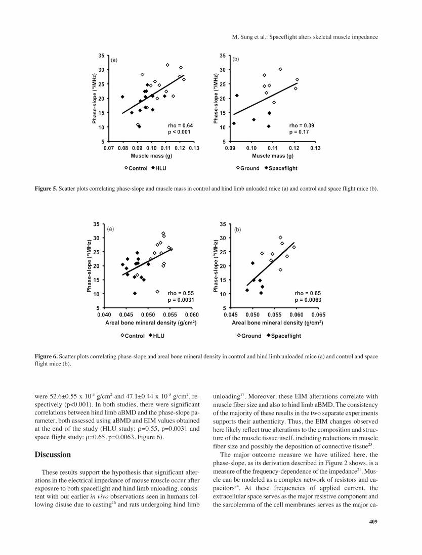
were 52.6±0.55 x 10⁻³ g/cm2 and 47.1±0.44 x 10⁻³ g/cm2, re-
spectively (p<0.001). In both studies, there were significant
correlations between hind limb aBMD and the phase-slope pa-
rameter, both assessed using aBMD and EIM values obtained
at the end of the study (HLU study: ρ=0.55, p=0.0031 and
space flight study: ρ=0.65, p=0.0063, Figure 6).
Discussion
These results support the hypothesis that significant alter-
ations in the electrical impedance of mouse muscle occur after
exposure to both spaceflight and hind limb unloading, consis-
tent with our earlier in vivo observations seen in humans fol-
lowing disuse due to casting16 and rats undergoing hind limb
unloading17. Moreover, these EIM alterations correlate with
muscle fiber size and also to hind limb aBMD. The consistency
of the majority of these results in the two separate experiments
supports their authenticity. Thus, the EIM changes observed
here likely reflect true alterations to the composition and struc-
ture of the muscle tissue itself, including reductions in muscle
fiber size and possibly the deposition of connective tissue23.
The major outcome measure we have utilized here, the
phase-slope, as its derivation described in Figure 2 shows, is a
measure of the frequency-dependence of the impedance21. Mus-
cle can be modeled as a complex network of resistors and ca-
pacitors24. At these frequencies of applied current, the
extracellular space serves as the major resistive component and
the sarcolemma of the cell membranes serves as the major ca-
Figure 5. Scatter plots correlating phase-slope and muscle mass in control and hind limb unloaded mice (a) and control and space flight mice (b).
Figure 6. Scatter plots correlating phase-slope and areal bone mineral density in control and hind limb unloaded mice (a) and control and space
flight mice (b).

M. Sung et al.: Spaceflight alters skeletal muscle impedance
410
pacitive component. The resulting voltages from such complex
circuits are typically very sensitive to current frequency and
thus even subtle alterations in the structure and composition of
the tissue are likely to be observed as shifts in the phase-slope
measurement. The phase itself represents a combination of both
the resistive and reactive elements in the circuit and has been
shown to be very sensitive to a variety of neuromuscular dis-
eases as well as to disuse atrophy16,21,25,26. Taken together, the
changes in the phase-slope measure likely represent a reduction
in overall surface area of sarcolemmal membrane due to re-
duced muscle fiber area, as supported by our quantitative mi-
croscopy data. Such reduced fiber area would likely be
associated with reduced muscle contractile force and power27.
Importantly, the alterations observed in the EIM data cor-
related with meaningful and potentially important measures,
including muscle fiber size and, very preliminarily, hind limb
aBMD. Previous studies have already identified EIM data cor-
relating significantly to muscle fiber size in rats, in both sciatic
crush and HLU models15,17. However, this is the first time that
a relationship between EIM and aBMD has been suggested.
Since muscle contractions provide much of the mechanical
loading experienced by bone, and prior studies have shown as-
sociations between muscle mass and aBMD28, it is perhaps not
surprising that calf muscle EIM measurements correlate with
leg aBMD. Longitudinal measurements of muscle mass, EIM,
and bone mass would allow a better understanding of the tem-
poral relationship between bone and muscle changes in re-
sponse to disuse.
The gastrocnemius was studied in these two experiments.
Previous studies have shown that in rodents, type 1 muscle
fibers tend to atrophy more than type 2 fibers during space-
flight29,30. Thus, the soleus, which consists mainly of type 1
fibers, generally shows greater alteration than the gastrocne-
mius which mostly consists of type 2 fibers31. Still, some al-
teration in the gastrocnemius muscle does occur, and it is
reassuring that measurement of this less affected muscle still
identified a change following disuse. The soleus was not stud-
ied for two major reasons. First, the soleus from the spaceflight
experiment was not available to this group of researchers. Sec-
ond, the mouse soleus is considerably smaller than the gas-
trocnemius and would have been technically challenging to
work with and measure ex vivo in the impedance measurement
cell even had it been available.
There are several limitations to this study worth highlighting,
most of which relate to the study design. First, no in vivo EIM
measurements were made. One of the potentially most impor-
tant aspects of EIM is its ability to measure and quantify muscle
health rapidly and non-invasively. Thus, it would be valuable
to monitor in vivo surface EIM change longitudinally, as was
done in the study on rats17, and to correlate in vivo EIM data
with in vivo bone mass data, as well as to ex vivo muscle his-
tology and bone microarchitecture. Second, we did not perform
any functional testing of the muscle unit or muscle fibers, which
would have allowed us to relate dynamic changes to the EIM
data. Third, the impedance-measuring device used here (the
Imp SFB7® from Impedimed, Inc) is limited in that it is not
specifically designed for this use, having been developed for
whole-body bioimpedance analysis measurements. It is likely
that a dedicated muscle impedance-measuring device would
have offered even greater sensitivity to these changes. Fourth,
it is possible that the DXA measurements were impacted by the
muscle loss to some extent; ideally, qCT measurements would
have been performed to more accurately assess bone mineral
density. Finally, it is impossible to exclude the possibility that
water shifts or dehydration from prolonged suspension/space-
flight may be contributing to the observed changes in the EIM
parameters. Although in both experiments the animals were al-
lowed to locomote normally for a period of time before
necropsy, simple shifts in muscle water content could have in-
fluenced the EIM results. We do note, however, that other work
has shown no evidence of impedance change even with 23%
reduction in total body weight over a 48-hour period of water
restriction (unpublished results, Rutkove and Li, 2013).
In summary, we have identified similar alterations in the
electrical impedance of muscle after exposure to either micro-
gravity or hind limb unloading and these alterations correlate
with both muscle fiber size (in space flight animals) and hind
limb aBMD. These results support the need for further study
of EIM technology for use in in vivo monitoring of muscle al-
teration during spaceflight and other conditions leading to
musculoskeletal disuse.
Acknowledgements
This work was funded by NASA grant NNX10AE39G. Additional fund-
ing for the STS-135 experiment was provided by NASA NNJ10GA25A;
NSBRI MA00002 and BL01302; Amgen/UCB Pharma; and BioServe
Space Technologies at the University of Colorado, Boulder CO.
References
1. Tischler ME, Slentz M. Impact of weightlessness on mus-
cle function. ASGSB Bull 1995;8:73-81.
2. Convertino VA, Sandler H. Exercise countermeasures for
spaceflight. Acta Astronaut 1995;35:253-70.
3. Smith SM, Heer MA, Shackelford LC, Sibonga JD,
Ploutz-Snyder L, Zwart SR. Benefits for bone from re-
sistance exercise and nutrition in long-duration space-
flight: Evidence from biochemistry and densitometry. J
Bone Miner Res 2012;27:1896-906.
4. Leblanc A, Matsumoto T, Jones J, et al. Bisphosphonates
as a supplement to exercise to protect bone during long-
duration spaceflight. Osteoporos Int 2013;24:2105-14.
5. Wimalawansa SM, Wimalawansa SJ. A novel pharmaco-
logical approach of musculoskeletal losses associated
with simulated microgravity. J Musculoskelet Neuronal
Interact 2000;1:35-41.
6. LeBlanc A, Schneider V, Shackelford L, et al. Bone min-
eral and lean tissue loss after long duration space flight.
J Musculoskelet Neuronal Interact 2000;1:157-60.
7. Lang T, LeBlanc A, Evans H, Lu Y, Genant H, Yu A. Cor-
tical and trabecular bone mineral loss from the spine and

M. Sung et al.: Spaceflight alters skeletal muscle impedance
411
hip in long-duration spaceflight. J Bone Miner Res 2004;
19:1006-12.
8. Scott JM, Martin DS, Ploutz-Snyder R, et al. Reliability
and validity of panoramic ultrasound for muscle quantifi-
cation. Ultrasound Med Biol 2012;38:1656-61.
9. Rutkove SB. Electrical Impedance Myography: Back-
ground, Current State, and Future Directions. Muscle
Nerve 2009;40:936-46.
10. Grimnes S, Martinsen OG. Bioimpedance and Bioelec-
tricity Basics. Second ed. London: Academic press; 2008.
11. Schwan HP. Electrical properties of tissue and cell sus-
pensions. Adv Biol Med Phys 1957;5:147-209.
12. Schwan HP. Mechanisms responsible for electrical prop-
erties of tissues and cell suspensions. Med Prog Technol
1993;19:163-5.
13. Ackmann JJ. Complex bioelectric impedance measure-
ment system for the frequency range from 5 Hz to 1 MHz.
Ann Biomed Eng 1993;21:135-46.
14. Rakhilin S, Turner G, Katz M, et al. Electrical impedance
as a novel biomarker of myotube atrophy and hypertro-
phy. J Biomol Screen 2011;16:565-74.
15. Ahad MA, Fogerson PM, Rosen GD, Narayanswami P,
Rutkove SB. Electrical characteristics of rat skeletal mus-
cle in immaturity, adulthood, and after sciatic nerve injury
and their relation to muscle fiber size. Physiol Meas 2009;
30:1415-27.
16. Tarulli AW, Duggal N, Esper GJ, et al. Electrical imped-
ance myography in the assessment of disuse atrophy.
Arch Phys Med Rehabil 2009;90:1806-10.
17. Li J, Spieker AJ, Rosen GD, Rutkove SB. Electrical im-
pedance alterations in the rat hind limb with unloading. J
Musculoskelet Neuronal Interact 2013;13(1):37-44.
18. Sun GS, Tou JC, Liittschwager K, et al. Evaluation of the
nutrient-upgraded rodent food bar for rodent spaceflight
experiments. Nutrition 2010;26:1163-9.
19. Dalton P, Gould M, Girten B, Stodieck LS, Bateman TA.
Preventing annoyance from odors in spaceflight: a
method for evaluating the sensory impact of rodent hous-
ing. Journal of applied physiology 2003;95:2113-21.
20. Riley D, Slocum G, Bain J, Sedlak F, Sowa T, Mellender J.
Rat hindlimb unloading: soleus histochemistry, ultrastruc-
ture, and electromyography. J Appl Physiol 1990;69:58-66.
21. Rutkove SB, Shefner JM, Gregas M, et al. Characterizing
spinal muscular atrophy with electrical impedance myo-
graphy. Muscle Nerve 2010;42:915-21.
22. Wang LL, Spieker AJ, Li J, Rutkove SB. Electrical im-
pedance myography for monitoring motor neuron loss in
the SOD1 G93A amyotrophic lateral sclerosis rat. Clin
Neurophysiol 2011;122:2505-11.
23. Jarvinen TA, Jozsa L, Kannus P, Jarvinen TL, Jarvinen M.
Organization and distribution of intramuscular connective
tissue in normal and immobilized skeletal muscles. An im-
munohistochemical, polarization and scanning electron mi-
croscopic study. J Muscle Res Cell Motil 2002;23:245-54.
24. Shiffman C, Aaron R, Altman A. Spatial dependence of
the phase in localized bioelectrical impedance analysis.
Phys Med Biol 2001;46:N97-104.
25. Rutkove SB, Caress JB, Cartwright MS, et al. Electrical
impedance myography as a biomarker to assess ALS pro-
gression. Amyotrophic Lateral Sclerosis 2012;13:439-45.
26. Tarulli A, Esper G, Lee K, Aaron R, Shiffman C, Rutkove S.
Electrical impedance myography in the bedside assessment
of inflammatory myopathy. Neurology 2005;65:451-2.
27. Hortobagyi T, Houmard JA, Stevenson JR, Fraser DD,
Johns RA, Israel RG. The effects of detraining on power
athletes. Med Sci Sports Exerc 1993;25:929-35.
28. Bevier WC, Wiswell RA, Pyka G, Kozak KC, Newhall
KM, Marcus R. Relationship of body composition, muscle
strength, and aerobic capacity to bone mineral density in
older men and women. J Bone Miner Res 1989;4:421-32.
29. Allen DL, Yasui W, Tanaka T, et al. Myonuclear number
and myosin heavy chain expression in rat soleus single
muscle fibers after spaceflight. Journal of applied physi-
ology 1996;81:145-51.
30. Harrison BC, Allen DL, Girten B, et al. Skeletal muscle
adaptations to microgravity exposure in the mouse. Jour-
nal of applied physiology 2003;95:2462-70.
31. Narici MV, de Boer MD. Disuse of the musculo-skeletal
system in space and on earth. Eur J Appl Physiol 2010;
111:403-20.


![GUANG - gdftu.org.cn · PDF file11 22 ! "#$%&'()*+,&-. ! /0%&123 456789:;? ! $@,1a&34bcdef45 ghief12345 ! jk"#$0lbmnop ! ! ! 11 13 ! /0%& "qrst # tu&v's)wxyz,&[\-. /0%& $]^$0%&o](https://static.fdocuments.net/doc/165x107/5a746cd17f8b9aa3688bc4e1/guang-gdftuorgcn-a-11-22-0123-456789-1a34bcdef45.jpg)

![· PDF fileB$4!gZ>)/(’1!’&+’$!/$,$!0(>14&)!5!)$W41!8$!0(1!0(>14&)G!B14$! $W131/1hT!O)’&G!5!+$!g"D$!$W131%/&)G!3>)/(’1-h!1)G!)1Mp+!]&+’G!8&! 41)B(1)’&!31!8&!V8](https://static.fdocuments.net/doc/165x107/5a8d4c307f8b9a7f398c80a3/4gz10145w41801014gb14-w1311htog5gdw131g31-h1g1mpg8.jpg)


