07 Human transport Biology Notes IGCSE Cambridge 2014 #69 ... · #69 Transport in humans - the...
27
07 – Human transport Biology Notes IGCSE Cambridge 2014 #69 Transport in humans - the circulatory system The main transport system of human is the circulatory system, a system of tubes (blood vessels) with a pump (the heart) and valves to ensure one-way flow of blood. Its functions: To transport nutrients and oxygen to the cells. To remove waste and carbon dioxide from the cells. To provide for efficient gas exchange. The right side of the heart collects deoxygenated blood form the body and pumps it to the lungs. The left side collects oxygenated blood from the lungs and pumps it to the body.
Transcript of 07 Human transport Biology Notes IGCSE Cambridge 2014 #69 ... · #69 Transport in humans - the...

07 – Human transport Biology Notes IGCSE Cambridge 2014
#69 Transport in humans - the circulatory system
The main transport system of human is the circulatory system, a system of tubes (blood
vessels) with a pump (the heart) and valves to ensure one-way flow of blood.
Its functions:
To transport nutrients and oxygen to the cells.
To remove waste and carbon dioxide from the cells.
To provide for efficient gas exchange.
The right side of the heart
collects deoxygenated blood form the body and pumps it to the lungs.
The left side collects oxygenated blood from the lungs and pumps it to the body.

The double circulation
Beginning at the lungs, blood flows into the left-hand side of the heart, and
then out to the rest of the body. It is brought back to the right-side of the heart, before going back to the lungs again.
This is call a double circulation system, because the blood travels through
the heart twice on one complete journey around the body:
one circuit links the heart and lungs (low pressure circulation) the other circuit links the heart with the rest of the body (high
pressure circulation).
The importance of a double circulation
Oxygenated blood is kept separate from deoxygenated blood. The septum in the heart ensures this complete separation. Oxygenated blood flows through the left side of the heart while deoxygenated
blood flows through the right.
The blood pressure in the systemic circulation is kept higher than
that in the pulmonary circulation. The left ventricle, with a thicker wall, pumps blood under higher pressure to the body and delivers
oxygenated blood effectively to all parts of the body. The right ventricle has a thinner wall and pumps blood to the lungs under lower
pressure, thereby avoiding any lung damage.
Video Circulatory system
https://www.youtube.com/watch?v=oE8tGkP5_tc
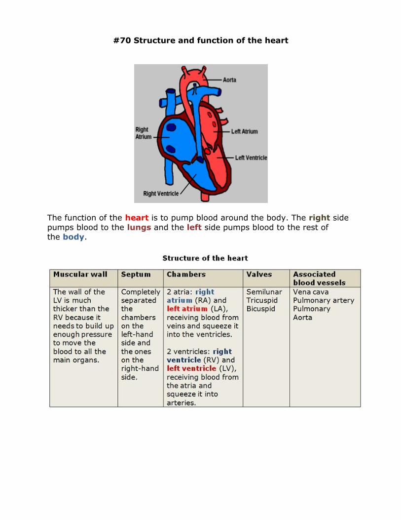
#70 Structure and function of the heart
The function of the heart is to pump blood around the body. The right side
pumps blood to the lungs and the left side pumps blood to the rest of the body.

Heart's function
Blood circulation
1. Blood in the right ventricle (RV) is pump to the lungs
2. Blood from the lungs flows back into the left atrium (LA) and then into the left ventricle (LV).
3. Blood in the LV is pumped through the body (except for the lungs) 4. Blood returns to the heart where it enters the right atrium (RA).

Muscular contraction
The heart is made of a special type of muscle called cardiac muscle which
contracts and relaxes regularly, throughout life.
The heart’s muscle is constantly active, so it needs its own blood supply, through the coronary artery, to provide it with oxygen and glucose.
Working of the valves
Valves in the heart prevent blood from being pushed backwards up into the
atria when the heart ‘beats’.
Animation: Heart Contraction and Blood Flow How the heart pumps blood

Try this
1.On a copy of the diagram of the double circulatory system, labell: i) The 4 main blood vessels (4 marks)
ii) The chambers of the heart (4 marks)
iii) The 2 valves shown (2 marks)
2.State 2 differences in composition between blood leaving the right ventricle and blood entering the left atrium. (2 marks)
Answers:
1.
2.Blood leaving the right ventricle has more CO2 and less O2 than blood entering the left atrium.
Common misconceptions
Remember that blood passing through the chambers of the heart
does not supply the heart muscle with oxygen or glucose. The heart muscles has its own blood supply - via the coronary arteries - to do this.

#71 Effect of exercise on heartbeat and causes
of coronary heart disease
A heartbeat is a contraction. Each contraction squeezes blood to the lungs and body. The heart
beats about 70 times a minute, more if you are younger, and the rate becomes lower the fitter you
are.
During exercise the heart rate increases to supply the muscles with more oxygen and
glucose à allow the muscles to respire
aerobically à they have sufficient energy to contract.
Regular exercise is important to keep the
heart muscle in good tone à heart is more efficient in maintaining blood pressure and↓risk of coronary heart disease and stroke.
Coronary arteries
The muscles of the heart are so thick that the nutrients and oxygen in the
blood inside the heart would not be able to diffuse to all the muscles quickly enough. The heart muscles needs a constant supply of nutrients so that it
can keep contracting and relaxing. The coronary arteries supply this.
If a coronary artery gets blocked – e.g. by a blood clot – the cardiac muscles run short of oxygen à they can not respire à can not obtain
energy to contract à heart stops beating. This is called a heart attack or cardiac arrest.
Main causes of a coronary heart disease and preventive measures

Blockage of the coronary arteries is called coronary heart disease.

#72 Arteries, veins and capillaries - structure and functions
There are 3 main kinds of blood vessels – arteries, veins and capillaries.
Arteries carry blood away from the heart. They divide again and again, and eventually form very tiny vessels called capillaries.
The capillaries gradually join up with one another to form large vessels called veins.
Veins carry blood towards the heart.
The comparison of blood vessels structure and functions

The transfer of materials between capillaries and tissue fluid

As blood enters capillaries from arterioles (small arteries), it slows down.
This allows substances in the plasma, as well as O2 from red blood cells, to diffuse through the capillary wall into the surrounding tissues (the
capillary wall is thin and permeable).
Liquid in the plasma also passes out. This forms tissue fluid, bathing the
cells. Waste products from the cells, e.g. CO2, diffuse back through the
capillary walls into the plasma. Some of the tissue fluid also passes back.
Diffusion is responsible for the
transfer of materials between capillaries and tissue fluid.
Plan of the main blood vessels in the human body

Sample questions
Figure above shows a section through the heart
i) Name the two blood vessels A and B [2 marks]
ii) Which of blood vessels A, B, C or D carry oxygenated blood [1 mark] iii) Name valve E and state its function [3
marks]
Student's answer
i) A, vena cava () B, pulmonary vein ()
ii) C ()
iii) name: semilunar valve () function: to stop blood going backwards ()
Examiner's comments
Blood vessels B is the pulmonary artery. Arteries of the heart always carry
blood from a ventricle.
Part ii) needs two answers (blood vessels C and D) to gain the mark.
D is the pulmonary vein, which carries oxygenated blood to the heart from the lung.
C is the aorta, which carries oxygenated blood from the heart to the body.
In part iii) the name of the valve is correct, but there are two marks for its
functions. This candidate has given only one statement: a second mark was available for stating that the valve prevents blood from going back into the
left ventricle.

#73 Blood composition and Plasma
If blood is allowed to stand without clotting, it separates out into 4 components: plasma, red blood cells, white blood cells and platelets.
The plasma and red blood cells play an important role in
the transportation of substances, around the body.
White blood cells and platelets are part of the body's immune system.
55% of the blood is plasma. This straw-coloured liquid contain water
with many important dissolved substances which must be carried around
the body. Most materials are carried by the blood plasma, except for oxygen.

Credit: moodle
Plasma transports:
blood cells soluble nutrients e.g. glucose (products of digestion) from the small
intestine to the organs
amino acids (plasma acts as a pool for amino acids for these cannot be stored in the body)
plasma proteins that are important in blood clotting (e.g. fibrinogen).
CO2 (waste gas produced by respiration in cells) from the organs to lungs
Other wastes of digestion (e.g. urea) from the liver to the kidneys.
Antibodies and antitoxins Hormones
Ions Heat from the liver and muscles to all parts of the body.
Video What is blood? https://www.youtube.com/watch?v=CRh_dAzXuoU

#74 Blood cells - structure and functions
Blood consists of cells floating in plasma.
Most of the cells are red blood cells. A much smaller number are white blood cells.
There are also fragments formed from special cells in the bone marrow,
called platelets.
Red and white blood cells as seen
under a light microscope.
Blood as seen through a microscope:
The largest cells are white cells.
The others are all red cells.
There are also a few platelets.

Functions of blood cells
Red blood cells transport oxygen.
White blood cells protect against disease. Blood platelets help the blood to clot.
1.Red blood cells (erythrocytes)
Made in the bone marrow of some bones, including ribs, vertebrae and some limb bones. Produced at a very fast rate – about 9000
million per hour!
Transport O2 from lungs to all respiring tissues. Prepare CO2 for
transport from all rerspiring tissues to lungs.
Contain haemoglobin (Hb), a red iron-containing pigment which can carry O2. In the lungs, Hb combines with O2 to form
oxyhaemoglobin. In other organs, oxyhaemoglobin splits up into Hb and O2
Have no nucleus à can fit more Hb inside the cytoplasm, but can lives only for about 4 months.
Have a special biconcave disc shape à increases the surface area and makes the diffusion of oxygen into & out of the cell easier.
Old red blood cells are broken down in the liver, spleen and bone
marrow. Some of the iron from the Hb is stored, and used for making new Hb, some of it is turned into bile pigment and excreted.

2. White blood cells (leukocytes)
Made in the bone marrow and in the lymph nodes.
Have a nucleus, often large and lobed.
Can move around and squeeze out through the walls of blood capillaries into all parts of the body.
There are many different kinds of white blood cells. They all have the
function of fighting pathogens (disease-causing bacteria and viruses) and to clear up any dead body cells in your body:
a.Phagocytes:
Have lobed nuclei and granular cytoplasm.
Can move out of capillaries to the site of an infection.
Remove any microorganisms that invade the body and might cause
infection, engulf (ingest) and kill them by digesting them.
b. Lymphocytes: produce antibodies to fight bacteria and foreign
materials.
Have large nuclei Responsible for immunity
There are two different types of lymphocytes:
B-lymphocytes: secrete special proteins called antibodies in
response to contact with their particular antigen, which may be an invading pathogen or a foreign tissue that has been transplanted.
T-lymphocytes attack foreign or infected cells and kill them.
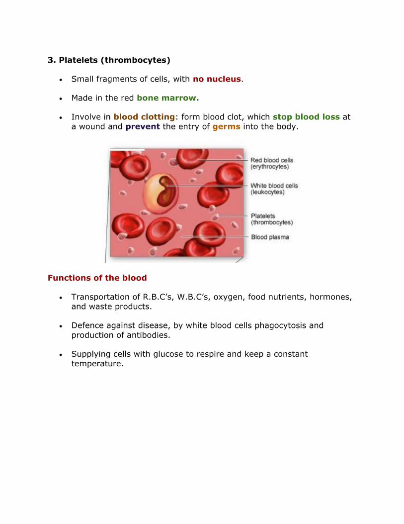
3. Platelets (thrombocytes)
Small fragments of cells, with no nucleus.
Made in the red bone marrow.
Involve in blood clotting: form blood clot, which stop blood loss at a wound and prevent the entry of germs into the body.
Functions of the blood
Transportation of R.B.C’s, W.B.C’s, oxygen, food nutrients, hormones, and waste products.
Defence against disease, by white blood cells phagocytosis and
production of antibodies.
Supplying cells with glucose to respire and keep a constant temperature.

#75 Blood clotting
When an injury causes a blood vessel wall to break, platelets are activated.
They change shape from round to spiny, stick to the broken vessel wall and each other, and begin to plug the break.
The platelets also interact with fibrinogen, a soluble plasma protein, to
form insoluble fibrin. Calcium is required for that.
Fibrin strands form a net that entraps
more platelets and other blood cells (red cells and white cells), producing
a clot that plugs the break.
Necessity for blood clotting
Prevent excessive blood loss from the body when there is a damage of the blood vessel.
Maintain the blood pressure.
Prevent the entry of microorganism and foreign particles into the body. Promote wound healing.
Video How does blood clot https://www.youtube.com/watch?v=--bZUeb83uU

#76 Immune system - antibody production,
tissue rejection & phagocytosis
The immune system is the body's defence against disease and foreign
bodies, under the form of antibody production, tissue rejection and phagocytosis.
Antibody production
Antibodies are produced by lymphocytes, which are formed in lymph
nodes. Lymphocytes produced antibodies in response to the presence of pathogens such as bacteria. This is because alien cells have chemicals
called antigen on their surface. A different antibody is produced for each antigen.
The antibodies make bacteria clump together in preparation for action
by phagocytes, or neutralise toxins produced by the bacteria. Once antibodies have been made, they remain in the blood to provide long-term
protection.
Some lymphocytes memorise the antigens the body has been exposed to.
They can rapidly reproduce and produce antibodies to respond to further infections by the same pathogen (disease-causing organism).

Tissue rejection
Transplants involve replacing a
damaged organ with a donor organ. Unfortunately, lymphocytes
and phagocytes will respond to any foreign cells in your body, even if
they are not pathogens.
If a person's kidneys fail, they can be given a new kidney taken from
another person. However the recipient's immune system will
recognise the cells in the new kidney as 'foreign', and will attack
and destroy them.
The transplanted organ triggers an immune response, antibodies
are secreted and the organ may be rejected. This is called tissue
rejection.
To prevent this happening:
The donor organ needs to be a similar tissue type to that of the
patient e.g. from a close relative.
Immunosuppressive drugs are uses, which switch off the body's
immune response. While recovering, transplant patients are
at risk of dying from any disease they are exposed to, so they need
to be kept in isolation.

Phagocytosis
Phagocytes have the ability to move out of capillaries to the site of an infection. They then engulf (ingest) the infecting pathogens and kill them by
digesting them.
Video: Phagocytosis of a Paramecium (unicellular ciliate protozoa)

#77 Functions of lymphatic system
The lymphatic system is a collection of lymph vessels and glands. It has 3 main roles:
Fluid balance: return tissue fluid to the blood
Protection from infection: produce white blood cells lymphocytes
Absorption of fats: transport digested fats from villi to blood stream
1. Lymph and Tissue Fluid
Tissue fluid is a fluid surrounding the cells of a tissue. It is leaked plasma - Plasma from the blood capillaries move to the tissue through gaps in the
walls and become tissue fluid.

Tissue fluid play an important role in substance exchange between blood
and cells. It supplies cells with O2 and nutrients and takes away waste products including CO2.
At the end of the capillary bed, the tissue fluid leaks back into the blood, and
becomes plasma again, but not all of it. A little of it is absorbed by the lymphatic vessel and becomes lymph.
The lymphatic vessel takes the lymph to the blood stream by secreting them in a vein near the heart, called subclavian vein. The lymph in the
lymphatic vessels are moved along by the squeeze of muscles against the vessel, just like some veins.
The return of tissue fluid to the blood in the form of lymph fluid prevents
fluid built up in the tissue.
2. Production of lymphocytes
The lymphatic system is an important component of the immune system, which fights infection. One group of white blood cells, the lymphocytes, are
made in lymph glands such as the tonsils, adenoids and spleen. The
glands become more active during an infection because they are producing and releasing large numbers of lymphocytes.
The lymphocytes can live and multiply in the lymphatic system, where they
attack and destroy foreign organisms. Lymphoid tissue scattered throughout the body filters out pathogens, other foreign matter and cellular
debris in body fluids.


3. The absorption of fatty acids and glycerol from the small
intestine
Following the chemical and mechanical breakdown of food in the digestive tract, most nutrients are absorbed into the blood through intestinal
capillaries. Many digested fats, however, are too large to enter the blood capillaries and are instead absorbed into lymphatic
capillaries by intestinal lacteals. Fats are added to the blood when lymph joins the bloodstream.
Each villus contains a lacteal - a blind ending lymph vessel.
Additional source: lubopitko-bg Xtremepapers
Related topic: Absorbtion – function of the small intestine and significance of villi
Video: Lymphatic system
https://www.youtube.com/watch?v=Kh-XdNnTZUo
#78 Summary of human transport
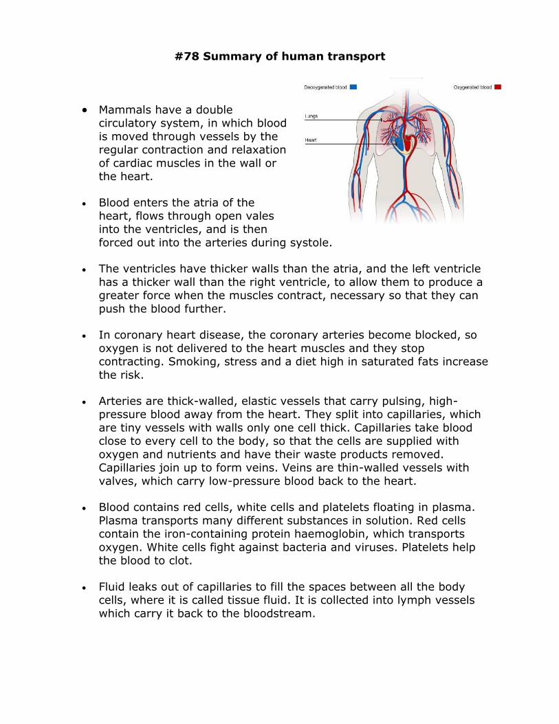
Mammals have a double circulatory system, in which blood
is moved through vessels by the regular contraction and relaxation
of cardiac muscles in the wall or the heart.
Blood enters the atria of the heart, flows through open vales into the ventricles, and is then
forced out into the arteries during systole.
The ventricles have thicker walls than the atria, and the left ventricle
has a thicker wall than the right ventricle, to allow them to produce a greater force when the muscles contract, necessary so that they can
push the blood further.
In coronary heart disease, the coronary arteries become blocked, so
oxygen is not delivered to the heart muscles and they stop contracting. Smoking, stress and a diet high in saturated fats increase
the risk.
Arteries are thick-walled, elastic vessels that carry pulsing, high-pressure blood away from the heart. They split into capillaries, which
are tiny vessels with walls only one cell thick. Capillaries take blood close to every cell to the body, so that the cells are supplied with
oxygen and nutrients and have their waste products removed. Capillaries join up to form veins. Veins are thin-walled vessels with
valves, which carry low-pressure blood back to the heart.
Blood contains red cells, white cells and platelets floating in plasma.
Plasma transports many different substances in solution. Red cells contain the iron-containing protein haemoglobin, which transports
oxygen. White cells fight against bacteria and viruses. Platelets help the blood to clot.
Fluid leaks out of capillaries to fill the spaces between all the body cells, where it is called tissue fluid. It is collected into lymph vessels
which carry it back to the bloodstream.