06. Action Potential
36
1 Introductory Medical Physiology Sept., 2008
Transcript of 06. Action Potential

8/16/2019 06. Action Potential
http://slidepdf.com/reader/full/06-action-potential 1/36
1
Introductory Medical Physiology
Sept., 2008

8/16/2019 06. Action Potential
http://slidepdf.com/reader/full/06-action-potential 2/36
2
i.i.i.i. Abstract
ii. Contentii. Contentii. Contentii. Content
1. Membrane potentials as information signal
1.1. The role of the nervous system in intercellular communication
1.2. From resting membrane potential to information signal
1.3. Membrane potentials and ion channels
1.4. Locations of the leakage and gated channels and their implication
2. Change in membrane potentials when stimulated: Graded potential
2.1. From resting potential to graded potential
2.2. Graphical representation of graded potential
2.3. Analysis of graded potential
3. Concept of threshold
4. Change in membrane potentials when stimulated: Action potential
5. Conduction/propagation/transmission of action potential
5.1. Mechanism of action potential propagation
5.2. The significance of refractory periods
5.3. Strategies to hasten action potential propagation
Resting membrane potential cannot carry any signal unless and until it is
transformed to graded potential and then to action potential. Only actionpotential can be propagated to carry the message about a stimulus so that
appropriate response can be elicited in the target cell. A stimulus triggers
transformation of resting membrane potential at special sites of anexcitable tissue (at a receptor or sensor, post-synaptic membrane, end-
plate surface of skeletal muscle, or pacemaker) to produce graded
potential. This is due to stimulation of chemical-gated or mechanically-gated ion channels that open and cause a change the permeability of the
membrane to certain ions. If the stimulus produces a graded potential
that depolarises enough to reach threshold, this voltage difference willstimulate voltage-gated ion channels located close to the receptor site.
The membrane will depolarize to form an action potential. Thisdepolarization will in turn stimulate the next voltage-gated channel along
the axon and another action potential is produced. This processcontinues to occur until the action potential reaches the axon terminal,
thus carrying the message about the stimulus to be communicated to thenext neuron or effector cell.

8/16/2019 06. Action Potential
http://slidepdf.com/reader/full/06-action-potential 3/36
3
iii. Checklist of topics and learning activities in
this module
Content Page Comments on
masteryi Abstract 4
ii Learning Resources 4
iii Background Knowledge 5
iv Terms to Know 5
v Objectives 6
vi Learning Activities 7
1 Membrane potentials as information signal 7
1.1. The role of the nervous system in
intercellular communication
7
ACTIVITY 6.1: Significance of intercellular
communication
7
1.2. From resting membrane potential to
information signal
8
ACTIVITY 6.2: Membrane potential asinformation signal
8
1.3. Membrane potentials and ion channels 9
ACTIVITY 6.3. Significance of ion channels 9
ACTIVITY 6.4: Implication of ionic diffusion
via gated channels
10
1.4. Locations of the leakage and gated channels
and their implication
11
ACTIVITY 6.5. Locations of membrane channels 11
2 Change in membrane potentials when
stimulated: Graded potential
12
2.1. From resting potential to graded potential 12
ACTIVITY 6.6: Equilibrium potential 12
ACTIVITY 6.7: Demonstration on change in
membrane potential
13
ACTIVITY 6.8: Demonstration of depolarisation 14
ACTIVITY 6.9: Graded potential 16
2.2. Graphical representation of graded potential 17
ACTIVITY 6.10: Analysing membrane potentialgraphically 18
2.3. Analysis of graded potential 19
ACTIVITY 6.11: Molecular mechanism of graded
potential
20
ACTIVITY 6.12: Types of graded potentials 20
ACTIVITY 6.13: EPSP and IPSP 20
ACTIVITY 6.14: Graphs of graded potential 21

8/16/2019 06. Action Potential
http://slidepdf.com/reader/full/06-action-potential 4/36
4
3 Concept of threshold 22
ACTIVITY 6.15: Stimulus-response relationship 22
4 Change in membrane potentials when
stimulated: Action potential
24
ACTIVITY 6.16: Action potential 24
ACTIVITY 6.17: Mechanism of action potentialproduction
25
5 Conduction/propagation/transmission of action
potential
28
5.1. Mechanism of action potential propagation 28
ACTIVITY 6.18: Mechanism of action potential
propagation
28
ACTIVITY 6.19: The domino effect of action
potential propagation
29
5.2. The significance of refractory periods 30
ACTIVITY 6.20: Relevance of refractory periods 31
5.3. Strategies to hasten action potential propagation
31
ACTIVITY 6.21. Saltatory conduction of action
potential
33
vii Summary 34
viii Conclusion 34
ix Assessment 35

8/16/2019 06. Action Potential
http://slidepdf.com/reader/full/06-action-potential 5/36
5
iv. Learning Resources
Please refer to the following sources for further information:
• Boron and Boulpaep, Ch. 2 & 3
• Guyton & Hall, pp 10-12 & Ch. 4
• Ganong, Ch. 1
• Marieb, Ch. 3. + Study partner
• Tortora & Grabowski, Ch. 3
• Vander, Sherman & Luciano, Ch. 3
• Supplementary materials provided
Animations:
1. http://www.tvdsb.on.ca/westmin/science/sbioac/homeo/action.htm action potential2. http://www.accessexcellence.org/RC/VL/GG/action_Potent.html Propagation of
action potentials
v. Background knowledge
To complete this module successfully, you should have the following background:
• Knowledge on the general structure and functions of cell membranes (Module 2).
• Knowledge on the mechanisms of solute transport across cell membrane (Module 3).• Knowledge on the basic mechanism of signal transduction (Module 4).• Understanding of the definition of chemical and electrical gradients (Module 5).
• Knowledge on membrane potentials of excitable tissues at rest (Module 5).
8/16/2019 06. Action Potential
http://slidepdf.com/reader/full/06-action-potential 6/36
6
vi. Terms to know
After studying the materials and doing the activities in this module, students should be
comfortable with the following terms:
action potentialactive transport
body fluid compartments
channelschemically activated gates
concentration
concentration gradientsconductance
cytoplasm
depolarization
diffusiondynamic equilibrium
electrical gradients
electrodeelectrochemical gradient
electrogenic pump
excitatory post-synaptic potential(EPSP)
fatty acid (tails)
gated channelshydrophilic
interstitial fluid (ISF)
hydrophobichyperpolarized
inhibitory post-synaptic potential (IPSP)
ion binding sitesmembrane permeability
membrane potentials
milliseconds (msec).Na + /K + -ATPase
net charge
net movement
passive channelsphospholipid bilayer
receptor site
resting membrane potentialsemi-permeable
solute
solventthreshold
voltage activated gates
voltmeter
Please add other terms that you feel are relevant to your understanding of this module.

8/16/2019 06. Action Potential
http://slidepdf.com/reader/full/06-action-potential 7/36
7
vii. Learning objectives
After studying the materials in Module 6, the students should be able to:
Please set up more specific objectives after you’ve thoroughly studied the material in this
module to help yourself in your revision later on. Make notes that meet the requirement of
the new objectives.
1. Explain the role of the nervous system in intercellular communication.
2. Predict the sequence of events that take place on a cell membrane when a
stimulus reaches a receptor.
3. Describe the significance of different ion channels of the membrane of a neuron
in terms of their effects on membrane potential.
4. Define graded potential, describe how it is established, and describe its
characteristics.
5. List down the types of graded potential and state the significance of each.
6. Compare and contrast between EPSP and IPSP.
7. Define threshold stimulus and threshold potential
8. Plot the membrane potential response to subthreshold, threshold, and
suprathreshold stimuli and explain the molecular basis of the response.
9. Explain the meaning of action potential.
10. Describe the mechanism of action potential production.
11. Compare and contrast between action potential and graded potential.
12. Describe the mechanism of action potential propagation.
13. Describe the molecular basis of absolute and relative refractory periods and
state the significance.
14. Describe the strategies that can hasten action potential propagation.

8/16/2019 06. Action Potential
http://slidepdf.com/reader/full/06-action-potential 8/36
8
Objectives from the American Physiological Society
CE 10. Based on the principle of ionic attraction, explain how a potential difference
across a membrane will influence the distribution of a cation and an anion.
CE 11. Define the term “steady state,” and differentiate it from “equilibrium.” Relate the
pump-leak model of steady-state ion content to cell solute gradients and cell volumemaintenance.
CE 12. Write the Nernst equation, and indicate how this equation accounts for both the
chemical and electrical driving forces that act on an ion.
CE 13. Based on the Nernst equilibrium potential, predict the direction that an ion will
move when the membrane potential a) is at its equilibrium potential, b) is higher than the
equilibrium potential, or c) is less than the equilibrium potential. List values in a typical
non-excitable cell for the membrane potential, for ENa, EK, ECl, and ECa.
CE 14. Define the concepts of electrochemical equilibrium and equilibrium potential, andgive internal and external ion concentrations. Be able to calculate an equilibrium potential
for that ion using the Nernst equation. Contrast the difference in EK (the Nernst potential
for K+) caused by a 5 mEq/l increase in extracellular K
+ with the change in ENa (the
Nernst potential for Na+) caused by a 5 mEq/l increase in extracellular Na+.
CE 15. Explain how the resting membrane potential is generated and calculate membranepotential by using either a) the Goldman-Hodgkin-Katz equation or b) the chord
conductance equation. Given an increase or decrease in the permeability of K, Na, or Cl,
predict how the membrane potential would change.

8/16/2019 06. Action Potential
http://slidepdf.com/reader/full/06-action-potential 9/36
9
viii. Learning activities
1. Membrane potentials as information signal
In Module 5 we learned about the membrane potential when the cell is not stimulated. Weknow that resting membrane potential exists in every cell, but resting potential does not
carry any message even in excitable cells, as there is no stimulation. Nevertheless, when
the cell is stimulated, the resting membrane potential is transformed into action potentialby excitable cells (non-excitable cells are not able to do this). Action potential can be
propagated along the excitable cell to affect adjacent cells. In this Module, we’ll explore
how excitable cells transform resting membrane potential in the presence of appropriatestimulus. This forms the basis of intercellular communication via the nervous system.
1.1. The role of the nervous system in intercellular communication
You have learnt about intercellular communication in Module 4. Activity 6.1 helps you toreview the significance of intercellular communication in body functions.
Receptor
Control
centre
Effector
Figure 6.1. Information pathway from receptor to effector
Human beings and animals must fulfill two basic physiological needs:a) to regulate chemical and physical factors in the internal environment, andb) to respond/adapt to external environment.
How do we regulate the chemical and physical factors in the internal environment? What arethe basic components of homeostasis? Give examples of homeostatic regulation, and name allthe specific components. Explain the mechanism of information flow from one component to
another.
Why do we need to respond to the external environment? Give examples. Explain theprocesses (information flow) that are involved in the perception of the external environment.Use Fig. 6.1 to help you in your answer.
From the above explanation, deduce the significance of intercellular communication.
ACTIVITY 6.1: Significance of intercellular communication

8/16/2019 06. Action Potential
http://slidepdf.com/reader/full/06-action-potential 10/36
10
1.2. From resting membrane potential to information signal
From Activity 6.1 you infer that intercellular communication may occur via the nervous
system. You also know that the neurons are excitable i.e. it can generate impulses that are
used as information signals. However, we know from Module 5 that resting membrane
potential does not carry any message (why?). Therefore, the resting potential must betransformed in such a manner so as to function as signal carrier.
Activity 6.2 helps you to predict the sequence of events that take place on a cell membrane
when a stimulus reaches a receptor.
The key molecules that are involved in producing information signal on a cell membrane
are the proteins such as receptor and channel proteins. Before we look at what happens tothe membrane potential when stimulated, let’s review the relationship between ion
channels and membrane potentials.
How is resting membrane potential changed? Hint:1. Firstly, there must be a stimulus (give examples of internal and external stimuli) to
trigger the change.2. Secondly, there must be an appropriate receptor that is associated with the ion
channels.3. Thirdly, the stimulus interacts with membrane receptors change the configuration of
ion channels on the membrane change the permeability of membrane to certain ionsby altering the characteristics of channel proteins.
Predict the consequence of the above events. Hint: Change in the pattern of ion movementacross the membrane. What happens to the membrane potential if the ion channels involvedare Na
+ channels?
Predict the implication of the above events. Hint: Resting potential is changed to some otherpotentials that can be used to carry information (Refer to Table 6.1).
ACTIVITY 6.2: Membrane potential as information signal

8/16/2019 06. Action Potential
http://slidepdf.com/reader/full/06-action-potential 11/36
11
1.3. Membrane potentials and ion channels
We know from Module 5 that:
• membrane potential is due to the unequal distribution of ions across the plasma
membrane
• ions move across the membrane through ion channels• channels are specific for each ion
We also know that there are two main types of ion channels (Fig 6.2):
• Passive, or leakage channels that are always open.
• Active, or gated channels that open or close only in response to various signals.
There are three main types of gated channels:
o Chemical-gated channel
o Voltage-gated channel
o Mechanically-gated channel
Fig. 6.2. Types of ion channels
Activity 6.3 helps you to understand the significance of ion channels.
Ion channels
Passive(leakage)
Active(gated)
Chemical-gated
Voltage-gated
Mechanically-gated
What is the implication of the number of leakage K+ and Na
+ channels in plasma membrane to
the resting potential? You have calculated the EK+ and ENa+ using the Nernst’s equation, andthe EM using the Goldman’s equation. Why is the resting membrane potential closer to theequilibrium potential of K
+ compared to that of Na
+?
Predict what happens to a cell that has only leakage channels (no gated channels) when it isstimulated by an appropriate stimulus. Which type of cell has no gated channels? Which typeof cell has gated channels? What is the advantage of having gated channels? Why don’t allcells have gated channels? What kind of gated channels exist in receptor cells that directlyrespond to internal and external environmental stimuli?
ACTIVITY 6.3: Significance of ion channels

8/16/2019 06. Action Potential
http://slidepdf.com/reader/full/06-action-potential 12/36
12
Ions move along chemical and electrical gradients (electrochemical gradients) through
the channels, carrying the +ve or –ve charges. This will cause a change in membranepotential. Activity 6.4 helps you to clarify this phenomenon.
Figure 6.3. Chemical-gated channel
Figure 6.4. Voltage-gated channel.
Refer to Fig. 6.3:What happens when acetylcholine binds to its receptor in the membrane? Draw a graph toshow resting membrane potential and the change in potential when acetylcholine binds to thereceptor.
Refer to Fig. 6.4:What happens to the voltage gated K
+ channel when the membrane potential is changed from
-70 mV to -50 mV? What is the implication in terms of the resulting membrane potential?
Draw a graph to show resting membrane potential and the change in potential when thevoltage-gated K+ channel is stimulated by an initial depolarization.
ACTIVITY 6.4: Implication of ionic diffusion via gated channels
8/16/2019 06. Action Potential
http://slidepdf.com/reader/full/06-action-potential 13/36
13
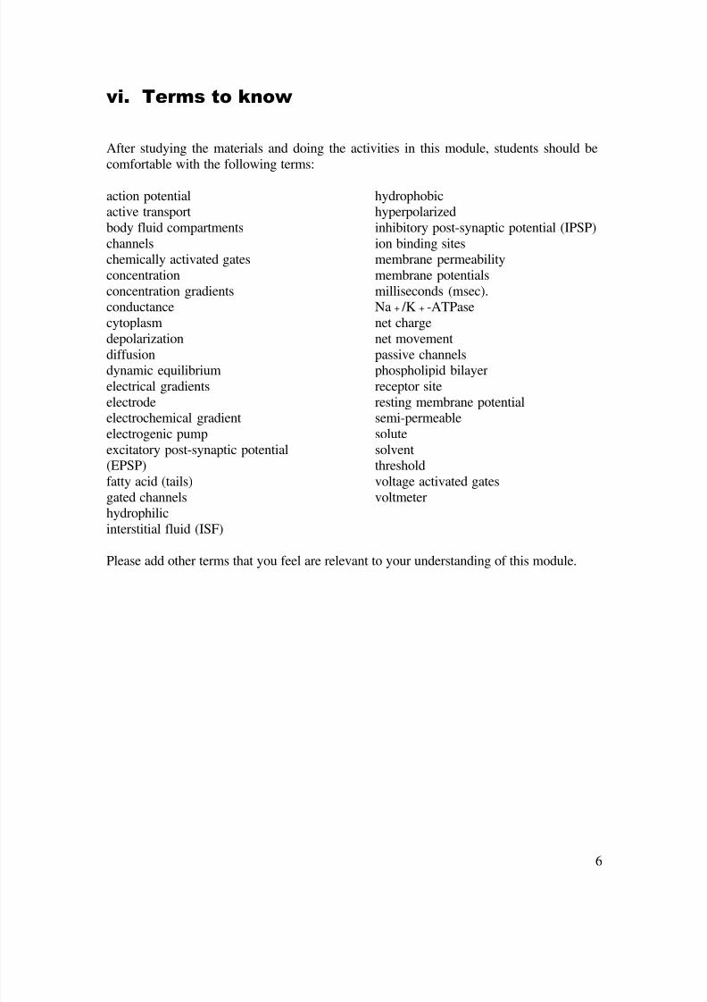
1.4. Locations of the leakage and gated channels and their implication
Leakage channels are found almost everywhere on a cell membrane. However, the
number of individual channels on a cell membrane may be different (refer to Activity 6.8).
Chemical/mechanical-gated channels are located on:• nerve endings or receptor (sensor) sites
• post-synaptic membrane• pacemaker cells
• muscle (neuromuscular junction)
Voltage gated channels are located at axon hillock and along the axon distal to receptor
region (Fig. 6.5)
Figure 6.5. The input zone, trigger zone, conducting zone, and output zone on a neuron.
List down the locations of leakage channels, chemical-gated and mechanical gated channels incell membrane. Hypothesise the significance of the locations of the leakage and gatedchannels.
Based on Fig. 6.5 suggest the function of:Input zone, Trigger zone, Conducting zone, Output zone
What ion channels are present at the above zones? What are the implications?
ACTIVITY 6.5: Locations of membrane channels

8/16/2019 06. Action Potential
http://slidepdf.com/reader/full/06-action-potential 14/36
14
2. Change in membrane potentials when stimulated: Gradedpotential
2.1. From resting potential to graded potential
In Module 5 you have worked on equilibrium potential and resting membranepotential using a manipulative model. You also know how to calculate equilibrium
potential and resting membrane potential using the Nernst and Goldman equations. Inaddition, you have also drawn graphs to represent the membrane potentials.
Activity 6.6 helps you to revise the significance of equilibrium potential.
In Activity 6.7 you will demonstrate the change in membrane potential by working on thetemplate model (use the model in Appendix A in Module 5).
Please draw line graphs showing EK, ENa, ECl, and EM complete with labeled X and Y axis whena cell is at rest.
What happens to the membrane potential when permeability to each of the ionsincreases/decreases? Illustrate by drawing the graphs superimposed on the previous graphs.
What may cause the permeability of the ions to change? Hint: what are the specific stimuli?
A change in the permeability of an ion will affect membrane potential.
ACTIVITY 6.6: Significance of equilibrium potential
Set up your membrane template (Appendix A in Module 5) with a K+ channel in one gap, and a
Na+ channel in the other (Appendix B in Module 5). To each of these channels lay a gate
across the opening on the outside of the membrane. The small notch in the gate should faceup.
Add the following ions to the ISF: 10 Na+, 1 K
+, and 3 Cl
- . Add the following ions to the
cytoplasm: 1 Na+ , 10 K
+ , and 1 Cl
-. Please do not disregard the 20 negative charges inside
the model cell due to protein. Note that these do not represent physiological proportions, butwill be useful for helping you do the simulations.
Determine by the counting technique you learned in Module 5 what the membrane potential is
for this model cell.
Observe the channels that you set up on your template. What types of channels are these?Specify both the ion and the means of opening/closing.
One type of gated channel that is vital for communication between cells in the body iscontrolled chemically. What does this mean?
ACTIVITY 6.7: Demonstration on change in membrane potential

8/16/2019 06. Action Potential
http://slidepdf.com/reader/full/06-action-potential 15/36
15
These chemical-gated (or ligand-gated) channels are found, for example, on the skeletal
muscle cells innervated by the motor neurons. So when you decide to flex your arm at theelbow (Fig. 6.1), the signal is sent out to the skeletal muscle cells of the biceps brachii
muscle by means of motor neurons that signal the muscle to contract. In this example, the
motor neurons release a chemical signal (a neurotransmitter called acetylcholine) that
attaches to the channel protein at a special receptor site. The binding of the chemicalsignal to the receptor causes the gate to open briefly, causing membrane depolarization.
Activity 6.8 helps you to understand the above concept better.
Continue your work from Activity 6.7. Choose the square signal molecule for your model,attach it to the receptor site on the gate of the Na
+ channel, and rotate the channel to an open
position. Demonstrate what happens to the sodium ions.
The attachment between the signal molecule and the receptor site is weak. Soon it
detaches and the gate closes. Demonstrate it. The signal molecule is then split apart by anenzyme (not shown here) in the cell membrane. You can simulate this by simply removing thesquare signal molecule.
Based upon what you have learnt so far, predict what will happen to membrane potential whenNa
+ gate is opened.
Test your prediction by running the following simulation: Attach the signal molecule to thechannel, open the gate, move 3 Na
+ into the cell, and close the gate. Determine the new
membrane potential and record it. What forces tend to move Na+ into the cell?
If you did it correctly, there is a change in membrane potential called depolarization of themembrane. What does depolarization mean? Draw a graph showing resting membranepotential and how this potential changes when Na
+ channel is open.
Confirm that the potential difference across the membrane was reduced (went closer to zero).Thus depolarization takes the membrane from its resting potential closer to zero.
Try to produce a computer animation of the above process.
ACTIVITY 6.8: Demonstration of depolarisation

8/16/2019 06. Action Potential
http://slidepdf.com/reader/full/06-action-potential 16/36
16
In Activity 6.8, the influx of Na+ ions creates a new gradient inside the cell (i.e. charge
difference underneath the membrane between the site of Na+ entry and the adjacent sites
i.e. the dark band in Fig. 6.6a). To understand this, consider the concentration of sodium
ions in the cytoplasm directly beneath the channel compared to the concentration in the
adjacent area. What is the gradient? Refer to Fig 6.6a. What do you expect will happen to
the extra Na
+
ions that just came in?
The resulting diffusion of ions creates a tiny current in the cell. It also will restore thelocal membrane potential back toward the resting value (Fig 6.6b). Fig. 6.7 shows what
happens in a graphical format.
Figure 6.6 a. Stimulation of the membrane causes deplarisation; b. The positive charges spread to
the neighbouring sites
Figure 6.7. Graphical representation of the spread of charges from the site of depolarization.

8/16/2019 06. Action Potential
http://slidepdf.com/reader/full/06-action-potential 17/36
17
Now you know what happens when the cell is stimulated by a ligand that sits on itsreceptor, causing the gated ion channel to open. If the stimulus causes Na+ channel to
open, Na+ will rush in, depolarizing the membrane from its resting state. The Na+ ions
will be attracted to the negative charges of the neighbouring sites and soon the membrane
potential falls back to the resting state. This type of transient depolarization is called
graded potential.
Activity 6.9 helps you to explore more about graded potentials.
Table 6.1 gives a brief description of different types of membrane potentials.
Table 6.1. Types of membrane potentials
Types of potential Description
Potential = potentialdifference
The voltage difference between two points
Membrane potential The voltage difference between the inside and outside of thecell
Equilibrium potential The voltage difference across a membrane that produces a flux
of a given ion species that is equal but opposite the flux due tothe concentration gradient affecting the same ion species
Resting potential The steady transmembrane potential of a cell that is notproducing an electrical signal
Graded potential A potential change of variable amplitude and duration that isconducted decrementally; it has no threshold or refractoryperiod
Action potential A brief all-or-none depolarisation of the membrane, reversingpolarity in neurons; it has a threshold and refractory period andis conducted without decrement
Synaptic potential A graded potential change produced in the postsynaptic neuronin response to release of a neurotransmitter by a presynapticterminal; it may be depolarising (EPSP) or hyperpolarising
(IPSP)Receptor potential A graded potential produced at the peripheral endings ofafferent neurons (or in separate receptor cells) in response to astimulus
Pacemaker potential A spontaneously occurring graded potential change that occursin certain specialised cells
What is graded potential? Can graded potential be used to carry information? Predict thefunction of graded potential.
ACTIVITY 6.9: Graded potential

8/16/2019 06. Action Potential
http://slidepdf.com/reader/full/06-action-potential 18/36
18
2.2. Graphical representation of graded potential
Membrane potentials change rapidly over time. In a matter of a few milliseconds (one-thousandth of a second) major shifts can occur in membrane potentials with the production
and transmission of signals among cells of the body. These changes are often represented
graphically as shown in Fig 6.8. This graph represents a recording, over time, of themembrane potential from a single cell. You may remember that the recording electrode is
inside the cell (cytoplasm) while the reference electrode is outside in the ISF (Fig 5.4,
Module 5).
Figure 6.8. Change in membrane potential when a cell is stimulated
The graphs in Fig 6.8 shows a recording of what happens to the membrane potential when
2 separate cells are stimulated (at the arrow, time=1 msec.)
Activity 6.10 helps you to analyse the graph clearly.
M e m b r a n e
o t e n t i a l ( m V )
Time (msec)
1 2 3 4 5 6 7 8 9 10
-100
-80
-60
-40
-20
0
20
11 12 13 14 15
ab c
d
ef g
h

8/16/2019 06. Action Potential
http://slidepdf.com/reader/full/06-action-potential 19/36
19
Figure 6.9. Membrane potential showing a) depolarization and b) hyperpolarisation
Refer to Fig. 6.8. Look at the top curve abcd and answer the following questions:a. What is the membrane potential at a called? What is its value?b. What is the name of the event when the curve goes up at b?b. Referring to your work in the prior section, what change in the membrane is likely to
have caused the shift in membrane potential at b?c. What is the value of membrane potential at the peak of the curve?d. What is the name of the event when the curve goes down at c?e. What change in the membrane is likely to have caused the shift in membrane potential at
c?f. Is there any difference between the values of a and d? Why?
The lower curve efgh in Fig 6.8 comes from a recording in a different cell.a. What is its resting membrane potential, e?b. What is the name of the event when the curve goes down at f?c. Referring to your work in the prior section, what change in the membrane is likely to have
caused the shift in membrane potential at f?d. What is the most negative reading on the lower curve?
e. What is the name of the event when the curve goes up at g?f. What change in the membrane is likely to have caused the shift in membrane potential atg?
g. Is there any difference between the values of e and g? Why?
Fig 6.9 repeats the same concept of depolarization and hyperpolarisation presented in Fig. 6.8.Study the graphs carefully. Where do you think the phenomenon represented by each graphoccurs in your body?
ACTIVITY 6.10: Anal sin membrane otential ra hicall

8/16/2019 06. Action Potential
http://slidepdf.com/reader/full/06-action-potential 20/36
20
2.3. Analysis of graded potential
In this section you will explore:
What is graded potential?
Where does it occur?What is it used for?
Characteristics of graded potential:
• Short-lived, local changes in membrane
potential (depolarisation orhyperpolarisation) due to opening of
chemical-gated ion channels (Fig
6.10a)
• Amplitude proportional to strength ofstimulus (Fig 6.10b)
•
Current flows decreases with distance;so, cannot be utilized for long distancesignals (Fig 6.10c)
• Can be summed spatially and
temporally (concept of temporal andspatial summation) (Fig 6.10d)
Activity 6.11 helps you to appreciate the
characteristics of graded potential by
explaining the molecular mechanismsinvolved.
Activity 6.12 helps you to identify the different types of graded potentials that occur inyour body.
Figure 6.10. Characteristics of graded potential
For each of the characteristics mentioned above, please explain the molecular mechanisminvolved.
ACTIVITY 6.11: Molecular mechanism of graded potential
Specific names of graded potential based on location/ function:• Receptor potential or generator potential• Postsynaptic potential (EPSP and IPSP)• Pacemaker potential• End plate potential
Please provide appropriate diagrams (your own sketch or drawing from a source) to show thelocation where the above graded potentials occur.
ACTIVITY 6.12: Types of graded potentials

8/16/2019 06. Action Potential
http://slidepdf.com/reader/full/06-action-potential 21/36
21
The graded potential events represented in Fig 6.10 are frequent occurrences in neuron and
muscle cells. For example, the brief depolarization of the membrane of a post-synapticneuron in response to a chemical signal (neurotransmitter) is called an excitatory post-
synaptic potential (EPSP). It is so named because it increases the likelihood that the cell
will produce an action potential, another type of communication signal (Section 4).
The hyperpolarization in Fig 6.10 is called an inhibitory post-synaptic potential (IPSP).
These signals have the opposite action of EPSPs. They decrease the likelihood that the cellwill produce an action potential. Some neurotransmitters cause depolarization while
others cause hyperpolarisation.
Activity 6.14 helps you to present graded potential in graphical format.
• Compare and contrast between EPSP and IPSP.• What is the relevance of EPSP and IPSP?
ACTIVITY 6.13: EPSP and IPSP
• In Fig 6.11 draw a graph from the data in the table provided. The recordings in the table arefrom two different cells. Plot the points and connect them to make the curve.
• When you have completed the graphing, label the segments of each curve by referring toFig 6.8. (A new segment begins with a change in direction of the curve). You may need touse more letters on the top curve than the bottom.
• Next, interpret in words what is happening in each segment of the curve. Suggest what is
happening in the membrane to bring about the changes from one segment to the next.
ACTIVITY 6.14: Graphs of graded potential

8/16/2019 06. Action Potential
http://slidepdf.com/reader/full/06-action-potential 22/36
22
What is the relevance of graded potential? If it can’t carry signals over long distance,
what is it for? Graded potential is actually important in initiating action potentials, the
long distance signals. How is this possible?
Before answering the above questions, let’s first look at the concept of threshold.
3. Concept of threshold
What is threshold stimulus? It is a stimulus which is strong enough to cause formation
of action potential.What is threshold potential? It is a membrane potential (voltage) which is strong enoughto stimulate voltage-gated channels to open. It is normally ~15mV above resting
potential.
To understand the concept of threshold stimulus and threshold potential, let’s carry outthe Activity 6.15:
Time(msec)
Membranepotential (mV)
Cell 1 Cell 2
0 -60 -750.5 -60 -75
1.0 -60 -751.5 -50 -75
2.0 +20 -842.5 -50 -883.0 -60 -84
3.5 -65 -754.0 -63 -75
4.5 -60 -755.0 -60 -75
M e m b r a n e
o t e n t i a l ( m V )
Time (msec)
1 2 3 4 5 6 7 8
-100
-80
-60
-40
-20
0
20
Figure 6.11. Membrane potential recordings for two cells

8/16/2019 06. Action Potential
http://slidepdf.com/reader/full/06-action-potential 23/36
23
Study the two panels in Fig. 6.12. Both panels are drawn on the same time scale. The Y-axisof the upper panel represents membrane potential, and that of the lower panel representsstimulus strength.
a. Label the Y-axis of the upper panel with -70 mV, -55 mV, 0 mV, and +30 mV. You candisregard the square markings .
b. For this simulation, let’s use an arbitrary unit for the stimulus strength (which could besolute concentration, strength of mechanical force, etc). Let’s assume that the thresholdstimulus is 10 units, and the threshold potential is -55 mV (i.e. 15 mV above restingpotential). Label the Y-axis of the lower panel with 0 units and 10 units.
c. Look at stimulus “a”. Since its strength is below threshold, it is called a “subthresholdstimulus”. When you give this stimulus to a cell, it’s going to open up a few chemical-gated Na
+ channels, therefore there is a little depolarization (observe the corresponding
membrane potential response in the upper panel). This depolarization is below thresholdpotential, and it dies off when the stimulus is taken off.
d. Look at stimulus “b”. It is still subthreshold, but the strength of the stimulus is greater than“a”. What do you expect to happen to the cell membrane? The corresponding membranepotential in the upper panel reflects an appropriate depolarization response to stimulus “b”.
e. Stimulus “c” is greater than “b”, but is still subthreshold. Draw the appropriatedepolarization on the upper panel.
f. Stimulus “d” is greater than “c”, but still subthreshold. Draw the appropriate depolarizationon the upper panel.Based on this activity so far, summarise the characteristics of graded potential.
g. Now, look at stimulus “e”. It has reached threshold, so it is called a threshold stimulus.That means the stimulus is strong enough to open enough ligand-gated channels todepolarize the membrane to a level where it will stimulate voltage-gated Na
+ channels to
open. The characteristic feature of this voltage-gated channel is that all of the channelspresent at the point of stimulation will open. What do you expect to happen to themovement of Na
+ ions?
This characteristic is also known as all-or-none, i.e. all of the voltage-gated channels
either open together under the influence of a threshold stimulus, or none of them will openwhen the stimulus is subthreshold. Another characteristic feature of the channel is that theopening is transient (only for 1 msec). What do you think will happen when the channelsare closed?The spike of depolarization due to simultaneous opening of voltage-gated channelstogether with repolarisation when the channels are closed is called an action potential.Please refer to the membrane potential (upper panel) in response to stimulus “e”.
h. Stimulus “f” is greater than threshold stimulus, so it is called a suprathreshold stimulus.Remember that with threshold stimulus, all the voltage-gated Na
+ channels are open.
What do you expect to happen when the cell is given a suprathreshold stimulus? Pleasedraw the membrane potential appropriately as a response to stimulus “f”.
i. Stimuli “g” and “h” are also suprathreshold. Please draw the membrane potentialappropriately as a response to the stimuli.
j. Label “subthreshold stimulus”, “threshold stimulus” and “suprathresold stimulus”, “gradedpotentials”, “action potentials”.
k. In addition, another characteristic of a graded potential is it can be summed temporally andspatially. Please refer to Fig. 6.13 and explain the meaning of the terms “spatial
summation” and “temporal summation”.
ACTIVITY 6.15: Stimulus-res onse relationshi

8/16/2019 06. Action Potential
http://slidepdf.com/reader/full/06-action-potential 24/36
24
c d e f hba
M e m b r a n e
p o t e n t
i a l ( m V )
S t i m
u l u s
s t r e n g t h
Time
Thresholdstimulus
Restingmembranepotential
Thresholdpotential
Figure 6.12. Stimulus-response relationship.
Figure 5.13. Temporal and spatial summation

8/16/2019 06. Action Potential
http://slidepdf.com/reader/full/06-action-potential 25/36
25
4. Change in membrane potentials when stimulated: Actionpotential
You have seen in the previous section the relationship between graded potential and actionpotential. Action potential is basically a brief reversal of membrane potential with a total
amplitude (change in voltage) of about 100mV (from –70 to +30 mV).
Review:• Action potentials occur in certain parts of the cell membrane where there are
voltage-gated channels.
• Nerve impulse is transmitted action potentials. It is used as a principal method ofcommunication by neurons
• Stimulus for generation of action potential is graded potential. It involves voltage-
gated channels at axon hillocks or proximal to receptor regions
• An action potential is of no use in terms of signal carrying capacity unless it can be
propagated as impulses. In neurons, action potentials trigger neurotransmitter
release; in muscle cell they cause contraction.
In interpreting the segments of the curve that make up the action potential, note that for abrief period of time, the membrane potential is actually reversed. Locate this portion of the
action potential and label it "reversal of membrane potential".
The upward section of the curve in Fig. 6.11 between 1.5-2.0 msec is due to the opening
of voltage-activated Na+ channels that allow Na
+ to flow briefly into the cell. The
downward segment from 2.0-2.5 msec is due to outward movement of K+ ions through
voltage-activated channels. The period of hyperpolarization between 3.0-4.5 msec results
from the fact that more K+ leave the cell than required to bring it back to resting potential.
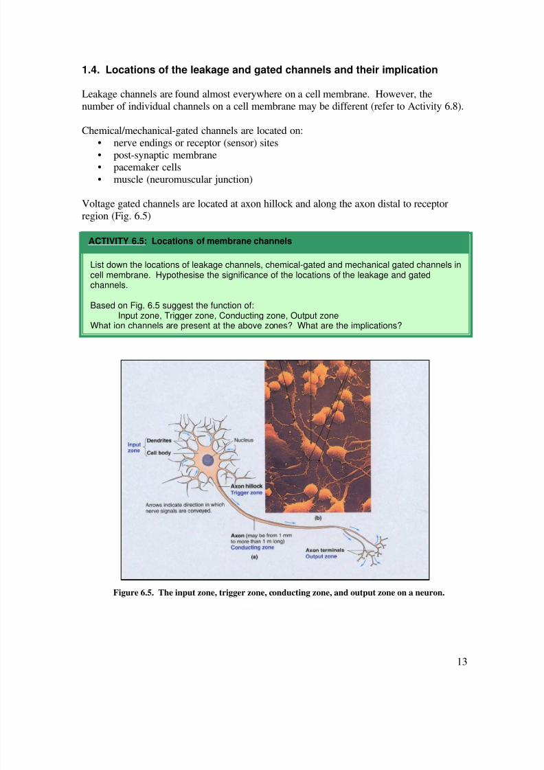
Let’s now look closely at the mechanism of action potential production:
Formation of action potential involves 3 consecutive but overlapping changes inmembrane permeability due to opening and closing of active ion gates:
i. Transient increase in Na+
permeability depolarisation
ii. Na+ impermeability
iii. Short-lived increase in K+ permeability
There are two types of gates on voltage dependent Na+ channels:
re olarisation
What does “reversal of membrane potential” mean?
Only cells with excitable membranes (neurons and muscle cells) can generate action potential.Why? Please give a molecular evidence.
ACTIVITY 6.16: Action potential
8/16/2019 06. Action Potential
http://slidepdf.com/reader/full/06-action-potential 26/36
26
i. Activation gate
ii. Inactivation gate
Please use Fig. 6.14 to figure out the involvement of the gates in the formation of an
action potential.
Figure 6.14. Mechanism of action potential formation.
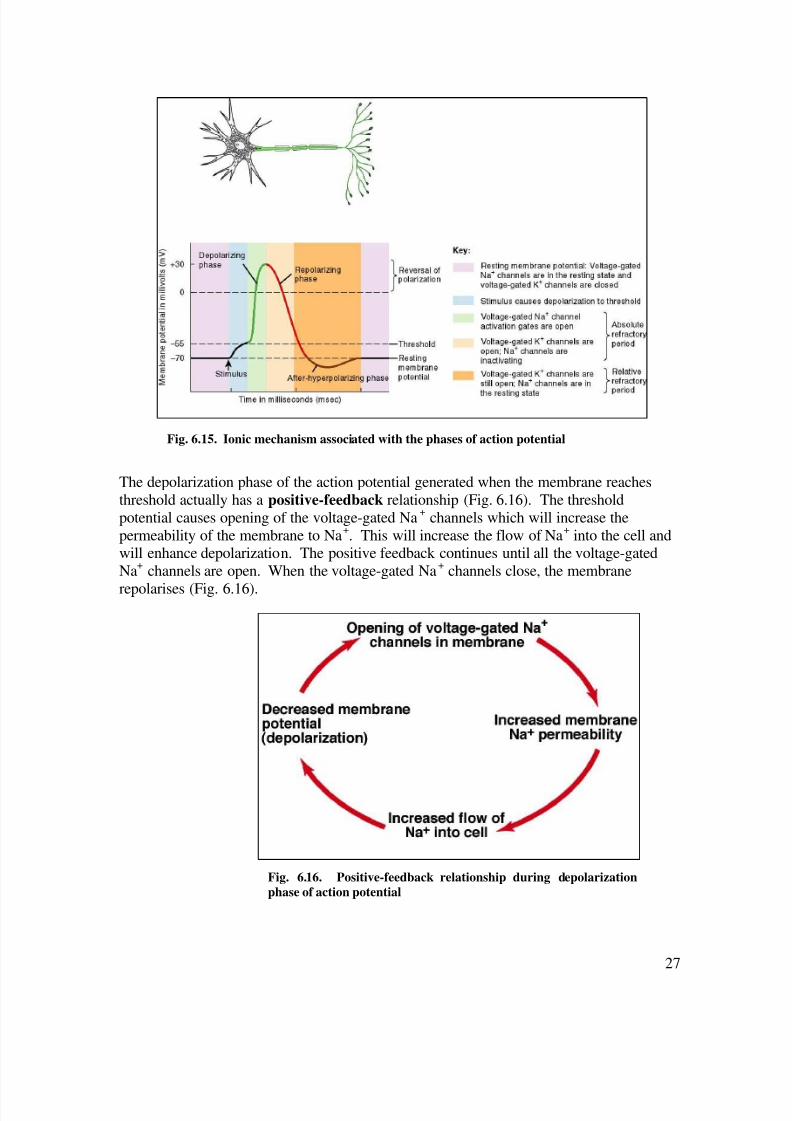
Study Fig 6.14 carefully and write a short assay on the mechanism of action potentialproduction. Fig 6.15 will help you to explain the ionic mechanism associated with the phases
(1-4) of action potential.
ACTIVITY 6.17: Mechanism of action potential production
8/16/2019 06. Action Potential
http://slidepdf.com/reader/full/06-action-potential 27/36
27
The depolarization phase of the action potential generated when the membrane reaches
threshold actually has a positive-feedback relationship (Fig. 6.16). The threshold
potential causes opening of the voltage-gated Na+ channels which will increase the
permeability of the membrane to Na+. This will increase the flow of Na
+ into the cell and
will enhance depolarization. The positive feedback continues until all the voltage-gated
Na+ channels are open. When the voltage-gated Na
+ channels close, the membrane
repolarises (Fig. 6.16).
Fig. 6.15. Ionic mechanism associated with the phases of action potential
Fig. 6.16. Positive-feedback relationship during depolarization
phase of action potential

8/16/2019 06. Action Potential
http://slidepdf.com/reader/full/06-action-potential 28/36
28
You should be comfortable with the relationship between graded potential and action
potential now. Table 6.2 describes the comparison between graded potential and actionpotential.
Table 6.2. Comparison between graded potential and action potential
Graded potential Action potential
Amplitude varies with conditions of theinitiating event
All-or-none once membrane is depolarisedto threshold. Amplitude is independent ofinitiating event
Can be summed Cannot be summed
Has no threshold Threshold is usually about 15 mVdepolarised relative to the resting potential
Has no refractory period Has refractory period
Conducted decrementally Conducted without decrement
Duration varies with initiating condition Duration constantCan be depolarised or hyperpolarised Depolarisation with overshoot
Initiated by environmental stimulus,neurotransmitter or spontaneously
Initiated by graded potential
Mechanism depends onchemical/mechanical-sensitive channel
Mechanism depends on voltage-gatedchannel
You know that action potential is of no use in terms of signal carrying capacity unless itcan be propagated. Let us now explore how action potential is propagated and form what
is known as impulses.

8/16/2019 06. Action Potential
http://slidepdf.com/reader/full/06-action-potential 29/36
29
5. Conduction/propagation/transmission of action potential
5.1. Mechanism of action potential propagation
An action potential is of no use if it cannot be
propagated along the entire length of theneuron or muscle fibre. Why? How is action
potential propagated?
The first action potential in a neuron results
from a graded potential (receptor potential,synaptic potential, end-plate potential, or
pacemaker potential) that is still at threshold
when it reaches the voltage-gated channel atthe axon hillock. If this action potential is not
propagated, the signal that establishes the
graded potential will not be transmitted.
To appreciate the above concept, let’s do
Activity 6.18.
Fig. 6.17. Formation of consecutive action
potential. Note the timing of action potential
formation in the three panels.Review the positions of chemical-gated (ormechanically-gated) and voltage-gated Na
+
channels on the membrane of neurons.
Follow the explanation below while referringto Fig. 6.17.
• Graded potential (formed as receptorpotential, synaptic potential, end-plate
potential, or pacemaker potential)
stimulates voltage-gated Na+ channel
formation of the first action potential(depolarisation i.e. Na
+ high inside): Top
panel of Fig. 6.17• Membrane depolarization of the first
action potential stimulates the adjacent voltage-dependent channels to open,
thus a new action potential is produced(Middle panel in Fig. 6.17).
• The second action potential stimulatedthe third, and so on (bottom panel in Fig.6.17).
• As long as the stimulus is there toproduce the initial action potential, it willcontinuously be propagated along theaxon of the neuron
• This is like a domino effect, which youwill work on in Activity 6.19.
ACTIVITY 6.18: Mechanism of actionpotential propagation
Figure 6.17. Mechanism of action potential
propagation

8/16/2019 06. Action Potential
http://slidepdf.com/reader/full/06-action-potential 30/36
30
Activity 6.19 helps you to feel the process of action potential propagation and to relate it
to the strength of the stimulus.
5.2. The significance of refractory periods
Why is action potential always propagated in a one-way direction i.e. away from the pointof origin towards the axon terminal? The answer is straightforward; it is because naturally
the neuron is always stimulated at the dendrite or soma region and therefore voltage-gated
Na+ channels immediately distal to it open consecutively from the point of stimulation
towards the axon terminal. Moreover, during the depolarizing phase of the action
potential, the positive charges cannot cause depolarization of the preceding site because
the site is in refractory period. This is the reason why propagation of action potential isunidirectional.
What is refractory period?
• Absolute refractory period: a period when no action potential can be formedregardless of the strength of the stimulus
• Relative refractory period: a period when action potential can only be formed when
the stimulus is stronger than the normal threshold.
Arrange about 20 dominos on the floor.
• First give a little push to the first domino; use a force that is not sufficient to cause thedomino to fall. What do you notice? Relate it to the subthreshold stimulus. Thestimulus you gave was not strong enough to cause the first domino to fall onto thesecond domino, just like a subthrehold stimulus is not strong enough to depolarize theneuron to open the voltage-gated channel.
• Give a stronger push onto the first domino until it is just sufficient to fall onto the seconddomino. What do you notice?
• Once initiated, the action potential is a self propagating process that continues along theaxon at a constant velocity (like domino effect).
• Relate this to the events that take place in a neuron when a threshold stimulus isapplied.
• Hypothesise what would happen if you push the first domino harder. Verify this bytiming the fall of the 20 dominos. Compare this to the threshold and suprathresholdeffects of stimuli.
• Please use the analogy of the connection between one domino and the next domino tothe connection between one action potential to the next action potential in a neuron.Please explain based on the molecular level.
ACTIVITY 6.19: The domino effect of action potential propagation

8/16/2019 06. Action Potential
http://slidepdf.com/reader/full/06-action-potential 31/36
31
Activity 6.17 helps you to understand more about the relevance of refractory periods.
Fig. 6.18. Absolute and relative refractory periods

8/16/2019 06. Action Potential
http://slidepdf.com/reader/full/06-action-potential 32/36
32
5.3. Strategies to hasten action potential propagation
Signals of some information need to be propagated very quickly, while others do not
require very fast delivery. For example, signals from peripheral pain receptors are
propagated quickly to the brain, whilst signals via the autonomic nervous system need notbe sent as quickly. What are the strategies that can hasten the rate of propagation of action
potentials?
a. the presence of myelin sheath
b. diameter of the axon: larger diameter permits faster conductionc. influence of chemicals
Myelin sheaths on axons have significant effect on the rate of action potential conduction
(Fig. 6.19). Voltage gated channels are mostly at the nodes of Ranvier. The myelinatedportion of the axon does not permit much ion exchange due to the sheath’s insulating
property. Therefore, current carried by Na+ and K
+ flows through the membrane mainly at
the nodes. In other words, action potentials occur only at the nodes. This hastens the rate
of propagation of action potentials. This is called saltatory conduction. Saltatory
conduction is analogous to walking with normal steps, whereas continuous conduction islike walking by arranging one heel to touch the toes of the other leg.
Fig. 6.19. Mechanism of salutatory conduction of action potentials
Refer to Fig. 6.18.
What is:
• Absolute refractory period?
• Relative refractory period?
Please explain the molecular mechanism that causes absolute refractory period and relativerefractory period. Hint: you must know what happens to the channel proteins duringdepolarization, repolarisation, and after repolarisation (undershoot).
Why can’t depolarization of the membrane immediately distal to the patch that is undergoingrefractory period produce action potential on that patch?
ACTIVITY 6.20: Relevance of refractory periods

8/16/2019 06. Action Potential
http://slidepdf.com/reader/full/06-action-potential 33/36
33
Axon diameter also affects the rate of action potential conduction. Larger diameter axonspropagate impulses faster than smaller ones. Therefore, information that requires vey fast
response (for example sensory inputs and motor outputs to muscles and joints) is
transmitted by thicker fibres.
Effect of axon diameter on speed of conduction: • Group A fibres: largest diameter ~5-20mm; all myelinated; somatic sensory (touch,
pressure) and motor fibres to skin, muscle and joints; speed of conduction: 12-
• 130m/sec
• Group B fibres: diameter 2-3mm; lightly myelinated; conduct sensory impulses to
CNS, and all ANS axons to autonomic ganglia; speed: up to 15 m/sec
• Group C fibres: diameter 0.5-1.5mm; all unmyelinated; conduct sensory impulses for
pain, touch, pressure, heat and cold from the skin, and pain impulses from the viscera.
Include autonomic fibres from autonomic ganglia to heart, smooth muscle and glands.Speed: 0.5-2m/sec)
Chemical factors can also affect impulse conduction. These are substances which alter
membrane permeability to ions, thus influence the rate of impulse conduction. Forexample:
• Ca2+
are required to close Na+ channels in axon membranes during an action potential.
Lack of Ca2+
Na+ channels remain open Na
+ diffuse in again and again
Based on Fig. 6.19 and 6.20:
• explain the molecular mechanism of saltatory conduction
• compare and contrast between continuous and saltatory conduction.• explain the significance of saltatory conduction.
• which neurons in the body are myelinated?
ACTIVITY 6.21. Saltatory conduction of action potential

8/16/2019 06. Action Potential
http://slidepdf.com/reader/full/06-action-potential 34/36
34
impulses transmitted repeatedly. If impulses to skeletal muscle muscle spasm
(tetanus). Usually occur in women during pregnancy, people with diet lacking Ca2+
orVit D, or prolong diarrhea.
• A small increase in concentration of extracellular K+ resting potential of nerve
fibres less negative threshold potential reached with smaller stimulus i.e. the
affected fibers are very excitable
convulsion may occur.• A big decrease in concentration of extracellular K+ resting potential very negative
action potential cannot occur muscles become paralysed.• Certain anaesthetic drugs eg. procaine decrease membrane permeability to Na+
prevent impulses from passing throughthe affected area prevent perception of touch
and pain.

8/16/2019 06. Action Potential
http://slidepdf.com/reader/full/06-action-potential 35/36
35
vii. Summary
The resting membrane potential itself cannot propagate impulses, thus it cannot transmitinformation signals. However, it is a basis for production of graded potentials when the
membrane is stimulated. This is due to the membrane characteristics of excitable cells i.e.
by having gated ion channels that are responsive to chemicals, change in voltage, ormechanical stimulation. The action potentials produced are propagated along the axon dueto the presence of voltage-gated ion channels on the axon membrane. This is how
information signal is transmitted from the receptor (sensor) to the central nervous system
(CNS) for processing, and from the CNS to the effector for appropriate response to thestimulus.
Draw a creative and comprehensive concept map that encompasses the main ideas in this
module.
Describe what you have learnt from this module, including non-academic outcomes.
Comments on the activities, and suggest innovations for improvement of the module.
ACTIVITY 6.22: Summary

8/16/2019 06. Action Potential
http://slidepdf.com/reader/full/06-action-potential 36/36
viii. Conclusions
Please make sure that you achieve all the objectives set up at the beginning of this module:
Objectives Comments
1. Explain the role of the nervous system in
intercellular communication.
2. Predict the sequence of events that take place
on a cell membrane when a stimulus reachesa receptor.
3. Describe the significance of different ion
channels of the membrane of a neuron in
terms of their effects on membrane potential.
4. Define graded potential, describe how it is
established, and describe its characteristics.
5. List down the types of graded potential and
state the significance of each.
6. Compare and contrast between EPSP and
IPSP.
7. Define threshold stimulus and thresholdpotential
8. Plot the membrane potential response tosubthreshold, threshold, and suprathreshold
stimuli and explain the molecular basis of the
response.
9. Explain the meaning of action potential.
10. Describe the mechanism of action potentialproduction.
11. Compare and contrast between action
potential and graded potential.
12. Describe the mechanism of action potential
propagation.
13. Describe the molecular basis of absolute and
relative refractory periods and state thesignificance.
14. Describe the strategies that can hasten actionpotential propagation.
Don’t forget the objectives that you have constructed yourselves!