EPITHELIAL TISSUE Squamous Cuboidal Columnar Simple Stratified.

! 129
!!
http://classes.midlandstech.edu/carterp/Courses/bio210/chap04/chap04.html

!130
http://classes.midlandstech.edu/carterp/Courses/bio210/chap04/chap04.html

! 131
http://classes.midlandstech.edu/carterp/Courses/bio210/chap04/chap04.html

!132
http://classes.midlandstech.edu/carterp/Courses/bio210/chap04/chap04.html
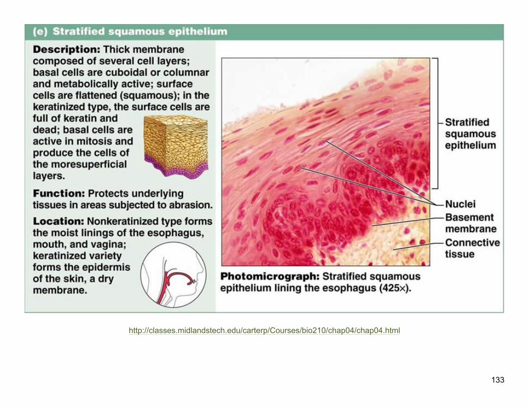
! 133
http://classes.midlandstech.edu/carterp/Courses/bio210/chap04/chap04.html
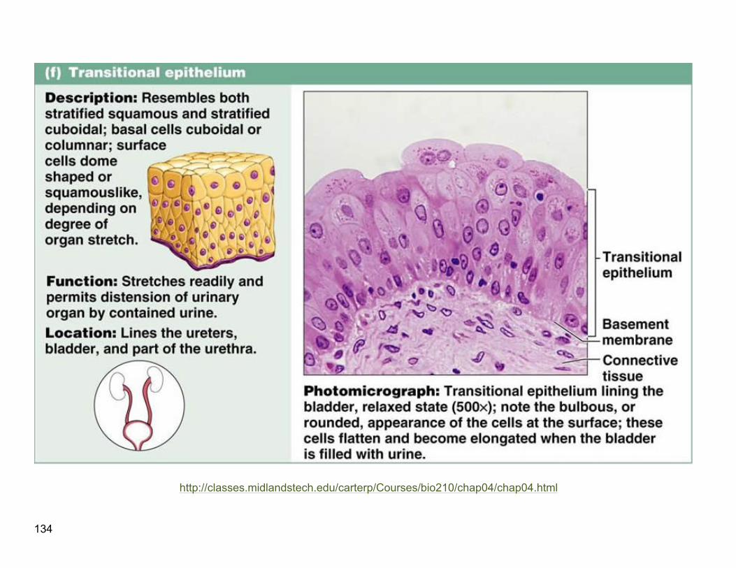
!134
http://classes.midlandstech.edu/carterp/Courses/bio210/chap04/chap04.html
! 135
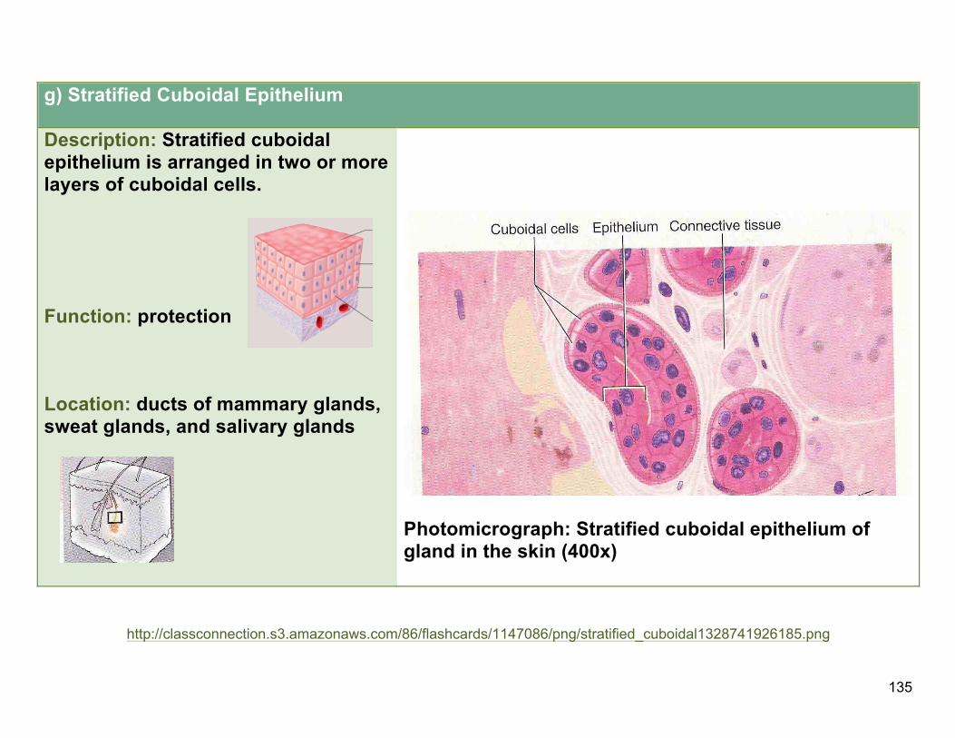
g) Stratified Cuboidal Epithelium
Description: Stratified cuboidal epithelium is arranged in two or more layers of cuboidal cells.
Function: protection
Location: ducts of mammary glands, sweat glands, and salivary glands
Photomicrograph: Stratified cuboidal epithelium of gland in the skin (400x)
http://classconnection.s3.amazonaws.com/86/flashcards/1147086/png/stratified_cuboidal1328741926185.png
!136
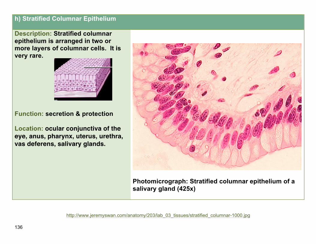
h) Stratified Columnar Epithelium
Description: Stratified columnar epithelium is arranged in two or more layers of columnar cells. It is very rare.
Function: secretion & protection
Location: ocular conjunctiva of the eye, anus, pharynx, uterus, urethra, vas deferens, salivary glands.
Photomicrograph: Stratified columnar epithelium of a salivary gland (425x)
!
http://www.jeremyswan.com/anatomy/203/lab_03_tissues/stratified_columnar-1000.jpg

! 137
Lab$4a,$Part$B:$$Color$Images$$
A.!! ! ! ! ! ! ! !!!!B.!
! !!! !http://science.tjc.edu/Course/BIOLOGY/1409/simple_columnar_ep.jpg http://sciweb.hfcc.edu/Biology/AP/134/lab/lab%20guide%20images/Histology/Bio%20134%20photomicrographs.400x/Trans%20E%20400x.3.jpg!! !
C.! ! ! ! ! ! ! !!!!D.!
!!!! !http://www.pathguy.com/histo/081.jpg http://stevegallik.org/sites/histologyolm.stevegallik.org/images/PseudoCilia4.jpg!E.!!! ! ! ! ! ! ! !!!!F.!
!!!! !http://science.tjc.edu/Course/BIOLOGY/1409/squamouslow.jpg http://im.glogster.com/media/4/21/13/65/21136509.jpg
!!!!!

!138
!G.! ! ! ! ! ! !!!!!!!!H.!
!!!!!!!!!!!!!!! ! http://bio.rutgers.edu/~gb102/lab_6/epithelium/cuboidal_high_kc.jpg http://science.tjc.edu/Course/BIOLOGY/1409/ciliatedpseudostrat.jpg
I.! ! ! ! ! ! ! J.!
!!!! !http://science.tjc.edu/Course/BIOLOGY/1409/cuboidal2.6-9.jpg http://classconnection.s3.amazonaws.com/112/flashcards/844112/jpg/simple_squamous_epithelium1336529145515.jpg
K.! ! ! ! ! ! ! !!!!L.!
!!!! !http://www.stegen.k12.mo.us/tchrpges/sghs/ksulkowski/images/13_Stratified_Squamous_Epithelial_Tissuetemp_000.jpg http://o.quizlet.com/i/_F3HzOtZs1wgdIybe41sXQ_m.jpg
!!!
!