04.11.13 0. General introduction - CIME · 04.11.13 1 Intensive SEM/TEM training: Introduction 1...
14
Intensive SEM/TEM training: Introduction Aïcha Hessler-Wyser 1 CiMe 0. General introduction Dr Aïcha Hessler-Wyser Bat. MXC 134, Station 12, EPFL+41.21.693.48.30. Centre Interdisciplinaire de Microscopie Electronique CIME Intensive SEM/TEM training: Introduction Aïcha Hessler-Wyser 2 CiMe Outline a. History of microscopy b. What can be done with a microscope? c. General bibliography
Transcript of 04.11.13 0. General introduction - CIME · 04.11.13 1 Intensive SEM/TEM training: Introduction 1...

04.11.13
1
Intensive SEM/TEM training: Introduction Aïcha Hessler-Wyser 1 CiMe
0. General introduction
Dr Aïcha Hessler-Wyser
Bat. MXC 134, Station 12, EPFL+41.21.693.48.30.
Centre Interdisciplinaire de Microscopie Electronique
CIME
Intensive SEM/TEM training: Introduction Aïcha Hessler-Wyser 2 CiMe
Outline
a. History of microscopy b. What can be done with a microscope? c. General bibliography

04.11.13
2
Intensive SEM/TEM training: Introduction Aïcha Hessler-Wyser 3 CiMe
a. Little history
1665 2007
Intensive SEM/TEM training: Introduction Aïcha Hessler-Wyser 4 CiMe
a. Little history: optics
Optical microscopy Antiquity: first etch of convex lenses
XII-XIIIth centuries: magnification power of convex lenses, magnifier, glasses
1590 Janssen, first composed microscope
1609 Galilei: occhiolino
1665 Hooke: first cell image
1801 Young: wave nature of light
1872 (~) Abbe: the resolution limit is linked to wave length of the used beam

04.11.13
3
Intensive SEM/TEM training: Introduction Aïcha Hessler-Wyser 5 CiMe
a. Little history: electrons?
Electron microscopy 1923 De Broglie: concept of wavelength associated to
particles, confirmation by Young's experiment 1927 Busch: focalisation low for magnetic fields
Davisson, Germer, Thomson: electron diffraction 1931 Ruska, Knoll: first images by electron microscopy
Intensive SEM/TEM training: Introduction Aïcha Hessler-Wyser 6 CiMe
a. Little history: electrons?
Electron microscopy 1923 De Broglie: concept of wavelength associated to
particles, confirmation by Young's experiment 1927 Busch: focalisation low for magnetic fields
Davisson, Germer, Thomson: electron diffraction 1931 Ruska, Knoll: first images by electron microscopy

04.11.13
4
Intensive SEM/TEM training: Introduction Aïcha Hessler-Wyser 7 CiMe
a. Little history: electrons?
1936 Scherzer: main electromagnetic lens aberrations cannot be avoided
1938 Von Ardenne: first microprobe scanning electron microscope
1939 Siemens: first industrial electron microscopes
1948 Gabor: holography invention
1951 Castaing: first X-ray micro-analyser
1960 XX: first MV microscope, competition for resolution
1965 Crewe: first scanning transmission electron microscope
1982 Binnig et Rohrer: scanning tunnelling microscope
1986 Ruska, Binnig et Rohrer: Prix Nobel Physics
1990 Rose: proposes the Cs corrector principle
1995 Haider: first realisation of the Cs corrector
Intensive SEM/TEM training: Introduction Aïcha Hessler-Wyser 8 CiMe
a. Little history: resolution?
Resolution evolution with time and new technologies
0.0001
0.001
0.01
0.1
1
1800 1840 1880 1920 1960 2000 2040
Res
olut
ion
(An
g.-1)
Year
Electron Microscope
Light Microscope
Corrected EM
Ross
Amici
Abbe
Ruska
Marton
Dietrich(200keV)
Haider(200keV)
H. Rose, 1994 TEAM project, 2002

04.11.13
5
Intensive SEM/TEM training: Introduction Aïcha Hessler-Wyser 9 CiMe
a detector (eye, photographic plate,
video camera...
an illumination section (lenses, apertures...)
a magnification section (lenses, apertures...)
a sample (+ a "goniometer")
A "light" source
b. What can be done with an EM?
Intensive SEM/TEM training: Introduction Aïcha Hessler-Wyser 10 CiMe
b. What can be done with an EM?
And SEM techniques…

04.11.13
6
Intensive SEM/TEM training: Introduction Aïcha Hessler-Wyser 11 CiMe
b. What can be done with an EM?
Different signals Different detectors
= Different informations!
1. Interaction of electrons with matter
Intensive SEM/TEM training: Introduction Aïcha Hessler-Wyser 12 CiMe
b. What can be done with an EM?
Electron source
Lenses
Detectors
Pumping unit
2. Components of an electron microscope
04.11.13
7
Intensive SEM/TEM training: Introduction Aïcha Hessler-Wyser 13 CiMe
b. What can be done with an EM?
Topographical contrast
Z contrast
3. SEM basics
Intensive SEM/TEM training: Introduction Aïcha Hessler-Wyser 14 CiMe
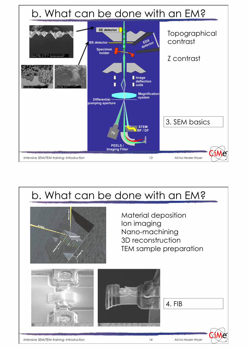
b. What can be done with an EM?
Material deposition Ion imaging Nano-machining 3D reconstruction TEM sample preparation
4. FIB

04.11.13
8
Intensive SEM/TEM training: Introduction Aïcha Hessler-Wyser 15 CiMe
b. What can be done with an EM?
5. ESEM
Partial pressure Hydrated or charging samples
Intensive SEM/TEM training: Introduction Aïcha Hessler-Wyser 16 CiMe
b. What can be done with an EM?
Back-scattered + diffracted e-
Orientation map
6. EBSD

04.11.13
9
Intensive SEM/TEM training: Introduction Aïcha Hessler-Wyser 17 CiMe
b. What can be done with an EM?
7. Advanced SEM
Different detectors, different informations
Intensive SEM/TEM training: Introduction Aïcha Hessler-Wyser 18 CiMe
b. What can be done with an EM?
X-ray detection Chemical analysis Chemical mapping
8. EDS in SEM 15. EDS in TEM

04.11.13
10
Intensive SEM/TEM training: Introduction Aïcha Hessler-Wyser 19 CiMe
b. What can be done with an EM?
Conventional TEM BF, DF Basics of electron diffraction
9. TEM principles 13. Diffraction
contrast 14b. Bio TEM
Intensive SEM/TEM training: Introduction Aïcha Hessler-Wyser 20 CiMe
b. What can be done with an EM?
Electron diffraction
10. Basics of diffraction
11. Diffraction

04.11.13
11
Intensive SEM/TEM training: Introduction Aïcha Hessler-Wyser 21 CiMe
b. What can be done with an EM?
3mm m
ax.
2.4
mm
.
3mm.
12. Sample preparation
Intensive SEM/TEM training: Introduction Aïcha Hessler-Wyser 22 CiMe
b. What can be done with an EM?
Phase contrast Atomic column imaging High resolution
14a. HRTEM

04.11.13
12
Intensive SEM/TEM training: Introduction Aïcha Hessler-Wyser 23 CiMe
b. What can be done with an EM?
Inelastically scattered electrons Z contrast T contrast
16. STEM
Intensive SEM/TEM training: Introduction Aïcha Hessler-Wyser 24 CiMe
b. What can be done with an EM?
Energy lost by electrons Chemical information Electronic properties
17-18. EELS

04.11.13
13
Intensive SEM/TEM training: Introduction Aïcha Hessler-Wyser 25 CiMe
b. What can be done with an EM?
New technologies Aberration correction 3D tomography Cathodoluminescence
19. Next generation Bibliography
Cs corrector “On”; Cs = 0.05 mm
Intensive SEM/TEM training: Introduction Aïcha Hessler-Wyser 26 CiMe
b. What can be done with an EM?
Cs corrector “On”; Cs = 0.05 mm

04.11.13
14
Intensive SEM/TEM training: Introduction Aïcha Hessler-Wyser 27 CiMe
c. General bibliography
SEM J.I. Goldstein et al., Scanning Electron Microscopy and X-ray Microanalysis, Springer (2007), 3rd edition L. Reimer, Scanning Electron Microscopy, (Springer 1998), 2nd edition L. Reimer, Image Formation in Low-Voltage Scanning Electron Microscopy, Spie Press (1993)
J.I. Goldstein et al., Practical scanning electron microscopy, Plenum Press (1977) Lyman et al., Scanning electron microscopy, X-ray microanalysis, and analytical electron microscopy, Plenum Press (1990) D.E. Newbury, Advanced Scanning Electron Microscopy and X-ray Microanalysis, Plenum Press (1986) D.B Holt, D.C. Joy, SEM microcharacterization of semiconductors, Academic Press (1989) B.G. Yacobi, D.B. Holt, Cathodoluminescence microscopy of inorganic solids, Plenum Press (1990)
J. F. Bresse (ed.), Travaux pratiques de microscopie electronique à balayage et de microanalyse X, ANRT Paris (1994)
Intensive SEM/TEM training: Introduction Aïcha Hessler-Wyser 28 CiMe
c. General bibliography
TEM D.B. Williams and C.B. Carter, Transmission electron microscopy, Springer (2009)
J.W. Edington, Practical electron microscopy in material science, (1976)
Hirsch, Electron microscopy of thin crystals , (1977)
Multi-techniques R.W. Cahn et al., Materials Science and Technology, Characterization of Materials (vol. 1 and 2), VCH (1992)
S. Amelinckx et al., Handbook of Microscopy: Applications in Material Science, Solid State Physics and Chemistry (vol. 1 and 2), VCH (1997)
E. Fuchs, H. Oppolzer, H. Rehme, Particle Beam Microanalysis, VCH Verlag 1990)
J.P. Eberhart, Analyse structurale et chimique des matériaux, Dunod, (1989)
M. Amou éd., Microcaractérisation des Solides, Ecole CRAM (CRAM 1989, 541 p) D.K. Bowen, C.R. Hall, Microscopy of Materials, McMillan (1975)