מבנה התא. what is in a cell? Cells are 90% fluid (cytoplasm) which consists of free amino...
95
הההה ההה
-
Upload
mattie-colling -
Category
Documents
-
view
218 -
download
2
Transcript of מבנה התא. what is in a cell? Cells are 90% fluid (cytoplasm) which consists of free amino...

מבנה התא

what is in a cell?
Cells are 90% fluid (cytoplasm) which consists of free amino acids, proteins, glucose, and numerous other molecules.
Elements:
59% Hydrogen (H) 24% Oxygen (O) 11% Carbon (C) 4% Nitrogen (N) 2% Others - Phosphorus (P), Sulphur (S), etc.
Molecules that make up the cell:
50% protein 15% nucleic acid 15% carbohydrates 10% lipids 10% Other
The cell is a unit of organization
Cells are classified by fundamental units of structure and by the way they obtain energy.
Cells are classified as prokaryotes or eukaryotes.
Living things are classified in six kingdoms based on structure:
Within prokaryotes , which appeared 3.5 billion years
ago, are the kingdoms Eubacteria and Archaea.
Within eukaryotes, which evolved 1.5 billion years ago,
are the kingdoms Protista (חד תאיים), Plantae, Fungae (פטריות), Animalia.

Cells are also defined according the need for energy.
Autotrophs are "self feeders" that use light or chemical energy to make food. Plants are an example of autotrophs.
In contrast, heterotrophs ("other feeders") obtain energy from other autotrophs or heterotrophs. Many bacteria and animals are heterotrophs.

Multicellular Organisms
Multicellular organisms are created from a complex organization of cooperating cells.
There must be new mechanisms for :
1. cell to cell communication and regulation.
2. single fertilized egg to develop into all the different kinds of tissues of the
body.
In humans, there are 1014 cells comprising 200 kinds of tissues!

The major differences between Prokaryotic and Eukarotic cells are that :
prokaryotes don't have a nucleus and rarely have membrane bound organelles, (the only exception is bacteria with vacuoles).
They both do have DNA for genetic material, have a exterior membrane, have ribosomes, accomplish similar functions, and are very diverse.
For instance, there are over 200 types of cells in the human body, that very greatly in size, shape, and function.

Prokaryotes:
Prokaryotes are cells without a nucleus.
They have genetic materials but are not enclosed within a membrane.
The genetic material is a single circular DNA and is contained in the cytoplasm, since there is no nucleus. Recombination happens through transfers of plasmids (short circles of DNA that pass from one bacterium to another).
Prokaryotes have a cell wall made up of peptidoglycan.
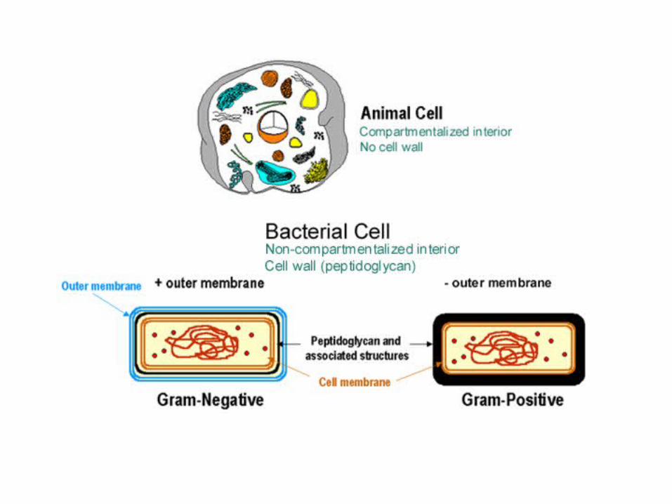

Eukaryotes:
These are cells with a nucleus, this is where the genetic material is surrounded by a membrane much like the cells membrane.
Eucaryotic cells are found in humans and other multicellular organisms (plants and animals) also algae, protazoa.
They have both a cellular membrane and a nuclear membrane, also the genetic material forms multiple chromosomes, that is linear and complexed with proteins that help it 'pack' and is involved in regulation.

Eukaryotes are composed of both plant and animal cells.
Plants vary from animal cells in that they have large vacuoles, cell wall, chloroplasts, and a lack of lysosomes, centrioles, pseudopods, and flagella or cilia (ריסים).
Animal cells do not have the chloroplasts, and may or may not have cilia, pseudopods or flagella, depending on the type of cell.
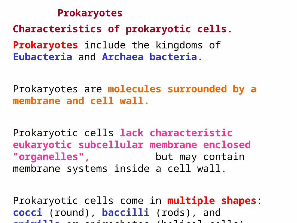
Prokaryotes
Characteristics of prokaryotic cells.
Prokaryotes include the kingdoms of Eubacteria and Archaea bacteria.
Prokaryotes are molecules surrounded by a membrane and cell wall.
Prokaryotic cells lack characteristic eukaryotic subcellular membrane enclosed "organelles", but may contain membrane systems inside a cell wall.
Prokaryotic cells come in multiple shapes: cocci (round), baccilli (rods), and spirilla or spirochetes (helical cells)

Characteristics of bacteria:
1. Microscopic size (0.2-20 micron).2. Simple cell structure.3. have 0.001 times as much DNA as a eukaryotic cell. 4. Can divide every twenty minutes.5. Also, at fairly high frequencies, have spontaneous mutations.
Bacteria is also used as a tool in a massive effort to understand genetics. The method of recombinant DNA is designed to exploit bacteria for formation of numerous valuable products .

מבנה תא פרוקריוטי

Cell Envelope - two to three layers: • in some species of bacteria - an outer capsule.• the cell wall• the interior cytoplasmic membrane

Capsule – polysaccharides (complex carbohydrates). keep the bacterium from drying outProtect it from phagocytosis (engulfing) by larger microorganisms.

Cell Wall - peptidoglycan, a protein-sugar (polysaccharide) molecule.
gives the cell its shape protects the cytoplasmic membrane from osmotic pressure.helps to anchor appendages like the pili and flagella
N-acetyl glucosamine -NAG
N-acetyl muramic acid -NAM

Cytoplasmic Membrane - phospholipids and proteins. regulates the flow of materials in and out of the cell.

Pili - small hairlike projections emerging from the outside cell surface. assist the bacteria in attaching to other cells and surfaces, such as teeth, intestines, and rocks.
Flagella (singular: flagellum) - hairlike structures locomotion for those bacteria that have them.

Cytoplasm - a gel-like matrix composed of water, enzymes, nutrients, wastes, and gases
contains cell structures such as ribosomes, a chromosome, and plasmids.
Nucleoid - The nucleoid is a region of cytoplasm where the chromosomal DNA is located.
Ribosomes - microscopic "factories" translate nucleic acid to amino acids -- the building blocks of proteins.

The Archaea
Extreme environment conditions:thermophiles, Halobacterium, Metanococcus The methanogenic archaeon, Metanococcus jannaschii:
Is found 3 km down, at 85 deg C
Has 1738 genes, 56% of which are new to science
has bacteria-like genes and operons
but with eukaryotic-like information processing and secretion systems
and eukaryotic protein synthesis

מבנה תא אאקריוטי

• The Cell Membrane • The Cell Wall • The nucleus • Cytoplasm • Vacuoles and vesicles • Ribosomes • Endoplasmic reticulum • Golgi Apparatus (and Dictyosomes)• Lysosomes • Mitochondria • Plastids
מבנה תא אאקריוטי

Membranes (קרום)vital because they separate the cell from the outside world.
They also separate compartments inside the cell to protect important processes and events.

Membranes are essential for the integrity and function of the cell
Protective
Regulate transport in and out of cell or subcellular domain
Allow selective receptivity and signal transduction
Allow cell recognition
Anchoring point for cytoskeletal or extracellular elements

Fluid Mosaic model
Membrane architecture is that of a lipid bilayer.
lipids are amphipathic :
hydrophilic polar heads pointing out
hydrophobic portion forming the core.
Proteins are embedded in the bilayer.


Fluid Mosaic model
.ניתן לדמות את הממברנה למוזאיקה נוזלית• - בשל שיבוצה במרכיבים שונים, מוזאיקה•
ליפידים, חלבונים, כולסטרול. – הודות לקיום דיפוזיה ותנועה אופקית נוזלית•
מתמדת של הליפידים והחלבונים בממברנה.

Membrane Phospholipids
One of the principal types of lipid in the membrane include the phospholipids .
These have a polar head group and two hydrocarbon tails which are hydrophobic.
The top region beginning with the NH3 is the polar group.
It is connected by glycerol to two fatty acid tails.
fatty acid tails
One of the tails is a straight chain fatty acid (saturated). The other has a kink (עיקול) in the tail because of a cis double bond (unsaturated).
This kink influences packing and movement in the lateral plane of the membrane.
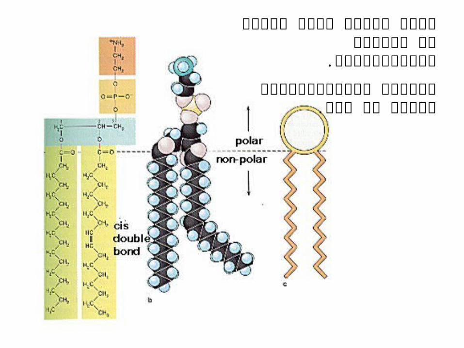
הראש הטעון פונה החוצה אל .הסביבה ההידרופילית
הזנבות ההידרופוביים פונים זה לזה.

The lipid bilayer gives the membranes its
fluid characteristics.
The following cartoon shows the effect of temperature on the packing of the hydrocarbons.
Note that a low temperatures, the bilayer is in a gel state and tightly packed.
At higher (body) temperatures, the bilayer actually "melts' and the interior is fluid allowing the lipid molecules to move around, rotate, exchange places.
This also allows movement of other components of the membrane.


Membrane Cholesterol
Another type of lipid in the membrane is cholesterol.
The amount of cholesterol may vary with the type of membrane.
Plasma membranes have nearly one cholesterol per
phospholipid molecule.

Cholesterol molecules have several functions in the membrane:
They immobilize the first few hydrocarbon groups of the phospholipid molecules.
This makes the lipid bilayer less deformable and decreases its permeability to small water-soluble molecules.
Without cholesterol (such as in a bacterium) a cell would need a cell wall.
Cholesterol prevents crystallization of hydrocarbons and phase shifts in the membrane.

Membrane Glycolipids
Glyco-lipids are also a constituent of membranes.
The sugar groups projecting into the extracellular space.
These components of the membrane may be protective, insulators, and sites of receptor binding.
Among the molecules bound by glycolipids include cell poisons such as cholera and tetanus toxins.
Sphingolipids and cholesterol work together to help cluster proteins in a region called a "microdomain".
They function as "rafts" or platforms for the attachment of proteins as membranes are moved around the cell and also during signal transduction.

Membrane Proteins
In different organelles, there are important membrane proteins that function for that particular organelle.

Transmembrane proteins or"integral proteins".
amphipathic- they have hydrophobic and hydrophilic regions that are oriented in the same regions in the lipid bilayer.
peripheral membrane proteins
linked only at the cytoplasmic surface, by attachment to a fatty acid chain,
or at the external cell surface, attached by a oligosaccharide.
Or, may be bound to other membrane proteins.

תפקידים שונים של חלבונים ממברנליים

סוכר

של הממברנה; הא-סימטריה•צד ציטוזולי הפונה לכל ממברנה תאית יש
, לציטופלזמה. וצד אקסופלסמטי הפונה לכיוון חוץ התא
קיים הבדל בין מרכיבי שני הצדדים:. המבנה הא-סימטרי של החלבונים הממברנלים1. הגליקוליפידים נמצאים רק בצד האקסופלזמטי 2
הממברנה. של
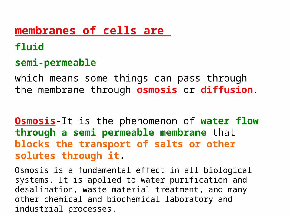
membranes of cells are fluid
semi-permeable
which means some things can pass through the membrane through osmosis or diffusion.
Osmosis-It is the phenomenon of water flow through a semi permeable membrane that blocks the transport of salts or other solutes through it. Osmosis is a fundamental effect in all biological systems. It is applied to water purification and desalination, waste material treatment, and many other chemical and biochemical laboratory and industrial processes.

diffusion -It is the phenomenon of soluble elements flow through a semi permeable membrane
The rate of diffusion will vary depending on the its:temperaturesize
polarity
charge
concentration on the inside of the membrane versus the concentration on the outside of the membrane.
When something is permeable it means that something can spread throughout.

Here is a list of some molecules and how they relate to passing through the membrane without assistance:
Hydrophobic Molecules: Large Uncharged Polar Molecules:
O2 - Oxygen Glucose
N2 - Nitrogen Sucrose
benzene
Small uncharged Polar Molecules: Ions:
H2O - Water H+ - Hydrogen ion
urea Na+ - Sodium ion
glycerol K+ - Potassium ion
C02 - Carbon Dioxide Ca²+ - Calcium ion
Cl- - Chloride ion

הממברנה –קרום בררני


Gap Junctionstype of junction allows communication between cells.
Small molecules or ions can pass through.

דופן התא(צמחים, פטריות)
Plasmodesmata

Nucleus גרעין

Nucleus -Structure/function correlations
The cell nucleus is a remarkable organelle because it forms the package for our genes and their controlling factors.
It functions to:
Store genes on chromosomes Organize genes into chromosomes to
allow cell division.Transport regulatory factors & gene
products via nuclear pores Produce messages (messenger
RiboNucleic Acid or mRNA) that code for proteins

מעטפת הגרעין
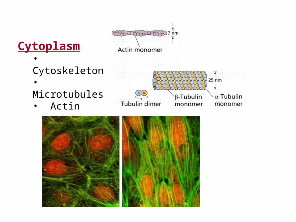
Cytoplasm • Cytoskeleton • Microtubules• Actin

MicrotubulesFunction
Microtubules are conveyer (מוביל) belts inside the cells.
They move vesicles, granules, organelles like mitochondria, and chromosomes via special attachment proteins.
They also serve a cytoskeletal role.

Structure
Microtubules are linear polymers of tubulin which is a globular protein.
These linear polymers are called protofilaments .
The tubulin molecules are the bead like structures.
A protofilaments is a linear row of tubulin beads.
Microtubules may work alone, or join with other proteins to form more complex structures called cilia, flagella or centrioles.
an alpha and a beta tubulin molecule join to form a heterodimer

Centrioles and Basal Bodies
centrioles are associated with the cytoskeleton.
A centriole is commonly found in the centrosome, which is where the asters (אזור הגרעין) originate during metaphase of cell division.
Centrioles are often known for their role in cell division.
Centrioles seem to determine the position of the pericentriolar material, which in turn affects the polarity of the cell.
A centriole is made up of nine (9) sets of triplet Microtubules and forms a short cylinder-like structure.
The centriole is important in the formation of cilia or flagella, then it is called a basal body

Actin
These are the smallest (in diameter) of the 3 cytoskeletal filaments that are found in all cells (microfilaments, intermediate filaments, microtubules).
Actin Filaments consist of two strands of globular molecules, called actin.
The molecules are about 4 nm in diameter and they are twisted to form a helix.
Another interesting fact is that actin filaments are polar, Meaning that one end different than the other.
Mammalian actin exists in 6 different forms (isoforms) 4 associated with different types of muscle and 2 cytoskeletal isoforms found in all non-muscle cells.

Intermediate filaments
Intermediate filaments, also called "thick filaments" are important components of the cell's cytoskeletal system.
They may stabilize organelles, like the nucleus, or they may be involved in specialized junctions.
They are distinguished from "thin filaments” -actin, by their size (8-10 nm) and the fact that thin filaments are motile.

There are five major types of intermediate filaments, each unique to a particular cell type:
Keratin Found in skin, produced by epidermal cells and completely fills upper cell layers of the epidermis.
Vimentin Found in fibroblasts; used as a cytochemical marker for fibroblasts in tissue culture
Glial fibrillary acidic protein GFAP is found in glial cells and is also used as a marker for this cell type.
Desmin An intermediate filament marker for muscle cells
Neurofilament proteins A marker for Nerve cells

השלד התוך תאי סיכום
תפקידים
גודל
מרכיבים
חלוקת התא,קביעת צורה,
ותנועה
מעורבים במגוון תנועה תאית
חוזק המבני של התאים, שמירה על צורה,
סוגים שונים נמצאים ברקמות שונות
25nm7.5nm12-8nm
קבוצות של אקטיןטובוליןחלבונים
מיקרוטובוליםMicrotubles
מיקרופילמנטיםMicrofilaments
קורי בינייםIntermediate
filaments


Vacuole
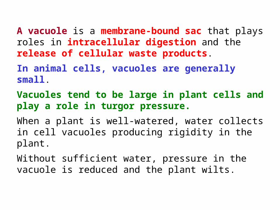
A vacuole is a membrane-bound sac that plays roles in intracellular digestion and the release of cellular waste products.
In animal cells, vacuoles are generally small.
Vacuoles tend to be large in plant cells and play a role in turgor pressure.
When a plant is well-watered, water collects in cell vacuoles producing rigidity in the plant.
Without sufficient water, pressure in the vacuole is reduced and the plant wilts.

Ribosomes

DNA is responsible for coding for all proteins.
Each amino acid is designated by one or more set of triplet nucleotides.
The code is produced from one strand of the DNA by a process called "transcription".
This produces mRNA which then is sent out of the nucleus where the message is translated into proteins.

Synthesis of proteins can be done in the cytoplasm on clusters of ribosomes, called "polyribosomes".
Or it can be done on the membranes of the rough endoplasmic reticulum.

The ribosomes provide the structural site for synthesis of proteins.
Ribosomes are composed of
ribosomal RNA (rRNA) molecules
more than 50 accessory proteins
with a general organization of a
small subunit (30S)
large subunit (50S).

PolyribosomesClusters of ribosomes may sit on a mRNA and make proteins, each making a strand of polypeptides.
These clusters are called polyribosomes.
When they are free in the cytoplasm, they are called free polyribosomes (linked by the mRNA).
Or, they may bind to rough endoplasmic reticulum

Endoplasmic Reticulum (ER)

The Endoplasmic Reticulum
Endoplasmic reticulum is a network of tubules, vesicles and sacs that are interconnected.
The ER is a made up of two phospholipid bilater membranes. The enclosed 'sac' is called the ER lumen, the internal space of the ER.
The ER membrane typically makes up more than half of the total membrane in the cell and is located between the nucleus and the cytosol and specifically the golgi apparatus.

The Endoplasmic Reticulum (ER) may serve specialized functions in the cell including protein synthesis, sequestration of calcium, production of steroids, storage and production of glycogen, and insertion of membrane proteins.
Rough endoplasmic reticulum gets its name from the presence of ribosomes on its surface.

Arrangement of polyribosomes on the rough endoplasmic reticulum.
Free polyribosomes connected by the mRNA.
the growing polypeptide chain is inserted through the membrane and into the cisterna of the rough endoplasmic reticulum.

Golgi Apparatus

The Golgi complex controls trafficking of different types of proteins.
Some are destined for secretion.
Others are destined for the extracellular matrix.
Finally, other proteins, such as lysosomal enzymes, may need to be sorted and sequestered from the remaining constituents because of their potential destructive effects.
This figure shows the two types of secretory pathways.
The regulated secretory pathway, as its name implies, is a pathway for proteins that requires a stimulus or trigger to elicit secretion. Some stimuli regulate synthesis of the protein as well as its release.
The constitutive pathway allows for secretion of proteins that are needed outside the cell, like in the extracellular matrix. It does not require stimuli, although growth factors may enhance the process.


There is an actual interface between the ER and the Golgi complex.
This drawing shows how the rough endoplasmic reticulum forms vesicles (without ribosomes attached) that carry the newly synthesized proteins to the Golgi complex.
The inside of the vesicle becomes continuous with the inside of the Golgi cisternae, so that protein groups pointing towards the inside, could eventually be directed to face the outside of the cell.
Carbohydrate groups are attached and any subunits may be joined in these cisternae.

The protein is then passed to the final region of the Golgi called the "trans face".
There it is placed in vacuoles that bud from this region of the Golgi complex.
The vacuoles continue to condense the proteins and the final mature secretory granule is then moved to the membrane for secretion.

Lysosomes
• Hydrolytic enzymes

LysosomesLysosomes are small organelles containing around 40 enzymes for intercellular digestion.
The lysosome membrane helps to protect the enzymes as much as it helps protect the cell.
This is because the optimal pH for these enzymes is around a pH of 5.
The structures vary in size from 0.2 to 2 micrometers in diameter.
The membrane of the lysosome is again a lipid bilayer and is thought to have a ATP hydrolysis to pump H+ into the lysosome to maintain the pH.

Mitochondria
ATP

The mitochondria major role is ATP production in the eukaryotic cell.
The fixed position is especially true of cells or locations in cells where there will be a need for a high amount of ATP production, such as, near flagella or between myofibrils of muscle.
Mitochondria are also self-reproducing, they have their own circular DNA, with a slightly modified version of the codons. This differs with theeukaryote codon.

Mitochondria and Endosymbiosis

Plastids (chloroplasts) Plants
Endosymbiosis

Chloroplasts are specialized organelles found in all higher plant cells.
These organelles contain the plant cell's chlorophyll, hence provide the green color.
They have a double outer membrane.
Within the stroma are other membrane structures - the thylakoids and grana (singular = granum) where photosynthesis takes place.

• The Cell Membrane • The Cell Wall • The nucleus • Cytoplasm • Vacuoles and vesicles • Ribosomes • Endoplasmic reticulum • Golgi Apparatus (and Dictyosomes)• Lysosomes • Mitochondria • Plastids
מבנה תא אוקריוטי - סיכום
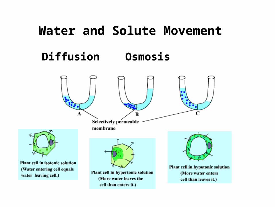
Water and Solute Movement
Diffusion Osmosis

המשך
• . היא דיפוזיה של מים דרך קרומיםאוסמוזה


סביבה איזוטונית

סביבה היפרטונית

סביבה היפוטונית

Osmosis……

)vesicles(הובלה בתוך שלפוחיות

הובלה פסיבית

הובלה אקטיבית

תא אנימלי
תא צמחי

תנועה של התא
The 9+2 arrangement of microtubules in a flagellum or cilium.
השלד התוך תאי