downloads.hindawi.comdownloads.hindawi.com/archive/2014/231418.f1.docx · Web viewPhosphoprotein...
37
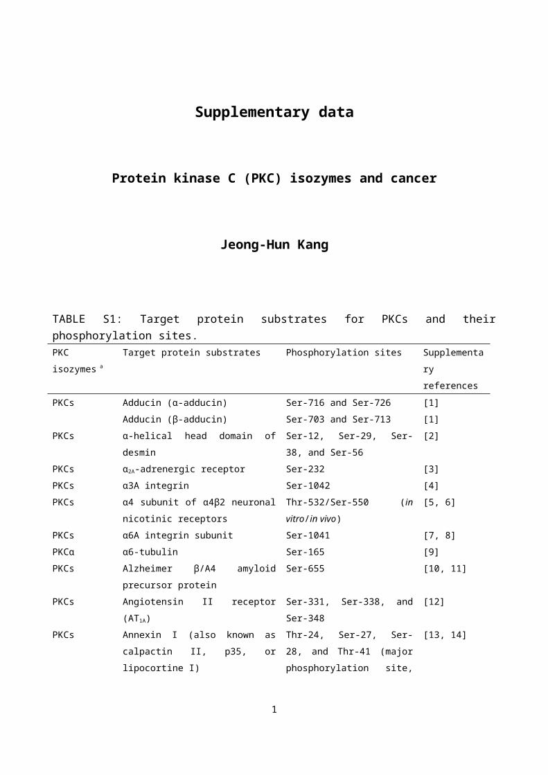
Supplementary data Protein kinase C (PKC) isozymes and cancer Jeong-Hun Kang TABLE S1: Target protein substrates for PKCs and their phosphorylation sites. PKC isozymes a Target protein substrates Phosphorylation sites Supplementa ry references PKCs Adducin (α-adducin) Adducin (β-adducin) Ser-716 and Ser-726 Ser-703 and Ser-713 [1] [1] PKCs α-helical head domain of desmin Ser-12, Ser-29, Ser- 38, and Ser-56 [2] PKCs α 2A -adrenergic receptor Ser-232 [3] PKCs α3A integrin Ser-1042 [4] PKCs α4 subunit of α4β2 neuronal nicotinic receptors Thr-532/Ser-550 (in vitro/in vivo) [5, 6] PKCs α6A integrin subunit Ser-1041 [7, 8] PKCα α6-tubulin Ser-165 [9] PKCs Alzheimer β/A4 amyloid precursor protein Ser-655 [10, 11] PKCs Angiotensin II receptor (AT 1A ) Ser-331, Ser-338, and Ser-348 [12] PKCs Annexin I (also known as calpactin II, p35, or lipocortine I) Thr-24, Ser-27, Ser- 28, and Thr-41 (major phosphorylation site, [13, 14] 1
Transcript of downloads.hindawi.comdownloads.hindawi.com/archive/2014/231418.f1.docx · Web viewPhosphoprotein...
Supplementary data
Protein kinase C (PKC) isozymes and cancer
Jeong-Hun Kang
TABLE S1: Target protein substrates for PKCs and their phosphorylation sites.PKC isozymes a Target protein substrates Phosphorylation sites Supplementary
references
PKCs Adducin (α-adducin)
Adducin (β-adducin)
Ser-716 and Ser-726
Ser-703 and Ser-713
[1]
[1]
PKCs α-helical head domain of desmin Ser-12, Ser-29, Ser-38, and Ser-56 [2]
PKCs α2A-adrenergic receptor Ser-232 [3]
PKCs α3A integrin Ser-1042 [4]
PKCs α4 subunit of α4β2 neuronal nicotinic
receptors
Thr-532/Ser-550 (in vitro/in vivo) [5, 6]
PKCs α6A integrin subunit Ser-1041 [7, 8]
PKCα α6-tubulin Ser-165 [9]
PKCs Alzheimer β/A4 amyloid precursor
protein
Ser-655 [10, 11]
PKCs Angiotensin II receptor (AT1A) Ser-331, Ser-338, and Ser-348 [12]
PKCs Annexin I (also known as calpactin II,
p35, or lipocortine I)
Thr-24, Ser-27, Ser-28, and Thr-
41 (major phosphorylation site,
Ser-27 and Thr-41)
[13, 14]
PKCα, βΙ, and
βII
Annexin II (also known as calpactin I,
p36, or lipocortine II)
Ser-25 [15, 16]
cPKCs Annexin IV (also known as endonexin I
or p33/40)
Thr-6 [17]
cPKCs Avian retrovirus matrix protein Ser-68 and Ser-106 [18]
PKCθ and ε Bad (Bcl-associated death protein) Ser-112 [19]
PKCι Ser-112, Ser-136, and Ser-155 [20]
PKCs basic fibroblast growth factor (also Ser-64 [21]
1
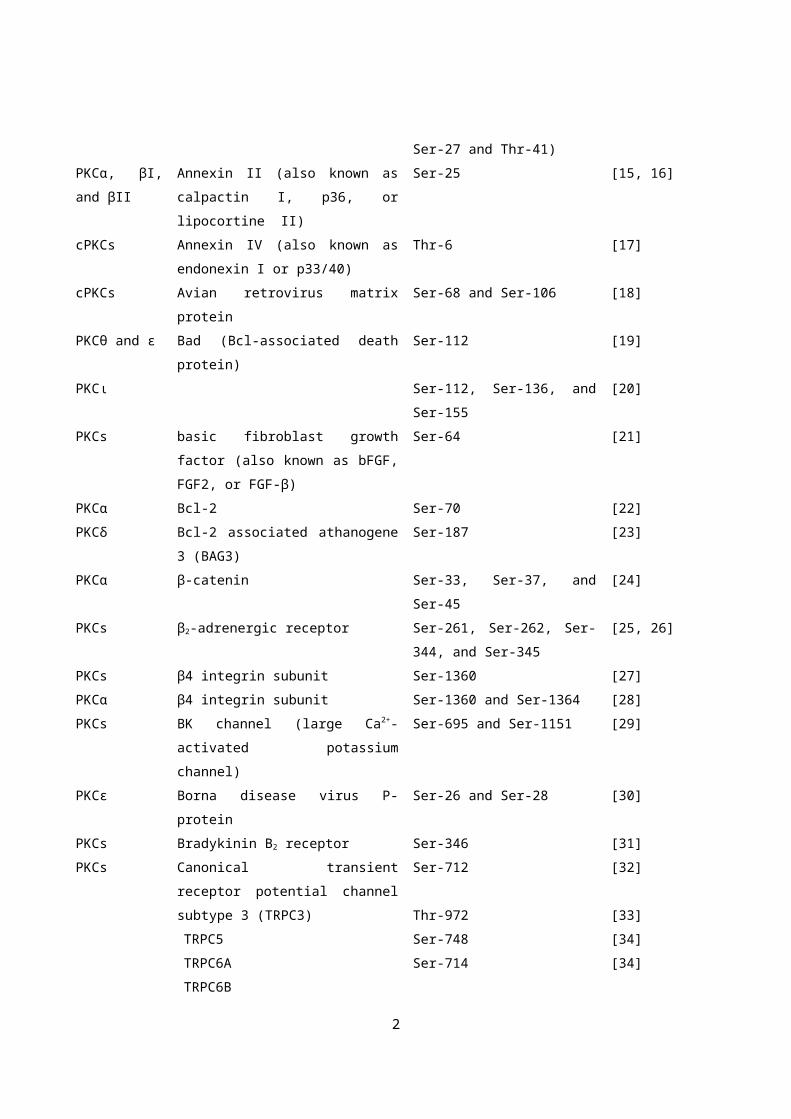
known as bFGF, FGF2, or FGF-β)
PKCα Bcl-2 Ser-70 [22]
PKCδ Bcl-2 associated athanogene 3 (BAG3) Ser-187 [23]
PKCα β-catenin Ser-33, Ser-37, and Ser-45 [24]
PKCs β2-adrenergic receptor Ser-261, Ser-262, Ser-344, and
Ser-345
[25, 26]
PKCs β4 integrin subunit Ser-1360 [27]
PKCα β4 integrin subunit Ser-1360 and Ser-1364 [28]
PKCs BK channel (large Ca2+-activated
potassium channel)
Ser-695 and Ser-1151 [29]
PKCε Borna disease virus P-protein Ser-26 and Ser-28 [30]
PKCs Bradykinin B2 receptor Ser-346 [31]
PKCs Canonical transient receptor potential
channel subtype 3 (TRPC3)
TRPC5
TRPC6A
TRPC6B
Ser-712
Thr-972
Ser-748
Ser-714
[32]
[33]
[34]
[34]
PKCδ TRPC6 Ser-448 [35]
PKCs Cardiac myosin-binding protein C
(cMyBP-c) (also known as MYBPC3)
Ser-273 and Ser-302/Ser-265 and
Ser-300 (human/chicken)
[36, 37]
PKCs Ca2+/calmodulin-dependent protein
kinases II (CaM kinases II)
Thr-286 [38]
PKCs CD33 (known as Siglec-3) Ser-307 [39]
PKCs cGMP-dependent protein kinase (PKG or
cGK) 1α
Thr-58 [40]
PKCs Chaperone or modulatory protein 14-3-3 Ser-58, Ser-59, and Ser-60 for 14-
3-3ζ, 14-3-3η, and 14-3-3β,
respectively
[41]
cPKCs Choline acetyltransferase Thr-255, Ser-346, Ser-347, Ser-
440, and Ser-476
(nPKCs and aPKCs
phosphorylate Ser-440 and Ser-
476)
[42]
PKCα c-kit-encoded tyrosine kinase receptor
for stem cell factor (Kit/SCFR)
Ser-741 and Ser-746 [43]
PKCζ c-Myc Ser-373 [44]
2

PKCε Connexin 43 (Cx43) (also known as gap
junction α-1 protein)
Ser-262
Ser-368
[45]
[46]
PKCs CPI-17 (protein kinase C-dependent
inhibitory of 17 kDa)
Thr-38 [47]
PKCs CTP synthetase 1 Ser-462 and Thr-455 [48]
PKCγ
cPKCs
Diacylglycerol kinase (DGK)γ accessory
domain
DGKδ1 PH domain
Ser-776 and Ser-779
Ser-22 and Ser-26
[49]
[50]
PKCα Dynamin I Ser-795 [51]
PKCs Dystrophin Ser-136 and Ser-147 [52]
PKCα ELAV-like protein HuR (HuA) Ser-158 and Ser-221 [53]
PKCs Endothelial nitric oxide synthase (eNOS) Thr-495/497 (human/bovine) [54, 55]
PKCs Epidermal growth factor receptor
(EGFR)
Thr-654 [56, 57]
PKCι Epithelial cell transformaing sequence 2
(Ect2)
Thr-328 [58]
PKCδ ErbB3-binding protein 1 (EBP1) Ser-360 [59]
PKCδ
PKCβΙ
Eukaryotic elongation factor 1α (eEF-1α) Thr-431
Ser-53
[60]
[61]
PKCs Eukaryotic initiation factor-2β (eIF-2β)
eIF-2γ
eIF-4E
Ser-13
Thr-66
Ser-209 and Thr-210
[62]
[63]
[64]
PKCα Farnesoid X receptor (FXR) (also known
as NR1H4)
Ser-135 and Ser-154 [65]
PKCs Fms-interacting protein (FMIP) Ser-5 and Ser-6 [66]
PKCs γ-aminobutyric acid type A (GABAA)
receptor β1 subunit
GABAA receptor β2 subunit
GABAA receptor γ2S subunit
GABAA receptor γ2L subunit
Ser-409
Ser-410
Ser-327
Ser-343
[67]
[68]
[67, 68]
[67]
PKCs GluA1 AMPA receptor subunit Ser-831 [69, 70]
PKCα and γ Glutathione S-transferase P1 (GSTP1) Ser-42 and Ser-148 in the absence
of glutathione
[71]
PKCα, βI, βII,
γ, δ, ε, η, and ζ
Ser-42 and Ser-148 in the
presence of glutathione
3

PKCα, βII, γ, δ,
and η
(PKCβII and δ
are very weak)
Glycogen synthase kinase (GSK)-3α Ser-21 [72]
PKCα, βI, and γ GSK-3β Ser-9 [73]
PKCα, βI, and η Ser-9 [72]
PKCs G protein α subunit Gzα Ser-25 and Ser-27 [74]
PKCα, δ, and ζ G protein-coupled receptor kinase-2
(GRK2) [also known as β-adrenergic
receptor kinase (βARK)]
Ser-29 [75, 76]
PKCs GRK5 Ser-566 and Ser-572 [77]
PKCα GTPase activating protein (GAP) p190A Ser-1221 and Thr-1226 [78]
PKCs GTP-binding protein γ12 Ser-1 [79]
PKCδ Heat-shock protein-25/27 Ser-15 and Ser-86 [80]
PKCs Hepatitis B virus surface M protein (C-
termimally truncated middle size protein)
Ser-28 [81]
PKCs Hepatitis C virus core protein Ser-116 [82]
PKCs Hepatocyte growth factor receptor kinase Ser-985 [83]
PKCζ Heterogeneous ribonuscleoprotein A1 Ser-199 + three unknown sites [84]
cPKCs and
PKCδ
High-mobility-group protein-1 Ser-44 and Ser-64 [85]
PKCs Histamin H1 receptor (H1R) Ser-398 [86]
PKCs Histone H1 Ser-103 [87]
PKCβΙ Histone H3 Thr-6 [88]
PKCε and β Ser-10 [89]
PKCs HIV-1 gag protein Ser-111 [90]
PKCδ HuR Ser-318 [91]
PKCε IKKβ Ser-177 [92]
PKCs Insulin receptor Thr-1336 [93]
PKCζ Insulin-responsive aminopeptidase
(IRAP)
Ser-80 and Ser-91 [94]
PKCδ
PKCθ
PKCβII
PKCδ
Insulin receptor substrate-1 (IRS-1)
(human)
IRS-1 (human)
IRS-1 (mouse)
Ser-24, Ser-307, Ser-323, and Ser-
574
Ser-1101
Ser-336
[95, 96]
[97]
[98]
[99]
4

PKCζ IRS-1 (rat)
IRS-1 (rat)
Ser 357
Ser-318, Ser-498, Ser-570, and
Ser-612 (major phosphorylation
site, Ser-318)
[100, 101]
PKCι Interleukin-1 receptor-associated kinase
(IRAK)
Thr-66 [102]
PKCs Interleukin-2 (IL-2) Ser-7 [103]
PKCs Iron-responsive element-binding protein Ser-138 and Ser-711 [104]
cPKCs Junctional adhesion molecule (JAM) Ser-284 [105]
PKCε Keratin 8 Ser-8 and Ser-23 [106]
PKCε Keratin 18 Ser-52 [107]
PKCδ and ε Kv3.1b potassium channel Ser-503 [108]
PKCα, βΙ, βII,
γ, δ, and θ
All PKCs
except PKCη
L-type Ca2+ channel (also known as
voltage-dependent Ca2+ channel or
Cav1.2 α1c)
Ser-1674
Ser-1928
[109]
[109, 110]
PKCs L-myc Ser 38 and Ser 42 [111]
PKCs Lens fiber major intrinsic protein Ser-245 [112]
PKCs Microtubule-associated protein 2
(MAP2)
MAP4
Ser-1703, Ser-1711, and Ser-1728
Ser-815
[113]
[114]
cPKCs Myelin basic protein (MBP) Ser-110 and Ser-115 [115]
PKCs Ser-8, Ser-11, Ser-46, Ser-55, Ser-
110, Ser-132, Ser-151, and Ser-
161
[116]
PKC βII Myristoylated alanin-rich kinase
substrate (MARCKS) (also known as
p80 and p87)
Ser-152 and Ser-156 (murine) [117]
PKCs Ser-45, Ser-80, Ser-99, and Ser-
116 (bovine)
Ser-152, Ser156, and Ser-163
(murine)
[118]
[119]
PKCs NADPH oxidase activator 1 (NOXA1) Ser-172 [120]
PKCζ
cPKCs
NADPH oxidase component p47phox Ser-303, Ser-304, and Ser-315
Ser-303, Ser-304, Ser-320, and
Ser-328
[121]
[122]
5

PKCs Na,K-ATPase α-subunit Ser-11 and Ser-18
Ser-774 and Thr-938
[123]
[124]
PKCs NAP-22 (also known as GAP-23 and
BASP1)
Ser-6 [125]
PKCθ NDRG2 (NDRG family member 2) Ser-332 [126]
PKCs Neurogranin (also known as p17,
BICKS, or RC3 protein)
Ser-36 [127, 128]
PKCs Neuronal growth-associated protein B-50
(also known as GAP-43, protein F1, p57,
pp46, or neuromodulin)
Ser-41 (mammals)/Ser-42 (chick)
Ser-209
[129, 130]
[131]
PKCα NG2 proteoglycan Thr-2256 [132]
PKCs N-methyl-D-aspartate (NMDA) receptor
1 subunit
Ser-890 and Ser-896 [133, 134]
PKCβII Nonmuscle myosin heavy chain IIA Ser-1917 [135]
PKCs Nonmuscle myosin heavy chain IIB Ser-362 and Ser-370/Ser-368 and
Ser-370 (βF47/αF47)
[136]
cPKCs
PKCβII
PKCα
Nuclear lamin (also known as class V
intermediate filament) A
Nuclear lamin B
Nuclear lamin C
Ser-5 and Ser-525
Ser-395 and Ser-405
Ser-572
[137]
[138, 139]
[140]
PKCs Nucleolar protein B23 (also known as
nucleophosmin, numatrin, or nucleolar
protein NO38)
Ser-225 [141]
PKCη Occludin Thr-403 and Thr-404 [142]
PKCs Opi1p Ser-26 [143]
aPKCs (PKCζ
and λ)
Par-1b kinase T595 [144, 145]
aPKCs Par-3 Ser-827 [146]
PKCs P-glycoprotein Ser-661 [147]
PKCs Phosphatidylinositol transfer protein (PI-
TP)α
Ser-166 [148]
PKCs Phosphodiesterase (PDE) 3A Ser-312, Ser-428, Ser-438, Ser-
465, and Ser-492
[149]
PKCζ 3-phosphoglycerate dehydrogenase
(PHGDH)
Ser-55, Thr-57, and Thr-78 [150]
6

PKCθ Phosphoinositide-dependent protein
kinase-1 (PDK1)
Ser-504 and Ser-532 [151]
PKCs Phospholemman (PLM) (also known as
FXYD1)
Ser-63 and Ser-68
Thr-69
[152]
[153]
PKCs Phospholipase D1 (PLD1) Ser-2, Thr-147, and Ser-561 [154]
PKCδ Phospholipid scramblase Thr-161 [155]
PKCs Phosphoprotein enriched in diabetes-15
(PED-15) [(also known as
phosphoprotein enriched in astrocytes
(PEA)-15]
Ser-104 [156]
PKCζ Phosphoprotein of human parainfluenza
virus type 3
Ser-333 [157]
PKCα, β, γ, and
ζ
Phosphoprotein of rabies virus Ser-162, Ser-210, and Ser-271 [158]
PKCs pp60src Ser-12 [159, 160]
PKCζ Profilin Ser-137 [161]
PKCs Protamine P1
Protamine P2 (stallion)
Ser-21 for human and Ser-29 for
stallion
Ser-32
[162, 163]
[162]
PKCα Protein interacting with C kinase 1
(PICK1)
Ser-77 [164]
PKCε and η/δ
PKCδ
Protein kinase D Ser-744 and Ser-748/Ser-738 and
Ser-742 (mouse/human)
Ser-412 (human)
[165, 166]
[167]
PKCs Protein phosphatase inhibitor-1 Ser-65 [168]
PKCs Protein tyrosine phosphate PTP-PEST Ser-39 and Ser-435 [169]
PKCs p120-catenin Ser-873 [170]
aPKCs (PKCζ) p21 [cyclin-dependent kinase (Cdk)
inhibitor]
Ser-146 [171]
PKCs Ser-153 [172]
PKCα
PKCδ
p53 tumor suppressor Ser-371 (human)
Ser-46
[173]
[174]
PKCs Ser-360, Thr-365, Ser-370, Thr-
371, Ser-372, and Thr-377
(mouse)
[175, 176]
PKCδ p73β (one of p53 homologs) Ser-289 [177]
7

PKCs Rad Ser-214, Ser-257, Ser-273, Ser-
290, and Ser-299
[178]
PKCα Raf-1 Ser-497 and Ser-499 [179]
PKCs Ser-497 and Ser-619 [180]
cPKCs and
aPKCs
Raf kinase inhibitory protein Ser-153 [181]
PKCθ
PKCθ
RasGRP2 (Ras guanine-releasing protein
2)
RasGRP3
Ser-960
Thr-133
[182]
[183]
PKCs Ras p21 (H-ras p21 and K-ras p21) Ser-177 for H-ras p21 and Ser
181 for K-ras p21
[184-186]
PKCs Receptor-like protein-tyrosine
phosphatase (RPTP)α
Ser-180 and Ser-204 [187]
PKCζ RelA Ser-311 [188]
PKCα Retinoic acid-related orphan nuclear
receptor (ROR)α
Ser-35 [189]
cPKCs Ribosomal protein S6 Ser-5, Ser-9, and Ser-11 [190]
PKCβΙ, βII, and
δ
Ribosomal protein S6 kinase (S6K) βII Ser-486 [191]
PKCε RhoA Thr-127 and Ser-188 [192]
PKCs Rhodopsin Ser-334 [193]
PKCα RLIP76 Thr-297 [194]
PKCs ROMK1 (also known as KIR1.1) Ser-4 and Ser-201 [195]
PKCs Seminal vesicle protein IV Ser-58 [196]
PKCs SM22 (also known as smooth muscle
protein 22 or transgelin)
Ser-181 [197]
PKCs Smooth muscle caldesmon Ser-127, Ser-587, Ser-600, Ser-
657, Ser-686, and Ser-726 (major
phosphorylation site, Ser-587 and
Ser-600)
[198, 199]
PKCs Smooth muscle myosin light chain Ser-1 and/or Ser-2 and Thr-9 [200, 201]
PKCζ Sp1 (specificity protein 1) Ser-641 [202]
PKCδ Stat3 (signal transducer and activator of
transcription 3)
Ser-727 [203]
PKCε Ser-727 [204]
8

PKCs Synaptosomal-associated protein of 25
kDa (SNAP-25)
Ser-187 [205]
PKCs Synaptotagmin Ι Thr-112 [206]
PKCs Syndecan cytoplasmic domain Ser-197 and Ser-339 [207]
PKCs Tac antigen (interleukin 2 receptor) Ser-247 and Ser-250 [208]
PKCs Talin (cytoskeletal protein) Thr-144, Thr-150, and Ser-425 [209]
PKCs Tau (microtubule-associated proteins) Ser-293, Ser-305, and Ser-324 [210, 211]
PKCs Transcription factor C/EBP
(CCAAT/enhancer-binding protein)
Ser-248 [212]
PKCε Transient receptor potential cation
channel subfamily V member 1 (TRPV1)
(known as capsaicin receptor or vanilloid
receptor)
Ser-800 [213]
PKCs Transferrin receptor Ser-24 [214]
PKCδ Troponin I (TnI) (bovine) Ser-23/Ser-24 [215]
PKCs Ser-43/45, Ser-78, and Thr-144 [216, 217]
PKCα, δ, and ε Troponin T (TnT) (bovine) Thr-190, Thr-194, Thr-199, and
Thr-280
[215]
PKCs Thr-190, Thr-199, and Thr-280
Thr-197, Ser-201, Thr-206, and
Thr-287 (functionally critical
phosphorylation site, Thr-206)
[216]
[218]
PKCε UDP-glucuronosyltransferase (UGT)
1A7
Ser-432 [219]
PKCs Vasodilator-stimulated phosphoprotein
(VASP)
Ser-157 [220]
PKCs Vimentin Ser-4, Ser-6, Ser-7, Ser-8, and
Ser-9
[221, 222]
PKCα Vinculin Ser-1033 and Ser-1045 [223]
PKCs Vitronectin Ser-362 [224]
PKCβ Vitamin D receptor Ser-51 [225]
PKCs Yeast choline kinase Ser-30 [226]a PKCs, unknown PKC isozymes; cPKCs, conventional or classic PKCs; and aPKCs, atypical PKCs.
9

Supplementary references
[1] Y. Matsuoka, C. A. Hughes, and V. Bennett V, “Adducin regulation. Definition of the calmodulin-binding
domain and sites of phosphorylation by protein kinases A and C,” The Journal of Biological Chemistry, vol.
271, no. 41, pp. 25157-25166, 1996.
[2] S. Kitamura, S. Ando, M. Shibata, K. Tanabe, C. Sato, and M. Inagaki, “Protein kinase C phosphorylation
of desmin at four serine residues within the non-α-helical head domain,” The Journal of Biological
Chemistry, vol. 264, no. 10, pp. 5674-5678, 1989.
[3] M. Liang, N. J. Freedman, C. T. Theiss, and S. B. Liggett, “Serine 232 of theα 2A-adrenergic receptor is a
protein kinase C-sensitive effector coupling switch,” Biochemistry, vol. 40, no. 49, pp. 15031-15037, 2001.
[4] X. A. Zhang, A. L. Bontrager, C. S. Stipp et al., “Phosphorylation of a conserved integrin α3 QPSXXE
motif regulates signaling, motility, and cytoskeletal engagement,” Molecular Biology of the Cell, vol. 12,
no. 2, pp. 351-365, 2001.
[5] L. Wecker and C. Q. Rogers CQ, “Phosphorylation sites within α4 subunits of α4β2 neuronal nicotinic
receptors: a comparison of substrate specificities for cAMP-dependent protein kinase (PKA) and protein
Kinase C (PKC),” Neurochemical Research, vol. 28, no. 3-4, pp. 431-436, 2003.
[6] V. V. Pollock, T. E. Pastoor, and L. Wecker, “Cyclic AMP-dependent protein kinase (PKA)
phosphorylates Ser362 and 467 and protein kinase C phosphorylates Ser550 within the M3/M4 cytoplasmic
domain of human nicotinic receptor α4 subunits,” Journal of Neurochemistry, vol. 103, no. 2, pp. 456-466,
2007.
[7] F. Hogervorst, I. Kuikman, E. Noteboom, and A. Sonnenberg, “The role of phosphorylation in activation
of the α6Aβ1 laminin receptor,” The Journal of Biological Chemistry, vol. 268, no. 25, pp. 18427-18430,
1993.
[8] C. Gimond, A. de Melker, M. Aumailley, and A. Sonnenberg, “The cytoplasmic domain of α6A integrin
subunit is an in vitro substrate for protein kinase C. Experimental Cell Research, vol. 216, no. 1, pp. 232-
235, 1995.
[9] T. P. Abeyweera, X. Chen, and S. Rotenberg, “Phosphorylation of α6-tubulin by protein kinase Cα
activates motility of human breast cells,” The Journal of Biological Chemistry, vol. 284, no. 26, pp. 17648-
17656, 2009.
[10] S. Gandy, A. J. Czernik, and P. Greengard, “Phosphorylation of Alzheimer disease amyloid precursor
peptide by protein kinase C and Ca2+/calmodulin-dependent protein kinase II,” Proceedings of the National
10

Academy of Sciences of the United States of America, vol. 85, no. 16, pp. 6218-6221, 1988.
[11] T. Suzuki, A. C. Nairn, S. E. and P. Gandy, Greengard, “Phosphorylation of Alzheimer amyloid
precursor protein by protein kinase C,” Neuroscience, vol. 48, no. 4, pp. 755-761, 1992.
[12] H. Qian, L. Pipolo, and W. G. Thomas, “Identification of protein kinase C phosphorylation sites in the
angiotensin II (AT1A) receptor,” Biochemical Journal, vol. 343, no. 3, pp. 637-644, 1999.
[13] D. D. Schlaepfer and H. T. Haigler, “In vitro protein kinase C phosphorylation sites of placental
lipocortin,” Biochemistry, vol. 27, no. 12, pp. 4253-4258, 1988.
[14] L, Varticovski, S. B. Chahwala, M. Whitman et al., “Location of sites in human lipocortin I that are
phosphorylated by protein tyrosine kinases and protein kinases A and C,” Biochemistry, vol. 27, no. 10, pp.
3682-3690, 1988.
[15] K. L. Gould, J. R. Woodgett, C. M. Isacke, and T. Hunter, “The protein-tyrosine kinase substrate p39 is
also a substrate for protein kinase C in vitro and in vivo,” Molecular and Cellular Biology, vol. 6, no. 7, pp.
2738-2744, 1986.
[16] W. Luo, G. Yan, L. Li et al., “Epstein-Barr virus latent membrane protein 1 mediates serine 25
phosphorylation and nuclear entry of annexin A2 via PI-PLC-PKCα/PKCβ pathway,” Molecular
Carcinogenesis, vol. 47, no. 12, pp. 934-946, 2008.
[17] K. Weber, N. Johnsson, U. Plessmann et al., “The amino acid sequence of protein II and its
phosphorylation site for protein kinase C; the domain structure Ca2+-modulated lipid binding proteins,” The
EMBO Journal, vol. 6, no. 6, pp. 1599-1604, 1987.
[18] J. Leis, N. Phillips, X. Fu, P. T. Tuazon, and J. A. Traugh, “Phosphorylation of avian retrovirus matix
protein by Ca2+/phospholipid-dependent protein kinase,” European Journal of Biochemistry, vol. 179, no. 2,
pp. 415-422, 1989.
[19] K. N. Thimmaiah, J. B. Easton, and P. J. Houghton, “Protection from rapamycin-induced apoptosis by
insulin-like growth factor-1 is partially dependent on protein kinase C signaling,” Cancer Research, vol. 70,
no. 5, pp. 2000-2009, 2010.
[20] S. Desai, P. Pillai, H. Win-Piazza, and M. Acevedo-Duncan, “PKC-ι promotes glioblastoma cell survival
by phosphorylating and inhibiting BAD through a phosphatidylinositol 3-kinase pathway,” Biochimica et
Biophysica Acta, vol. 1813, no. 6, pp. 1190-1197, 2011.
[21] J. J. Feige and A. Baird, “Basic fibroblast growth factor is a substrate for protein phosphorylation and is
phosphorylated by capillary endothelial cells in culture,” Proceedings of the National Academy of Sciences
of the United States of America, vol. 86, no. 9, pp. 3174-3178, 1989.
[22] J. Villar, H. S. Quadri, I. Song, Y. Tomita, O. M. Tirado, and V. Notario, “PCPH/ENTPD5 expression
confers to prostate cancer cells resistance against cisplatin-induced apoptosis through protein kinase Cα-
mediated Bcl-2 stabilization,” Cancer Research, vol. 69, no. 1, pp. 102-110, 2009.
[23] N. Li, Z. X. Du, Z. H. Zong et al., “KCδ-mediated phosphorylation of BAG3 at Ser187 site induces
epithelial-mesenchymal transition and enhances invasiveness in thyroid cancer FRO cells,” Oncogene, vol.
11

32, no. 88, pp. 4539-4548, 2013.
[24] J. Gwak, M. Cho, S. J. Gong et al., “Protein-kinase-C-mediated β-catenin phosphorylation negatively
regulates the Wnt/β-catenin pathway,” Journal of Cell Science, vol. 119, no. 22, pp. 4702-4709, 2006.
[25] M. Bouvier, N. Gulbault, and H. Bonin, “Phorbol-ester-induced phosphorylation of the β2-adrenergic
receptor decreases its coupling to Gs,” FEBS Letters, vol. 279, no. 2, pp. 243-248, 1991.
[26] N. Yuan, J. Friedman, B. S. Whaley, and R. B. Clark, “cAMP-dependent protein kinase and protein
kinase C consensus site mutations of the β-adrenergic receptor: Effect on desensitization and stimulation of
adenylylcyclase,” The Journal of Biological Chemistry, vol. 269, no. 37, pp. 23032-23038, 1994.
[27] K. Wilhelmsen, S. H. M. Litjens, I. Kuikman, C. Margadant, J. van Rheenen, and A. Sonnenberg,
“Serine phosphorylation of the integrin β4 subunit is necessary for epidermal growth factor recptor-
induced hemidesmosome disruption,” Molecular Biology of the Cell, vol. 18, no. 9, pp.35512-3522, 2007.
[28] I. Rabinovitz, L. Tsomo, and A. M. Mercurio, “Protein kinase C-α phosphorylation of specific serines in
the connecting segment of the β4 integrin regulates the dynamics of type II hemidesmosomes,” Molecular
Biology of the Cell, vol. 24, no. 10, pp. 4351-4360, 2004.
[29] X. B. Zhou, I. Wulfsen, E. Utku et al., “Dual role of protein kinase C on BK channel regulation,”
Proceedings of the National Academy of Sciences of the United States of America, vol. 107, no. 17, pp.
8005-8010, 2010.
[30] M. Schwemmle, B. De, L. Shi, A. Banerjee, and I. Lipkin, “Borna disease virus P-protein is
phosphorylated by protein kinase Cε and casein kinase II,” The Journal of Biological Chemistry, vol. 272,
no. 35, pp. 21818-21823, 1997.
[31] A. Blaukat, A. Pizard, A. Breit et al., “Determination of bradykinin B2 receptor in vivo phosphorylation
sites and their role in receptor function,” The Journal of Biological Chemistry, vol. 276, no. 44, pp. 40431-
40440, 2001.
[32] M. Trebak, N. Hempel, B. J. Wedel, J. T. Smyth, G. S. Bird, and J. W. Jr Putney, “Negative regulation of
TRPC3 channels by protein kinase C-mediated phosphorylation of serine 712,” Molecular Pharmacology,
vol. 67, no. 2, pp. 558-563, 2005.
[33] M. H. Zhu, M. R. Chae, H. J. Kim et al., “Desensitization of canonical transient receptor potential
channel 5 by protein kinase C,” American Journal of Physiology Cell Physiology, vol. 289, no. 3, pp. C591-
C600, 2005.
[34] J. Y. Kim and D. Saffen, “Activation of M1 muscarinic acetylcholine receptors stimulates the formation
of a multiprotein complex centered on TRPC6 channels,” The Journal of Biological Chemistry, vol. 280,
no. 36, pp. 32035-32047, 2005.
[35] S. M. Bousquet, M. Monet, and G. Boulay, “Protein kinase C-dependent phosphorylation of transcient
receptor potential canonical 6 (TRPC6) on serine 448 causes channel inhibition,” The Journal of Biological
Chemistry, vol. 285, no. 52, pp. 40534-40543, 2010.
[36] A. S. Mohamed, J. D. Dignam, and K. K. Schlender, “Cardiac myosin-binding protein C (MyBP-C):
12

identification of protein kinase A and protein kinase C phosphorylation sites,” Archives of Biochemistry and
Biophysics, vol. 358, no. 2, pp. 313-319, 1998.
[37] S. Sadayappan, J. Gulick, H. Osinska et al., “A critical function of Ser-282 in cardiac myosin binding
protein-C phosphorylation and cardiac function,” Circulation Research, vol. 109, no. 2, pp. 141-150, 2011.
[38] M. N. Waxham and J. Aronowski, “Ca2+/calmodulin-dependent protein kinase II is phosphorylated by
protein kinase C in vitro,” Biochemistry, vol. 32, no. 11, pp. 2923-2930, 1993.
[39] K. Grobe and L. D. Powell, “Role of protein kinase C in the phosphorylation of CD33 (Siglec-3) and its
effect on lectin activity,” Blood, vol. 99, no. 9, pp. 3188-3196, 2002.
[40] Y. Hou, J. Lascola, N. O. Dulin, R. D. Ye, and D. D. Browning, “Activation of cGMP-dependent protein
kinase by protein kinase C,” The Journal of Biological Chemistry, vol. 278, no. 19, pp. 16706-16712, 2003.
[41] T. Megidish, J. Cooper, L. Zhang, H. Fu, and S. Hakomori, “A novel sphingosine-dependent protein
kinase (SDK1) specifically phosphorylates certain isoforms of 14-3-3 protein,” The Journal of Biological
Chemistry, vol. 273, no. 34, pp. 21834-21845, 1998.
[42] T. Dobransky, A. Doherty-Kirby, A. R. Kim, D. Brewer, G. Lajoie, and R. J. Rylett, “Protein kinase C
isoforms differentially phosphorylate human choline acetyltransferase regulating its catalytic activity,” The
Journal of Biological Chemistry, vol. 279, no. 50, pp. 52059-52068, 2004.
[43] P. Blume-Jensen, C. Wernstedt, C. H. Heldin, and L. Rönnstrand, “Identification of the major
phosphorylation sites for protein kinase C in kit/stem cell factor receptor in vitro and in intact cells,” The
Journal of Biological Chemistry, vol. 270, no. 23, pp. 14192-14200, 1995.
[44] J. Y. Kim, T. Valencia, S. Abu-Baker et al., “c-Myc phosphorylation by PKCζ represses prostate
tumorigenesis,” Proceedings of the National Academy of Sciences of the United States of America , vol. 110,
no. 16, pp. 6418-6423, 2013.
[45] W. Srisakuldee, M. M. Jeyaraman, B. E. Nickel, S. Tanguy, Z. S. Jiang, and E. Kardami,
“Phosphorylation of connexin-43 at serine 262 promotes a cardiac injury-resistant state,” Cardiovascular
Research, vol. 83, no. 4, pp. 672-681, 2009.
[46] P. D. Lampe, E. M. TenBroek, J. M. Burt, W. E. Kurata, R. G. Johnson, and A. F. Lau, “Phosphorylation
of connexin43 on serine368 by protein kinase C regulates gap junctional communication,” The Journal of
Cell Biology, vol. 149, no. 7, pp. 1503-1512, 2000.
[47] M. Eto, S. Senba, F. Morita, and M. Yazawa, “Molecular cloning of a novel phosphorylation-dependent
inhibitory protein of protein phosphatase-1 (CPI17) in smooth muscle: its specific localization in smooth
muscle,” FEBS Letters, vol. 410, no. 2-3, pp. 356-360, 1997.
[48] Y. F. Chang, S. S. Martin, E. P. Baldwin, and G. M. Carman, “Phosphorylation of human CTP
synthetase 1 by protein kinase C: identification of Ser462 and Thr455 as major sites of phosphorylation,” The
Journal of Biological Chemistry, vol. 282, no. 24, pp. 17613-17622, 2007.
[49] Y. Yamaguchi, Y. Shirai, T. Matsubara et al., “Phosphorylation and up-regulation of diacylglycerol
kinaseγ via its interaction with protein kinase Cγ,” The Journal of Biological Chemistry, vol. 281, no. 42,
13

pp. 31627-31637, 2006.
[50] S. Imai, M. Kai, K. Yamada, H. Kanoh, and F. Sakane, “The plasma membrane translocation of
diacylglycerol kinase δ1 is negatively regulated by conventional protein kinase C-dependent
phosphorylation at Ser-22 and Ser-26 within the pleckstrin homology domain,” Biochemical Journal, vol.
382, no. 3, pp. 957-966, 2004.
[51] K. A. Powell, V. A. Valova, C. S. Malladi, O. N. Jensen, M. R. Larsen, and P. J. Robinson,
“Phosphorylation of dynamin I on Ser-795 by protein kinase C blocks its association with phospholipids,”
The Journal of Biological Chemistry, vol. 275, no. 16, pp. 11610-11617, 2000.
[52] M. Luise, C. Presotto, L. Senter et al., “Dystrophin is phosphorylated by endogeneous protein kinases,”
Biochemical Journal, vol. 293, no. 1, pp. 243-247, 1993.
[53] A. Doller, A. Huwiler, R. Müller, H. H. Radeke, J. Pfeilschifter, and W. Eberhardt W, “Protein kinase
Cα-dependent phosphorylation of the mRNA-stabilizing factor HuR: implications for posttranscriptional
regulation of cyclooxygenase-2,” Molecular Biology of the Cell, vol. 18, no. 6, pp. 2137-2148, 2007.
[54] B. J. Michell, Z. Chen, T. Tiganis et al., “Coordinated control of endothelial nitric-oxide synthase
phosphorylation by protein kinase C and the cAMP-dependent protein kinase,” The Journal of Biological
Chemistry, vol. 276, no. 21, pp. 17625-17628, 2001.
[55] M. Matsubara, N. Hayashi, T. Jing, and K. Titani, “Regulation of endothelial nitric oxide synthase by
protein kinase C,” The Journal of Biochemistry, vol. 133, no. 6, pp. 773-781, 2003.
[56] T. Hunter, N. Ling, and A. Cooper, “Protein kinase C phosphorylation of the EGF receptor at a
threonine residue close to the cytoplasmic face of the plasma membrane,” Nature, vol. 311, no. 5985, pp.
480-483, 1984.
[57] J. Bao, I. Alroy, H. Waterman et al., “ Threonine phosphorylation diverts internalized epidermal growth
factor receptors from a degradative pathway to the recycling endosome,” The Journal of Biological
Chemistry, vol. 275, no. 34, pp. 26178-26186, 2000.
[58] V. Justilien, L. Jameison, C. J. Der, K. L. Rossman, and A. P. Fields, “Oncogenic activity of Ect2 is
regulated through protein kinase Cι-mediated phosphorylation,“ The Journal of Biological Chemistry, vol.
286, no. 10, pp. 8149-8157, 2011.
[59] Z. Liu, X. Liu, K. I. Nakayama, K. Nakayama, and K. Ye, “Protein kinase C-δ phosphorylates Ebp1 and
prevents its proteolytic degradation, enhancing cell survival,” Journal of Neurochemistry, vol. 100, no. 5,
pp. 1278-1288, 2007.
[60] K. Kielbassa, H. J. Müller, H. E. Meyer, F. Marks, and M. Gschwendt, “Protein kinase Cδ-specific
phosphorylation of the elongation factor eEF-α and an eEF-1 α peptide at threonine 431,” The Journal of
Biological Chemistry, vol. 270, no. 11, pp. 6156-6162, 1995.
[61] M. Piazzi, A. Bavelloni, I. Faenza et al., “eEF1A phosphorylation in the nucleus of insulin-stimulated
C2C12 myoblasts: Ser53 is a novel substrate for protein kinase C βI,” Molecular & Cellular Proteomics, vol.
9, no. 12, pp. 2719-2728, 2010.
14

[62] G. I. Welsh, N. T. Price, B. A. Bladergroen, G. Bloomberg, and C. G. Proud, “Identification of novel
phosphorylation sites in the β-subunit of translation initiation factor eIF-2,” Biochemical and Biophysical
Research Communications, vol. 201, no. 3, pp. 1279-1288, 1994.
[63] A. Andaya, W. Jia, M. Sokabe, C. S. Fraser, J. W. B. Hershey, and L. A. Leary, “Phosphorylation of
human eukaryotic initiation factor 2γ: novel site indentification and targeted PKC involvement,” Journal of
Proeome Research, vol. 10, no. 10, pp. 4613-4623, 2011.
[64] A. Makkinje, H. Xiong, M. Li, and Z. Damuni, “Phosphorylation of eukaryotic protein synthesis
initiation factor 4E by insulin-stimulated protamine kinase,” The Journal of Biological Chemistry, vol. 270,
no. 24, pp. 14824-14828, 1995.
[65] R. Gineste, A. Sirvent, R. Paumelle et al., “Phosphorylation of farnesoid X receptor by protein kinase C
promotes its transcriptional activity,” Molecular Endocrinology, vol. 22, no. 11, pp. 2433-2447, 2008.
[66] A. Mancini, A. Koch, A. D. Whetton, and T. Tamura, “The M-CSF receptor substrate and interacting
protein FMIP is governed in its subcellular localization by protein kinase C-mediated phosphorylation, and
thereby potentiates M-CSF-mediated differentiation,” Oncogene, vol. 23, no. 39, pp. 6581-6589, 2004.
[67] S. J. Moss, C. A. Doherty, and R. L. Huganir, “Identification of the cAMP-dependent protein kinase and
protein kinase C phosphorylation sites within the major intracellular domains of the β1, γ2S, and γ2L
subunits of the γ-aminobutyric acid type A receptor,” The Journal of Biological Chemistry, vol. 267, no. 20,
pp. 14470-14476, 1992.
[68] S. Kellenberger, P. Malherbe, and E. Sigel, “Function of the α1β2γ2S γ-aminobutyric acid type A
receptor is modulated by protein kinase C via multiple phosphorylation sites,” The Journal of Biological
Chemistry, vol. 267, no. 36, pp. 25660-25663, 1992.
[69] I. M. Brooks and S. J. Tavalin, “Ca2+/calmodulin-dependent protein kinase II inhibitors disrupt
AKAP79-dependent PKC signaling to GluA1 AMPA receptors,” The Journal of Biological Chemistry, vol.
286, no. 8, pp. 6697-6706, 2011.
[70] M. A. Jenkins and S. F. Traynelis, “PKC phosphorylates GluA1-Ser831 to enhance AMPA receptor
conductance,” Channels, vol. 6, no. 1, pp. 60-64, 2012.
[71] H. W. Lo, G. R. Antoun, and F. Ali-Osman, “The human glutathione S-transferase P1 protein is
phosphorylated and its metabolic function enhanced by the Ser/Thr protein kinases, cAMP-dependent
protein kinase and protein kinase C, in glioblastoma cells,” Cancer Research, vol. 64, no. 24, pp. 9131-
9138, 2004.
[72] X. Fang, S. Yu, J. L. Tanyi, Y. Lu, J. R. Woodgett, and G. B. Mills, “Convergence of multiple signaling
cascades at glycogen synthase kinase 3: Edg receptor-mediated phosphorylation and inactivation by
lysophosphatidic acid through a protein kinase C-dependent intracellular pathway,” Molecular Cellular
Biology, vol. 22, no. 7, pp. 2099-2110, 2002.
[73] N. Goode, K. Hughes, J. R. Woodgett, and P. J. Parker, “Differential regulation of glycogen synthase
kinase-3 beta by protein kinase C isotypes,” The Journal of Biological Chemistry, vol. 267, no. 24, pp.
15

16878-16882, 1992.
[74] K. M. Lounsbury, B. Schlegel, M. Poncz, L. F. Brass, and D. R. Manning, “Analysis of Gz alpha by
site-directed mutagenesis. Sites and specificity of protein kinase C-dependent phosphorylation,” The
Journal of Biological Chemistry, vol. 268, no. 5, pp. 3494-3498, 1993.
[75] C. Krasel, S. Dammeier, R. Winstel, J. Brockmann, H. Mischak, and M. J. Lohse, “Phosphorylation of
GRK2 by protein kinase C abolishes its inhibition by calmodulin,” The Journal of Biological Chemistry,
vol. 276, no. 3, pp. 1911-1915, 2001.
[76] R. Malhotra, K. M. D’Souza, M. L. O. Staron, K. G. Birukov, I. Bodi, and S. A. Akhter, “Gαq-mediated
activation of GRK2 by mechanical stretch in cardiac myocytes,” The Journal of Biological Chemistry, vol.
285, no. 18, pp. 13748-13760, 2010.
[77] A. N. Pronin and J. L. Benovic, “Regulation of the G protein-coupled receptor kinase GRK5 by protein
kinase C,” The Journal of Biological Chemistry, vol. 272, no. 6, pp. 3806-3812, 1997.
[78] M. Lévay, J. Settleman, and E. Ligeti, “Regulation of the substrate preference of p190RhoGAP by
protein kinase C-mediated phosphorylation of a phospholipid binding site,” Biochemistry, vol. 48, no. 36,
pp. 8615-8623, 2009.
[79] T. Asano, R. Morishita, H. Ueda, M. Asano, and K Kato, “GTP-binding protein γ12 subunit
phosphorylation by protein kinase C: Identification of the phosphorylation site and factors involved in
cultured cells and rat tissues in vivo,” European Journal of Biochemistry, vol. 251, no. 1-2, pp. 314-319,
1998.
[80] E. T. Maizels, C. A. Peters, M. Kline, R. E. Cutler, M. Shanmugam, and M. Hunzicker-Dunn, “Heat-
shock protein-25/27 phosphorylation by the δ isoform of protein kinase C,” Biochemical Journal, vol. 332,
no. 3, pp. 703-712, 1998.
[81] E. Hildt, B. Munz, G. Saher, K. Reifenberg, and P. H. Hofschneider, “The PreS2 activator MHBs t of
hepatitis B virus activates c-raf-1/Erk2 signaling in transgenic mice,” The EMBO Journal, vol. 21, no. 4,
pp. 525-535, 2002.
[82] W. Lu and J. H. Ou, “Phosphorylation of hepatitis C virus core protein by protein kinase A and protein
kinase C,” Virology, vol. 300, no. 1, pp. 20-30, 2002.
[83] L. Gandino, P. Longati, E. Medico, M. Prat, and P. M. Comoglio, “Phosphorylation of serine 985
negatively regulates the hepatocyte growth factor receptor kinase,” The Journal of Biological Chemistry,
vol. 269, no. 3, pp. 1815-1820, 1994.
[84] M. Municio, J. Lozano, P. Sánchez, J. Moscat, and D. Diaz-Meco, “Identification of heterogeneous
ribonucleoprotein A1 as a novel substrate for protein kinase C ζ,” The Journal of Biological Chemistry, vol.
270, no. 26, pp. 15884-15891, 1995.
[85] D. M. Xiao, J. H. Pak, X. Wang et al., “Phosphorylation of HMG-1 by protein kinase C attenuates its
binding affinity to the promoter regions of protein kinase C γ and neurogranin/RC3 genes,” Journal of
Neurochemistry, vol. 74, no. 1, pp. 392-399, 2000.
16
[86] K. Fujimoto, K. Ohta, K. Kangawa, U. Kikkawa, S. Ogino, and H. Fukui, “Identification of protein
kinase C phosphorylation sites involved in phorbol ester-induced desensitization of the histamine H1
receptor,” Molecular Pharmacology, vol. 55, no. 4, pp. 735-742, 1999.
[87] S. Jakes, T. G. Hastings, E. M. Reimann, and K. K. Schlender, “Identification of the phosphoserine
residue in histone H1 phosphorylated by protein kinase C,” FEBS Letters, vol. 234, no. 1, pp. 31-34, 1988.
[88] E. Metzger, A. Imhof, D. Patel et al., “Phosphorylation of histone H3T6 by PKCβI controls
demethylation at histone H3K4,” Nature, vol. 464, no. 7289, pp. 792-796, 2010.
[89] W. Huang, V. Mishra, S. Batra, I. Dillon, and K. D. Mehta, “Phorbol ester promotes histone H3-Ser10
phosphorylation at the LDL receptor promoter in a protein kinase C-dependent manner,” Journal of Lipid
Research, vol. 45, no. 8, pp. 1519-1527, 2004.
[90] B. Burnette , G. Yu, and R. L. Felsted, “Phosphorylation of HIV-1 gag proteins by protein kinase C,”
The Journal of Biological Chemistry, vol. 268, no. 12, pp. 8698-8703, 1993.
[91] A. Doller, S. Schulz, J. Pfeilschifter, and W. Eberhardt, “RNA-dependent association with myosin IIA
promotes F-actin-guided trafficking of the ELAV-like protein HuR to polysomes,” Nucleic Acids Research,
vol. 41, no. 19, pp. 9152-9167, 2013.
[92] W. Yang, Y. Xia, Y. Cao et al., “EGFR-induced and PKCε monoubiquitylation-dependent NF-κB
activation upregulates PKM2 expression and promotes tumorigenesis,” Molecular Cell, vol. 48, no. 5, pp.
771-784, 2012.
[93] R. E. Lewis, L. Cao, D. Perregaux, and M. P. Czech, “Threonine 1336 of the human insulin receptor is a
major target for phosphorylation by protein kinase C,” Biochemistry, vol. 29, no. 7, pp. 1807-1813, 1990.
[94] J. Ryu, J. S. Hah, J. S. Park, W. Lee, A. L. Rampal, and C. Y. Jung, “Protein kinase C-ζ phosphorylates
insulin-responsive aminopeptidase in vitro at Ser-80 and Ser-91,” Archives Biochemistry Biophysics, vol.
403, no. 1, pp. 71-82, 2002.
[95] M. W. Greene, N. Morrice, R. S. Garofalo, and R. A. Roth, “Modulation of human insulin receptor
substrate-1 tyrosine phosphorylation by protein kinase Cδ,” Biochemical Journal, vol. 378, no. 1, pp. 105-
116, 2004.
[96] M. W. Greene, M. S. Ruhoff, R. A. Roth, J. Kim, M. J. Quon, and J. A. Krause, “PKCδ-mediated IRS-1
Ser24 phosphorylation negatively regulates IRS-1 function,” Biochemical and Biophysical Research
Communications, vol. 349, no. 3, pp. 976-986, 2006.
[97] Y. Li, T. J. Soos, X. Li et al., “ Protein kinase C θ inhibits insulin signaling by phosphorylating IRS1 at
Ser1101,” The Journal of Biological Chemistry, vol. 279, no. 44, pp. 45304-45307, 2004.
[98] Z. Liberman, B. Plotkin, T. Tennenbaum, and H. Eldar-Finkelman, “Coordinated phosphorylation of
insulin receptor substrate-1 by glycogen synthase kinase-3 and protein kinase in the diabetic fat tissue,”
American Journal of Physiology Endocrinology and Metabolism, vol. 294, no. 6, pp. E1169-E1177, 2008.
[99] R. S. Waraich, C. Weigert, H. Kalbacher et al., “Phosphorylation of Ser357 of rat insulin receptor
substrate-1 mediates adverse effects of protein kinase C-δ on insulin action in skeletal muscle cells,” The
17
Journal of Biological Chemistry, vol. 283, no. 17, pp. 11226-11233, 2008.
[100] A. Beck, K. Moeschel, M. Deeg et al., “Identification of an in vitro insulin receptor substrate-1
phosphorylation by negative-ion-muLC/ES-API-CID-MS hybrid scan technique,” Journal of the American
Society for Mass Spectrometry, vol. 14, no. 4, pp. 401-405, 2003.
[101] K. Moeschel, A. Beck, C. Weigert et al., “Protein kinase C-ζ-induced phosphorylation of Ser318 in
insulin receptor substrate-1 (IRS-1) attenuates the interaction with the insulin receptor and the tyrosine
phosphorylation of IRS-1,” The Journal of Biological Chemistry, vol. 279, no. 24, pp. 25157-25163, 2004.
[102] W. Mamidipudi, C. Lin, M. L. Seibenhener, and M. W. Wooten, “Regulation of interleukin receptor-
associated kinase (IRAK) phosphorylation and signaling by iota protein kinase C,” The Journal of
Biological Chemistry, vol. 279, no. 6, pp. 4161-4165, 2004.
[103] H. F. Kung, I. Calvert, E. Bekesi et al., “Phosphorylation of human interleukin-2 (IL-2),” Molecular
and Cellular Biochemistry, vol. 89, no. 1, pp. 29-35, 1989.
[104] R. S. Eisenstein, P. T. Tuazon, K. L. Schalinske, S. A. Anderson, and J. A. Traugh, “Iron-responsive
element-binding protein phosphorylation by protein kinase C,” The Journal of Biological Chemistry, vol.
268, no. 36, pp. 27363-27370, 1993.
[105] H. Ozaki, K. Ishii, H. Arai et al., “Junctional adhesion molecule (JAM) is phosphorylated by protein
kinase C upon platelet activation,” Biochemical and Biophysical Research Communications, vol. 276, no. 3,
pp. 873-878, 2000.
[106] Y. Akita, H. Kawasaki, S. Imajoh-Ohmi et al., “Protein kinase C ε phosphorylates keratin 8 at Ser8 and
Ser23 in GH4C1 cells stimulated by thyrotropin-releasing hormone,” The FEBS Journal, vol. 274, no. 13,
pp. 3270-3285, 2007.
[107] N. O. Ku and B. Omary, “Identification of the major physiologic phosphorylation site of human keratin
18: potential kinases and a role in filament reorganization,” The Journal of Cell Biology, vol. 127, no. 1, pp.
161-171, 1994.
[108] P. Song and L. K. Kaczmarek, “Modulation of Kv3.1b postassium channel phosphorylation in auditory
neurons by conventional and novel protein kinase C isozymes,” The Journal of Biological Chemistry, vol.
281, no. 22, pp. 15582-15591, 2006.
[109] L. Yang, D. Doshi, J. Morrow, A. Katchman, X. Chen, and S. Marx, “PKC isoforms differentially
phosphorylate Cav1.2 α1c,” Biochemistry, vol. 48, no. 28, pp. 6674-6683, 2009.
[110] L. Yang, G. Liu, S. I. Zakharov et al., “Ser1928 is a common site for Cav1.2 phosphorylation by
protein kinase isoforms,” The Journal of Biological Chemistry, vol. 280, no. 1, pp. 207-214, 2005.
[111] K. Saksela, T. P. Mäkelä, K. Hughes, J. R. Woodgett, and K. Alitalo, “Activation of protein kinase C
increases phosphorylation of the L-myc trans-activator domain at a GSK-3 target site,” Oncogene, vol. 7,
no. 2, pp. 347-353, 1992.
[112] P. D. Lampe and R. G. Johnson, “Amino acid sequence of in vivo phosphorylation sites in the main
intrinsic protein (MIP) of lens membranes,” European Journal of Biochemistry, vol. 194, no. 2, pp. 541-
18

547, 1990.
[113] A. M. Ainsztein and D. L. Purich, “Stimulation of tubulin polymerization by MAP-2. Control by
protein kinase C-mediated phosphorylation at specific sites in the microtubule-binding region,” The Journal
of Biological Chemistry, vol. 269, no. 45, pp. 28465-28471, 1994.
[114] A. Mori, H. Aizawa, T. C. Saido et al., “Site-specific phosphorylation by protein kinase C inhibits
assembly-promoting activity of microtubule-associated protein 4. Biochemistry, vol. 30, no. 38, pp. 9341-
9346, 1991.
[115] R. S. Turner, B. E. Kemp, H. Su, and J. F. Kuo, “Substrate specificity of phospholipid/Ca2+-dependent
protein kinase as probed with synthetic peptide fragments of the bovine myelin basic protein,” The Journal
of Biological Chemistry, vol. 260, no. 21, pp. 11503-11507, 1985.
[116] A. Kishimoto, K. Nishiyama, H. Nakanishi et al., “Studies on the phosphorylation of myelin basic
protein by protein kinase C and adenosine 3ʹ:5ʹ-monophosphate-dependent protein kinase,” The Journal of
Biological Chemistry, vol. 260, no. 23, pp. 12492-12499, 1985.
[117] D. S. Chappell, N. A. Patel, K. Jiang et al., “Functional involvement of protein kinase CβII and its
substrate, myristoylated alanine-rich C-kinase substrate (MARCKS), in insulin-stimulated glucose transport
in L6 rat skeletal muscle cells,” Diabetologia, vol. 52, no. 5, pp. 901-911, 2009.
[118] H. Taniguchi. S. Manenti, M. Suzuki, and K. Titani, “Myristoylated alanine-rich C kinase substrate
(MARCKS), a major protein kinase C substrate, is an in vivo substrate of proline-directed protein kinase(s).
A mass spectroscopic analysis of the post-translational modifications,” The Journal of Biological
Chemistry, vol. 269, no. 28, pp. 18299-18302, 1994.
[119] T. Herget, S. A. Oehrlein, D. J. C. Pappin, E. Rozengurt, and P. J. Parker, “The myristoylated alanine-
rich C-kinase substrate (MARCKS) is sequentially phosphorylated by conventional, novel and atypical
isotypes of protein kinase C,” European Journal of Biochemistry, vol. 233, no. 2, pp. 448-457, 1995.
[120] Y. Kroviarski, M. Debbabi, R. Bachoual et al., “Phosphorylation of NADPH oxidase activator 1
(NOXA1) on serin 282 by MAP kinases and on serine172 by protein kinase C and protein kinase A prevents
NOX1 hyperactivation,” The FASEB Journal, vol. 24, no. 6, pp. 2077-2092, 2010.
[121] P. M. C. Dang, A. Fontayne, J. Hakim, E. EL Benna, and A. Périanin, “Protein kinase Cζ
phosphorylates a subset of selective site of the NADPH oxidase component p47phox and participates in
formyl peptide-mediated neutrophil respiratory burst,” The Journal of Immunology, vol. 166, no. 2, pp.
1206-1213, 2001.
[122] J. El Benna, L. P. Faust, and B. M. Babior, “The phsophorylation of the respiratory burst oxidase
component p47phox during neutrophil activation,” The Journal of Biological Chemistry, vol. 269, no. 38, pp.
23431-23436, 1994.
[123] M. S. Feschenko and K. J. Sweadner, “Structural basis for species-specific differences in the
phosphorylation of Na,K-ATPase by protein kinase C,” The Journal of Biological Chemistry, vol. 270, no.
23, pp. 14072-14077, 1995.
19

[124] Y. A. Mahmmoud and F. Cornelius, “Protein kinase C phosphorylation of purified Nsa,K-ATPase: c-
terminal phosphorylation sites at the α- and γ-subunits close to the inner face of the plasma membrane,”
Biophysical Journal, vol. 82, no. 4, pp. 1907-1919, 2002.
[125] S. Maekawa, H. Murofushi, and S. Nakamura, “Inhibitory effect of calmodulin on phosphorylation of
NAP-22 with protein kinase C,” The Journal of Biological Chemistry, vol. 269, no. 30, pp. 19462-19465,
1994.
[126] J. G. Burchfield, A. J. Lennard, S. Narasimhan et al., “Akt mediates insulin-stimulated phosphorylation
of Ndrg2: evidence for cross-talk with protein kinase C θ,” The Journal of Biological Chemistry, vol. 279,
no. 18, pp. 18623-18632, 2004.
[127] J. Baudier, J. C. Deloulme, A. Van Dorsselaer, D. Black, and H. W. D. Mattes, “Purification and
characterization of a brain-specific protein kinase C substrate, neurogranin (p17). Identification of a
consensus amino acid sequence between neurogranin and neuromodulin (GAP43) that corresponds to the
protein kinase C phosphorylation site and the calmodulin-binding domain,” The Journal of Biological
Chemistry, vol. 266, no. 1, pp. 229-237, 1991.
[128] I. Domínguez-González, S. N. Vázquez-Cuesta, A. Algaba, and F. J. Díez-Guerra, “Neurogranin binds
to phosphatidic acid and associates to cellular membranes,” Biochemical Journal, vol. 404, no. 1, pp. 31-43,
2007.
[129] P. J. Coggins and H. Zwiers, “Evidence for a single protein kinase C-medicated phosphorylation site in
rat brain proein B-50,” Journal of Neurochemistry, vol. 53, no. 6, pp. 1895-1901, 1989.
[130] M. A. N. Edger, P. Pasinelli, M. DeWit et al., “Phosphorylation of the casein kinase II domain of B-50
(GAP-43) in rat cortical growth cones,” Journal of Neurochemistry, vol. 69, no. 5, pp. 2206-2215, 1997.
[131] H. M. Azzazy, G. W. Gross, and M. C. Wu, “Production and characterization of antibodies against C-
terminal peptide of protein F1: a novel phosphorylation at serine 209 of the peptide by protein kinase C,”
Neurochemical Research, vol. 19, no. 3, pp. 275-282, 1994.
[132] I. T. Makagiansar, S. Williams, T. Mustelin and W. B. Stallcup, “Differential phosphorylation of NG2
proteoglycan by ERK and PKCα helps balance cell proliferation and migration,” The Journal of Cell
Biology, vol. 178, no. 1, pp. 155-165, 2007.
[133] W. G. Tingley, M. D. Ehlers, K. Kameyama et al., “Characterization of protein kinase A and protein
kinase C phosphorylation of the N-methyl-D-aspartate receptor NR1 subunit using phosphorylation site-
specific antibodies,” The Journal of Biological Chemistry, vol. 272, no. 8, pp. 5157-5166, 1997.
[134] G. J. Brenner, R. R. Ji, S. Shaffer, and C. J. Woolf, “Peripheral noxious stimulation induces
phosphorylation of the NMDA receptor NR1 subunit at the PKC-dependent site, serine-896, in spinal cord
dorsal horn neurons,” European Journal of Neuroscience, vol. 20, no. 2, pp. 375-384, 2004.
[135] R. I. Ludowyke, Z. Elgundi, T. Kranenburg et al., “Phosphorylation of nonmuscle myosin heavy chain
IIA on Ser1917 is mediated by protein kinase CβII and coincides with the onset of stimulated degranulation of
RBL-2H3 mast cells,” The Journal of Immunology, vol. 177, no. 3, pp. 1492-1499, 2006.
20

[136] N. Murakami, V. P. S. Chauhan, and M. Elzinga, “Two nonmuscle myosin II heavy chain isoforms
expressed in rabbit brains: Filament forming properties, the effects of phosphorylation by protein kinase C
and casein kinase II, and location of the phosphorylation sites,” Biochemistry, vol. 37, no. 7, pp. 1989-2003,
1998.
[137] M. Eggert, N. Radomski, D. Linder, D. Tripier, P. Traub, and E. Jost E, “Identification of novel
phosphorylation sites in murine A-type lamins,” European Journal of Biochemistry, vol. 213, no. 2, pp.
659-671, 1993.
[138] B. A. Hocevar, D. J. Burns, and A. P. Fields, “Identification of protein kinase C (PKC) phosphorylation
sites on human lamin B. Potential role of PKC in nuclear lamina structural dynamics,” The Journal of
Biological Chemistry, vol. 268, no. 10, pp. 7545-7552, 1993.
[139] N. R. Murray, D. J. Burns, and A. P. Fields, “Presence of a β II protein kinase C-selective nuclear
membrane activation factor in human leukemia cells,” The Journal of Biological Chemistry, vol. 269, no.
33, pp. 21385-21390, 1994.
[140] M. Eggert, N. Radomski, D. Tripier, P. Traub, and E. Jost, “Identification of phosphorylation sites on
murine nuclear lamin C by RT-HPLC and microsequencing,” The FEBS Letters, vol. 292, no. 1-2, pp. 205-
209, 1991.
[141] R. Beckmann, K. Buchner, P. R. Jungblut et al., “Nuclear substrates of protein kinase C,” European
Journal of Biochemistry, vol. 210, no. 1, pp. 45-51, 1992.
[142] T. Suzuki, A. C. Nairn, S. E. Gandy, and P. Greengard, “Phosphorylation of Alzheimer amyloid
precursor protein by protein kinase C,” Neuroscience, vol. 48, no. 4, pp. 755-761, 1992.
[143] A. Sreenivas, M. J. Villa-Garcia, S. A. Henry, and G. M. Carman, “Phophorylation of the yeast
phospholipid synthesis regulatory protein Opi1p by protein kinase C,” The Journal of Biological Chemistry,
vol. 276, no. 32, pp. 29915-29923, 2001.
[144] A. Suzuki, M. Hirata, K. Kamimura et al., “aPKC acts upstream of Par-1b in both the establishment
and maintenance of mammalian epithelial polarity,” Current Biology, vol. 14, no. 16, pp. 1425-1435,
2004.
[145] J. L. Watkins, K. T. Lewandowski, S. E. Meek, P. Storz, A. Toker, and H. Piwnica-Worms,
“Phosphorylation of the Par-1 polarity kinase by protein kinase D regulates 14-3-3 binding and membrane
association,” Proceedings of the National Academy of Sciences of the United States of America , vol. 105,
no. 47, pp. 18378-18383, 2008.
[146] C. Wang, Y. Shang, J. Yu, and M. Zhang, “Substrate recognition mechanism of atypical protein kinase
Cs revealed by the structure of PKCι in complex with a substrate peptide for Par-3,” Structure, vol. 20, no.
5, pp.791-801, 2012.
[147] T. C. Chambers, J. Pohl, D. B. Glass, and J. F. Kuo, “Phosphorylation by protein kinase C and cyclic
AMP-dependent protein kinase of synthetic peptides from the linker region of human P-glycoprotein,”
Biochemical Journal, vol. 299, no. 1, pp. 309-315, 1994.
21
[148] C. M. van Tiel, J. Westerman, M. Paasman, K. W. Wirtz, and G. T. Snoek, “The protein kinase C-
dependent phosphorylation of serine 166 is controlled by the phospholipid species bound to the
phosphatidylinositol transfer protein α,” The Journal of Biological Chemistry, vol. 275, no. 28, pp. 21532-
21538, 2000.
[149] R. W. Hunter, C. Mackintosh, and I. Hers, “Protein kinase C-mediated phosphorylation and activation
of PDE3A regulate cAMP levels in human platelets,” The Journal of Biological Chemistry, vol. 284, no. 18,
pp. 12339-12348, 2009.
[150] L. Ma, Y. Tao, A. Duran et al., “Control of nutrient stress-induced metabolic reprogramming by PKCζ
in tumorigenesis,” Cell, vol. 152, no. 3, pp. 599-611, 2013.
[151] C. Wang, M. Liu, R. A. Riojas et al., “Protein kinase C θ (PKCθ)-dependent phosphorylation of PDK1
at Ser504 and Ser532 contributes to palmitate-induced insulin resistance,” The Journal of Biological
Chemistry, vol. 284, no. 4, pp. 2038-2044, 2009.
[152] S. I. Walaas, A. J. Czernik, O. K. Olstad, K. Sletten, and O. Walaas, “Protein kinase C and cyclic
AMP-dependent protein kinase phosphorylate phospholemman, an insulin and adrenaline-regulated
membrane phosphoprotein, at specific sites in the carboxy terminal domain,” Biochemical Journal, vol.
304, no.2 , pp. 635-640, 1994.
[153] W. Fuller, J. Howie, L. M. McLatchie et al., “FXYD1 phosphorylation in vitro and in adult rat cardiac
myocytes: threonine 69 is a novel substrate for protein kinase C,” American Journal of Physiology and Cell
Physiology, vol. 296, no. 6, pp. C1346-C1355, 2009.
[154] Y. Kim, J. M. Han, J. B. Park et al., “Phosphorylation and activation of phospholipase D1 by protein
kinase C in vivo: determination of multiple phosphorylation sites,” Biochemistry, vol. 38, no. 32, pp.
10344-10351, 1999.
[155] S. C. Frasch, P. M. Henson, J. M. Kailey et al., “Regulation of phospholipid scramblase activity during
apoptosis and cell activation by protein kinase Cδ,” The Journal of Biological Chemistry, vol. 275, no. 30,
pp. 23065-23073, 2000.
[156] H. Araujo, N. Danziger, J. Cordier, J. Glowinski, and H. Chneiweiss, “Characterization of PEA-15, a
major substrate for protein kinase C in astrocytes,” The Journal of Biological Chemistry, vol. 268, no. 8, pp.
5911-5920, 1993.
[157] C. C. Huntley, B. P. De, N. R. Murray, A. P. Fields, and A. K. Banerjee, “Human parainfluenza virus
type 3 phosphoprotein: identification of serine 333 as the major site for PKCζ phosphorylation,” Virology,
vol. 211, no. 2, pp. 561-567, 1995.
[158] A. Gupta, D. Blondel, S. Choudhary, and A. K. Banerjee, “The phosphoprotein of rabies virus is
phosphorylated by a unique cellular protein kinase and specific isomers of protein kinase C,” Journal of
Virology, vol. 74, no. 1, pp. 91-98, 2000.
[159] K. L. Gould, J. R. Woodgett, J. A. Cooper, J. E. Buss, D. Shalloway, and T. Hunter, “Protein kinase C
phosphorylates pp60src at a novel site,” Cell, vol. 42, no. 3, pp. 849-857, 1985.
22

[160] J. S. Moyers, A. H. Bouton, and S. J. Parsons, “The sites of phosphorylation by protein kinase C and an
intact SH2 domain are required for the enhanced response to β-adrenergic agonists in cells overexpressing
c-src,” Molecular and Cellular Biology, vol. 13, no. 4, pp. 2391-2400, 1993.
[161] B. Vemuri and S. S. Singh, “Protein kinase C isozyme-specific phosphorylation of profilin,” Cellular
Signalling, vol. 13, no. 6, pp. 433-439, 2001.
[162] A. Pirhonen, A. Linnala-Kankkuneu, and P. H. Mäenpää, “P2 protamines are phosphorylated in vitro
by protein kinase C, whereas P1 porotamines prefer cAMP-dependent protein kinase. A comparative study
of five mammalian species,” European Journal of Biochemistry, vol. 223, no. 1, pp. 165-169, 1994.
[163] A. Pirhonen, P. Valtonen, A. Linnala-Kankkuneu, and P. H. Mäenpää, “In vitro phosphorylation sites of
stallion and bull P1-protamines for cyclic adenosine 3ʹ,5ʹ-monophosphate-dependent protein kinase and
protein kinase C,” Biology of Reproduction, vol. 48, no. 4, pp. 821-827, 1993.
[164] I. Ammendrup-Johnsen, T. S. Thorsen, U. Gether, and K. L. Madsen, “Serine 77 in the PDZ domain of
PICK1 is a protein kinase Cα phosphorylation site regulated by lipid membrane binding,” Biochemistry,
vol. 51, no. 2, pp. 586-596, 2012.
[165] R. T. Waldron and E. Rozengurt, “Protein kinase C phosphorylates protein kinase D activation loop
Ser744 and Ser748 and releases autoinhibition by the pleckstrin homology domain,” The Journal of
Biological Chemistry, vol. 278, no. 1, pp. 154-163, 2003.
[166] P. Storz, H. Döppler, and A. Toker, “Protein kinase Cδ selectively regulates protein kinase D-
dependent activation of NF-κB in oxidative stress signaling,” Molecular and Cellular Biology, vol. 24, no.
7, pp. 2614-2626, 2004.
[167] D. Phan, M. S. Stratton, Q. K. Huynh, and T. A. McKinsey, “A novel protein kinase C target site in
protein kinase D is phosphorylated in response to signals for cardiac hypertrophy,” Biochemical and
Biophysical Research Communications, vol. 411, no. 2, pp. 335-341, 2011.
[168] B. Sahin, H. Shu, J. Fernandez et al., “Phosphorylation of protein phosphatase inhibitor-1 by protein
kinase C,” The Journal of Biological Chemistry, vol. 281, no. , 34, pp. 24322-24335, 2006.
[169] A. J. Garton and N. Tonks, “PTP-PEST: a protein tyrosine phosphatase regulated by serine
phosphorylation,” EMBO Journal, vol. 13, no. 16, pp. 3763-3771, 1994.
[170] X. Xia, D. J. Mariner, and A. B. Reynolds, “Adhesion-associated and PKC-modulated changes in
serin/threonine phosphorylation of p120-catenin,” Biochemistry, vol. 42, no. 30, pp. 9195-9204, 2003.
[171] M. T. Scott, A. Ingram, and K. L. Ball, “PDK 1-dependent activation of atypical PKC leads to
degradation of the p21 tumour modifier protein,” EMBO Journal, vol.21, no. 24, pp. 6771-6780, 2002.
[172] A. Rodríguez-Vilarrupla, M. Jaumot, N. Abella et al., “Binding of calmodulin to the carboxy-terminal
region of p21 induces nuclear accumulation via inhibition of protein kinase C-mediated phosphorylation of
Ser153,” Molecular and Cellular Biology, vol. 25, no. 16, pp. 7364-7374, 2005.
[173] M. Youmell, S. J. Park, S. Basu, and B. D. Price, “Regulation of the p53 protein by protein kinase C
alpha and protein kinase Cζ,” Biochemical and Biophysical Research Communications, vol. 245, no. 2, pp.
23

514-518, 1998.
[174] K. Yoshida, H. Liu, and Y. Miki, “Protein kinase C δ regulates Ser 46 phosphorylation of p53 tumor
suppressor in the apoptotic response to DNA damage,” The Journal of Biological Chemistry, vol. 281, no.
9, pp. 5734-5740, 2006.
[175] D. M. Milne, L. McKendrick,L. J. Jardine, E. Deacon, J. M. Lord, and D. W. Meek, “Murine p53 is
phosphorylated within the PAb421 epitope by protein kinase C in vitro, but not in vivo, even after
stimulation with the phorbol ester O-tetradecanoylphorbol 13-acetate,” Oncogene, vol. 3, no. 1, pp. 205-
211, 1996.
[176] C. Delphin, K. P. Huang, C. Scotto et al., “The in vitro phosphorylation of p53 by calcium-dependent
protein kinase C: Characterization of a protein-kinase-C-binding site on p53,” European Journal of
Biochemistry, vol. 245, no. 3, pp. 684-692, 1997.
[177] J. Ren, R. Datta, H. Shioya et al., “p73β is regulated by protein kinase Cδ catalytic fragment generated
in the apoptotic response to DNA damage,” The Journal of Biological Chemistry, vol. 277, no. 37, pp.
33758-33765, 2002.
[178] J. S. Moyers, J. Zhu, and C. R. Kahn, “Effects of phosphorylation on function of the Rad GTPase,”
Biochemical Journal, vol. 333, no. 3, pp. 609-614, 1998.
[179] G. Kochs, R. Hummel, D. Meyer, H. Hug, D. Marmé, and T. F. Sarre, “Activation and substrate
specificity of the human protein kinase C α and ζ isoenzymes,” European Journal of Biochemistry, vol. 216,
no. 2, pp. 597-606, 1993.
[180] M. P. Carroll and W. S. May, “Protein kinase C-medicated serine phosphorylation directly activates
Raf-1 in murine hematopoietic cells,” The Journal of Biological Chemistry, vol. 269, no. 2, pp. 1249-1256,
1994.
[181] K. C. Corbit, N. Trakul, E. M. Eves, B. Diaz, M. Marshall, and M. R. Rosner, “Activation of Raf-1
signaling by protein kinase C through a mechanism involving Raf kinase inhibitory protein,” The Journal of
Biological Chemistry, vol. 278, no. 15, pp. 13061-13068, 2003.
[182] T. Letschka, V. Kollmann, C. Pfeifhofer-Obermair et al., “PKC-θ selectively controls the adhesion-
stimulating molecule Rap1,” Blood, vol. 112, no. 12, pp. 4617-4627, 2008.
[183] Y. Zheng, H. Liu, J. Coughlin, J. Zheng, L. Li, and J. C. Stone, “Phophorylation of RasGRP3 on
thereonine 133 porovids a mechanistic link between PKC and Ras signaling systems in B cells,” Blood, vol.
105, no. 9, pp. 3648-3654, 2005.
[184] R. Ballester, M. E. Furth, and O. M. Rosen, “Phorbol ester- and protein kinase C-mediated
phosphorylation of the cellular Kirsten Ras gene product,” The Journal of Biological Chemistry, vol. 262,
no. 6, pp. 2688-2695, 1987.
[185] P. Saikumar, L. S. Ulsh, D. J. Clanton, K. P. Huang, and T. Y. Shih, “Novel phosphorylation of c-ras
p21 by protein kinases,” Oncogene Research, vol. 3, no. 3, pp. 213-222, 1988.
[186] B. Alvarez-Moya, C. López-Alcalá, M. Drosten, and N. Agell, “K-ras4B phosphorylation at Ser181 is
24

inhibited by calmodulin and modulates K-ras activity and function,” Oncogene, vol. 29, no. 44, pp. 5911-
5922, 2010.
[187] S. Tracy, P. van der Geer, and T. Hunter, “The receptor-like protein-tyrosine phosphatase, RPTPα, is
phosphorylated by protein kinase C on two serines close to the inner face of the plasma membrane,” The
Journal of Biological Chemistry, vol. 270, no. 18, pp. 10587-10594, 1995.
[188] A. Duran, M. T. Diaz-Meco, and J. Moscat, “Essential role of RelA Ser311 phosphorylation by ζPKC
in NF-κB transcription activation,” EMBO Journal, vol. 22, no. 15, pp. 3910-3918, 2003.
[189] J. M. Lee, I. S. Kim, H. Kim et al., “RORα attenuates Wnt/β-catenin signaling by PKCα-dependent
phosphorylation in colon cancer,” Molecular Cell, vol. 37, no. 2, pp. 183-195, 2010.
[190] Y. Sakanoue, E. Hashimoto, K. Mizuta, H. Kondo, and H. Tamamura, “Comparative studies on
phosphorylation of synthetic peptide analogue of ribosomal protein S6 and 40-S ribosomal subunits
between Ca2+/phospholipid-dependent protein kinase and its protease-activated form,” European Journal of
Biochemistry, vol. 168, no. 3, pp. 669-677, 1987.
[191] T. Valovka, F. Verdier, R. Cramer et al., “Protein kinase C phosphorylates ribosomal protein S6 kinase
βII and regulates its subcellular localization,” Molecular and Cellular Biology, vol. 23, no. 3, pp. 852-863,
2003.
[192] T. Su, S. Straight, L. Bao et al., “PKC ε Phosphorylates and Mediates the Cell Membrane Localization
of RhoA,” ISRN Oncology, vol. 2013, pp. Article ID 329063, 2013.
[193] N. M. Greene, D. S. Williams, and A. C. Newton, “Identification of protein kinase C phosphorylation
sites on bovine rhodopsin,” The Journal of Biological Chemistry, vol. 272, no. 16, pp. 10341-10344, 1997.
[194] S. S. Singhal, S. Yadav, J. Singhal, K. Drake, Y. C. A. and S. Awasthi, “The role of PKCα and RLIP76
in transport-mediated doxorubicin-resistance in lung cancer,” FEBS Letters, vol. 579, no. 21, pp. 4635-
4641, 2005.
[195] D. Lin, H. Sterling, K. M. Lerea, G. Giebisch , and W. H. Wang, “Protein kinase C (PKC)-induced
phosphorylation of ROMK1 is essential for the surface expression of ROMK1 channels,” The Journal of
Biological Chemistry, vol. 277, no. 46, pp. 44278-44284, 2002.
[196] V. Metafora, P. Franco, O. Massa et al., “Phosphorylation of seminal vesicle protein IV on Ser58
enhances its peroxidase-stimulating activity,” European Journal of Biochemistry, vol. 268, no. 13, pp. 3858-
3869, 2001.
[197] Y. Fu, H. W. Liu, S. M. Forsythe et al., “Mutagenesis analysis of human SM22: characterization of
actin binding,” Journal of Applied Physiology, vol. 9, no. 5, pp. 1985-1990, 2000.
[198] M. Ikebe and T. Hornick, “Determination of the phosphorylation sites of smooth muscle caldesmon by
protein kinase C,” Archives of Biochemistry and Biophysics, vol. 288, no. 2, pp. 538-542, 1991.
[199] A. V. Vorotnikov, N. B. Gusev, S. Hua, J. H. Collins, C. S. Redwood, and S. B. Marston,
“Phosphorylation of aorta caldesmon by endogenous proteolytic fragments of protein kinase C,” Journal
Muscle Research and Cell Motility, vol. 15, no. 1, pp. 37-48, 1994.
25

[200] A. R. Bengur, E. A. Robinson, E. Appella, and J. R. Sellers, “Sequence of the sites phosphorylated by
protein kinase C in the smooth muscle myosin light chain,” The Journal of Biological Chemistry, vol. 262,
no. 20, pp. 7613-7617, 1987.
[201] M. Ikebe, D. J. Hartshorne, and M. Elzinga, “Phosphorylation of the 20,000-dalton light chain of
smooth muscle myosin by the calcium-activated, phospholipid-dependent protein kinase. Phosphorylation
sites and effects of phosphorylation,” The Journal of Biological Chemistry, vol. 262, no. 20, pp. 9569-9573,
1987.
[202] Y. Zhang, M. Liao, and M. L. Dufau, “Phosphatidylinositol 3-kinase/protein kinase Cζ-induced
phosphorylation of Sp1 and p107 repressor release have a critical role in histone deacetylase inhibitor-
mediated depression of transcription of the luteinizing hormone receptor gene,” Molecular and Cellular
Biology, vol. 26, no. 18, pp. 6748-6761, 2006.
[203] N. Jain, T. Zhang, W. H. Kee, W. Li, and X. Cao, “Protein kinase C δ associates with and
phsphorylation Stat3 in an interleukin-6-dependent manner,” The Journal of Biological Chemistry, vol. 274,
no. 34, pp. 24392-34400, 1999.
[204] M. H. Aziz, B. B. Hafeez, J. M. Sand et al., “Protein kinase Cε mediates Stat3Ser727 phosphorylation,
Stat3-regulated gene expression, and cell invasion in various human cancer cell lines through integration
with MAPK cascade (RAF-1, MEK1/2, and ERK1/2),” Oncogene, vol. 29, no. 21, pp. 3100-3109, 2010.
[205] C. G. Lau, Y. Takayasu, A. Rodenas-Ruano et al., “SNAP-25 is a target of protein kinase C
phosphorylation critical to NMDA receptor trafficking,” Journal of Neuroscience, vol. 30, no. 1, pp. 242-
254, 2010.
[206] S. Hilfiker, V. A. Pieribone, C. Nordstedt, P. Greengard, and A. J. Czernik, “Regulation of
synaptotagmin Ι phosphorylation by multiple protein kinases,” Journal of Neurochemistry, vol. 73, no. 3,
pp. 921-932, 1999.
[207] T. Prasthofer, B. Ek, P. Ekman, R. Owens, M. Hook, and S. Johansson, “Protein kinase C
phosphorylates two of the four known syndecan cytoplasmic domains in vitro,” Biochemistry and
Molecular Biology International, vol. 36, no. 4, pp. 793-802, 1995.
[208] D. A. Shackelford and I. S. Trowbridge, “Identification of lymphocyte integral membrane proteins as
substrates for protein kinase C. Phosphorylation of the interleukin-2 receptor, class I HLA antigens, and
T200 glycoprotein,” The Journal of Biological Chemistry, vol. 261, no. 18, pp. 8334-8341, 1986.
[209] B. Ratnikov, C. Ptak, J. Han, J. shabanowitz, D. F. Hunt, and M. H. Ginsberg, “Talin phosphorylation
sites mapped by mass spectrometry,” Journal of Cell Science, vol. 118, no. 21, pp. 4921-4923, 2005.
[210] I. Correas, J. Diaz-Nido, and J. Avila, “Microtubule-associated protein tau is phosphorylated by protein
kinase C on its tubulin binding domain,” The Journal of Biological Chemistry, vol. 267, no. 22, pp. 15721-
15728, 1992.
[211] G. Drewes, B. Trinczek, S. Illenberger et al., “Microtubule-associated protein/microtubule affinity-
regulating kinase (p110mark). A novel protein kinase that regulates tau-microtubule interactions and
26

dynamic instability by phosphorylation at the Alzheimer-specific site serine 262,” The Journal of Biological
Chemistry, vol. 270, no. 13, pp. 7679-7688, 1995.
[212] G. Behre, S. M. Singh, H. Liu et al., “Ras signaling enhances the activity of C/EBPα to induce
granulocytic differentiation by phosphorylation of serine 248,” The Journal of Biological Chemistry, vol.
277, no. 29, pp. 26293-26299, 2002.
[213] S. Mandadi, T. Tominaga, M. Numazaki et al., “Increased sensitivity of desensitized TRPV1 by PMA
occurs through PKCε-mediated pshophorylation at S800,” Pain, vol. 123, no. 1-2, pp. 106-116, 2006.
[214] R. J. Davis, G. L. Johnson, D. J. Kelleher, J. K. Anderson, J. E. Mole, and M. P. Czech, “Identification
of serine 24 as the unique site on the transferring receptor phosphorylation by protein kinase C,” The
Journal of Biological Chemistry, vol. 261, no. 19, pp. 9034-9041, 1986.
[215] N. M. Jideama, T. A. Jr. Noland, R. L. Raynor et al., “Phosphorylation specificities of protein kinase C
isozymes for bovine cardiac troponin I and troponin T and sites within these proteins and regulation of
myofilament properties,” The Journal of Biological Chemistry, vol. 271, no. 38, pp. 23277-23283, 1996.
[216] T. A. Jr. Noland, R. L. Raynor, and J. F. Kuo, “Identification of sites phosphorylated in bovine cardiac
troponin I and troponin T by protein kinase C and comparative substrate activity of synthetic peptides
containing the phosphorylation sites,” The Journal of Biological Chemistry, vol. 264, no. 34, pp. 20778-
20785, 1989.
[217] T. A. Jr. Noland, X. Guo, R. L. Raynor RL et al., “Cardiac troponin I mutants. Phosphorylation by
protein kinases C and A and regulation of Ca2+-stimulated MgATPase of reconstituted actomyosin S-1,” The
Journal of Biological Chemistry, vol. 270, no. 43, pp. 25445-25454, 1995.
[218] M. P. Sumandea, W. G. Pyle, T. Kobayashi, P. P. de Tombe, and R. J. Solaro, “Identification of a
functionally critical protein kinase C phosphorylation residue of cardiac troponin T,” The Journal of
Biological Chemistry, vol. 278:, no. 37, pp. 35135-35144, 2003.
[219] N. K. Basu, M. Kovarova, A. Garza et al., “Phosphorylation of a UDP-glucuronosyltransferase
regulates substrate specificity,” Proceedings of the National Academy of Sciences of the United States of
America, vol. 102, no. 18, pp. 6285-6290, 2005.
[220] J. K. Wentworth, G. Pula, and A. W. Poole, “Vasodilator-stimulated phosphoprotein (VASP) is
phosphorylated on Ser157 by protein kinase C-dependent and -independent mechanisms in thrombin-
stimulated human platelets,” Biochemical Journal, vol. 393, no. 2, pp. 555-564, 2006.
[221] M. Inagaki, Y. Gonda, M. Matsuyama, K. Nishizawa, Y. Nishi, and C. Sato, “Intermediate filament
reconstitution in vitro: The role of phosphorylation on the assembly-disassembly of desmin,” The Journal
of Biological Chemistry, vol. 263, no. 12, pp. 5970-5978, 1988.
[222] J. E. Eriksson, T. He, A. V. Trejo-Skalli et al., “Specific in vivo phosphorylatin sites determine the
assembly dynamics of vimentin intermediate filaments,” Journal of Cell Science, vol. 117, no. 6, pp. 919-
932, 2004.
[223] W. H. Ziegler, U. Tigges, A. Zieseniss, and B. M. Jockusch, “A lipid-regulated docking site on vinculin
27

for protein kinase C,” The Journal of Biological Chemistry, vol. 277, no. 9, pp. 7396-7404, 2002.
[224] Z. Gechtman and S. Shaltiel, “Phosphorylation of vitronectin on Ser362 by protein kinase C attenuates
its cleavage by plasmin,” European Journal of Biochemistry, vol. 243, no. 1-2, pp. 493-501, 1997.
[225] J. C. Hsieh, P. W. Jurutka, M. A. Galligan et al., “Human vitamin D receptor is selectively
phosphorylated by protein kinase C on serine 51, a residue crucial to its trans-activation function,”
Proceedings of the National Academy of Sciences of the United States of America, vol. 88, no. 20, pp. 9315-
9319, 1991.
[226] M. G. Choi, V. Kurnov, M. C. Kersting, A. Sreenivas, and G. M. Carman, “Phosphorylation of the
yeast choline kinase by protein kinase C. Identification of Ser25 and Ser30 as major sites of
phosphorylation,” The Journal of Biological Chemistry, vol. 280, no. 28, pp. 26105-26112, 2005.
28