· Web viewThe type of operative procedure undertaken, such as a simple or radical hysterectomy,...
54
Required/ Recommende d Element name Values Commentary Implementati on notes Recommende d PRIOR TREATMENT Single selection value list: Previous procedure performed • Information not provided • No prior procedure • Loop • Cone • Trachelectomy (simple or radical) • Other (specify) Previous therapy • Information not provided • No prior therapy • Chemotherapy • Radiation • Chemoradiation • Other (specify) Prior chemotherapy, chemoradiation and radiation therapy may significantly alter the original tumour size. Patients with clinical stage IB2 to IIB cervical cancer usually receive chemotherapy, radiation or chemoradiation and this is followed by hysterectomy in some institutions.1-6 Studies have shown that the cervical tumour totally disappears in the majority of the cases with only a third of hysterectomy specimens containing residual tumour after neoadjuvant chemoradiation. Chemotherapy, chemoradiation or radiation may also introduce histological changes that were not present in the untreated tumour, such as multinucleate tumour giant cells and degenerate nuclei. Metastatic carcinomas may mimic primary cervical malignancies and knowledge of the patient’s cancer history is important for the diagnostic workup (immunohistochemistry or molecular studies) of a newly discovered cervical malignancy. Finally, histological findings (tumour size, histological type and grade and sometimes other parameters) in a prior cervical loop or cone excision may be important for the ultimate tumour staging and grading in a hysterectomy specimen. In patients with a prior loop excision, the size of the tumour in the original loop has to be taken into consideration in determining the overall tumour size (see section on TUMOUR DIMENSIONS).1-6 References 1 Landoni F, Maneo A, Colombo A, Placa F, Milani R, Perego P, Favini G, Ferri L and Mangioni C (1997). Randomised study of radical surgery versus radiotherapy for stage Ib-IIa cervical cancer. Lancet 350(9077):535-540. 2 Benedet JL, Odicino F, Maisonneuve P, Beller U, Creasman WT, Heintz AP, Ngan HY and Pecorelli S (2003). Carcinoma of the cervix uteri. Int J Gynaecol Obstet 83 Suppl 1:41-78. 3 Mabuchi S, Isohashi F, Yoshioka Y, Temma K, Takeda T, Yamamoto T, Enomoto T, Morishige K, Inoue T and Kimura T (2010). Prognostic factors for survival in patients with recurrent cervical cancer previously treated with radiotherapy. Int J Gynecol Cancer 20(5):834-840. 4 McCluggage WG, Hurrell DP and Kennedy K (2010). Metastatic carcinomas in the cervix mimicking primary cervical adenocarcinoma and adenocarcinoma in situ: report of a series of cases. Am J Surg Pathol 34(5):735-741. 5 Monnier L, Touboul E, Darai E, Lefranc JP, Lauratet B, Ballester M and Huguet F (2016). [Stage IB2, IIA and IIB cervical carcinoma without lymph node extension treated with neoadjuvant chemoradiotherapy]. Bull Cancer
Transcript of · Web viewThe type of operative procedure undertaken, such as a simple or radical hysterectomy,...

Required/ Recommended
Element name Values Commentary Implementation notes
Recommended PRIOR TREATMENT Single selection value list:Previous procedure performed• Information not provided• No prior procedure• Loop • Cone • Trachelectomy (simple or radical)• Other (specify)
Previous therapy• Information not provided• No prior therapy• Chemotherapy • Radiation • Chemoradiation• Other (specify)
Prior chemotherapy, chemoradiation and radiation therapy may significantly alter the original tumour size. Patients with clinical stage IB2 to IIB cervical cancer usually receive chemotherapy, radiation or chemoradiation and this is followed by hysterectomy in some institutions.1-6 Studies have shown that the cervical tumour totally disappears in the majority of the cases with only a third of hysterectomy specimens containing residual tumour after neoadjuvant chemoradiation. Chemotherapy, chemoradiation or radiation may also introduce histological changes that were not present in the untreated tumour, such as multinucleate tumour giant cells and degenerate nuclei. Metastatic carcinomas may mimic primary cervical malignancies and knowledge of the patient’s cancer history is important for the diagnostic workup (immunohistochemistry or molecular studies) of a newly discovered cervical malignancy. Finally, histological findings (tumour size, histological type and grade and sometimes other parameters) in a prior cervical loop or cone excision may be important for the ultimate tumour staging and grading in a hysterectomy specimen. In patients with a prior loop excision, the size of the tumour in the original loop has to be taken into consideration in determining the overall tumour size (see section on TUMOUR DIMENSIONS).1-6
References1 Landoni F, Maneo A, Colombo A, Placa F, Milani R, Perego P, Favini G, Ferri L and Mangioni C (1997). Randomised study of radical surgery versus radiotherapy for stage Ib-IIa cervical cancer. Lancet 350(9077):535-540.2 Benedet JL, Odicino F, Maisonneuve P, Beller U, Creasman WT, Heintz AP, Ngan HY and Pecorelli S (2003). Carcinoma of the cervix uteri. Int J Gynaecol Obstet 83 Suppl 1:41-78.3 Mabuchi S, Isohashi F, Yoshioka Y, Temma K, Takeda T, Yamamoto T, Enomoto T, Morishige K, Inoue T and Kimura T (2010). Prognostic factors for survival in patients with recurrent cervical cancer previously treated with radiotherapy. Int J Gynecol Cancer 20(5):834-840.4 McCluggage WG, Hurrell DP and Kennedy K (2010). Metastatic carcinomas in the cervix mimicking primary cervical adenocarcinoma and adenocarcinoma in situ: report of a series of cases. Am J Surg Pathol 34(5):735-741.5 Monnier L, Touboul E, Darai E, Lefranc JP, Lauratet B, Ballester M and Huguet F (2016). [Stage IB2, IIA and IIB cervical carcinoma without lymph node extension treated with neoadjuvant chemoradiotherapy]. Bull Cancer 103(2):164-172.6 Musaev A, Guzel AB, Khatib G, Gulec UK, Vardar MA, Altintas A and Gumurdulu D (2015). Assessment of primary radical hysterectomy and neoadjuvant chemotherapy followed by radical hysterectomy in Stage IB2, IIA bulky cervical cancer. Eur J Gynaecol Oncol 36(5):579-584.
Required SPECIMEN(S) SUBMITTED Multi select value list (choose all that apply):• Not specified• Loop excision*• Cone biopsy• Trachelectomy o Simple o Radical o Type not specified• Hysterectomy
The type of operative procedure undertaken, such as a simple or radical hysterectomy, is defined by the surgeon. A radical trachelectomy or hysterectomy includes parametrectomy with resection of the para-uterine node-bearing tissue. While the nature of the specimen(s) submitted for pathological assessment can usually be deduced from the procedure, in some cases the tissue submitted may be incomplete or include more components than expected and therefore specifying the anatomical structures included in the specimen(s) provides complementary information and confirmation that entire organ(s) have been resected and submitted.
Gynaecological oncologists typically divide lymph nodes into anatomical sub-groups and this should be
Notes
*Loop excision includes – loop electrosurgical excision procedure (LEEP) and large loop excision of the transformation
Required/ Recommended
Element name Values Commentary Implementation notes
o Simple o Radical o Part of exenteration o Type not specified• Left tube • Right tube• Left ovary • Right ovary• Left parametrium • Right parametrium• Vaginal cuff• Pelvic exenteration o Urinary bladder o Rectum o Vagina o Sigmoid colon o Other, specify• Lymphadenectomy specimen/s o Sentinel node/s > Left > Right o Regional nodes: pelvic > Left > Right o Non-regional nodes: inguinal > Left > Right o Non-regional: para-aortic o Other node group, (specify) • Other (specify)
documented in the report. zone (LLETZ)
Required SPECIMEN DIMENSION Cervical specimens include loop/cone excisions, simple and radical hysterectomies, simple and radical trachelectomies, and pelvic exenterations. The cervix is a cylindrical structure and taking into account the various surgical procedures that are carried out to remove it, this means that a conventional approach to measuring the size of the cervix in 3 dimensions is difficult to apply. Measurement is further complicated by differences between laboratories in how they fix and grossly examine the specimens. In loop/cone excisions and trachelectomies, the diameter of the ectocervix (two dimensions) and the depth (thickness) of the specimen should be recorded in millimetres (mm). The metric should be accurate and reproducible since it may be important for documentation, diagnostic and prognostic purposes and therapeutic decision-making. The minimum and maximum cranio-caudal lengths of the vaginal cuff, when present, should be measured in mm. If a parametrectomy has been performed, a measurement from the side of the uterus to the lateral edge of each unstretched parametrium (lateral extent) should be recorded in mm. Surgically dissected
HeadingNotes:*Applicable to loop/cone biopsies only**Applicable to loop/cone biopsies and trachelectomy specimens only***Applicable to
Required/ Recommended
Element name Values Commentary Implementation notes
parametrium (formal parametrectomy) is not part of a simple hysterectomy specimen. Fragments of paracervical/parametrial soft tissue may be included in the sections of cervix from a simple hysterectomy. Some pathologists may submit this tissue as a paracervical/parametrial shave. Although paracervical/parametrial tissue is present, this does not represent a formal parametrectomy.
trachelectomy and hysterectomy specimens
Required Number of tissue pieces* Numeric:___
Required Tissue piece dimensions* Numeric: __x__x__mm Record for each piece
Required CERVIX**
Required Diameter of ectocervix Numeric: __x__mm
Required Depth of specimen Numeric:__mm
Required VAGINAL CUFF*** Not applicableOR complete the following
Required Minimum length Numeric:__mm
Required Maximum length Numeric:__mm
Recommended Left parametrium Not applicableOR complete the following
Recommended Lateral extent Numeric:__mm
Recommended Right parametrium Not applicableOR complete the following
Recommended Lateral extent Numeric:__mm
Recommended MACROSCOPIC APPEARANCE OF TUMOUR(S)
No macroscopically visible tumourORMulti select value list (choose all that apply):• Exophytic/polypoid• Flat• Ulcerated• Circumferential/barrel shaped cervix
Documentation of the macroscopic appearance of cervical tumours allows correlation with the clinical and radiological assessment of the tumour. Clinically visible cervical cancers are, by definition, FIGO stage IB1 at least.1 Exophytic/polypoid carcinomas may have a growth pattern that results in very little or even no invasion of the underlying stroma and ulcerated tumours may entirely or predominantly supplant the surface epithelium. In both these circumstances, it may be necessary to measure tumour “Thickness” rather than “Depth of Invasion” and it is helpful to document the macroscopic appearance to provide context and explanation for
Required/ Recommended
Element name Values Commentary Implementation notes
• Other, (specify) the use of the alternative measurements. In large circumferential tumours, there is a risk of overestimating the maximum horizontal extent of the tumour (see section on TUMOUR DIMENSIONS). Bulky (≥4 cm) barrel-shaped cervical tumours had a significantly worse overall and disease-free survival in one study, but bulky exophytic cervical tumours had the same surgical morbidity, overall and disease-free survival as non-bulky cervical tumours.2 The macroscopic appearance of the tumour influences tumour sampling. For cases where there is no macroscopically visible tumour either because there has been a prior surgical procedure or prior therapy the entire cervix should be blocked. For cases with a large visible tumour, it is not necessary to block the whole tumour, but instead careful block selection ensuring representative sampling of the tumour, accurate assessment of margins and tumour extent is required. The blocks should be taken to include the nearest margin(s) and show the maximum depth of stromal invasion. In departments where the facility for processing oversize blocks is available, a good overview of the tumour and resection margins can be obtained. In departments where this facility is not available, large blocks may need to be subdivided; in such cases, the relationship of the blocks to one another should be clearly documented.References
1 Pecorelli S, Zigliani L and Odicino F (May 2009). Revised FIGO staging for carcinoma of the cervix. Int J Gynaecol Obstet. 105(2):107-108. Epub 2009 Apr 2001.2 Trimbos JB, Lambeek AF, Peters AA, Wolterbeek R, Gaarenstroom KN, Fleuren GJ and Kenter GG (2004). Prognostic difference of surgical treatment of exophytic versus barrel-shaped bulky cervical cancer. Gynecol Oncol 95(1):77-81.
Required MACROSCOPIC TUMOUR SITE(S)
No macroscopically visible tumourORIndeterminateORMulti select value list (choose all that apply):• Ectocervix o Anterior o Posterior o Left lateral o Right lateral o Circumference of cervix• Endocervix o Anterior o Posterior o Left lateral o Right lateral o Circumference of cervix• Vagina• Uterus o Lower uterine segment
The gross location of cervical tumours in all resection specimens, including hysterectomy specimens and trachelectomies, must be documented. In addition to providing the tumour dimensions (see section on TUMOUR DIMENSIONS below) and the proximity of the tumour to surgical resection margins, the relationship to local anatomical structures such as the vaginal cuff, the resected parametrial tissue (if present) as well as involvement of the lower uterine segment and uterine corpus should be documented. Because there may be an increased risk of para-aortic lymph node spread1 and a higher rate of ovarian metastases2 in cases with invasion of the uterine corpus, the presence of macroscopic involvement of the uterine corpus should be recorded. The exact anatomical location of the cervical tumour should be stated (e.g. anterior or posterior cervical lip, right or left lateral, ectocervix or endocervix) and it may be helpful to provide a precise location according to the position on a clock face for localised tumours, or to specify circumferential cervical involvement when appropriate. Specifying the exact site of the tumour allows detailed comparison with radiological findings and also facilitates careful block selection and embedding of tissue slices with respect to the resection margins. Sometimes in cases where a previous loop excision has been undertaken or prior chemotherapy, chemoradiation or radiation therapy has been administered, no grossly visible tumour is identified in the hysterectomy or trachelectomy specimen. In the event that sub-categorisation of the tumour site with respect to laterality or anterior/ posterior location is not possible (for example, in an unorientated trachelectomy specimen), then only the main/primary site, (ectocervix, endocervix etc) should be recorded.
Required/ Recommended
Element name Values Commentary Implementation notes
o Corpus• Parametrium o Left o Right• Other organs or tissues, (specify)
References1 Mileshkin L, Paramanathan A, Kondalsamy-Chennakesavan S, Bernshaw D, Khaw P and Narayan K (2014). Smokers with cervix cancer have more uterine corpus invasive disease and an increased risk of recurrence after treatment with chemoradiation. Int J Gynecol Cancer 24(7):1286-1291.2 Kato T, Watari H, Takeda M, Hosaka M, Mitamura T, Kobayashi N, Sudo S, Kaneuchi M, Kudo M and Sakuragi N (2013). Multivariate prognostic analysis of adenocarcinoma of the uterine cervix treated with radical hysterectomy and systematic lymphadenectomy. J Gynecol Oncol 24(3):222-228.
Recommended BLOCK IDENTIFICATION KEY
Text The origin/designation of all tissue blocks should be recorded. This information should ideally be documented in the final pathology report and is particularly important should the need for internal or external review arise. The reviewer needs to be clear about the origin of each block in order to provide an informed specialist opinion. If this information is not included in the final pathology report, it should be available on the laboratory computer system and relayed to the reviewing pathologist. It may be useful to have a digital image of the specimen and record of the origin of the tumour blocks in some cases.Recording the origin/designation of tissue blocks also facilitates retrieval of blocks for further immunohistochemical or molecular analysis, research studies or clinical trials.
List overleaf or separately with an indication of the nature and origin of all tissue blocks
Required TUMOUR DIMENSIONS Tumour dimensions cannot be determined OR complete the following
Reasons for accurate tumour measurementMeasurement of tumour dimensions in cervical carcinomas is important for accurate FIGO staging of early cervical cancers, patient management and patient prognostication. Tumours should be measured in mm in three dimensions, namely two measurements of horizontal extent and the depth of invasion (Figure 1). There are multiple problems with regard to measuring cervical tumours and these are discussed in detail in this section. In addition, it may not be possible to provide accurate tumour dimensions in fragmented or thermally damaged specimens. In situations where the tumour extends to resection margins, the tumour dimensions should be qualified by use of the term ‘at least’ to indicate that the measurements may not indicate the true/final tumour size.In most datasets, separate gross and microscopic measurements are mandated but this may result in confusion if different measurements are given. Some tumours (especially larger ones) are more accurately measured grossly while others (especially smaller tumours and some larger tumours with a diffusely infiltrative pattern or with marked tumour associated fibrosis) are best measured (or can only be measured) microscopically. In this dataset, separate gross and microscopic measurements are not included but rather one set of measurements is required which is based on a correlation of the gross and microscopic features with gross examination being more important in some cases and microscopic examination in others. A few other points are emphasised:-1. In providing the final tumour dimensions, the measurements in any prior specimens, for example loop/cone excisions, will need to be taken into account. Although it may overestimate the maximum horizontal extent, it is recommended to add together the maximum horizontal measurement in different specimens when calculating the final horizontal extent. The depth of invasion can be taken as the maximum depth of invasion in the two different specimens. Similar comments pertain if loop/cone excisions are received in more than one piece and where multifocal tumour can be excluded. 2. Many cervical carcinomas of large size or advanced stage are treated by chemoradiation, without surgical resection, once the diagnosis has been confirmed on a small biopsy specimen. In such cases, the tumour
HeadingNotes: If separate tumours specify the dimensions for each tumour.** It is advisable to include “at least” for the tumour measurements in loop or cone excisions when tumour is present at a resection margin/s. If not applicable, delete “at least”.
Required/ Recommended
Element name Values Commentary Implementation notes
dimensions will be derived from clinical examination and the radiological appearances. As indicated previously, this dataset applies only to excision/resection specimens and not to small biopsy specimens.3. Occasionally resections are undertaken following chemoradiation for cervical carcinoma. In such cases, there may be no residual tumour or only small microscopic foci making it impossible to assess the tumour dimensions. In such cases, the pre-treatment clinical or radiological tumour dimensions should be used for staging.
Specific situations where tumour measurements are importantThese include:-1. Small carcinomas where accurate measurement is paramount in distinguishing between FIGO stage IA1, IA2 and IB1 neoplasms.1 As well as providing an accurate stage, this may also be critical in dictating patient management. For example, FIGO IA1 neoplasms are often treated by local excision ensuring that the margins are clear of pre-invasive and invasive disease while IA2 and IB1 neoplasms are usually treated by radical surgery (radical hysterectomy or trachelectomy).2. In patients with FIGO stage IB tumours treated by radical hysterectomy, the tumour size is often one of the parameters used (in conjunction with tumour differentiation, presence or absence of lymphovascular invasion and distance to margins) in assessing the need for adjuvant therapy.3. The tumour measurements may be important in helping to determine whether radical hysterectomy or trachelectomy is performed; sometimes a cut-off size of 2 cm is used for performing a radical trachelectomy, although some surgeons would still perform this procedure for larger size lesions. Following radical trachelectomy, the recurrence rate is statistically higher with tumour size greater than 2 cm and rates of adjuvant treatment are higher.2,3 There is also a trend towards more conservative surgery (simple as opposed to radical hysterectomy) in patients with tumours less than 2 cm as the probability of parametrial infiltration is very low.4. Several studies have shown that in FIGO stage IB1 cervical carcinomas, a cut-off size of 2 cm may be of prognostic value.4,5 5. A cut-off of 4 cm is similarly of prognostic significance in distinguishing between FIGO IB1 and IB2 neoplasms and between IIA1 and IIA2 neoplasms.1,6
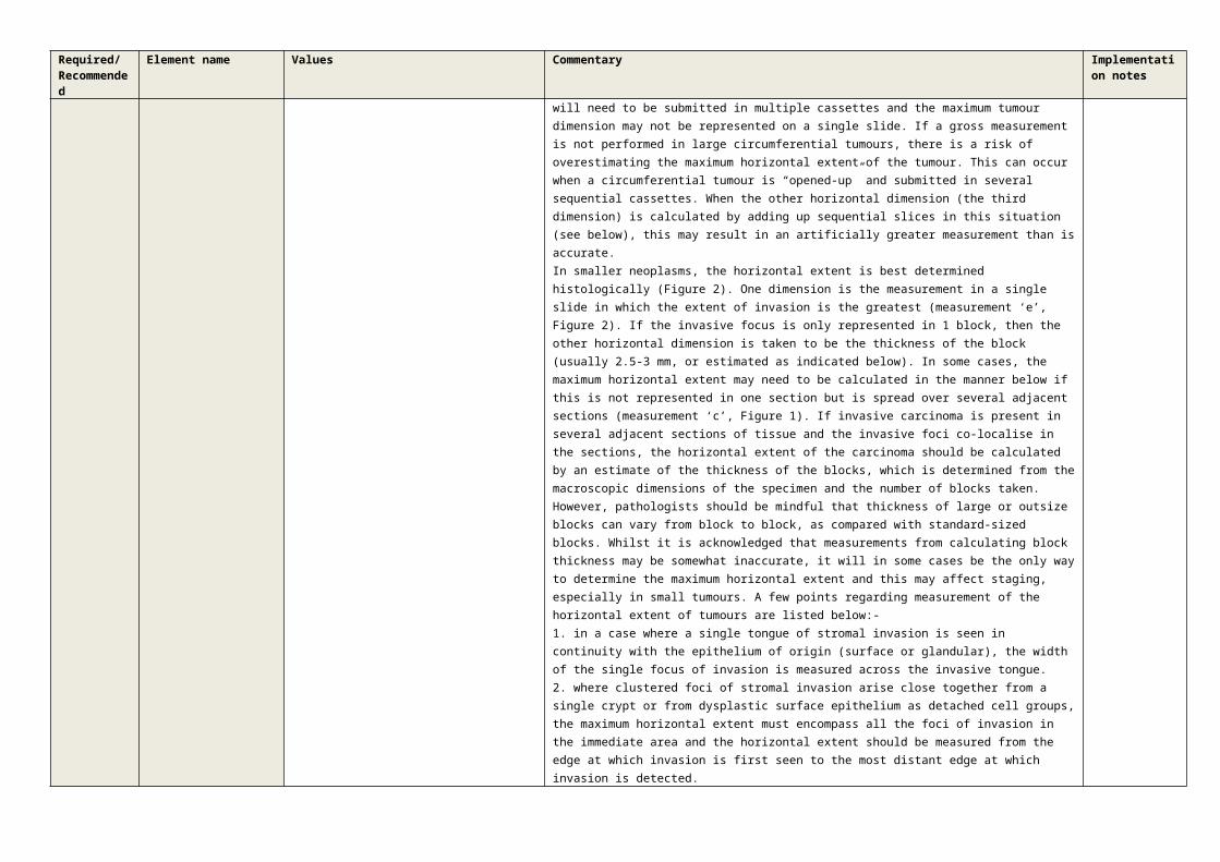
Measurement of horizontal extent of tumour (Figures 1 and 2)The horizontal extent (two dimensions, i.e. both tumour length and width, measurements ‘b’ and ‘c’ in Figure 1) must be measured in all cases. As discussed earlier, in large tumours, this may best be done grossly if large block processing is not available, because in many cases these neoplasms will need to be submitted in multiple cassettes and the maximum tumour dimension may not be represented on a single slide. If a gross measurement is not performed in large circumferential tumours, there is a risk of overestimating the maximum horizontal extent of the tumour. This can occur when a circumferential tumour is “opened-up” and submitted in several sequential cassettes. When the other horizontal dimension (the third dimension) is calculated by adding up sequential slices in this situation (see below), this may result in an artificially greater measurement than is accurate. In smaller neoplasms, the horizontal extent is best determined histologically (Figure 2). One dimension is the measurement in a single slide in which the extent of invasion is the greatest (measurement ‘e’, Figure 2). If the invasive focus is only represented in 1 block, then the other horizontal dimension is taken to be the thickness
Required/ Recommended
Element name Values Commentary Implementation notes
of the block (usually 2.5-3 mm, or estimated as indicated below). In some cases, the maximum horizontal extent may need to be calculated in the manner below if this is not represented in one section but is spread over several adjacent sections (measurement ‘c’, Figure 1). If invasive carcinoma is present in several adjacent sections of tissue and the invasive foci co-localise in the sections, the horizontal extent of the carcinoma should be calculated by an estimate of the thickness of the blocks, which is determined from the macroscopic dimensions of the specimen and the number of blocks taken. However, pathologists should be mindful that thickness of large or outsize blocks can vary from block to block, as compared with standard-sized blocks. Whilst it is acknowledged that measurements from calculating block thickness may be somewhat inaccurate, it will in some cases be the only way to determine the maximum horizontal extent and this may affect staging, especially in small tumours. A few points regarding measurement of the horizontal extent of tumours are listed below:-1. in a case where a single tongue of stromal invasion is seen in continuity with the epithelium of origin (surface or glandular), the width of the single focus of invasion is measured across the invasive tongue.2. where clustered foci of stromal invasion arise close together from a single crypt or from dysplastic surface epithelium as detached cell groups, the maximum horizontal extent must encompass all the foci of invasion in the immediate area and the horizontal extent should be measured from the edge at which invasion is first seen to the most distant edge at which invasion is detected.3. where several foci of invasion arise in one single piece of cervical tissue as separate foci of invasion, but in close proximity (see section below on MEASUREMENT OF MULTIFOCAL CARCINOMAS), either as contiguous tongues of invasion or detached epithelial groups, the maximum horizontal extent is taken from the edge at which invasion is first seen to the most distant edge at which invasion is detected. The small amount of intervening tissue with no invasion (usually with in situ neoplasia) is included in the measurement.
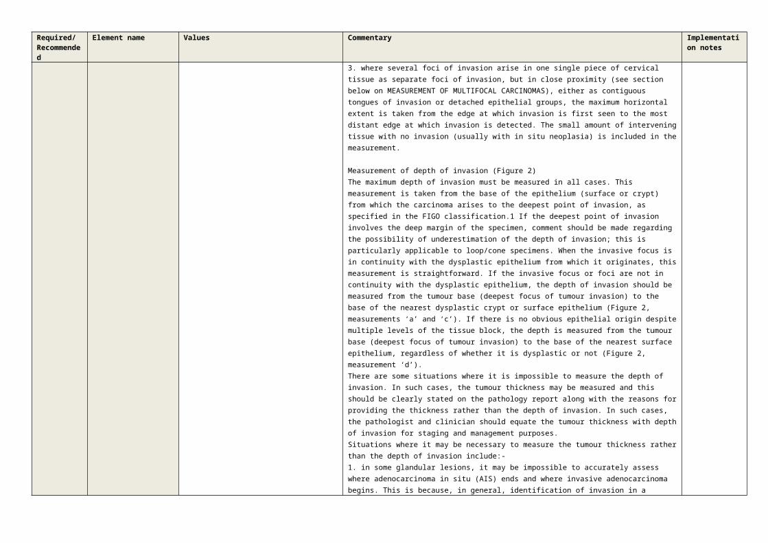
Measurement of depth of invasion (Figure 2)The maximum depth of invasion must be measured in all cases. This measurement is taken from the base of the epithelium (surface or crypt) from which the carcinoma arises to the deepest point of invasion, as specified in the FIGO classification.1 If the deepest point of invasion involves the deep margin of the specimen, comment should be made regarding the possibility of underestimation of the depth of invasion; this is particularly applicable to loop/cone specimens. When the invasive focus is in continuity with the dysplastic epithelium from which it originates, this measurement is straightforward. If the invasive focus or foci are not in continuity with the dysplastic epithelium, the depth of invasion should be measured from the tumour base (deepest focus of tumour invasion) to the base of the nearest dysplastic crypt or surface epithelium (Figure 2, measurements ‘a’ and ‘c’). If there is no obvious epithelial origin despite multiple levels of the tissue block, the depth is measured from the tumour base (deepest focus of tumour invasion) to the base of the nearest surface epithelium, regardless of whether it is dysplastic or not (Figure 2, measurement ‘d’).There are some situations where it is impossible to measure the depth of invasion. In such cases, the tumour thickness may be measured and this should be clearly stated on the pathology report along with the reasons for providing the thickness rather than the depth of invasion. In such cases, the pathologist and clinician should equate the tumour thickness with depth of invasion for staging and management purposes.Situations where it may be necessary to measure the tumour thickness rather than the depth of invasion include:-1. in some glandular lesions, it may be impossible to accurately assess where adenocarcinoma in situ (AIS)
Required/ Recommended
Element name Values Commentary Implementation notes
ends and where invasive adenocarcinoma begins. This is because, in general, identification of invasion in a glandular lesion is more difficult than in a squamous lesion and this is an area where a specialist opinion may be of value. In some cases where the thickness is measured (from the epithelial surface to the deepest point of the tumour) because the point of origin is impossible to establish, this may result in overestimation of the depth of invasion. 2. in ulcerated tumours with no obvious origin from overlying epithelium, the thickness may need to be measured. In this situation, measurement of tumour thickness may result in an underestimate of the depth of invasion.3. uncommonly, squamous carcinomas, adenocarcinomas and other morphological subtypes are polypoid with an exclusive or predominant exophytic growth pattern. In such cases, the carcinoma may project above the surface with little or even no invasion of the underlying stroma. These should not be regarded as in-situ lesions and the tumour thickness will need to be measured in such cases (from the surface of the tumour to the deepest point of invasion). Depth of invasion i.e. the extent of infiltration below the level of the epithelial origin, should not be provided in these cases as it may not be a true reflection of the biological potential of such tumours.
Avoid the term microinvasive carcinoma The term “microinvasive carcinoma” does not appear in the FIGO staging system for cervical cancer.1 Furthermore, use of the term “microinvasive carcinoma” has different connotations in different geographical areas. For example, in the United Kingdom, microinvasive carcinoma was considered to be synonymous with FIGO stage IA1 and IA2 disease in most, but not all, institutions (some used the term “microinvasive carcinoma” to denote only FIGO stage IA1 tumours). In the United States and Canada where the Lower Anogenital Squamous Terminology (LAST)7 recommendations have been adopted, the term superficially invasive squamous cell carcinoma (SISCCA) is used to describe FIGO stage 1A1 tumours with negative margins, and the term “microinvasive squamous cell carcinoma” is no longer in routine use. Confusingly, however, the American Society of Gynecologic Oncology (SGO) has its own definition of stage IA tumours, which is limited not only by the depth of tumour invasion, but, in contrast to FIGO and TNM, also by the absence of lymphovascular invasion.8 According to the SGO, cancers invading less than 3 mm but with lymphovascular involvement are classified as FIGO stage IB1. Thus, in order to avoid confusion, it is recommended to avoid using the term “microinvasive carcinoma” for all morphological subtypes and to use the specific FIGO stage.
Measurement of multifocal carcinomasEarly invasive carcinomas of the cervix, especially squamous, are sometimes multifocal comprising tumours that show multiple foci of invasion arising from separate sites in the cervix and separated by uninvolved cervical tissue. Specifically, multifocal tumours should be diagnosed if foci of invasion are: • separated by blocks of uninvolved cervical tissue (levels must be cut to confirm this)• located on separate cervical lips with discontinuous tumour, not involving the curvature of the canal • situated far apart from each other in the same section (see below). The individual foci of stromal invasion may be attached to, or discontinuous from, the epithelium from which they arise. Multifocal carcinomas should not be confused with the scenario in which tongues or buds of invasion originate from more than one place in a single zone of transformed epithelium and will, over time, coalesce to form a single invasive tumour which represents unifocal disease (and should be measured as
Required/ Recommended
Element name Values Commentary Implementation notes
indicated above, in three dimensions).The frequency of multifocality in FIGO stage IA1 cervical squamous carcinomas has been reported to be between 12 and 25%9-11 although multifocality in larger, advanced tumours is uncommon. There are few (and some rather dated) guidelines regarding measurement of multifocal carcinomas. Although pre-invasive disease may be present, when foci of stromal invasion arise from separate sites or are separated by cervical tissue without invasion (after levels/deeper sections have been cut to confirm this), the foci of invasion should be measured separately, in 3 dimensions, as described above, and staged according to the dimensions of the larger/largest tumour with a clear statement that the tumour is multifocal. However, in the last of the scenarios mentioned above (foci of stromal invasion situated far apart from each other in the same section) measurement of the multifocal disease is problematical. Options include measuring from the edge of one invasive focus to the edge of the furthest invasive focus according to FIGO guidelines (irrespective of the distance between foci of invasion), adding the maximum horizontal extent of each invasive focus together (which clearly does not reflect the biological potential of the individual invasive foci) or regarding widely separated foci as representing small independent areas of invasion.9-13 For tumours with a shallow depth of invasion (up to 3mm), the assessment and measurement of multifocal disease have implications for staging. It is in the context of these early, shallow tumours in loop/cone excisions that management may be significantly affected if the maximum horizontal extent is taken from the edge of one invasive focus to the edge of the furthest invasive focus, when the invasive foci are separate from each other. This may upstage a small superficially invasive carcinoma to FIGO stage IB1, leading to radical surgery (radical hysterectomy or trachelectomy) in patients who are often young and wish to retain their fertility. An alternative view is that when widely separated, these foci of invasion could be regarded as separate foci of IA1 disease, which can be treated by local excision or simple hysterectomy. The SHAPE trial14 sets out to address this problematic issue. However, two recent studies have regarded such lesions as representing multiple foci of invasion (multifocal FIGO IA1 carcinomas) if the foci of invasion are clearly separated. However, the distance of separation is not defined and FIGO provides no guidance on this matter. An arbitrary minimum distance of 2 mm between each separate focus of invasion has been applied in the 2 studies.9,10 Follow-up of patients in these two studies, which include a combined total of 46 cases of ‘‘multifocal IA1 cervical squamous carcinomas’’ treated by local excisional methods (loop/cone excision) with margins clear of premalignant and malignant disease, has shown no evidence of recurrent premalignant or malignant disease with median follow-up periods of 45 months and 7 years respectively.9,10 Moreover, one of the studies also showed that the prevalence of residual pre-invasive (20%) and invasive disease (5%) on repeat excision were comparable to data available for unifocal FIGO stage IA1 cases.10 These studies included cases which would have been regarded as FIGO stage IB1 had the horizontal extent been measured from the edge of one invasive focus to the edge of the furthest invasive focus, as per FIGO guidelines. Although limited by a relatively small number of cases and the selection of an arbitrary distance of separation of 2 mm, the findings support the hypothesis that with regard to tumour staging and management, it may be appropriate to consider superficial, widely separated foci of invasion as representing multifocal lesions, to measure each focus separately, and to determine the FIGO stage on the basis of the invasive focus with the higher/highest FIGO stage. Of course, the possibility that such lesions behave as FIGO stage IA1 tumours may reflect the shallow depth of invasion, which clinicians do not seem to take account of when faced with a tumour whose maximum horizontal width is 7 mm or more, and the spectre of a FIGO IB1 tumour is raised. Although the ICCR does not have a mandate to implement an approach based only on 2 studies involving 46
Required/ Recommended
Element name Values Commentary Implementation notes
patients in total, the ICCR recommends that this approach be considered and discussed at the Tumour Board/multidisciplinary team (MDT) meetings to avoid unnecessary surgery in young patients who wish to preserve their fertility in this specific clinical situation. This approach needs to be verified by additional larger collaborative studies and trials. It is also stressed that in such cases, the tissue blocks containing the invasive foci and those in between should be levelled to confirm that the invasive foci are truly separate and ensure that there is no occult stromal invasion in the intervening areas. If this approach is adopted, the pathology report should clearly indicate how the measurements have been obtained to arrive at a diagnosis of multifocal invasion, provide the dimensions of the separate foci of invasion and indicate how the FIGO stage has been ascertained. Such cases may need to be referred to Cancer Centres for review and, as indicated above, should be discussed individually at the tumour board/MDT meeting. There have been no similar studies for multifocal adenocarcinomas but anecdotally these are less common than multifocal squamous carcinomas and until further evidence becomes available, a similar approach is recommended.Measurement of tumour volume In most studies, tumour size is based on measurement of two dimensions but in a few studies, tumour volume (based on the three measured tumour dimensions) has been shown to predict prognosis more reliably than measurements in only one or two dimensions. Some older studies have suggested tumour volume as a reliable prognostic factor for early stage tumours: a volume of less than 420 mm3 has been suggested to be associated with no lymph node metastasis.15-17 This is one of the main reasons for recommending that three tumour dimensions (two of horizontal extent and one of depth of invasion or tumour thickness) are provided. However, only a few centres continue to routinely factor tumour volume into patient management.
Figure 1: Measurement of cervical tumours in three dimensions CIN3 with involvement of endocervical gland crypts is represented by the dark blue-coloured areas, non-dysplastic squamous epithelium is pink, and grey areas indicate foci of stromal invasion. The depth of invasion, (a), and horizontal tumour dimension/width, (b) are measured in unifocal disease.Third dimension: when stromal invasion is present in three or more consecutive blocks of a loop or cone biopsy the third tumour dimension, (c), may exceed 7 mm, i.e. the carcinoma may be more than FIGO stage IA2. This dimension is determined by calculating the block thickness (usually 2.5 - 3.0 mm) from the macroscopic specimen dimensions and multiplying this by the number of sequential blocks through which the invasion extends. Figure 2: Measurement of width and depth of invasion in cervical tumours The dark blue-coloured areas represent CIN3 with involvement of endocervical gland crypts, non-dysplastic squamous epithelium is pink, and grey areas indicate foci of stromal invasion.Depth of invasion: when invasion originates from the surface epithelium, (a), or gland crypts (b and c), the depth of invasion is taken from the base of the epithelium from which the invasive carcinoma arises, to the deepest focus of invasion, as specified in the FIGO classification. Measurements are taken in the same way, regardless of whether the invasive foci remain attached to the gland crypt (b) or not (c). Where invasion occurs and no obvious surface (or crypt) epithelial origin is seen, the depth of invasion is measured from the deepest focus of tumour invasion, to the base of the nearest non-neoplastic surface epithelium, (d).
Required/ Recommended
Element name Values Commentary Implementation notes
Horizontal dimension/width in unifocal tumours, (e): this is measured in the slice of tissue in which the width is greatest (from the edge at which invasion is first seen, to the most distant edge at which invasion is identified), in sections where the foci of invasion arise in close proximity to each other, even if those foci are separated by short stretches of normal epithelium.
References1 Pecorelli S, Zigliani L and Odicino F (May 2009). Revised FIGO staging for carcinoma of the cervix. Int J Gynaecol Obstet. 105(2):107-108. Epub 2009 Apr 2001.2 Plante M, Gregoire J, Renaud MC and Roy M (2011). The vaginal radical trachelectomy: an update of a series of 125 cases and 106 pregnancies. Gynecol Oncol 121(2):290-297.3 Park JY, Joo WD, Chang SJ, Kim DY, Kim JH, Kim YM, Kim YT and Nam JH (2014). Long-term outcomes after fertility-sparing laparoscopic radical trachelectomy in young women with early-stage cervical cancer: an Asan Gynecologic Cancer Group (AGCG) study. J Surg Oncol 110(3):252-257.4 Turan T, Yildirim BA, Tulunay G, Boran N and Kose MF (2010). Prognostic effect of different cut-off values (20mm, 30mm and 40mm) for clinical tumor size in FIGO stage IB cervical cancer. J Surg Oncol 19(2):106-113.5 Horn LC, Bilek K, Fischer U, Einenkel J and Hentschel B (2014). A cut-off value of 2 cm in tumor size is of prognostic value in surgically treated FIGO stage IB cervical cancer. Gynecol Oncol 134(1):42-46.6 Horn LC, Fischer U, Raptis G, Bilek K and Hentschel B (2007). Tumor size is of prognostic value in surgically treated FIGO stage II cervical cancer. Gynecol Oncol 107(2):310-315.7 Darragh TM, Colgan TJ, Cox JT, Heller DS, Henry MR, Luff RD, McCalmont T, Nayar R, Palefsky JM, Stoler MH, Wilkinson EJ, Zaino RJ, Wilbur DC and Members of LAST Project Work Groups (2012). The Lower Anogenital Squamous Terminology Standardization Project for HPV-Associated Lesions: background and consensus recommendations from the College of American Pathologists and the American Society for Colposcopy and Cervical Pathology. Arch Pathol Lab Med 136(10):1266-1297.8 Girardi F, Burghardt E and Pickel H (1994). Small FIGO stage IB cervical cancer. Gynecol Oncol 55(3 Pt 1):427-432.9 McIlwaine P, Nagar H and McCluggage WG (2014). Multifocal FIGO stage 1A1 cervical squamous carcinomas have an extremely good prognosis equivalent to unifocal lesions. Int J Gynecol Pathol 33(3):213-217.10 Day E, Duffy S, Bryson G, Syed S, Shanbhag S, Burton K, Lindsay R, Siddiqui N and Millan D (2016). Multifocal FIGO Stage IA1 Squamous Carcinoma of the Cervix: Criteria for Identification, Staging, and its Good Clinical Outcome. Int J Gynecol Pathol.11 Reich O, Pickel H, Tamussino K and Winter R (2001). Microinvasive carcinoma of the cervix: site of first focus of invasion. Obstet Gynecol 97(6):890-892.12 Reich O and Pickel H (2002). Multifocal Stromal Invasion in Microinvasive Squamous Cell Carcinoma of the Cervix: How to Measure and Stage these Lesions. Int J Gynecol Pathol 21:416-417.13 Hirschowitz L, Nucci M and Zaino RJ (2013). Problematic issues in the staging of endometrial, cervical and vulval carcinomas. Histopathology 62(1):176-202.14 Cancer Research UK (2015). A trial looking at surgery for cervical cancer (SHAPE). Available at: http://www.cancerresearchuk.org/about-cancer/find-a-clinical-trial/a-trial-looking-at-surgery-for-cervical-cancer-shape. (Accessed 11th May 2016).15 Burghardt E, Baltzer J, Tulusan AH and Haas J (1992). Results of surgical treatment of 1028 cervical cancers studied with volumetry. Cancer 70(3):648-655.
Required/ Recommended
Element name Values Commentary Implementation notes
16 Trattner M, Graf AH, Lax S, Forstner R, Dandachi N, Haas J, Pickel H, Reich O, Staudach A and Winter R (2001). Prognostic factors in surgically treated stage ib-iib cervical carcinomas with special emphasis on the importance of tumor volume. Gynecol Oncol 82(1):11-16.17 Burghardt E and Holzer E (1977). Diagnosis and treatment of microinvasive carcinoma of the cervix uteri. Obstet Gynecol 49(6):641-653.
Required Horizontal extent Numeric: __x__mm At least**
Required Depth of invasion Numeric:__mm At least**ORNot assessable
If not assessable record thickness
Required Thickness Numeric:__mm
Required HISTOLOGICAL TUMOUR TYPE
Text All cervical carcinomas should be typed according to the 2014 WHO classification.1 Carcinosarcoma is also included since, although it is included in the category of mixed epithelial and mesenchymal tumours, it is essentially a carcinoma which has undergone sarcomatous differentiation/metaplasia. The major subtypes of cervical carcinoma are squamous cell carcinoma (SCC), adenocarcinoma (with various subtypes), adenosquamous carcinoma and neuroendocrine tumours. While it is beyond the remit of this document to detail the morphological appearances of the different tumour types in detail, a few points should be noted.SCCs are nearly all caused by high-risk human papillomavirus (HPV) with rare exception2,3 and are subclassified by the WHO based on their histological growth pattern and the presence of keratinization. However, the subclassification of SCC seems to have little or no bearing on clinical behaviour and so it is not considered necessary to specify the subtype (keratinizing, papillary, basaloid, warty, verrucous etc). However, it may be useful to record unusual subtypes, for example lymphoepithelioma-like, since the behaviour of these is not well established.There are several subtypes of cervical adenocarcinoma, the most common being the usual type which, in the majority of cases, is associated with high-risk HPV. The other, less common subtypes (gastric type, mesonephric, clear cell and others) are generally unassociated with HPV infection and have different and distinct histologic appearances. While there is limited information regarding the clinical behaviour of the adenocarcinoma subtypes, it is now well established that gastric type adenocarcinomas of the cervix (adenoma malignum or mucinous variant of minimal deviation adenocarcinoma is the morphologically well differentiated end of the spectrum of gastric type adenocarcinoma) have a particularly aggressive behaviour with poor prognosis, even in early stage disease.4-6 Therefore, it is extremely important from both a prognostic stance as well as an aetiologic and epidemiologic perspective (in light of widespread HPV vaccination programs) to correctly identify these tumour subtypes. The ubiquitous use of and reliance on p16 immunohistochemistry to diagnose cervical adenocarcinoma may cause diagnostic problems for HPV negative tumours, since these do not exhibit the diffuse block-type immunoreactivity characteristic of HPV-related tumours (see section on ANCILLARY STUDIES).7,8 In addition, in the era of molecular characterization and targeted therapy, correct identification of the tumour subtypes will be even more crucial for understanding tumour biology and discovery of potential therapeutic targets. Adenosquamous carcinomas (defined in WHO 2014 blue book as a malignant epithelial tumour comprising
Required/ Recommended
Element name Values Commentary Implementation notes
both adenocarcinoma and squamous carcinoma1) are usually related to high-risk HPV. To make a diagnosis of adenosquamous carcinoma, malignant squamous and glandular components should be identifiable on routine haematoxylin and eosin stained sections. The demonstration of foci of intracytoplasmic mucin by mucin stains in an otherwise typical squamous carcinoma should not result in diagnosis of an adenosquamous carcinoma. Carcinomas which lack evidence of squamous differentiation (intercellular bridges, keratinisation) but have abundant mucin-producing cells should be diagnosed as poorly-differentiated adenocarcinomas. Adenosquamous carcinoma should also be distinguished from a spatially separate squamous carcinoma and adenocarcinoma, which occasionally occurs. While some studies have indicated a worse outcome than pure squamous or adenocarcinomas, there is not robust evidence to confirm these findings.9,10 Primary serous carcinoma of the cervix is exceedingly rare and some doubt its existence, although it is included in the 2014 WHO Classification. Most cases reported as primary cervical serous carcinoma are likely to represent a metastasis from the corpus or extrauterine sites or a usual HPV-related adenocarcinoma with marked nuclear atypia. Metastasis should be excluded before diagnosing a primary cervical serous carcinoma. Usual type cervical adenocarcinomas can have a papillary growth pattern and may show high-grade nuclear atypia, which can mimic serous carcinoma. Whether true p53 mutation-associated serous carcinoma of the cervix exists is unresolved at this time.While endometrioid type adenocarcinoma of the cervix is a subtype listed in the 2014 WHO classification, in the past this has been an over-used diagnostic category and some even doubt its existence as a primary cervical neoplasm. Most adenocarcinomas classified as primary cervical endometrioid adenocarcinomas in the literature represent usual type cervical adenocarcinomas with mucin depletion. These are different from true endometrioid type adenocarcinomas of the uterine corpus or adnexa which are driven by hormones and not HPV-associated. If endometrioid adenocarcinoma occurs as a primary neoplasm in the cervix, it is most likely in the setting of endometriosis and has the same histologic and immunohistochemical profiles as endometrioid adenocarcinomas of the uterine corpus or ovary. As with serous carcinoma, extreme caution should be exercised before diagnosing a primary cervical endometrioid adenocarcinoma. Neuroendocrine carcinomas (NECs) (small cell and large cell neuroendocrine carcinoma) are uncommon but well described in the cervix and can occur in pure form or associated with another tumour type, typically adenocarcinoma, squamous carcinoma or adenosquamous carcinoma. These are referred to in the WHO 2014 blue book as high-grade neuroendocrine carcinomas. The term ‘small cell neuroendocrine carcinoma’ is preferred to ‘small cell carcinoma’ since a small cell variant of squamous carcinoma occurs and if the term “neuroendocrine” is not applied, this may result in confusion. When mixed with another tumour type, the percentage of the neuroendocrine component should be given. Regardless of the percentage of NEC, it is recommended that the tumour be reported as mixed since all tumours containing a component of NEC have a very poor prognosis and the NEC component may be underestimated in a limited sample.11 Several studies of small cell neuroendocrine carcinomas of the cervix have shown that adjuvant chemotherapy after surgery for early stage disease provides significant clinical benefit compared to surgery alone and therefore, it is extremely important to correctly diagnose any component of NEC. Additionally, in many institutions surgical resection is not undertaken for a NEC even if early stage but instead chemotherapy treatment is given. Diagnosing NEC or a component of NEC can be difficult, especially in small samples, but a combination of synaptophysin, chromogranin, CD56, TTF1 and p63 has been shown to be helpful in making the distinction between NEC and poorly-differentiated non-NEC (see section on ANCILLARY STUDIES).12,13
Required/ Recommended
Element name Values Commentary Implementation notes
References1 Kurman RJ, Carcangiu ML, Herrington CS and Young RH (2014). WHO classification of tumours of the female reproductive organs. IARC press, Lyon.2 Casey S, Harley I, Jamison J, Molijn A, van den Munckhof H and McCluggage WG (2015). A rare case of HPV-negative cervical squamous cell carcinoma. Int J Gynecol Pathol 34(2):208-212.3 Rodriguez-Carunchio L, Soveral I, Steenbergen RD, Torne A, Martinez S, Fuste P, Pahisa J, Marimon L, Ordi J and del Pino M (2015). HPV-negative carcinoma of the uterine cervix: a distinct type of cervical cancer with poor prognosis. Bjog 122(1):119-127.4 Kojima A, Mikami Y, Sudo T, Yamaguchi S, Kusanagi Y, Ito M and Nishimura R (2007). Gastric morphology and immunophenotype predict poor outcome in mucinous adenocarcinoma of the uterine cervix. Am J Surg Pathol 31(5):664-672.5 Karamurzin YS, Kiyokawa T, Parkash V, Jotwani AR, Patel P, Pike MC, Soslow RA and Park KJ (2015). Gastric-type Endocervical Adenocarcinoma: An Aggressive Tumor With Unusual Metastatic Patterns and Poor Prognosis. Am J Surg Pathol 39(11):1449-1457.6 Park KJ, Kiyokawa T, Soslow RA, Lamb CA, Oliva E, Zivanovic O, Juretzka MM and Pirog EC (2011). Unusual endocervical adenocarcinomas: an immunohistochemical analysis with molecular detection of human papillomavirus. Am J Surg Pathol 35(5):633-646.7 Howitt BE, Herfs M, Brister K, Oliva E, Longtine J, Hecht JL and Nucci MR (2013). Intestinal-type endocervical adenocarcinoma in situ: an immunophenotypically distinct subset of AIS affecting older women. Am J Surg Pathol 37(5):625-633.8 Vang R, Gown AM, Farinola M, Barry TS, Wheeler DT, Yemelyanova A, Seidman JD, Judson K and Ronnett BM (2007). p16 expression in primary ovarian mucinous and endometrioid tumors and metastatic adenocarcinomas in the ovary: utility for identification of metastatic HPV-related endocervical adenocarcinomas. Am J Surg Pathol 31(5):653-663.9 Look KY, Brunetto VL, Clarke-Pearson DL, Averette HE, Major FJ, Alvarez RD, Homesley HD and Zaino RJ (1996). An analysis of cell type in patients with surgically staged stage IB carcinoma of the cervix: a Gynecologic Oncology Group study. Gynecol Oncol 63(3):304-311.10 Lea JS, Coleman RL, Garner EO, Duska LR, Miller DS and Schorge JO (2003). Adenosquamous histology predicts poor outcome in low-risk stage IB1 cervical adenocarcinoma. Gynecol Oncol 91(3):558-562.11 Horn LC, Hentschel B, Bilek K, Richter CE, Einenkel J and Leo C (2006). Mixed small cell carcinomas of the uterine cervix: prognostic impact of focal neuroendocrine differentiation but not of Ki-67 labeling index. Ann Diagn Pathol 10(3):140-143.12 Ganesan R, Hirschowitz L, Dawson P, Askew S, Pearmain P, Jones PW, Singh K, Chan KK and Moss EL (2016). Neuroendocrine Carcinoma of the Cervix: Review of a Series of Cases and Correlation With Outcome. Int J Surg Pathol 24(6):490-496.13 McCluggage WG, Kennedy K and Busam KJ (2010). An immunohistochemical study of cervical neuroendocrine carcinomas: Neoplasms that are commonly TTF1 positive and which may express CK20 and P63. Am J Surg Pathol 34(4):525-532.
Recommended HISTOLOGICAL TUMOUR GRADE
Single selection value list:• Not graded/applicable• G1: Well differentiated
Grading of cervical carcinomaTumour grade is regularly included in histopathology reports of cervical squamous cell carcinoma (SCC) and adenocarcinoma (ACA). However, at present no particular grading system(s) has achieved universal
Required/ Recommended
Element name Values Commentary Implementation notes
• G2: Moderately differentiated• G3: Poorly differentiated• GX: Cannot be graded
acceptance and grading of these tumours remains of uncertain clinical value.1-3 For example, grade is not amongst the factors considered in determining the Gynecology Oncology Group (GOG) score which is used to assess the need for adjuvant therapy following surgery for low-stage cervical carcinomas.4 Not uncommonly, studies that assess grade as a potential prognostic variable provide no details of the grading system employed, and this is also true of large multicentre investigations such as SEER analyses.5,6 For these and other reasons (discussed below), tumour grading is not listed as a required but rather a recommended element. Furthermore, no particular grading system for squamous carcinoma or adenocarcinoma is recommended.General considerations 1. As with tumours arising in other anatomical sites, grading of cervical carcinomas has a considerable subjective component and this probably explains, at least in part, the variable proportion of well, moderately, and poorly-differentiated tumours reported in different studies. However, some investigators have demonstrated reasonable intra- and inter-observer agreement using more complex multifactor grading schemes in SCC (discussed below). 2. Almost all cervical SCCs are HPV-associated and given that HPV-associated SCCs very commonly have a “basaloid” morphology with minimal keratinisation, they are very commonly poorly-differentiated.3. Most clinically advanced cervical carcinomas are treated with primary chemoradiation rather than surgery and histological sampling may be limited to a small diagnostic biopsy. This may not be fully representative due to tumour heterogeneity and could be potentially misleading as regards tumour differentiation or grade.1 This may be particularly relevant since less differentiated appearing tumour elements may be located more deeply towards the invasive margin.2 4. There is an implicit correlation between tumour subtype and grade in certain cervical carcinomas and therefore a separate grade may not be applicable. For example, pure villoglandular ACA of the cervix is by definition a low-grade neoplasm while serous and clear cell carcinoma, as in the endometrium, are considered high-grade by default. Similarly, ‘gastric-type’ cervical ACAs and NECs are clinically aggressive regardless of their histological pattern and therefore are best considered high-grade automatically.7,8 There is no published grading system for cervical mesonephric ACAs. Several variants of cervical SCC are also recognised, although most do not differ from conventional SCC in terms of prognosis or therapy.9 5. It is uncertain whether a truly ‘undifferentiated’ cervical carcinoma should be regarded as a separate tumour subtype analogous, for example, to similar tumours arising in the endometrium. 6. Grading of very small superficially (‘early’) invasive carcinomas of either squamous or glandular type is probably not possible or relevant. Grading of Cervical SCCHistorically, cervical SCCs were graded using Broder’s system or modifications thereof based upon the degree of keratinisation, cytological atypia and mitotic activity. In some schemes, the pattern of invasion (pushing versus infiltrating) has also been taken into account. Traditionally, SCCs have also been subclassified into large cell keratinising, large cell non-keratinising and small cell non-keratinising categories, with these sometimes being regarded as approximately equivalent to well, moderately and poorly-differentiated, respectively. As noted above, this raises the issue whether such categorisation represents a tumour subtype (arguably not further graded), or a grade within a spectrum of a single type of tumour. It should be noted that some studies have found that the keratinising variant of large cell SCC actually has a poorer prognosis than the non-keratinising variant, an apparently paradoxical finding if keratinisation is deemed to be evidence of better differentiation. It is also uncertain what proportion of “small cell SCCs” reported in the older literature would

Required/ Recommended
Element name Values Commentary Implementation notes
now be classified as high-grade NECs (small cell NEC), and this could potentially bias the supposedly poor outcome of this tumour category.More complex multifactor grading systems (MGS) that include both tumour and host/stromal parameters have been assessed in cervical carcinomas, mainly SCC.10-14 For example, the system employed by Stendahl et al,10 based upon that used in head and neck SCC, comprised eight features, 4 of which were tumour-related (growth pattern, differentiation, pleomorphism and mitoses) and four of which were stromal-related (pattern of invasion, stage/depth of invasion, vascular invasion and inflammatory reaction). Each factor was scored from 1 to 3 and thus the potential total MGS score ranged from 8-24 points. Simplified modifications to the MGS have also been described including systems that selectively focus upon the invasive tumour border or the patterns of tumour invasion.15-18 However, the “cut-off value” for tumour grade has varied in different studies and not all have demonstrated a correlation with prognosis.2,19,20 At present, none of these grading systems has been widely adopted in routine diagnostic practice. Grading of Cervical ACAAs with SCC, it is controversial whether grading has independent prognostic value in cervical ACA. Whilst a correlation between higher grade and adverse outcomes has been reported,21-25 at least for poorly differentiated tumours, this has not been a universal finding.26,27 It should also be noted that some studies have included a variable proportion of less common histological subtypes such as adenosquamous carcinoma, mesonephric, gastric-type and clear cell carcinoma21,24,25 and often tumour details are not provided. Therefore, it is not clear whether the reported grading data are applicable to usual-type cervical ACA or have been biased by the inclusion of other more aggressive tumour subtypes (for example, gastric-type ACA). Most grading systems for cervical ACA have been based upon the relative proportion of glandular differentiation, typically following the FIGO system for endometrial endometrioid adenocarcinoma (EEC). However, the maximum permitted extent of solid growth for a grade 1 cervical ACA has been variably specified to be 5%28,29 or 10%.25,30 As with EEC, an upward grade adjustment has been suggested for those tumours exhibiting more marked cytological atypia. However, it is pertinent that usual-type cervical ACAs typically demonstrate more marked nuclear atypia, mitotic and apoptotic activity than architecturally similar EECs. 31 There are no separate grading systems for the various non-HPV related cervical ACAs.Recently, a system of assessing cervical ACAs based upon their invasive growth pattern has been developed, and this has been shown to be reproducible amongst pathologists and to correlate with the risk of lymph node metastasis and patient outcomes.32-35 If these findings are confirmed by additional studies it may be argued whether this system could be considered a complement to, or even an alternative to, conventional grading. The latter has traditionally been based upon the cytoarchitectural pattern of the neoplasm itself but as noted above, tumour-stromal relationships including the pattern of stromal invasion have been included in earlier grading schemes of cervical SCC. Grading of Cervical Adenosquamous CarcinomaAlthough it has been suggested that adenosquamous carcinomas are graded on the basis of the degree of differentiation of both the glandular and squamous components, there is no well-established grading system for these neoplasms which has been shown to be of prognostic significance.
References1 Benda JA (1996). Histopathologic prognostic factors in early stage cervical carcinoma. J Natl Cancer Inst
Required/ Recommended
Element name Values Commentary Implementation notes
Monogr(21):27-34.2 Zaino R, Ward S, Delgado G, Bundy B, Gore H, Fetter G, Ganjei P and Frauenhofer E (1992). Histopathologic Predictors of the Behavior of Surgically Treated Stage IB Squamous Cell Carcinoma of the Cervix. A Gynecologic Oncology Group Study. Cancer 69(7):1750-1758.3 Tiltman AJ (2005). The pathology of cervical tumours. Best Pract Res Clin Obstet Gynaecol 19(4):485-500.4 Delgado G, Bundy B, Zaino R, Sevin BU, Creasman WT and Major F (1990). Prospective surgical-pathological study of disease-free interval in patients with stage IB squamous cell carcinoma of the cervix: a Gynecologic Oncology Group study. Gynecol Oncol 38:352-357.5 Vinh-Hung V, Bourgain C, Vlastos G, Cserni G, De Ridder M, Storme G and Vlastos AT (2007). Prognostic value of histopathology and trends in cervical cancer: a SEER population study. BMC Cancer 7:164.6 Macdonald OK, Chen J, Dodson M, Lee CM and Gaffney DK (2009). Prognostic significance of histology and positive lymph node involvement following radical hysterectomy in carcinoma of the cervix. Am J Clin Oncol 32(4):411-416.7 Kojima A, Mikami Y, Sudo T, Yamaguchi S, Kusanagi Y, Ito M and Nishimura R (2007). Gastric morphology and immunophenotype predict poor outcome in mucinous adenocarcinoma of the uterine cervix. Am J Surg Pathol 31(5):664-672.8 Karamurzin YS, Kiyokawa T, Parkash V, Jotwani AR, Patel P, Pike MC, Soslow RA and Park KJ (2015). Gastric-type Endocervical Adenocarcinoma: An Aggressive Tumor With Unusual Metastatic Patterns and Poor Prognosis. Am J Surg Pathol 39(11):1449-1457.9 Stoler M, Bergeron C and Colgan TJ et al (2014). Squamous cell tumours and precursors. In: WHO Classification of Tumours of Female Reproductive Organs, 4th ed., Kurman RJ, Carcangiu ML, Herrington CS and Young RH (eds), IARC, Lyon, 172-182.10 Stendahl U, Eklund G and Willen R (1983). Prognosis of invasive squamous cell carcinoma of the uterine cervix: a comparative study of the predictive values of clinical staging IB--III and a histopathologic malignancy grading system. Int J Gynecol Pathol 2(1):42-54.11 Bichel P and Jakobsen A (1985). Histopathologic grading and prognosis of uterine cervical carcinoma. Am J Clin Oncol 8(3):247-254.12 Kristensen GB, Abeler VM, Risberg B, Trop C and Bryne M (1999). Tumor size, depth of invasion, and grading of the invasive tumor front are the main prognostic factors in early squamous cell cervical carcinoma. Gynecol Oncol 74(2):245-251.13 Lindahl B, Ranstam J and Willen R (2007). Prospective malignancy grading of invasive squamous carcinoma of the uterine cervix. Prognostic significance in a long-term follow-up. Anticancer Res 27(4c):2829-2832.14 Eggen T, Arnes M, Moe B, Straume B and Orbo A (2007). Prognosis of early cervical cancer (FIGO Stages IA2, IB, and IIA) in northern Norway predicted by malignancy grading score and objective morphometric image analysis. Int J Gynecol Pathol 26(4):447-456.15 Stendahl U, Eklund G and Willen R (1981). Invasive squamous cell carcinoma of the uterine cervix. IV. Analysis of a histopathologic malignancy grading system and construction of a partial index. Acta Radiol Oncol 20(5):289-284.16 Graflund M, Sorbe B, Hussein A, Bryne M and Karlsson M (2002). The prognostic value of histopathologic grading parameters and microvessel density in patients with early squamous cell carcinoma of the uterine cervix. Int J Gynecol Cancer 12(1):32-41.17 Horn LC, Fischer U, Raptis G, Bilek K, Hentschel B, Richter CE, Braumann UD and Einenkel J (2006). Pattern
Required/ Recommended
Element name Values Commentary Implementation notes
of invasion is of prognostic value in surgically treated cervical cancer patients. Gynecol Oncol 103(3):906-911.18 Horn LC, Hentschel B and Braumann UD (2008). Malignancy grading, pattern of invasion, and juxtatumoral stromal response (desmoplastic change) in squamous cell carcinoma of the uterine cervix. Int J Gynecol Pathol 27(4):606-607.19 Crissman JD, Budhraja M, Aron BS and Cummings G (1987). Histopathologic prognostic factors in stage II and III squamous cell carcinoma of the uterine cervix. An evaluation of 91 patients treated primarily with radiation therapy. Int J Gynecol Pathol 6(2):97-103.20 Samlal RA, van der Velden J, Schilthuis MS, Gonzalez Gonzalez D, Ten Kate FJ, Hart AA and Lammes FB (1997). Identification of high-risk groups among node-positive patients with stage IB and IIA cervical carcinoma. Gynecol Oncol 64(3):463-467.21 McLellan R, Dillon MB, Woodruff JD, Heatley GJ, Fields AL and Rosenshein NB (1994). Long-term follow-up of stage I cervical adenocarcinoma treated by radical surgery. Gynecol Oncol 52(2):253-259.22 Goodman HM, Buttlar CA, Niloff JM, Welch WR, Marck A, Feuer EJ, Lahman EA, Jenison EL and Knapp RC (1989). Adenocarcinoma of the uterine cervix: prognostic factors and patterns of recurrence. Gynecol Oncol 33(2):241-247.23 Lea JS, Sheets EE, Wenham RM, Duska LR, Coleman RL, Miller DS and Schorge JO (2002). Stage IIB-IVB cervical adenocarcinoma: prognostic factors and survival. Gynecol Oncol 84(1):115-119.24 Eifel PJ, Morris M, Oswald MJ, Wharton JT and Delclos L (1990). Adenocarcinoma of the uterine cervix. Prognosis and patterns of failure in 367 cases. Cancer 65(11):2507-2514.25 Baalbergen A, Ewing-Graham PC, Hop WC, Struijk P and Helmerhorst TJ (2004). Prognostic factors in adenocarcinoma of the uterine cervix. Gynecol Oncol 92(1):262-267.26 Alfsen GC, Kristensen GB, Skovlund E, Pettersen EO and Abeler VM (2001). Histologic Subtype Has Minor Importance for Overall Survival in Patients with Adenocarcinoma of the Uterine Cervix. A Population-Based Study of Prognostic Factors in 505 Patients with Nonsquamous Cell Carcinomas of the Cervix. Cancer 92(9):2471–2483.27 Leminen A, Paavonen J, Forss M, Wahlstrom T and Vesterinen E (1990). Adenocarcinoma of the uterine cervix. Cancer 65(1):53-59.28 Silverberg SG and Ioffe OB (2003). Pathology of cervical cancer. Cancer J 9(5):335-347.29 D’Angelo E and Prat J (2014). Cervical glandular neoplasia. In: Pathology of the Female Reproductive Tract, 3rd edition, Mutter GL and Prat J (eds), Churchill Livingstone, 251-289.30 Lawrence WD, Abdul-Karim FW, Crum C and Fu Y-S (2000). Recommendations for the Reporting of Surgical Specimens Containing Uterine Cervical Neoplasms. Mod Pathol 13(4):1029-1033.31 Young RH and Clement PB (2002). Endocervical adenocarcinoma and its variants: their morphology and differential diagnosis. Histopathology 41:185-207.32 Roma AA, Diaz De Vivar A, Park KJ, Alvarado-Cabrero I, Rasty G, Chanona-Vilchis JG, Mikami Y, Hong SR, Teramoto N, Ali-Fehmi R, Rutgers JK, Barbuto D and Silva EG (2015). Invasive endocervical adenocarcinoma: a new pattern-based classification system with important clinical significance. Am J Surg Pathol 39(5):667-672.33 Diaz De Vivar A, Roma AA, Park KJ, Alvarado-Cabrero I, Rasty G, Chanona-Vilchis JG, Mikami Y, Hong SR, Arville B, Teramoto N, Ali-Fehmi R, Rutgers JK, Tabassum F, Barbuto D, Aguilera-Barrantes I, Shaye-Brown A, Daya D and Silva EG (2013). Invasive endocervical adenocarcinoma: proposal for a new pattern-based classification system with significant clinical implications: a multi-institutional study. Int J Gynecol Pathol 32(6):592-601.
Required/ Recommended
Element name Values Commentary Implementation notes
34 Roma AA, Mistretta TA, De Vivar AD, Park KJ, Alvarado-Cabrero I, Rasty G, Chanona-Vilchis JG, Mikami Y, Hong SR, Teramoto N, Ali-Fehmi R, Barbuto D, Rutgers JK and Silva EG (2016). New pattern-based personalized risk stratification system for endocervical adenocarcinoma with important clinical implications and surgical outcome. Gynecol Oncol 141(1):36-42.35 Rutgers JK, Roma AA, Park KJ, Zaino RJ, Johnson A, Alvarado I, Daya D, Rasty G, Longacre TA, Ronnett BM and Silva EG (2016). Pattern classification of endocervical adenocarcinoma: reproducibility and review of criteria. Mod Pathol 29(9):1083-1094.
Required LYMPHOVASCULAR INVASION
Single selection value list:• Not identified• Present• Indeterminate
Lymphovascular invasion (LVI) does not affect FIGO or TNM staging (for example if there is LVI in tissues outside the cervix but the tumour itself is confined to the cervix, this is still FIGO stage I) but should be clearly documented in the pathology report. The significance of LVI in cervical carcinoma has been debated for predicting overall survival (OS), disease free interval (DFI), recurrence free survival (RFS) and regional lymph node metastasis for decades. Although studies conflict, there is general agreement that LVI is an independent predictor of adverse outcome.1-11 Early studies indicated that LVI was an independent predictor of DFI with one study reporting a 1.7 times higher rate of recurrence in patients with LVI compared to those without LVI in low-stage cervical carcinoma.3 This has been confirmed in later studies, particularly in low-stage (FIGO stage IB) cervical carcinoma.5 The significance of LVI in superficially invasive squamous cell carcinoma (SISCCA) is unclear, likely due to the rarity of adverse outcomes including lymph node metastasis in SISCCA. Studies have shown that LVI does not predict lymph node metastasis in cases of SISCCA with a depth of invasion of ≤3 mm.12-15, Lack of standardised criteria and marked variability in recognition of LVI have undoubtedly lead to conflicting outcomes in previous studies. Fixation retraction around tumour cell groups is a well-recognized artifact which mimics LVI. Features that may help in the recognition of LVI include a tumour nest within a space associated with other vascular structures, the presence of an endothelial lining, adherence of the tumour cell group to the side of the space, the contour of the intravascular component matching the contour of the vessel and the presence of adherent fibrin. Immunohistochemical demonstration of an endothelial cell lining may assist but is not performed routinely. D2-40 (recognizing lymphatic endothelium) and CD31 and CD34 (recognizing both lymphatic and blood vascular endothelium) may be useful in confirming the presence of LVI.16-19In rare situations when specimens are severely traumatised or diathermied, LVI may be suspected but it may not be possible to reliably determine whether or not LVI is present. In these circumstances ‘indeterminate’ should be recorded in the reporting guide, although it is expected this will be a rare response. Most studies which have examined the significance of LVI in cervical carcinoma have not distinguished between lymphatic and blood vessel invasion and there is little evidence to support separating out the type of invasion, especially since this is not reliable in haematoxylin and eosin stained sections. Occasional studies have found blood vessel invasion to have a worse prognosis than lymphatic invasion and to be a predictor of ovarian involvement.20 However, there is insufficient evidence to warrant inclusion of blood vessel and lymphatic invasion as separate data items.
References1 Benson WL and Norris HJ (1977). A critical review of the frequency of lymph node metastasis and death from microinvasive carcinoma of the cervix. Obstet Gynecol 49(5):632-638.
Required/ Recommended
Element name Values Commentary Implementation notes
2 Maiman MA, Fruchter RG, DiMaio TM and Boyce JG (1988). Superficially invasive squamous cell carcinoma of the cervix. Obstet Gynecol 72(3 Pt 1):399-403.3 Delgado G, Bundy BN, Fowler WC, Jr., Stehman FB, Sevin B, Creasman WT, Major F, DiSaia P and Zaino R (1989). A prospective surgical pathological study of stage I squamous carcinoma of the cervix: a Gynecologic Oncology Group Study. Gynecol Oncol 35(3):314-320.4 Copeland LJ, Silva EG, Gershenson DM, Morris M, Young DC and Wharton JT (1992). Superficially invasive squamous cell carcinoma of the cervix. Gynecol Oncol 45(3):307-312.5 Zaino R, Ward S, Delgado G, Bundy B, Gore H, Fetter G, Ganjei P and Frauenhofer E (1992). Histopathologic Predictors of the Behavior of Surgically Treated Stage IB Squamous Cell Carcinoma of the Cervix. A Gynecologic Oncology Group Study. Cancer 69(7):1750-1758.6 Buckley SL, Tritz DM, Van Le L, Higgins R, Sevin BU, Ueland FR, DePriest PD, Gallion HH, Bailey CL, Kryscio RJ, Fowler W, Averette H and van Nagell JR, Jr. (1996). Lymph node metastases and prognosis in patients with stage IA2 cervical cancer. Gynecol Oncol 63(1):4-9.7 Obermair A, Wanner C, Bilgi S, Speiser P, Reisenberger K, Kaider A, Kainz C, Leodolter S, Breitenecker G and Gitsch G (1998). The influence of vascular space involvement on the prognosis of patients with stage IB cervical carcinoma: correlation of results from hematoxylin and eosin staining with results from immunostaining for factor VIII-related antigen. Cancer 82(4):689-696.8 Morimura Y, Nishiyama H, Hashimoto T, Fujimori K, Yamada H, Yanagida K and Sato A (1999). Re-assessment of stage I uterine cervical carcinoma according to revised JSGO (1997) staging. Fukushima J Med Sci 45(2):109-116.9 Elliott P, Coppleson M, Russell P, Liouros P, Carter J, MacLeod C and Jones M (2000). Early invasive (FIGO stage IA) carcinoma of the cervix: a clinico-pathologic study of 476 cases. Int J Gynecol Cancer 10(1):42-52.10 Morice P, Piovesan P, Rey A, Atallah D, Haie-Meder C, Pautier P, Sideris L, Pomel C, Duvillard P and Castaigne D (2003). Prognostic value of lymphovascular space invasion determined with hematoxylin-eosin staining in early stage cervical carcinoma: results of a multivariate analysis. Ann Oncol 14(10):1511-1517.11 Singh N and Arif S (2004). Histopathologic parameters of prognosis in cervical cancer - a review. Int J Gynecol Cancer. 14(5):741-750.12 Benedet JL (1997). Cervical cancer staging systems: the endless debate. Gynecol Oncol 65(1):6-7.13 Lee SW, Kim YM, Son WS, You HJ, Kim DY, Kim JH, Kim YT and Nam JH (2009). The efficacy of conservative management after conization in patients with stage IA1 microinvasive cervical carcinoma. Acta Obstet Gynecol Scand 88(2):209-215.14 Ostor AG and Rome RM (1994). Micro-invasive squamous cell carcinoma of the cervix: a clinico-pathologic study of 200 cases with long-term follow-up. Int J Gynecol Cancer 4(4):257-264.15 Robert ME and Fu YS (1990). Squamous cell carcinoma of the uterine cervix--a review with emphasis on prognostic factors and unusual variants. Semin Diagn Pathol 7(3):173-189.16 Birner P, Obermair A, Schindl M, Kowalski H, Breitenecker G and Oberhuber G (2001). Selective immunohistochemical staining of blood and lymphatic vessels reveals independent prognostic influence of blood and lymphatic vessel invasion in early-stage cervical cancer. Clin Cancer Res 7(1):93-97.17 Alexander-Sefre F, Singh N, Ayhan A, Salveson HB, Wilbanks G and Jacobs IJ (2003). Detection of tumour lymphovascular space invasion using dual cytokeratin and CD31 immunohistochemistry. J Clin Pathol 56(10):786-788.18 Urabe A, Matsumoto T, Kimura M, Sonoue H and Kinoshita K (2006). Grading system of lymphatic invasion
Required/ Recommended
Element name Values Commentary Implementation notes
according to D2-40 immunostaining is useful for the prediction of nodal metastasis in squamous cell carcinoma of the uterine cervix. Histopathology 49(5):493-497.19 Lim CS, Alexander-Sefre F, Allam M, Singh N, Aleong JC, Al-Rawi H and Jacobs IJ (2008). Clinical value of immunohistochemically detected lymphovascular space invasion in early stage cervical carcinoma. Ann Surg Oncol 15(9):2581-2588.20 Sakuragi N, Takeda N, Hareyama H, Fujimoto T, Todo Y, Okamoto K, Takeda M, Wada S, Yamamoto R and Fujimoto S (2000). A Multivariate Analysis of Blood Vessel and Lymph Vessel Invasion as Predictors of Ovarian and Lymph Node Metastases in Patients with Cervical Carcinoma. Cancer 88:2578-2583.
Required COEXISTENT PATHOLOGY Carcinomas of the cervix are often associated with premalignant precursor lesions, which are mostly squamous or glandular in type. Their pathology is well described and illustrated in the WHO 2014 Classification of Tumours of Female Reproductive Organs and a number of published reviews.1-3 There are also numerous benign squamous or glandular lesions which can be broadly classified as inflammatory, metaplastic and neoplastic. Their importance is in recognizing the lesions as benign as they can morphologically mimic premalignant or malignant glandular or squamous lesions, and result in a false positive diagnosis. It is important to report co-existing premalignant lesions and document whether they involve resections margins since this may influence patient management and follow up. All clearly defined premalignant lesions are caused by HPV. The terminology of HPV-associated premalignant squamous lesions was revised in WHO 2014 to Squamous Intraepithelial Lesion (SIL).4 The change also harmonizes with The Bethesda System for the reporting of cytological abnormalities in cervical smears. Squamous intraepithelial lesions are divided into low-grade SIL (LSIL) which is a viral infection with a high spontaneous resolution rate, and high-grade SIL (HSIL) which is a true premalignant lesion that can progress to SCC. The corresponding cervical intraepithelial neoplasia (CIN) terms can be included in parentheses.Adenocarcinoma in situ (AIS) is the HPV-associated precursor lesion of usual HPV-related cervical adenocarcinoma. High-grade cervical glandular intraepithelial neoplasia (HG CGIN) is an alternative terminology used in some jurisdictions.5 Stratified mucin producing intraepithelial lesion (SMILE) is a premalignant lesion with morphological overlap between SIL and AIS. In WHO 2014, it is regarded as a variant of AIS (and should be coded as such) but others consider it a form of high-grade reserve cell dysplasia and report it separately.6,7 The precursor lesions of non-HPV-related cervical adenocarcinomas are not well defined but lobular endocervical glandular hyperplasia (LEGH), atypical lobular endocervical glandular hyperplasia (ALEGH) and adenocarcinoma in situ of gastric type have been proposed as likely precursor lesions of gastric type adenocarcinoma of the cervix.8
References1 Kurman RJ, Carcangiu ML, Herrington CS and Young RH (2014). WHO classification of tumours of the female reproductive organs. IARC press, Lyon.2 McCluggage WG (2013). Premalignant lesions of the lower female genital tract: cervix, vagina and vulva. Pathology 45(3):214-228.3 Nucci MR (2014). Pseudoneoplastic glandular lesions of the uterine cervix: a selective review. Int J Gynecol Pathol 33(4):330-338.
HeadingNotes:Required for loop/cone excisions/trachelectomies only and recommended for other specimens.
Required/ Recommended
Element name Values Commentary Implementation notes
4 Darragh TM, Colgan TJ, Cox JT, Heller DS, Henry MR, Luff RD, McCalmont T, Nayar R, Palefsky JM, Stoler MH, Wilkinson EJ, Zaino RJ, Wilbur DC and Members of LAST Project Work Groups (2012). The Lower Anogenital Squamous Terminology Standardization Project for HPV-Associated Lesions: background and consensus recommendations from the College of American Pathologists and the American Society for Colposcopy and Cervical Pathology. Int J Gynecol Pathol 32:76-115.5 McCluggage WG (2013). New developments in endocervical glandular lesions. Histopathology 62(1):138-160.6 Park JJ, Sun D, Quade BJ, Flynn C, Sheets EE, Yang A, McKeon F and Crum CP (2000). Stratified mucin-producing intraepithelial lesions of the cervix: adenosquamous or columnar cell neoplasia? Am J Surg Pathol 24(10):1414-1419.7 Boyle DP and McCluggage WG (2015). Stratified mucin-producing intraepithelial lesion (SMILE): report of a case series with associated pathological findings. Histopathology 66(5):658-663.8 Mikami Y and McCluggage WG (2013). Endocervical glandular lesions exhibiting gastric differentiation: an emerging spectrum of benign, premalignant, and malignant lesions. Adv Anat Pathol 20(4):227-237.
Required Squamous intraepithelial lesion (SIL) (CIN)
Single selection value list:• Not identified• Present
If present, record GRADE
Required Grade Single selection value list:• Low-grade SIL (LSIL) (CIN 1)• High-grade SIL (HSIL) (CIN 2/3)
Required Adenocarcinoma in-situ (AIS)/High-grade cervical glandular intraepithelial neoplasia (HG CGIN)
Single selection value list:• Not identified • Present
Required Stratified mucin-producing intraepithelial lesion (SMILE)
Single selection value list:• Not identified • Present
Recommended Other possible precursor lesions
Single selection value list:• Not identified • Present (Multi select value list (choose all that apply)): o Lobular endocervical glandular hyperplasia o Adenocarcinoma in situ of gastric type o Other, (specify)
Required EXTENT OF INVASION Not applicableOR complete the following
The involvement of any extracervical structures by invasive tumour should be documented. Documentation of the involvement of various extracervical tissues is prognostically significant and is important for tumour staging. Involvement of the pelvic side-wall, vagina, ovary, fallopian tube, parametria, rectum and bladder upstage the tumour. Involvement of the uterine body, whilst not formally part of FIGO or TNM staging, has
Required/ Recommended
Element name Values Commentary Implementation notes
also been shown to be of prognostic significance.1 Documentation of the extent of invasion is also important for correlation with clinical and radiological findings.The parametria are composed of fibrous tissue, which surrounds the supravaginal part of the cervix and separates this part of the cervix anteriorly from the bladder and posteriorly from the rectum. The fibrous parametrial tissue extends onto the sides of the supravaginal cervix and between the layers of the broad ligaments. The fibrous connective tissue around the isthmus at the cervix/lower uterine segment junction should be regarded as part of the parametria and included in the sampling of parametrial tissue. Lymph nodes and the uterine blood vessels and lymphatics that supply and drain the cervix are contained within the fibrous parametrial tissue. The uterine body includes both endometrial (glandular/stromal) and myometrial structures.If the bladder or rectum is involved, the pathologist should state which compartments are infiltrated; in particular, if the bladder or rectal mucosa is involved, this implies that the tumour is stage IVA at least.Lymphovascular invasion (LVI) should be documented wherever it is identified, but anatomical structures where there is only LVI and no direct stromal infiltration, should not be recorded as being involved by tumour and the presence of LVI should not alter the FIGO stage.
References1 Narayan K, Fisher R and Bernshaw D (2006). Significance of tumor volume and corpus uteri invasion in cervical cancer patients treated by radiotherapy. Int J Gynecol Cancer 16:623-630.
Required Vagina Single selection value list:• Not applicable• Not involved • Involved (Multi select value list (choose all that apply)): o Upper two thirds o Lower third
Required Lower uterine segment Single selection value list:• Not applicable• Not involved • Involved
Required Endometrium Single selection value list:• Not applicable• Not involved • Involved
Required Myometrium Single selection value list:• Not applicable• Not involved
Required/ Recommended
Element name Values Commentary Implementation notes
• Involved
Required Parametrium Single selection value list:• Not applicable• Not involved • Involved (Multi select value list (choose all that apply)): o Left o Right
Required Fallopian tube Single selection value list:• Not applicable• Not involved • Involved (Multi select value list (choose all that apply)): o Left o Right
Required Ovary Single selection value list:• Not applicable• Not involved • Involved (Multi select value list (choose all that apply)): o Left o Right
Required Bladder Single selection value list:• Not applicable• Not involved • Involved (specify compartment)
Required Rectum Single selection value list:• Not applicable• Not involved • Involved (specify compartment)
Required Other organs or tissues Single selection value list:• Not applicable• Not involved • Involved (specify)
Required/ Recommended
Element name Values Commentary Implementation notes
Required PATHOLOGICALLY CONFIRMED DISTANT METASTASES
Single selection value list:• Not involved • Present (specify sites)
Documentation of known metastatic disease is an important part of the pathology report and is important for tumour staging, patient management and prognostication. Such information, if available, should be recorded in as much detail as is possible including the site of involvement and reference to any relevant prior surgical pathology or cytopathology specimens.
Required MARGIN STATUS The status of all surgical resection margins should be recorded (ectocervical, endocervical, radial/deep stromal and vaginal cuff). At the time of specimen grossing, it may be useful to ink the various resection margins with different colours to assist precise margin recognition.The recording of margin involvement by tumour is a REQUIRED data element. When invasive carcinoma is close to a surgical margin, documentation of the distance to the margin is RECOMMENDED. No data are available to indicate the optimal margin of clearance of carcinoma in simple hysterectomy, trachelectomy, cone or loop biopsy specimens. Consistent recording of the distance to the margins will enable data to be collected prospectively and provide evidence for future practice. A small number of retrospective studies has assessed the impact of close margins on local and overall recurrence in patients undergoing radical hysterectomy for cervical cancer.1 The crude local recurrence rate was 20% in 284 patients with FIGO stage IB carcinomas with ‘close’ margins (close was defined as <1 cm) in one study.2 In the same study, patients with negative margins, defined as a clearance of ≥1 cm, had a crude recurrence rate of 11%. Another study of close surgical margins after radical hysterectomy in early-stage cervical cancer3 found that close surgical margins, defined as ≤5 mm, were associated with recurrence rates of 24% as compared with recurrence rates of only 9% in patients with negative margins. In the same study, close surgical margins were significantly associated with positive lymph nodes, parametrial involvement, larger tumour size, deeper stromal invasion and lymphovascular invasion.In occasional cases where tumour involvement of the margin cannot be determined for various reasons (processing artifact, multiple pieces or poor tissue orientation), the margin status should be specified as “cannot be assessed” and the reason explained. In hysterectomy or trachelectomy specimens, the lateral radial margin may consist of parametrial soft tissue, which should be measured (see section on SPECIMEN DIMENSIONS), based on gross examination, and calculated into the margin evaluation. In contrast, anterior and posterior radial/deep stromal margins in a hysterectomy specimen will consist of cervical stromal tissue. The presence of margin involvement by HSIL, AIS or SMILE should be documented (REQUIRED element); if not involved, the distance to the resection margin is a RECOMMENDED element, although, as with invasive tumour, there are no data available to indicate the optimal margin of clearance. In hysterectomy specimens with stage IA or small IB carcinomas, the entire cervix should be assessed histologically to ensure an accurate measurement of the extent of the disease and surgical margins.4-7References
1 Khanna N, Rauh LA, Lachiewicz MP and Horowitz IR (2016). Margins for Cervical and Vulvar Cancer. J Surg Oncol 113:304-309.2 Viswanathan AN, Lee H, Hanson E, Berkowitz RS and Crum CP (2006). Influence of margin status and radiation on recurrence after radical hysterectomy in Stage IB cervical cancer. Int. J. Radiation Oncology Biol. Phys 65(5):1501-1507.3 McCann GA, Taege SK, Boutsicaris CE, Phillips GS, Eisenhauer EL, Fowler JM, O'Malley DM, Copeland LJ, Cohn DE and Salani R (2013). The impact of close surgical margins after radical hysterectomy for early-stage cervical
HeadingNotes:*This is required only for trachelectomy specimens**Use for loop/cone biopsies where it is not possible to say whether the margin is ectocervical or endocervical
Required/ Recommended
Element name Values Commentary Implementation notes
cancer. Gynecol Oncol. 128(1):44-48.4 Tanquay C, Plante M, Renauld M-C, Roy M and Tetu B (2004). Vaginal radical trachelectomy in the treatment of cervical cancer: the role of frozen section. Int J Gynecol Pathol 23:170-175.5 Andikyan V, Khoury-Collado F, Denesopolis J, Park KJ, Hussein YR, Brown CL, Sonoda Y, Chi DS, Barakat RR and Abu-Rustum NR (2014). Cervical conization and sentinel lymph node mapping in the treatment of stage I cervical cancer: is less enough? Int J Gynecol Cancer 24(1):113-117.6 Tierney KE, Lin PS, Amezcua C, Matsuo K, Ye W, Felix JC and Roman LD (2014). Cervical conization of adenocarcinoma in situ: a predicting model of residual disease. Am J Obstet Gynecol 210(4):366.e361-365.7 Lea JS, Shin CH, Sheets EE, Coleman RL, Gehrig PA, Duska LR, Miller DS and Schorge JO (2002). Endocervical curettage at conization to predict residual cervical adenocarcinoma in situ. Gynecol Oncol 87(1):129-132.
Required FOR CARCINOMA - HYSTERECTOMY/TRACHELECTOMY SPECIMEN
Heading
Required Ectocervical/vaginal cuff Single selection value list:• Cannot be assessed• Not involved • Involved
If not involved consider recording the distance to the margin
Required Endocervical Single selection value list:• Cannot be assessed• Not involved • Involved
If not involved consider recording the distance to the margin
Required Radial/deep stromal Single selection value list:• Cannot be assessed• Not involved • Involved
If not involved consider recording the distance to the margin
Required Closest lateral Single selection value list:• Left• RightANDSingle selection value list:• Cannot be assessed• Not involved
If not involved consider recording the distance to the margin
Required/ Recommended
Element name Values Commentary Implementation notes
• Involved
Required FOR CARCINOMA - LOOP/CONE
Heading
Required Ectocervical Single selection value list:• Cannot be assessed• Not involved • Involved
If not involved consider recording the distance to the margin
Required Endocervical Single selection value list:• Cannot be assessed• Not involved • Involved
If not involved consider recording the distance to the margin
Required Radial/deep stromal Single selection value list:• Cannot be assessed• Not involved • Involved
If not involved consider recording the distance to the margin
Required Unspecified** Single selection value list:• Cannot be assessed• Not involved • Involved
If not involved consider recording the distance to the margin
Required FOR PRE-INVASIVE DISEASE Heading
Required HSIL Heading
Required Ectocervical/vaginal cuff Margin is not applicable to specimenORSingle selection value list:• Cannot be assessed• Not involved • Involved
If not involved consider recording the distance to the margin
Required/ Recommended
Element name Values Commentary Implementation notes
Required Endocervical Margin is not applicable to specimenORSingle selection value list:• Cannot be assessed• Not involved • Involved
If not involved consider recording the distance to the margin
Required Radial/deep stromal Margin is not applicable to specimenORSingle selection value list:• Cannot be assessed• Not involved • Involved
If not involved consider recording the distance to the margin
Required Unspecified** Margin is not applicable to specimenORSingle selection value list:• Cannot be assessed• Not involved • Involved
If not involved consider recording the distance to the margin
Required AIS Heading
Required Ectocervical/vaginal cuff Margin is not applicable to specimenORSingle selection value list:• Cannot be assessed• Not involved • Involved
If not involved consider recording the distance to the margin
Required Endocervical Margin is not applicable to specimenORSingle selection value list:• Cannot be assessed• Not involved • Involved
If not involved consider recording the distance to the margin

Required/ Recommended
Element name Values Commentary Implementation notes
Required Radial/deep stromal Margin is not applicable to specimenORSingle selection value list:• Cannot be assessed• Not involved • Involved
If not involved consider recording the distance to the margin
Required Unspecified** Margin is not applicable to specimenORSingle selection value list:• Cannot be assessed• Not involved • Involved
If not involved consider recording the distance to the margin
Required SMILE Heading
Required Ectocervical/vaginal cuff Margin is not applicable to specimenORSingle selection value list:• Cannot be assessed• Not involved • Involved
If not involved consider recording the distance to the margin
Required Endocervical Margin is not applicable to specimenORSingle selection value list:• Cannot be assessed• Not involved • Involved
If not involved consider recording the distance to the margin
Required Radial/deep stromal Margin is not applicable to specimenORSingle selection value list:• Cannot be assessed• Not involved • Involved
If not involved consider recording the distance to the margin
Required Unspecified** Margin is not applicable to specimenORSingle selection value list:• Cannot be assessed• Not involved
If not involved consider recording the distance to the
Required/ Recommended
Element name Values Commentary Implementation notes
• Involved margin
Recommended Distance to the margin Numeric:___mm Repeat for each uninvolved margin
Required LYMPH NODE STATUS Not submitted OR complete the following
Lymph node status is one of the most important prognostic factors for survival in patients with cervical cancer.1 The 5-year survival rate decreases from 85 to 50% when lymph node metastases are identified.2 Radical hysterectomy or trachelectomy and pelvic lymphadenectomy are the standard of treatment in most centres for FIGO stage IB1 and IIA1 cervical carcinomas and, in some centres, for stage IA2 carcinomas. There is an increasing trend for a more conservative approach, such as loop/cone excision, in the treatment of FIGO stage IA2 and small stage IB1 carcinomas, particularly if additional risk factors such as lymphovascular invasion are absent. In such cases, lymphadenectomy is often performed. Lymphadenectomy may also occasionally be performed for bulky nodal metastases (>2 cm) which are resistant to radiotherapy and/or chemotherapy; debulking of enlarged pelvic nodes has been shown to reduce the risk of pelvic recurrence but does not benefit survival.3,4 Required data items regarding lymph node status are restricted to the number of lymph nodes identified from the various sites and the number involved by tumour. However, some of the other parameters included below may be recorded if locally agreed and recording these parameters (size of lymph node metastasis, extracapsular spread, lymph node ratio) may be useful for future research.Resected lymph nodes are categorised as regional (paracervical, parametrial and various pelvic lymph node groups, including obturator, internal, common or external iliac, presacral and lateral sacral) or non-regional nodes (para-aortic and inguinal and other nodes)5 The FIGO staging system does not include lymph node status (see section on PATHOLOGICAL STAGING). However, lymph node status is part of TNM staging. In applying a TNM stage, regional lymph node metastases contribute to the N category, but non-regional node involvement is regarded as distant metastasis. According to the Union for International Cancer Control (UICC), a pelvic lymphadenectomy specimen will ordinarily include 6 or more lymph nodes, but if this node count is not met and the resected lymph nodes are negative, the carcinoma should still be classified as pN0. The mean or median number of lymph nodes removed during pelvic lymphadenectomy varies widely in different studies and ranges from 13 to 56 nodes. Apart from the arbitrary minimum number of nodes proposed by the UICC, there is no internationally accepted minimum for the number of resected lymph nodes required as part of a lymphadenectomy for cervical cancer. A study by Inoue et al reported that the number of positive nodes was of greater prognostic significance than the presence of nodal metastasis per se6 and a more recent study showed that the number of lymph nodes with metastases is an independent risk factor for reduced survival in patients with cervical cancer.7In many centres, sentinel lymph node biopsy is now being undertaken in patients with presumed low-stage cervical carcinoma.8-10 Overall, in stage I cervical cancer the incidence of pelvic lymph node metastasis is approximately 10%11 and if the sentinel lymph node is negative, this avoids the morbidity associated with full pelvic lymphadenectomy in the remaining 90% of patients, i.e. sentinel lymph node biopsy is of value in reducing the requirement for a complete lymphadenectomy with its attendant morbidity in a patient population at low risk for lymph node metastases. With regard to the issue of “micrometastases” (which should be staged as pN1) and the use of immunohistochemistry (usually cytokeratin AE1/AE3), a study by
Notes:** If the actual number of lymph nodes examined or the number of positive nodes cannot be determined due, for example, to fragmentation, then this should be indicated in the response.
Required/ Recommended
Element name Values Commentary Implementation notes
Juretzka et al found immunohistochemically-detected micrometastases in 8.1% of patients with initially reported “negative” nodes (comprising 4 of 976 or 0.41% of pelvic lymph nodes examined).12 The immunohistochemically-detected micrometastases were more frequent in tumours with lymphovascular invasion; another study showed that immunohistochemically-detected micrometastases were a risk factor for tumour recurrence.13 Other studies have shown higher rates of lymph node micrometastases in early stage cervical carcinomas for example, 10.1% of cases in a study by Cibula et al14 and 15% in a study by Lentz and co-workers.15 The latter study also showed that micrometastases were more likely in patients in whom larger numbers of lymph nodes were removed. A study by Horn et al revealed that lymph node micrometastases were prognostically significant; patients with micrometastases had a reduced 5-year survival rate compared with node-negative patients, but fared better than those patients with macrometastases.16 In the study by Cibula et al14 isolated tumour cells (ITCs) were detected in 4.5% of cases and were found to be of no prognostic significance. If sentinel lymph node biopsy is carried out, the number of nodes examined and the number of positive nodes should be recorded. It should be noted that in the various studies, the definition of micrometastases and ITCs has generally been the same as that used more commonly in breast cancer. Note that micrometastases are regarded as lymph node involvement and pN1. ITCs, in common with TNM staging practices at other tumour sites, are regarded as node negative (pN0(i+)). It is acknowledged that there are few published data regarding ITCs in cervical cancer and until further data emerge it is recommended that these should be reported in the same way as ITCs at other sites.The size of lymph nodes with metastatic carcinoma has been reported to be a prognostic factor in one study; patients with lymph nodes >15 mm in short-axis diameter had significantly lower survival rates than nodes of smaller size.17 Lymph node ratio (LNR), the ratio of positive to negative lymph nodes, has been assessed in a wide range of different cancers. The significance of LNR in cervical carcinoma has only recently been evaluated and there is insufficient evidence to include this as a data item in the current dataset. However, in early stage cervical cancer, the LNR identifies node-positive patients with a worse prognosis18 and has been found to be an independent prognostic indicator of overall survival and disease-free survival in patients with SCC.19 There are very few studies that assess the significance of extracapsular/extranodal spread of metastatic cervical carcinoma, and the item has not been included in this dataset. One study showed extracapsular spread to correlate with advanced stage disease, the number of involved nodes and the size of metastatic deposits.20 In another study, patients with extracapsular lymph node spread had a significantly lower 5-year recurrence-free survival rate compared to patients whose nodes showed no extracapsular spread.21 The lymph node parameters considered in the last 3 paragraphs have not been included as specific data items due to a lack of supporting evidence. However, as indicated above, individual pathologists or institutions may choose to include some or all of these items in their own protocols. This may be useful for prospective data collection.
References1 Uno T, Ito H, Itami J, Yasuda S, Isobe K, Hara R, Sato T, Minoura S, Shigematsu N and Kubo A (2000). Postoperative radiation therapy for stage IB-IIB carcinoma of the cervix with poor prognostic factors. Anticancer Res 20(3b):2235-2239.2 Peters WA, 3rd, Liu PY, Barrett RJ, 2nd, Stock RJ, Monk BJ, Berek JS, Souhami L, Grigsby P, Gordon W, Jr. and Alberts DS (2000). Concurrent chemotherapy and pelvic radiation therapy compared with pelvic radiation
Required/ Recommended
Element name Values Commentary Implementation notes
therapy alone as adjuvant therapy after radical surgery in high-risk early-stage cancer of the cervix. J Clin Oncol 18(8):1606-1613.3 Cheung TH, Lo KW, Yim SF, Yau SH, Yu MM and Yeung WK (2011). Debulking metastatic pelvic nodes before radiotherapy in cervical cancer patients: a long-term follow-up result. Int J Clin Oncol 16(5):546-552.4 Yaegashi N, Sato S, Inoue Y, Noda K and Yajima A (1994). Conservative surgical treatment in cervical cancer with 3 to 5 mm stromal invasion in the absence of confluent invasion and lymph-vascular space involvement. Gynecol Oncol 54(3):333-337.5 Sobin L, Gospodarowicz M, Wittekind C and International Union against Cancer (eds) (2009). TNM Classification of Malignant Tumours, Wiley-Blackwell, Chichester, UK and Hoboken, New Jersey.6 Inoue T and Morita K (1990). The prognostic significance of number of positive nodes in cervical carcinoma stages IB, IIA, and IIB. Cancer 65(9):1923-1927.7 Park JW and Bae JW (2016). Prognostic significance of positive lymph node number in early cervical cancer. Mol Clin Oncol 4(6):1052-1056.8 Gortzak-Uzan L, Jimenez W, Nofech-Mozes S, Ismiil N, Khalifa MA, Dube V, Rosen B, Murphy J, Laframboise S and Covens A (2010). Sentinel lymph node biopsy vs. pelvic lymphadenectomy in early stage cervical cancer: is it time to change the gold standard? Gynecol Oncol 116(1):28-32.9 van de Lande J, Torrenga B, Raijmakers PG, Hoekstra OS, van Baal MW, Brolmann HA and Verheijen RH (2007). Sentinel lymph node detection in early stage uterine cervix carcinoma: a systematic review. Gynecol Oncol 106(3):604-613.10 Andikyan V, Khoury-Collado F, Denesopolis J, Park KJ, Hussein YR, Brown CL, Sonoda Y, Chi DS, Barakat RR and Abu-Rustum NR (2014). Cervical conization and sentinel lymph node mapping in the treatment of stage I cervical cancer: is less enough? Int J Gynecol Cancer 24(1):113-117.11 Steed H, Rosen B, Murphy J, Laframboise S, De Petrillo D and Covens A (2004). A comparison of laparascopic-assisted radical vaginal hysterectomy and radical abdominal hysterectomy in the treatment of cervical cancer. Gynecol Oncol 93(3):588-593.12 Juretzka MM, Jensen KC, Longacre TA, Teng NN and Husain A (2004). Detection of pelvic lymph node micrometastasis in stage IA2-IB2 cervical cancer by immunohistochemical analysis. Gynecol Oncol 93(1):107-111.13 Colturato LF, Signorini Filho RC, Fernandes RC, Gebrim LH and Oliani AH (2016). Lymph node micrometastases in initial stage cervical cancer and tumoral recurrence. Int J Gynaecol Obstet 133(1):69-75.14 Cibula D, Abu-Rustum NR, Dusek L, Zikan M, Zaal A, Sevcik L, Kenter GG, Querleu D, Jach R, Bats AS, Dyduch G, Graf P, Klat J, Lacheta J, Meijer CJ, Mery E, Verheijen R and Zweemer RP (2012). Prognostic significance of low volume sentinel lymph node disease in early-stage cervical cancer. Gynecol Oncol 124(3):496-501.15 Lentz SE, Muderspach LI, Felix JC, Ye W, Groshen S and Amezcua CA (2004). Identification of micrometastases in histologically negative lymph nodes of early-stage cervical cancer patients. Obstet Gynecol 103(6):1204-1210.16 Horn LC, Hentschel B, Fischer U, Peter D and Bilek K (2008). Detection of micrometastases in pelvic lymph nodes in patients with carcinoma of the cervix uteri using step sectioning: Frequency, topographic distribution and prognostic impact. Gynecol Oncol 111(2):276-281.17 Song S, Kim JY, Kim YJ, Yoo HJ, Kim SH, Kim SK, Lim MC, Kang S, Seo SS and Park SY (2013). The size of the metastatic lymph node is an independent prognostic factor for the patients with cervical cancer treated by definitive radiotherapy. Radiotherapy and oncology 108(1):168-173.
Required/ Recommended
Element name Values Commentary Implementation notes
18 Fleming ND, Frumovitz M, Schmeler KM, dos Reis R, Munsell MF, Eifel PJ, Soliman PT, Nick AM, Westin SN and Ramirez PT (2015). Significance of lymph node ratio in defining risk category in node-positive early stage cervical cancer. Gynecol Oncol 136(1):48-53.19 Li C, Liu W and Cheng Y (2016). Prognostic significance of metastatic lymph node ratio in squamous cell carcinoma of the cervix. Onco Targets Ther 9:3791-3797.20 Horn L-C, Hentschel B, Galle D and Bilek K (2008). Extracapsular extension of pelvic lymph node metastases is of prognostic value in carcinoma of the cervix uteri. Gynecologic Oncology 108:63-67.21 Metindir J and Bilir Dilek G (2008). Evaluation of prognostic significance in extracapsular spread of pelvic lymph node metastasis in patients with cervical cancer. Eur J Gynaecol Oncol 29(5):476-478.
Required Sentinel nodes
Required Left Number of lymph nodes examined**ANDNumber of positive lymph nodes**
Required Right Number of lymph nodes examined**ANDNumber of positive lymph nodes**
Required Regional nodes:pelvic
Required Left Number of lymph nodes examined**ANDNumber of positive lymph nodes**
Required Right Number of lymph nodes examined**ANDNumber of positive lymph nodes**
Required Non-regional nodes:inguinal
Required Left Number of lymph nodes examined**ANDNumber of positive lymph nodes**
Required Right Number of lymph nodes examined**ANDNumber of positive lymph nodes**
Required/ Recommended
Element name Values Commentary Implementation notes
Required Non-regional nodes:para-aortic
Number of lymph nodes examined**ANDNumber of positive lymph nodes**
Required Other node group Text, specify node groupAND recordNumber of lymph nodes examined**ANDNumber of positive lymph nodes**
Recommended ANCILLARY STUDIES Single selection value list:• Not performed• Performed
HPV testingHuman papillomavirus (HPV) is universally accepted to play an aetiological role in cervical carcinogenesis and HPVs are detectable in over 95% of pre-invasive and invasive cervical carcinomas, with HPV 16 and 18 being the most frequent types.1 Molecular testing for HPV may occasionally be useful in a diagnostic scenario. For example, this may be useful in primary diagnosis when the differential includes an HPV-related cervical cancer and a non HPV-related neoplasm or in confirmation of a metastatic HPV-related cervical neoplasm. ImmunohistochemistryIt is beyond the scope of this document to provide a detailed review of the immunophenotype of cervical neoplasms but some relevant issues should be noted.p16 ImmunohistochemistryDiffuse immunoreactivity (nuclear and cytoplasmic) for p16 is a surrogate marker for malignant or high-grade, premalignant epithelial lesions associated with high-risk HPV infections.2 In high-grade premalignant squamous lesions, this is referred to as “block type” immunoreactivity. AIS and high-risk HPV-associated cervical cancers also show strong diffuse p16 nuclear and cytoplasmic staining. However, it should be remembered that other gynaecological malignancies, for example uterine serous carcinoma and high-grade serous carcinoma of the ovary/fallopian tube typically exhibit such strong diffuse immunoreactivity with p16. This should be distinguished from focal/patchy (so-called “mosaic-type”) staining, which is not in keeping with a high-risk HPV associated neoplasm.Immunohistochemistry: Cervical versus Endometrial AdenocarcinomaImmunohistochemistry can be helpful in the differential diagnosis between a cervical and an endometrial adenocarcinoma.3 In the distinction between an endometrial and a cervical origin for an adenocarcinoma, the panels of markers which are useful will depend on the morphological subtype and not just the site of origin. In the distinction between a high-risk HPV-related (usual type) cervical adenocarcinoma and a low-grade endometrial endometrioid adenocarcinoma, the most useful immunohistochemical markers are p16 and hormone receptors (oestrogen receptor (ER) and progesterone receptor (PR)) with cervical adenocarcinomas exhibiting diffuse immunoreactivity with p16 and usually being negative or only focally positive with hormone receptors. In contrast, low-grade endometrial endometrioid adenocarcinomas are usually diffusely positive with hormone receptors and exhibit patchy “mosaic-type” staining with p16. Even when low-grade endometrial endometrioid adenocarcinomas exhibit diffuse positivity with p16, this is still usually patchy with alternating positive and negative areas. Vimentin (usually positive in low-grade endometrial endometrioid adenocarcinoma and negative in cervical adenocarcinomas) and CEA (usually positive in cervical adenocarcinomas and negative in low-grade endometrial endometrioid adenocarcinomas) may also be of
If performed complete the following
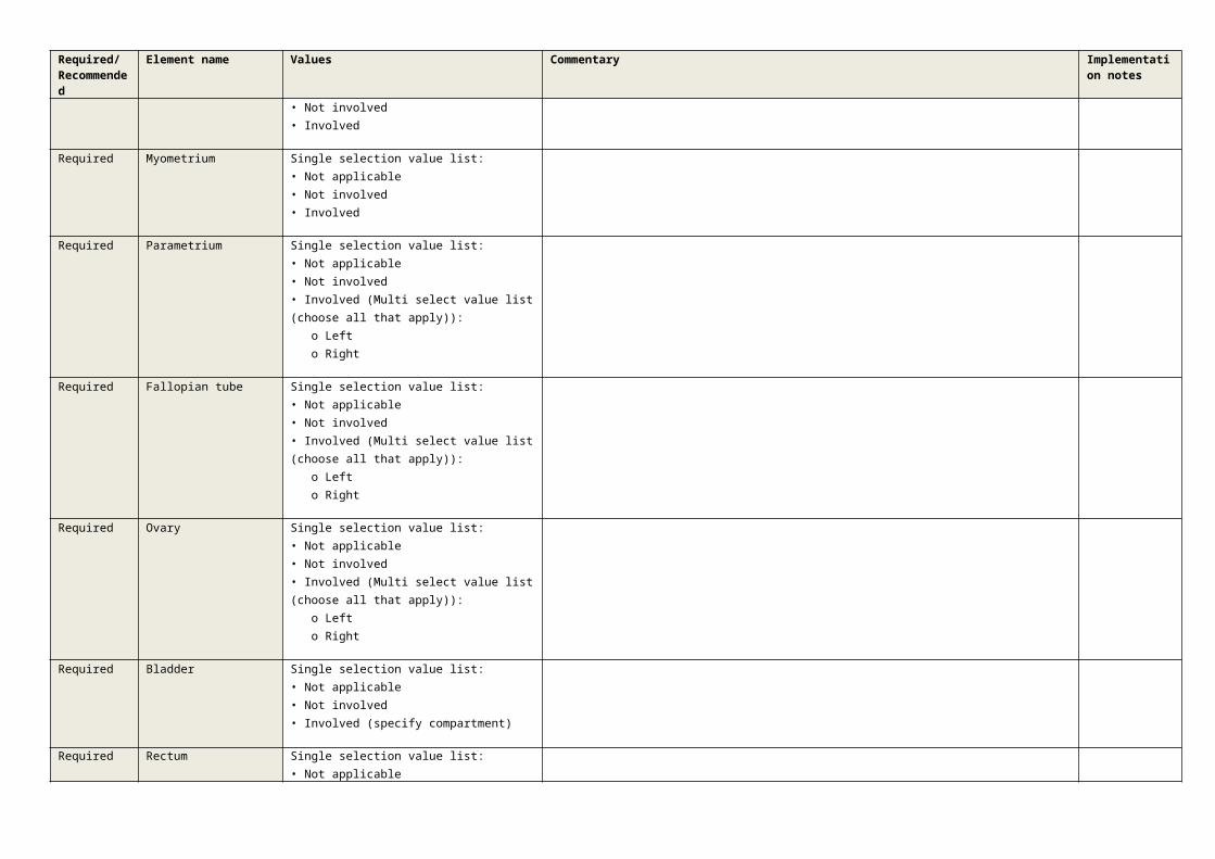
Required/ Recommended
Element name Values Commentary Implementation notes
value. However, it is stressed that there may be unexpected positive and negative staining reactions with any of the markers. HPV studies will be of value in such cases. In the distinction between a high-risk HPV-related (usual type) cervical adenocarcinoma and a high-grade endometrial adenocarcinoma, p16 and hormone receptors are often of limited value. p53 immunohistochemistry and HPV studies may be of value in this scenario. Most uterine serous carcinomas and many other high-grade endometrial carcinomas exhibit mutation-type p53 staining (“all or nothing” staining) and are HPV negative. High-risk HPV-related cervical adenocarcinomas rarely, if ever, exhibit “mutation-type” p53 expression.Immunohistochemistry of Non-HPV Related Cervical AdenocarcinomasNon-HPV related cervical adenocarcinomas have a different immunophenotype than usual HPV related adenocarcinomas. They tend to be negative or only focally positive with p16 and some, such as gastric type adenocarcinomas, may exhibit mutation-type staining with p53.4 Gastric type adenocarcinomas are usually positive with gastric markers such as MUC6 and HIK1083 and are flat negative with hormone receptors.4 There is no specific immunohistochemical marker of mesonephric adenocarcinomas but they tend to be flat negative with hormone receptors and may stain with CD10 and GATA3.5,6 Clear cell carcinomas are usually hormone receptor negative, exhibit wild-type staining with p53 and may be positive with Napsin A and hepatocyte nuclear factor 1-beta.Immunohistochemistry of Cervical Neuroendocrine CarcinomasCervical neuroendocrine carcinomas are variably positive with the neuroendocrine markers chromogranin, CD56, synaptophysin and PGP9.5. Of these, CD56 and synaptophysin are the most sensitive but CD56 lacks specificity. Chromogranin is the most specific neuroendocrine marker but lacks sensitivity with only about 50% of these neoplasms being positive.7 Chromogranin positivity is often very focal in small cell neuroendocrine carcinomas with punctate cytoplasmic immunoreactivity which is only visible on high-power magnification. A diagnosis of small cell neuroendocrine carcinoma can be made in the absence of neuroendocrine marker positivity if the morphological appearances are typical. Small cell neuroendocrine carcinoma may be only focally positive (often punctate cytoplasmic staining) or even negative with broad-spectrum cytokeratins. A diagnosis of large cell neuroendocrine carcinoma requires neuroendocrine marker positivity and most of these neoplasms are diffusely positive with broad-spectrum cytokeratins.A high percentage of primary cervical neuroendocrine carcinomas are TTF1 positive, including some with diffuse immunoreactivity, and this marker is of no value in distinction from a pulmonary metastasis.7 Most cervical neuroendocrine carcinomas are diffusely positive with p16 secondary to the presence of high-risk HPV.7 Diffuse p63 nuclear positivity is useful in confirming a small cell variant of squamous carcinoma rather than small cell neuroendocrine carcinoma. However, occasional cervical neuroendocrine carcinomas exhibit p63 nuclear immunoreactivity.7
References1 Wheeler CM, Hunt WC, Joste NE, Key CR, Quint WG and Castle PE (2009). Human papillomavirus genotype distributions: implications for vaccination and cancer screening in the United States. J Natl Cancer Inst 101(7):475-487.2 Kalof AN and Cooper K (2006). p16INK4a immunoexpression: surrogate marker of high-risk HPV and high-grade cervical intraepithelial neoplasia. Adv Anat Pathol 13(4):190-194.3 Castrillon DH, Lee KR and Nucci MR (2002). Distinction between endometrial and endocervical
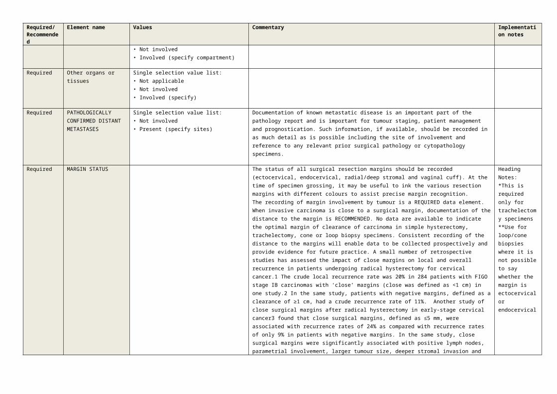
Required/ Recommended
Element name Values Commentary Implementation notes
adenocarcinoma: an immunohistochemical study. Int J Gynecol Pathol 21:4-10.4 Carleton C, Hoang L, Sah S, Kiyokawa T, Karamurzin YS, Talia KL, Park KJ and McCluggage WG (2016). A Detailed Immunohistochemical Analysis of a Large Series of Cervical and Vaginal Gastric-type Adenocarcinomas. Am J Surg Pathol 40(5):636-644.5 Howitt BE, Emori MM, Drapkin R, Gaspar C, Barletta JA, Nucci MR, McCluggage WG, Oliva E and Hirsch MS (2015). GATA3 Is a Sensitive and Specific Marker of Benign and Malignant Mesonephric Lesions in the Lower Female Genital Tract. Am J Surg Pathol 39(10):1411-1419.6 Kenny SL, McBride HA, Jamison J and McCluggage WG (2012). Mesonephric adenocarcinomas of the uterine cervix and corpus: HPV-negative neoplasms that are commonly PAX8, CA125, and HMGA2 positive and that may be immunoreactive with TTF1 and hepatocyte nuclear factor 1-beta. Am J Surg Pathol 36(6):799-807.7 McCluggage WG, Kennedy K and Busam KJ (2010). An immunohistochemical study of cervical neuroendocrine carcinomas: Neoplasms that are commonly TTF1 positive and which may express CK20 and P63. Am J Surg Pathol 34(4):525-532.
Recommended HPV testing Text, (specify details)
Recommended Immunohistochemistry Text, (specify details)
Recommended Other Text, (specify details)
Required PROVISIONAL PATHOLOGICAL STAGING PRE-MDTM
There are several difficulties inherent in the staging of carcinoma of the uterine cervix: (i) FIGO staging does not include lymph node status, (ii) there are difficulties in obtaining precise tumour measurements in low-stage disease (FIGO IA and IB), (iii) clinical staging, as recommended by FIGO, may under or overestimate true anatomical extent of disease as it does not include information obtained from post-surgical pathology specimens or radiological/surgical techniques which may not be universally available. Reliance on clinical staging tends to occur in underdeveloped or under-resourced countries where surgical facilities and ancillary investigations (such as radiology and pathology) may be limited. In developed countries, patient management tends to be based on the highest recorded anatomical extent of disease, which is based on surgical, pathological and radiological information, and not necessarily the clinical stage. Wherever possible, therefore, pathologists must provide accurate and comprehensive assessment of the pathological extent of disease to optimise patient management. The term "pathological staging” is used in this dataset to indicate that the specific staging data that are provided represent only one component of the composite staging process (which is carried out at the tumour board/multidisciplinary team meeting) and may not represent the final tumour stage, nor indeed, the clinical FIGO stage.As many cervical carcinomas are not treated surgically, the lymph node status cannot be assessed by routine histologic examination. Rather, imaging techniques (CT scan, PET scan or MRI) are commonly used to detect lymph nodes suspicious or positive for metastatic carcinoma. Since many regions in the world do not have these advanced imaging technologies, lymph node status is not included in the current FIGO staging.1 Therefore, a patient may be staged as FIGO stage I with or without lymph node metastases. The current FIGO staging does not account for significant differences in prognosis and treatment within the same stage grouping based on the presence or absence of lymph node status. Given this, it is REQUIRED that both FIGO staging and TNM pN category are included on the pathology report when the lymph node status is known. Other TNM
Heading
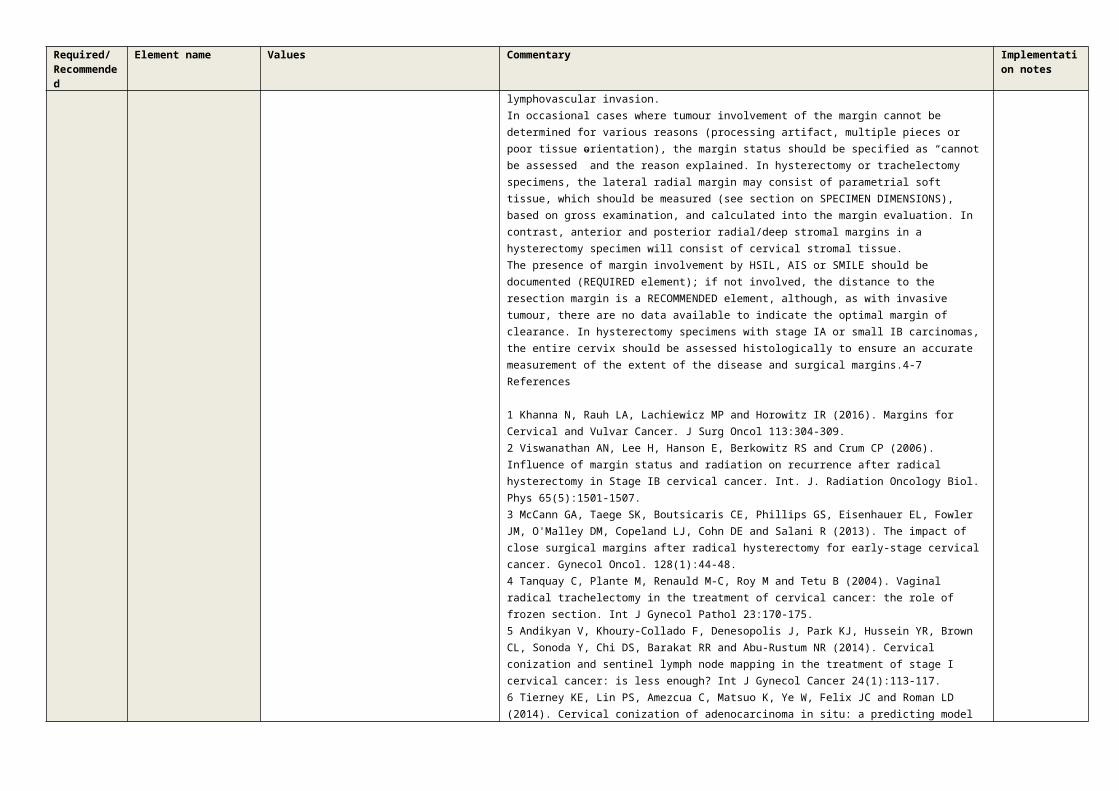
Required/ Recommended
Element name Values Commentary Implementation notes
parameters are not required elements as is the case for other gynaecological cancers where FIGO staging is a required element and TNM is not.The difficulties in obtaining precise dimensions for stage I tumours has been discussed in the TUMOUR DIMENSION section.
References1 FIGO Committee on Gynecologic Oncology (2014). FIGO staging for carcinoma of the vulva, cervix, and corpus uteri. Int J Gynaecol Obstet 125(2):97-98.
Required FIGO (2009 edition) Heading under Provisional Pathological Staging Pre-MDMTNote that permission to publish the FIGO cancer staging tables may be needed in your implementation. It is advisable to check with FIGO
Recommended & Required
TNM (UICC/AJCC 8th edition 2016)
Note that permission to publish the TNM cancer staging tables may be needed in your implementation.
Recommended Suffixes Choose if applicable:• m - multiple primary tumours • r - recurrent • y - posttreatment
Recommended Primary tumour (T) TNM 8th edition
Required Regional lymph nodes (N) TNM 8th edition
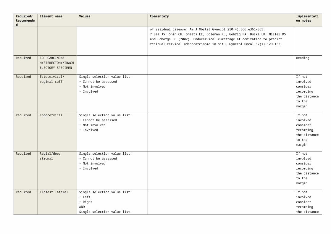
Required/ Recommended
Element name Values Commentary Implementation notes
Recommended Distant metastasis (M) TNM 8th edition or Not applicable