γ-secretase directly sheds the survival receptor BCMA ... · ARTICLE Received 27 Feb 2015 |...
12
ARTICLE Received 27 Feb 2015 | Accepted 28 Apr 2015 | Published 11 Jun 2015 g-secretase directly sheds the survival receptor BCMA from plasma cells Sarah A. Laurent 1 , Franziska S. Hoffmann 1, *, Peer-Hendrik Kuhn 2,3, *, Qingyu Cheng 4 , Yuanyuan Chu 5 , Marc Schmidt-Supprian 5 , Stefanie M. Hauck 6 , Elisabeth Schuh 1 , Markus Krumbholz 1 , Heike Ru ¨bsamen 1 , Johanna Wanngren 2,3 , Mohsen Khademi 7 , Tomas Olsson 7 , Tobias Alexander 4 , Falk Hiepe 4 , Hans-Walter Pfister 8 , Frank Weber 9 , Dieter Jenne 10 , Hartmut Wekerle 11,12 , Reinhard Hohlfeld 1,12 , Stefan F. Lichtenthaler 2,3,12 & Edgar Meinl 1 Survival of plasma cells is regulated by B-cell maturation antigen (BCMA), a membrane-bound receptor activated by its agonist ligands BAFF and APRIL. Here we report that g-secretase directly cleaves BCMA, without prior truncation by another protease. This direct shedding is facilitated by the short length of BCMA’s extracellular domain. In vitro, g-secretase reduces BCMA-mediated NF-kB activation. In addition, g-secretase releases soluble BCMA (sBCMA) that acts as a decoy neutralizing APRIL. In vivo, inhibition of g-secretase enhances BCMA surface expression in plasma cells and increases their number in the bone marrow. Furthermore, in multiple sclerosis, sBCMA levels in spinal fluid are elevated and associated with intracerebral IgG production; in systemic lupus erythematosus, sBCMA levels in serum are elevated and correlate with disease activity. Together, shedding of BCMA by g-secretase controls plasma cells in the bone marrow and yields a potential biomarker for B-cell involvement in human autoimmune diseases. DOI: 10.1038/ncomms8333 OPEN 1 Institute of Clinical Neuroimmunology, Ludwig Maximilian University Munich, 81377 Munich, Germany. 2 Neuroproteomics, Klinikum rechts der Isar, and Institute of Advanced Study, Technische Universita ¨t Mu ¨nchen, 81377 Munich, Germany. 3 German Center for Neurodegenerative Diseases (DZNE), 81377 Munich, Germany. 4 Department of Rheumatology and Clinical Immunology, Charite ´ - Universita ¨tsmedizin Berlin and Deutsches Rheuma-Forschungszentrum Berlin-a Leibniz Institute, 10117 Berlin, Germany. 5 Department of Internal Medicine III, Klinikum Rechts der Isar, Technische Universita ¨t Mu ¨nchen, 81675 Munich, Germany. 6 Research Unit Protein Science, Helmholtz Zentrum Mu ¨nchen (GmbH), German Research Center for Environmental Health, 85764 Neuherberg, Germany. 7 Karolinska University Hospital, Division of Clinical Neuroscience, 17176 Stockholm, Sweden. 8 Department of Neurology, Klinikum Grosshadern, Ludwig Maximilian University Munich, 81377 Munich, Germany. 9 Max-Planck-Institute of Psychiatry, 80804 Munich, Germany. 10 CPC Helmholtz Zentrum Mu ¨nchen (GmbH), 81377 Munich, Germany. 11 Max-Planck-Institute of Neurobiology, 82152 Martinsried, Germany. 12 Munich Cluster for Systems Neurology (SyNergy), 81377 Munich, Germany. *These authors contributed equally to this work. Correspondence and requests for materials should be addressed to E.M. (email: [email protected]). NATURE COMMUNICATIONS | 6:7333 | DOI: 10.1038/ncomms8333 | www.nature.com/naturecommunications 1 & 2015 Macmillan Publishers Limited. All rights reserved.
Transcript of γ-secretase directly sheds the survival receptor BCMA ... · ARTICLE Received 27 Feb 2015 |...

ARTICLE
Received 27 Feb 2015 | Accepted 28 Apr 2015 | Published 11 Jun 2015
g-secretase directly sheds the survival receptorBCMA from plasma cellsSarah A. Laurent1, Franziska S. Hoffmann1,*, Peer-Hendrik Kuhn2,3,*, Qingyu Cheng4, Yuanyuan Chu5,
Marc Schmidt-Supprian5, Stefanie M. Hauck6, Elisabeth Schuh1, Markus Krumbholz1, Heike Rubsamen1,
Johanna Wanngren2,3, Mohsen Khademi7, Tomas Olsson7, Tobias Alexander4, Falk Hiepe4, Hans-Walter Pfister8,
Frank Weber9, Dieter Jenne10, Hartmut Wekerle11,12, Reinhard Hohlfeld1,12, Stefan F. Lichtenthaler2,3,12
& Edgar Meinl1
Survival of plasma cells is regulated by B-cell maturation antigen (BCMA), a membrane-bound
receptor activated by its agonist ligands BAFF and APRIL. Here we report that g-secretase
directly cleaves BCMA, without prior truncation by another protease. This direct shedding is
facilitated by the short length of BCMA’s extracellular domain. In vitro, g-secretase reduces
BCMA-mediated NF-kB activation. In addition, g-secretase releases soluble BCMA (sBCMA)
that acts as a decoy neutralizing APRIL. In vivo, inhibition of g-secretase enhances BCMA
surface expression in plasma cells and increases their number in the bone marrow.
Furthermore, in multiple sclerosis, sBCMA levels in spinal fluid are elevated and associated with
intracerebral IgG production; in systemic lupus erythematosus, sBCMA levels in serum are
elevated and correlate with disease activity. Together, shedding of BCMA by g-secretase
controls plasma cells in the bone marrow and yields a potential biomarker for B-cell
involvement in human autoimmune diseases.
DOI: 10.1038/ncomms8333 OPEN
1 Institute of Clinical Neuroimmunology, Ludwig Maximilian University Munich, 81377 Munich, Germany. 2 Neuroproteomics, Klinikum rechts der Isar, andInstitute of Advanced Study, Technische Universitat Munchen, 81377 Munich, Germany. 3 German Center for Neurodegenerative Diseases (DZNE), 81377Munich, Germany. 4 Department of Rheumatology and Clinical Immunology, Charite - Universitatsmedizin Berlin and Deutsches Rheuma-ForschungszentrumBerlin-a Leibniz Institute, 10117 Berlin, Germany. 5 Department of Internal Medicine III, Klinikum Rechts der Isar, Technische Universitat Munchen, 81675Munich, Germany. 6 Research Unit Protein Science, Helmholtz Zentrum Munchen (GmbH), German Research Center for Environmental Health, 85764Neuherberg, Germany. 7 Karolinska University Hospital, Division of Clinical Neuroscience, 17176 Stockholm, Sweden. 8 Department of Neurology, KlinikumGrosshadern, Ludwig Maximilian University Munich, 81377 Munich, Germany. 9 Max-Planck-Institute of Psychiatry, 80804 Munich, Germany. 10 CPCHelmholtz Zentrum Munchen (GmbH), 81377 Munich, Germany. 11 Max-Planck-Institute of Neurobiology, 82152 Martinsried, Germany. 12 Munich Cluster forSystems Neurology (SyNergy), 81377 Munich, Germany. * These authors contributed equally to this work. Correspondence and requests for materials shouldbe addressed to E.M. (email: [email protected]).
NATURE COMMUNICATIONS | 6:7333 | DOI: 10.1038/ncomms8333 | www.nature.com/naturecommunications 1
& 2015 Macmillan Publishers Limited. All rights reserved.

Bcells are of pathogenic relevance and serve as a therapeutic
target both in generalized immunopathological diseasessuch as systemic lupus erythematosus (SLE), and in
organ-specific diseases such as multiple sclerosis (MS)1,2.Activation and survival of B cells is largely regulated via theBAFF–APRIL system that comprises three receptors (BAFF-R,TACI and BCMA (B-cell maturation antigen)) and two ligands(BAFF and APRIL)3. Membrane-bound BCMA (mBCMA) isexpressed on some activated B cells4 and Ig-secreting cells5,6; itbinds both BAFF and APRIL3. BCMA is essential for themaintenance of long-lived plasma cells7,8, an effect mediated byAPRIL or BAFF9,10. These plasma cells produce IgG that protectnot only against pathogens but are also critically involved inautoimmune diseases11,12. Further, mBCMA engagement onactivated B cells induces MHC class II, enhancing their ability topresent antigen4.
The BAFF–APRIL system is targeted for therapeutic interven-tion. In SLE, where BAFF levels in the serum are elevated, anantibody-binding BAFF is already approved13,14. On the otherhand, the recombinant soluble receptor atacicept, which targetsboth BAFF and APRIL, unexpectedly worsened MS15, indicatingthat essential features of this system are not fully understood.Other clinical trials targeting the BAFF–APRIL system inimmunopathological disorders have been launched13,14.
We aimed at identifying features of humoral immunity thatmight be altered in autoimmune diseases. Thereby, we found thata soluble form of BCMA (sBCMA) is regularly detectable inhuman blood. We then analysed sBCMA in human autoimmunediseases. In MS, sBCMA was elevated in the cerebrospinalfluid (CSF) and linked to local IgG production inside the brain.In SLE, sBCMA was systemically elevated and associated withdisease activity. We went on to uncover the underlyingbiochemical mechanism and found that BCMA was directly shedby g-secretase, a ubiquitous intramembranous protease. Directshedding of a membrane protein without prior processing byanother protease is a novel function of g-secretase, which is bestknown for processing of amyloid precursor protein (APP) inAlzheimer’s disease and Notch16,17. To analyse the functionalrelevance of this shedding in vivo, we treated mice with ag-secretase inhibitor; this enhanced surface BCMA on plasmacells and increased their number in the bone marrow. Thus,shedding of BCMA via g-secretase is an immunoregulatorymechanism limiting plasma cells.
ResultssBCMA shows local IgG production in MS and activity in SLE.We found sBCMA as a regular component of human blood fromhealthy subjects (Fig. 1a). In MS, sBCMA was elevated in the CSF,but not in the blood (Fig. 1a,b). sBCMA levels in the plasma andserum were very similar as seen in 14 controls (SupplementaryFig. 1a). Levels of sBCMA in the CSF correlated strongly withlocal IgG production in MS patients (Fig. 1c). This strongcorrelation was confirmed in a second cohort of 25 MS patients(Po0.0001, r¼ 0.80, Spearman rank correlation). Also inneuroborreliosis, an infectious neuroinflammatory disease char-acterized by chronic IgG production within the CNS, sBCMAlevels were increased and correlated with local IgG production(Fig. 1b,c). In CSF from MS patients, there was a slight inversecorrelation of sBCMA with its high-affinity ligand APRIL(r¼ � 0.35, P¼ 0.033, Spearman rank correlation), butno association to the plasma level of sBCMA (r¼ 0.07,P¼ 0.667, Spearman rank correlation). We analysed effects ofimmunosuppressive treatment on sBCMA levels longitudinally intwo cohorts of MS patients. Natalizumab, which blocks entry oflymphocytes into the CNS18 and local IgG production2, reduced
sBCMA in the CSF (Supplementary Fig. 1b). High-dose steroidsfor treatment of acute relapses reduced serum sBCMA levelstransiently (Supplementary Fig.1c).
In SLE, serum levels of sBCMA were elevated as seen in anuntreated and in a treated cohort (Fig. 1d). Immunosuppressivetreatment of SLE patients reduced sBCMA levels (Fig. 1d). Wenoted a strong correlation of serum sBCMA and disease activity(Fig. 1e) and an inverse correlation with the paraclinical markercomplement factor 3 (Fig. 1f). There was a trend (P¼ 0.0767,r¼ 0.23, Spearman rank correlation) for correlation betweenanti-dsDNA titre and sBCMA in these SLE patients. Serum BAFFlevels were elevated in our SLE cohort (mean¼ 0.61±0.13 ng ml� 1
in healthy controls; mean¼ 2.11±4.32 ng ml� 1 in SLE patients,P¼ 0.0004, Wilcoxon–Mann–Whithney test), and correlated withsBCMA levels (P¼ 0.0013, r¼ 0.47, Spearman rank correlation).
sBC
MA
(ng
ml–
1 )
IgG
inde
x
0.0 0.5 1.0 1.5 2.00
1
2
3
4
0
0.5
1.0
1.5
2.0***
***
sBC
MA
(ng
ml–
1 )
sBC
MA
(ng
ml–
1 )
SLE
DA
I
0 50 100 1500
10
20
30
40
sBCMA (ng ml–1)
C3
(mg
dl–1
)
0 50 100 1500
50
100
150
200
sBCMA (ng ml–1)
sBCMA (ng ml–1)
****
*
0
50
100
150
0
10
20
30
40
HCn=26
CIS/MSn=37
ONDn=20
NBn=5
CIS/MSn=37
ONDn=20
HCn=29
SLEn=17
SLEn=22
Figure 1 | sBCMA as a biomarker. (a) sBCMA plasma concentrations
were determined using ELISA in healthy controls (HC), patients with a
clinically isolated syndrome (CIS) or MS, or other neurological diseases
(OND). (b) sBCMA in the CSF was determined in patients with OND,
CIS/MS or neuroborreliosis (NB) (***Po0.001, Kruskal–Wallis test
followed by Dunn’s Multiple Comparison Test). (c) sBCMA in the CSF
correlated strongly with the intrathecal IgG production represented by the
IgG Index. This correlation was evident when all analysed CSF samples were
considered (Po0.0001, r¼0.85) and in the MS/CIS group (Po0.0001,
r¼0.77, Spearman rank correlation, CIS/MS n¼ 36; OND n¼ 20,
NB n¼ 5). (d) sBCMA in the serum was determined with ELISA in HC,
untreated (red) and treated (black) patients with SLE. sBCMA was elevated
in SLE patients and in the untreated SLE patients compared with the treated
patients (***Po0.001 and *Po0.05, Kruskal–Wallis test followed by
Dunn’s Multiple Comparison Test). (e) sBCMA in the serum of SLE patients
correlated strongly with disease activity quantified with SLE disease activity
index (SLEDAI; Po0.001; r¼0.54, Spearman correlation). (f) sBCMA in
the serum of SLE patients inversely correlated with the level of the
complement factor C3 (P¼0.0374, r¼ �0.29, Spearman correlation).
Bars represent means.
ARTICLE NATURE COMMUNICATIONS | DOI: 10.1038/ncomms8333
2 NATURE COMMUNICATIONS | 6:7333 | DOI: 10.1038/ncomms8333 | www.nature.com/naturecommunications
& 2015 Macmillan Publishers Limited. All rights reserved.

BCMA is shed during differentiation to Ig-secreting cells. SincemBCMA is known to be expressed on some activated B cells andIg-secreting cells5,6, we analysed the release of sBCMA byprimary human B cells. We applied two different protocols toactivate human primary B cells in order to differentiate themtowards Ig-secreting cells. First, purified blood-derived humanB cells were activated via CD40L and further differentiated toIg-secreting cells by adding interleukin (IL)-21 (ref. 5). Whilehuman B cells activated via CD40L alone released low, butdetectable, levels of sBCMA, this was strongly enhanced byaddition of IL-21 (Fig. 2a). In the second primary B-cell culturesystem we activated peripheral blood mononuclear cells (PBMC)with the TLR7þ 8 ligand R848 and IL-2, which inducesdifferentiation of human memory B cells to Ig-secreting cells19.
Again, differentiation towards IgG-secreting cells wasaccompanied by the appearance of sBCMA (Fig. 2b).
After activation with CD40Lþ IL-21, two B-cell populations,CD19þCD38� and CD19þCD38þ , cells could be distin-guished. We directly compared mBCMA and release of sBCMAby these different B-cell subsets. While mBCMA was absent onunstimulated B cells, CD19þCD38� cells weakly and CD19þ
CD38þ cells strongly expressed mBCMA (Fig. 2c). We sortedCD38þ and CD38� cells, cultured them for another 24 hwithout further stimulation and determined the amount of shedsBCMA (Fig. 2d). This revealed a close correlation betweenreleased sBCMA and surface expression of mBCMA (Fig. 2e).
The transcription of BCMA in these B-cell subsets was furthersubstantiated with qPCR. BCMA transcript levels in the CD38þ
4.6
10
15
1 2 3
kDa
4
3.5
10
2030
4050
kDa
sBCMA
IgH
IgL
0.0
0.5
1.0
1.5
2.0
2.5
3.0
3.5
0
1
2
3
4
5
6
7
R848IL-2
Vehicle0
2
4
6
8
10
12
14
0
1
2
3
4
5
6
+CD40L +CD40LIL-21
IgGsBCMA
IgGsBCMA
sBC
MA
(ng
ml–1
)
0 50 100 150 200 2500
2
4
6
8
BCMA surface expression (MFI)
sBC
MA
(ng
/ml)
0
2
4
6
8
No stimulation CD40L+IL-21
0
20
40
60
80
100
% o
f Max
0 102 103 104 105 0 102 103 104 105 0 102 103 104 1050
20
40
60
80
100
% o
f Max
0
20
40
60
80
100
% o
f Max
BCMA
CD40L+IL-21 stimulation
CD19+ CD38– CD38+
Vehicle
Extracellular domain Transmembranous domain
IgG
(μg
ml–1
)
IgG
(μg
ml–1
)
sBC
MA
(ng
ml–1
)
sBC
MA
(ng
ml–1
)CD19+ CD19+CD38– CD19+CD38+
No stimulation
Nt-MLQMA GQCSQ NEYFD SLLHA CIPCQ LRCSS NTPPL TCQRY CNASV TNSVK GTNAI LWTCL GLSLI ISLAV FVLMF LLRKI…
Figure 2 | sBCMA is released when B cells differentiate towards plasma cells and comprises the extracellular domain plus part of the
transmembranous region of BCMA. (a) Human purified B cells were activated for 5 days as indicated; IgG and sBCMA in the supernatant were quantified
using ELISA. Combined data of three independent experiments (mean±s.e.m., P¼0.0073, paired t-test). (b) PBMCs were stimulated with R848þ IL-2 for
7 days. IgG and sBCMA in the supernatant were quantified using ELISA. Combined data of three independent experiments (mean±s.e.m., P¼0.0227,
paired t-test). (c–e) Human purified B cells were stimulated with CD40Lþ IL-21. (c) surface BCMA was measured using flow cytometry on unstimulated B
cells, CD19þCD38� cells and CD19þCD38þ cells. (d) Sorted CD38þ and CD38� cells were cultured for another 24 h and the amount of shed sBCMA
was measured using ELISA, combined data of two independent experiments. (e) Correlation between sBCMA release and surface expression of BCMA for
a single replicate. (f,g) sBCMA was immunoprecipitated from supernatant of plasmacytoma cells (f, lanes 1, 2), serum (f, lane 3) and from supernatant of
human purified B cells cultured with CD40L plus IL-21 (f, lane 4) with anti-BCMA monoclonal antibodies (mAbs) A7D12.2 (f, lanes 1 and 4) or C12A3.2
(f, lane 2) or goat-anti-BCMA (f, lane 3). Western blot analysis for BCMA (f) and silver staining of sBCMA immunoprecipitated from plasmacytoma
supernatant (g) was performed. (h) The band at 6 kDa (from g) and sBCMA obtained using immunoprecipitation were analysed with mass spectrometry.
The aa sequences of BCMA and peptides identified after tryptic (blue) or chymotryptic (red) digestion are shown. No peptide was detected with a
C-terminal aa that was not a site for either tryptic or chymotryptic cleavage, indicating that the precise cleavage site of g-secretase within the membrane
needs to be identified.
NATURE COMMUNICATIONS | DOI: 10.1038/ncomms8333 ARTICLE
NATURE COMMUNICATIONS | 6:7333 | DOI: 10.1038/ncomms8333 | www.nature.com/naturecommunications 3
& 2015 Macmillan Publishers Limited. All rights reserved.

cells reached 15.9±5.2% peptidyl-prolyl isomerase A (PPIA),while BCMA expression in the CD38� cells subset accounted for2.5±1.3% PPIA (mean±s.e.m. of three independent replicates).Thus, in these B-cell subsets BCMA surface expression reflectedBCMA transcription.
We also analysed BCMA shedding in tumour cell lines andtransfectants. The human plasmacytoma cell line JK-6L sponta-neously shed sBCMA (Supplementary Fig. 2a). In BCMA-transfected HeLa cells surface expression of mBCMA wasaccompanied by release of sBCMA (157±6 ng ml� 1) withoutrequiring any further stimulus. HeLa cells did not secretedetectable amounts of APRIL or BAFF, neither spontaneouslynor after transfection with BCMA or an empty vector. Together,our observations with primary human B-cell cultures, plasmacy-toma cells and BCMA-transfected cells indicate that release ofsBCMA is a direct consequence of surface expression of mBCMA;it does not require additional stimulation or ligand binding.
sBCMA comprises extracellular and intramembranous part.sBCMA was isolated by immunoprecipitation from the super-natant of primary Ig-secreting cells, plasmacytoma cells or serum;in all these sources, sBCMA had a molecular weight (MW) ofB6 kDa as seen using western blot analysis (Fig. 2f). This size wasconfirmed when silver staining was applied to detect materialobtained by immunoprecipitation with anti-BCMA from thesupernatant of plasmacytoma cells (Fig. 2g). This corresponds tothe extracellular part of BCMA (54 amino acid (aa), calculatedMW 5.8 kDa). Unexpectedly, mass spectrometry revealed thatsBCMA comprised not only the complete extracellular domainwith an intact N terminus, but also part of the transmembraneregion (Fig. 2h). This indicated that it was released by an intra-membranous protease.
c-secretase inhibitors block BCMA shedding from B cells. SincemBCMA is a type-I oriented transmembrane protein with anextracellular N terminus, g-secretase was a candidate for itsintramembranous cleavage. We applied the g-secretase inhibitorDAPT and compared it with the metalloprotease inhibitor TAPI-1, which reduces the shedding of other TNFR-SF members. Weactivated human B cells either via CD40Lþ IL-21 (Fig. 3a,b) orvia R848þ IL-2 (Fig. 3c,d), and used both fluorescence-activatedcell sorting (FACS) and enzyme-linked immunosorbent assay(ELISA) as read-out systems to quantify mBCMA and sBCMA.DAPT blocked the release of sBCMA even at low concentrations,while TAPI-1 had little or no effect (Fig. 3a,c). After CD40Lþ IL-21 application, a high surface expression of mBCMA was noted inthe CD27þ þCD38þ subset (Fig. 3b), previously classified as lateplasmablasts20. DAPT enhanced surface expression of mBCMAin these cells, while TAPI-I had little or no effect (Fig. 3b). Whenhuman PBMCs were activated with R848þ IL-2, B20% of thecells were CD19þCD38þ after 7 days (Fig. 3d). These cellsstrongly expressed mBCMA on their surface and this was greatlyenhanced by the g-secretase inhibitor DAPT; again, TAPI-I hadlittle effect (Fig. 3d). Similar to primary human B cells, weobserved differential effects of DAPT and TAPI-1 on the releaseof sBCMA and surface expression of mBCMA on humanplasmacytoma cells (Supplementary Fig. 2a,b).
Further, we compared the effect of transition (LY-411575-I andLY685,458) and non-transition state (DAPT and RO4929097)inhibitors of the g-secretase on BCMA shedding from human Bcells. Human PBMCs were first stimulated with R848þ IL2 for 7days, and then CD19þ B cells were positively selected andcultured overnight in the absence of these g-secretase inhibitors.We found that RO4929097, LY-411575-I and LY685,458 hadsimilar effects as DAPT on the shedding of mBCMA as seen withboth read-out systems, FACS and ELISA (Supplementary Fig. 3).
c-secretase directly sheds BCMA. Presenilin (PS)1 or PS2 is thecatalytical component of the g-secretase complex14,15. To finallyprove that sBCMA is released by g-secretase, we switched from Bcells to mouse embryonic fibroblasts (MEF) deficient for both PS1and PS2 (PS� /� )21. These MEF cells were transduced with
DAPT (μM)
sBC
MA
rel
ease
(%
)
Vehicle 0.01 0.03 0.1 0.3 10
20
40
60
80
100
Vehicle 1 3 10 25 500
20
40
60
80
100
TAPI-1 (μM)
sBC
MA
rel
ease
(%
)
0
20
40
60
80
100%
Of m
ax
0
20
40
60
80
100
% O
f max
CD19
CD
38
BCMA
VehicleDAPT
VehicleTAPI-1
CD27
CD
38
0
0102
102
103
103
104
104
105
105 0 102 103 104 1050
20
40
60
80
100
% O
f max
0
20
40
60
80
100
% O
f max
Isotype control Isotype control
Vehicle 0.01 0.03 0.1 0.3 10
20
40
60
80
100
120
DAPT (μM)
Vehicle 1 3 10 25 500
20
40
60
80
100
120
sBC
MA
rel
ease
(%
)
sBC
MA
rel
ease
(%
)
TAPI-1 (μM)
BCMA
VehicleDAPT Isotype control
VehicleTAPI-1Isotype control
0 102 103 104 105
0102
103
104
105
0 102 103 104 105 0 102 103 104 105 0 102 103 104 105
Figure 3 | c-secretase inhibitor DAPT reduces release of sBCMA and
enhances surface expression of BCMA on activated human B cell.
(a,b) Human B cells were differentiated into Ig-secreting cells via
CD40Lþ IL-21. (a) Release of sBCMA on treatment with DAPT or TAPI-1
was measured using ELISA. sBCMA release was normalized to the amount
of sBCMA shed under vehicle conditions, which was set as 100%.
Combined data of three independent experiments (mean±s.e.m.).
(b) These activated primary human B cells were subgrouped on the basis of
expression of CD27 and CD38. A high surface expression of BCMA was
seen on the CD27þ þCD38þ subset. Surface expression of BCMA was
enhanced using DAPT treatment (1mM), while TAPI-I (50mM) had little
effect. (c,d) Human PBMCs were stimulated with R848þ IL-2 for 7 days.
(c) Release of sBCMA on treatment with DAPT or TAPI-1 was measured
using ELISA. sBCMA release was normalized to the amount of sBCMA shed
under vehicle conditions, which was set as 100%. Combined data of three
independent experiments (mean±s.e.m.). (d) High surface expression of
BCMA was seen on the CD19þCD38þ subset; this was further enhanced
by DAPT (1mM), while TAPI-I (50mM) had little effect.
ARTICLE NATURE COMMUNICATIONS | DOI: 10.1038/ncomms8333
4 NATURE COMMUNICATIONS | 6:7333 | DOI: 10.1038/ncomms8333 | www.nature.com/naturecommunications
& 2015 Macmillan Publishers Limited. All rights reserved.
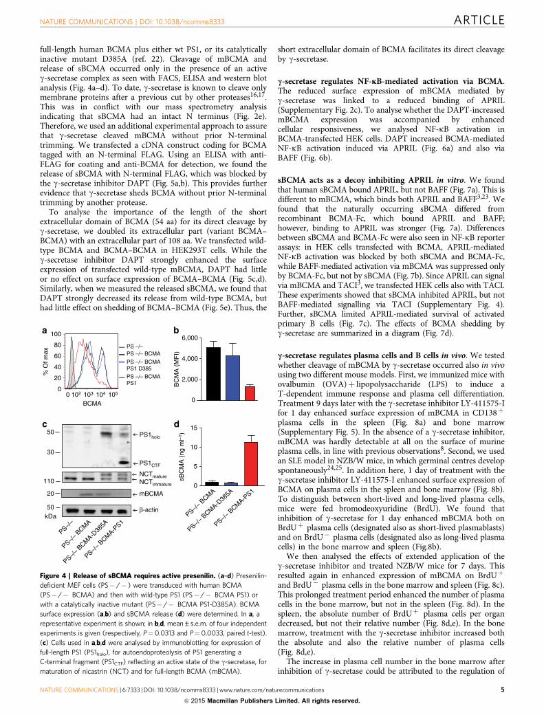
full-length human BCMA plus either wt PS1, or its catalyticallyinactive mutant D385A (ref. 22). Cleavage of mBCMA andrelease of sBCMA occurred only in the presence of an activeg-secretase complex as seen with FACS, ELISA and western blotanalysis (Fig. 4a–d). To date, g-secretase is known to cleave onlymembrane proteins after a previous cut by other proteases16,17.This was in conflict with our mass spectrometry analysisindicating that sBCMA had an intact N terminus (Fig. 2e).Therefore, we used an additional experimental approach to assurethat g-secretase cleaved mBCMA without prior N-terminaltrimming. We transfected a cDNA construct coding for BCMAtagged with an N-terminal FLAG. Using an ELISA with anti-FLAG for coating and anti-BCMA for detection, we found therelease of sBCMA with N-terminal FLAG, which was blocked bythe g-secretase inhibitor DAPT (Fig. 5a,b). This provides furtherevidence that g-secretase sheds BCMA without prior N-terminaltrimming by another protease.
To analyse the importance of the length of the shortextracellular domain of BCMA (54 aa) for its direct cleavage byg-secretase, we doubled its extracellular part (variant BCMA–BCMA) with an extracellular part of 108 aa. We transfected wild-type BCMA and BCMA–BCMA in HEK293T cells. While theg-secretase inhibitor DAPT strongly enhanced the surfaceexpression of transfected wild-type mBCMA, DAPT had littleor no effect on surface expression of BCMA–BCMA (Fig. 5c,d).Similarly, when we measured the released sBCMA, we found thatDAPT strongly decreased its release from wild-type BCMA, buthad little effect on shedding of BCMA–BCMA (Fig. 5e). Thus, the
short extracellular domain of BCMA facilitates its direct cleavageby g-secretase.
c-secretase regulates NF-jB-mediated activation via BCMA.The reduced surface expression of mBCMA mediated byg-secretase was linked to a reduced binding of APRIL(Supplementary Fig. 2c). To analyse whether the DAPT-increasedmBCMA expression was accompanied by enhancedcellular responsiveness, we analysed NF-kB activation inBCMA-transfected HEK cells. DAPT increased BCMA-mediatedNF-kB activation induced via APRIL (Fig. 6a) and also viaBAFF (Fig. 6b).
sBCMA acts as a decoy inhibiting APRIL in vitro. We foundthat human sBCMA bound APRIL, but not BAFF (Fig. 7a). This isdifferent to mBCMA, which binds both APRIL and BAFF3,23. Wefound that the naturally occurring sBCMA differed fromrecombinant BCMA-Fc, which bound APRIL and BAFF;however, binding to APRIL was stronger (Fig. 7a). Differencesbetween sBCMA and BCMA-Fc were also seen in NF-kB reporterassays: in HEK cells transfected with BCMA, APRIL-mediatedNF-kB activation was blocked by both sBCMA and BCMA-Fc,while BAFF-mediated activation via mBCMA was suppressed onlyby BCMA-Fc, but not by sBCMA (Fig. 7b). Since APRIL can signalvia mBCMA and TACI3, we transfected HEK cells also with TACI.These experiments showed that sBCMA inhibited APRIL, but notBAFF-mediated signalling via TACI (Supplementary Fig. 4).Further, sBCMA limited APRIL-mediated survival of activatedprimary B cells (Fig. 7c). The effects of BCMA shedding byg-secretase are summarized in a diagram (Fig. 7d).
c-secretase regulates plasma cells and B cells in vivo. We testedwhether cleavage of mBCMA by g-secretase occurred also in vivousing two different mouse models. First, we immunized mice withovalbumin (OVA)þ lipopolysaccharide (LPS) to induce aT-dependent immune response and plasma cell differentiation.Treatment 9 days later with the g-secretase inhibitor LY-411575-Ifor 1 day enhanced surface expression of mBCMA in CD138þ
plasma cells in the spleen (Fig. 8a) and bone marrow(Supplementary Fig. 5). In the absence of a g-secretase inhibitor,mBCMA was hardly detectable at all on the surface of murineplasma cells, in line with previous observations8. Second, we usedan SLE model in NZB/W mice, in which germinal centres developspontaneously24,25. In addition here, 1 day of treatment with theg-secretase inhibitor LY-411575-I enhanced surface expression ofBCMA on plasma cells in the spleen and bone marrow (Fig. 8b).To distinguish between short-lived and long-lived plasma cells,mice were fed bromodeoxyuridine (BrdU). We found thatinhibition of g-secretase for 1 day enhanced mBCMA both onBrdUþ plasma cells (designated also as short-lived plasmablasts)and on BrdU� plasma cells (designated also as long-lived plasmacells) in the bone marrow and spleen (Fig.8b).
We then analysed the effects of extended application of theg-secretase inhibitor and treated NZB/W mice for 7 days. Thisresulted again in enhanced expression of mBCMA on BrdUþ
and BrdU� plasma cells in the bone marrow and spleen (Fig. 8c).This prolonged treatment period enhanced the number of plasmacells in the bone marrow, but not in the spleen (Fig. 8d). In thespleen, the absolute number of BrdUþ plasma cells per organdecreased, but not their relative number (Fig. 8d,e). In the bonemarrow, treatment with the g-secretase inhibitor increased boththe absolute and also the relative number of plasma cells(Fig. 8d,e).
The increase in plasma cell number in the bone marrow afterinhibition of g-secretase could be attributed to the regulation of
BC
MA
(M
FI)
0 1051041031020
20
40
60
80
100
% O
f max
PS –/– BCMAPS1 D385
PS –/– BCMAPS1
PS –/– BCMAPS –/–
PS–/– B
CMA
PS–/– B
CMA-D38
5A
PS–/– B
CMA-PS1
PS–/–
PS–/–
BCMA
PS–/–
BCMA-D
385A
PS–/–
BCMA-P
S1
NCTmatureNCTimmature
30
50
kDa
110
β-actin50
BCMA
0
5
0
20 mBCMA
6,000
4,000
2,000
sBC
MA
(ng
ml–
1 )
15
10
PS1holo
PS1CTF
Figure 4 | Release of sBCMA requires active presenilin. (a–d) Presenilin-
deficient MEF cells (PS� /� ) were transduced with human BCMA
(PS� /� BCMA) and then with wild-type PS1 (PS� /� BCMA PS1) or
with a catalytically inactive mutant (PS� /� BCMA PS1-D385A). BCMA
surface expression (a,b) and sBCMA release (d) were determined. In a, a
representative experiment is shown; in b,d, mean±s.e.m. of four independent
experiments is given (respectively, P¼0.0313 and P¼0.0033, paired t-test).
(c) Cells used in a,b,d were analysed by immunoblotting for expression of
full-length PS1 (PS1holo), for autoendoproteolysis of PS1 generating a
C-terminal fragment (PS1CTF) reflecting an active state of the g-secretase, for
maturation of nicastrin (NCT) and for full-length BCMA (mBCMA).
NATURE COMMUNICATIONS | DOI: 10.1038/ncomms8333 ARTICLE
NATURE COMMUNICATIONS | 6:7333 | DOI: 10.1038/ncomms8333 | www.nature.com/naturecommunications 5
& 2015 Macmillan Publishers Limited. All rights reserved.

mBCMA on plasma cells (Fig. 8, Supplementary Fig. 4) or ondecoy effects of sBCMA, which we observed in vitro (Fig. 7b,c).To analyse possible immunoregulatory effects of the shed sBCMAin vivo, we produced recombinant sBCMA and applied itsystemically. We prepared sBCMA as a fusion protein witha1-antitrypsin (AT; Supplementary Fig. 6a) to increase itsmolecular weight and therefore its half live in vivo.The monomeric structure of sBCMA-AT was confirmed usinggel filtration (Supplementary Fig. 6b). Having shown thatrecombinant sBCMA-AT blocked APRIL-mediated NF-kB
activation in vitro (Supplementary Fig. 6c), we gave sBCMA-ATthree times a week for 4 weeks to NZB/W mice, which hadbeen immunized with OVA before. The applied amount ofsBCMA-AT corresponded to the dose previously used forBCMA-Fc26,27. Nevertheless, under these conditions sBCMA-AT did not modulate the total number of plasma cells, thenumber of OVA-specific plasma cells or the amount ofcirculating IgG, IgA and IgM (Supplementary Fig. 6d–f).
Notably, inhibition of g-secretase had additional effects onB cells not expressing BCMA. Short-term treatment withthe g-secretase inhibitor reduced GC B cells (defined asCD38intFashigh) in the spleen as seen in our immunization model(Supplementary Fig. 7a,b). Moreover, after 7 days of treatment,NZB/W mice had fewer B cells in the spleen and bone marrowand showed a striking reduction of pre-B cells in their bonemarrow (Supplementary Fig. 7c).
DiscussionWe report that g-secretase directly sheds mBCMA and regulatesthe number of plasma cells in the bone marrow. Further, thereleased sBCMA reflects B-cell activation in human autoimmu-nity, namely compartmentalized Ig production in MS and diseaseactivity in SLE.
When B cells are activated and differentiated towardsIg-secreting cells, they start to express mBCMA5,6, which isthen shed by g-secretase as we show here. Our experiments withactivated primary human B cells, plasmacytoma cells and BCMA
O.D
. 450
nm
DAPT
Empty vector
DAPT
BCMA
DAPT
BCMA-FLAG
Biotinylated anti-BCMA
sBCMA-FLAG
Anti-FLAG
Biotinylatedanti-BCMA
sBCMA
Anti-BCMA
DAPT
Empty vector
DAPT
BCMA
DAPT
BCMA-FLAG
sBC
MA
(ng
ml–1
)140
0
40
20
60
80
100
120
Anti-FLAGControl-Ig
0
0.1
0.2
0.3
0.4
0.5
/ / /
/ / /
VehicleDAPT Isotype control
0
20
40
60
80
100
% O
f max
0
20
40
60
80
100
% O
f max
0 102 103 104 105
BCMA wt BCMA-BCMA
BC
MA
exp
ress
ion
(fol
d in
crea
se)
on D
AP
T
BCMA wt
BCMA–BCMA
**
0
1
2
3
4
sBC
MA
rel
ease
(fol
d de
crea
se)
on D
AP
T
0
5
10
15 **
BCMA wt
BCMA–BCMA
BCMA
0 102 103 104 105
Figure 5 | Release of sBCMA occurs without prior N-terminal trimming and is facilitated by the short extracellular domain of BCMA. (a,b) HeLa cells
were transfected with plasmids coding for full-length human BCMA or BCMA with an N-terminal FLAG and then cultured with increasing amounts of the
g-secretase inhibitor DAPT (0.02, 0.1 and 0.5 mM). Twenty-four hours after transfection supernatants were harvested and the released sBCMA was
analysed using ELISA. In (a), ELISA wells were coated with anti-BCMA, and in (b) with anti-FLAG or a control IgG (anti-myelin oligodendrocyte
glycoprotein (MOG) 8.18 C5), both were developed with anti-BCMA. Schemes of the ELISAs are shown on the right. Combined data of two independent
experiments (mean±s.e.m.). (c–e) Human BCMA wild type (wt) or BCMA–BCMA with a doubled extracellular domain of BCMA were transfected into
HEK293T cells. (c,d) Surface expression of BCMA was determined in the absence or presence of the g-secretase inhibitor DAPT (1mM). (d) The combined
data of three experiments (P¼0.0049, **Po0.01, unpaired t-test). (e) The effect of DAPT on the released sBCMA after transfection with BCMA wt or
BCMA–BCMA (mean±s.e.m. of three experiments), P¼0.0081, **Po0.01, unpaired t-test.
10
20
30
40
BAFF BAFFEmpty BCMA
/ /PlasmidLigand
*P=0.05VehicleDAPT 1 μM
NF
-κB
(fo
ld in
duct
ion) Vehicle
DAPT 1 μM
NF
-κB
(fo
ld in
duct
ion)
20
15
10
5
APRIL APRILEmpty BCMA
/ /PlasmidLigand
a b
Figure 6 | c-secretase regulates BCMA-mediated NF-jB activation.
(a,b) HEK293T cells were transfected with full-length human BCMA or an
empty vector. DAPT, APRIL (a) or BAFF (b) were added, and NF-kB
activation was determined. Combined data of three independent
experiments (mean±s.e.m., *Po0.05, paired t-test).
ARTICLE NATURE COMMUNICATIONS | DOI: 10.1038/ncomms8333
6 NATURE COMMUNICATIONS | 6:7333 | DOI: 10.1038/ncomms8333 | www.nature.com/naturecommunications
& 2015 Macmillan Publishers Limited. All rights reserved.

transfectants indicate that BCMA shedding by g-secretase is aconsequence of surface expression of mBCMA; it does not requireadditional activation or ligand binding; however, we cannotexclude that this shedding can be further enhanced by yetunknown mechanisms. g-secretase is ubiquitously expressed16,17
and accessibility of its transmembranous substrates is largelyregulated by the length of their extracellular part. Whilemembrane proteins with an extracellular domain exceeding 100aa are not efficiently cleaved, extracellular domains of B50 aapermit effective cleavage28,29. g-secretase may remove proteinstubs for further degradation and was hence called ‘proteasome ofthe membrane’16, although this notion is controversiallydiscussed29. Probably due to these length restrictions, it hasbeen a unifying feature of all substrates of g-secretase (for
example, NOTCH and APP) that they become accessible tocleavage only after their extracellular domain has been trimmedby another protease16,17. We now show that g-secretase cancleave mBCMA directly and that prolonging its extracellular partgreatly reduced this cleavage. Thus, constitutive cleavage ofmBCMA by g-secretase is facilitated by its short (54 aa)extracellular domain. Further we found that g-secretase reducedBAFF and APRIL-mediated NF-kB activation via BCMA in vitro.
To get insight into the in vivo relevance of mBCMA sheddingby g-secretase, we used an immunization protocol and an SLEmodel with spontaneous formation of germinal centres andplasma cells24,25. We found that inhibition of g-secretaseenhances mBCMA on plasma cells in the bone marrow andspleen. This was observed in both BrdUþ and BrdU� plasma
0.0
0.5
1.0
1.5
2.0
LigandPBS
Anti-BCMA
sBCMA / BCMA-Fc
Anti-FLAG
Ligand-FLAG
sBCMABCMA-Fc
sBCMA BCMA-Fc+ – –+ –––– +
– –APRIL-FLAG
O.D
. 450
nm
0.0
0.1
0.2
0.3
0.4
0.5
sBCMA BCMA-Fc+ – –+ –––– +
– –BAFF-FLAG
O.D
. 450
nm
123456789
APRIL
BCMA
/
Plasmid
Ligand
NF
-κB
(fo
ld in
duct
ion)
sBCMA BCMA-Fc sBCMA BCMA-Fc
2
4
6
8
10
12
14
16
BAFF/Ligand
w/o sBCMAsBCMABCMA-Fc
w/o sBCMAsBCMABCMA-Fc
NF
-κB
(fo
ld in
duct
ion)
*
*
***
AP
RIL
-indu
ced
surv
ival
(%
)
0
20
40
60
80
100
*****
sBCMA
BCMA
APRIL
γ-secretaseMembrane
GSI
γγ
B-cell activation and survivalB-cell activation and survival
sBCMA BCMA-Fc sBCMA BCMA-Fc
BCMAPlasmid
Figure 7 | sBCMA is a decoy for APRIL in vitro. (a) A scheme of the ELISA is shown on the left; it detects BCMA–APRIL–FLAG (left panel) or
BCMA–BAFF–FLAG (right panel) complexes, but neither BCMA nor APRIL–FLAG nor BAFF–FLAG alone alone (*Po0.05, paired t-test). sBCMA was
derived from the supernatant of plasmacytoma cultured under serum-free conditions. Combined data of three independent experiments (mean±s.e.m.).
(b) HEK293T cells were transfected with human BCMA and activated with APRIL (left panel) or BAFF (right panel). sBCMA (50 and 200 ng ml� 1) was
applied as indicated. sBCMA and control supernatant were obtained as mentioned above. BCMA-Fc (50 and 200 ng ml� 1) was used as a positive control.
Combined data of three independent experiments (mean±s.e.m., *Po0.05; **Po0.01 paired test). (c) Murine B cells were activated via anti-IgM and
cultured for 2 days with APRIL in the presence or absence of sBCMA (200 and 400 ng ml� 1). APRIL-induced survival was calculated as described in the
Methods section. sBCMA was obtained from supernatant from HEK293T cells transfected with full-length BCMA (black bars). Control supernatant was
obtained after transfection with an empty vector (white bars). sBCMA significantly inhibited APRIL-mediated survival (***Po0.001 and **Po0.01, paired
t-test). Combined data of six independent experiments (mean±s.e.m.). (d) Illustration of the consequences of sBCMA shedding by g-secretase: left: an
active g-secretase cleaves mBCMA. This reduces the number of membrane-bound BCMA molecules and releases sBCMA, which binds its ligand APRIL
functioning as a decoy. Right: g-secretase inhibitors (GSIs) result in elevated mBCMA on the surface and increased APRIL-mediated activation and survival.
NATURE COMMUNICATIONS | DOI: 10.1038/ncomms8333 ARTICLE
NATURE COMMUNICATIONS | 6:7333 | DOI: 10.1038/ncomms8333 | www.nature.com/naturecommunications 7
& 2015 Macmillan Publishers Limited. All rights reserved.

cells indicating that both short-lived plasmablasts and long-livedplasma cells are regulated. Application of a g-secretase inhibitorin vivo for 7 days enhanced the number of plasma cells in thebone marrow, but not in the spleen. We assume that theenhanced number of plasma cells in the bone marrow afterg-secretase inhibition is at least partly caused by an enhancedpresence of mBCMA, since mBCMA mediates the induction ofthe survival protein Mcl-1 in bone marrow plasma cells7,8. Thedifferential effect of g-secretase inhibition on the plasma cellnumber in the spleen and bone marrow might be explained by theprevious observation that BCMA induced high expression ofMcl-1 in bone marrow but not in spleen plasma cells8. Inaddition, BCMA� /� mice had reduced plasma cell numbers inthe bone marrow, but not in the spleen8.
Reduction of plasma cell number in the bone marrow byg-secretase could be based on reducing membrane-boundmBCMA or, alternatively, on the shed decoy sBCMA. We testedthe decoy potential of sBCMA in vitro and in vivo. In vitro,sBCMA blocked APRIL, but had little or no effect on BAFF, whileBCMA-Fc inhibited both BAFF and APRIL similarly. Thefunctional features we observed for sBCMA are similar to those
described previously for human recombinant monomeric BCMA,which bound and blocked only APRIL, while recombinanthuman dimeric BCMA-Fc bound both APRIL and BAFF30.Binding assays revealed an avidity contribution of dimeric versusmonomeric recombinant BCMA, which resulted in a41,000-foldincrease in apparent affinity of BCMA binding to BAFF30. To getinsight into the immunoregulatory capacity of sBCMA in vivo, weproduced a recombinant variant of sBCMA (sBCMA-AT).sBCMA-AT blocked APRIL-mediated NF-kB activationin vitro, but did, in contrast to what was observed with BCMA-Fc previously26, not affect plasma cell numbers in the bonemarrow when given systemically. Murine bone marrow plasmacells receive survival signals either via BAFF or APRIL10, which isconsistent with our finding that sBCMA, which blocks mainlyAPRIL, has no effect on plasma cell numbers in the bone marrow.Together, we conclude that the effect of BCMA shedding on theplasma cell number in the bone marrow is largely based on thereduction of surface mBCMA. This would not exclude a functionof sBCMA, especially in a mucosal environment as a decoy forAPRIL, since APRIL-deficient mice had a reduced IgA responseto antigens encountered via the mucosal route31.
0Vehicle LY-411575-I
***
BCMA
0102 103 104 1050
20
40
60
80
100
% O
f max
0
20
40
60
80
100
% O
f max
Vehicle LY-411575-I
0
50
100
150
200
250
0
50
100
150
Spleen BM
***
Spleen BM
* **
0
200
400
600
800
0.0
0.5
1.0
1.5
2.5
0
2
4
6
8
10
Spleen BM
BC
MA
exp
ress
ion
(MF
I)
BC
MA
exp
ress
ion
(MF
I)
BC
MA
exp
ress
ion
(MF
I)
BC
MA
exp
ress
ion
(MF
I)
PC BrdU+
PC
num
ber
(×10
5 )
Spleen BM
BrdU–PC BrdU+ BrdU–
200
B220 PC BrdU+ BrdU– B220 PC BrdU+ BrdU–
PC
num
ber
(×10
5 )
PC BrdU+ BrdU–PC BrdU+ BrdU–
Fre
quen
cy o
f PC
(%
)
500
1,000
1,500***
* *
BC
MA
(M
FI)
0102 103 104 105
200
150
100
50
*******
1.0
0.8
0.6
0.4
0.2
0.0
**
*** *** ** ** *** ***
***
**** ****
Figure 8 | c-secretase regulates plasma cells in mice. (a) Immunized (OVA–LPS in alum) C57/BL6 mice were treated with the g-secretase inhibitor
LY-411575-I or vehicle, and the surface display of BCMA in splenocytes was measured using flow cytometry 1 day later. BCMA expression on gated
B220þCD138þ cells is shown, a representative example (left) and compiled data from all 17 analysed animals (mean, ***Po0.001, unpaired t-test; right).
The black symbols on the right indicate the samples shown on the left. Closed histograms indicate isotype controls. (b) NZB/W mice pretreated with BrdU
received the g-secretase inhibitor LY-411575-I (red) or vehicle (blue) for 1 day. Surface expression of BCMA on all CD138þ plasma cells (PC) and the
BrdUþ and BrdU� PC subgroups in the spleen and bone marrow (BM) was determined using flow cytometry. (c–e) Seven-day treatment of NZB/W mice
with LY-411575-I. (c) BCMA surface expression in the spleen and BM on B220, and BrdUþ and BrdU� plasma cells was determined. (d) Absolute number
of plasma cells, BrdUþ and BrdU� plasma cells in the spleen and BM. (e) Frequency (% of all cells in the organ) of plasma cells in the spleen and BM.
Compiled data from 10 analysed animals per group (mean; *Po0.05; **Po0.01; ***Po0.001; ****Po0.0001, unpaired t-test).
ARTICLE NATURE COMMUNICATIONS | DOI: 10.1038/ncomms8333
8 NATURE COMMUNICATIONS | 6:7333 | DOI: 10.1038/ncomms8333 | www.nature.com/naturecommunications
& 2015 Macmillan Publishers Limited. All rights reserved.

We further found that inhibition of g-secretase affected B-cellsubsets in vivo that do not express mBCMA, such as germinalcentre B cells. This might be based on a blockade of NOTCHpathways, which are known to determine T–B-cell lineagecommitment32, maintenance of marginal zone B cells33,survival of GC B cells34 and other, not yet identified, substratesof g-secretase, which also contribute to B-cell activation35.Together, our data indicate that inhibition of g-secretaseenhances the survival receptor BCMA on plasma cells, increasesthe number of plasma cells in the bone marrow and disturbs theB-cell compartment in the spleen.
Release of membrane receptors is a general regulatorymechanism of inflammatory responses36. TNFR1 is aprominent example, and the immunoregulatory features ofTNFR1 shedding were previously explored in vivo with aknock-in mouse expressing a mutated nonsheddable TNFR1,which resulted in autoinflammatory features and enhancedexperimental autoimmune encephalitis37. Biochemically, therelease of sBCMA by g-secretase is different from the sheddingof TNFR1, which is performed by the metalloprotease ADAM17(ref. 36), and of TACI that is shed by ADAM10 (ref. 38).
Does shedding of BCMA also occur in human autoimmunediseases? We measured sBCMA levels in a compartmentalizedorgan-specific autoimmune disease, MS, and in a systemic diseasecharacterized by general activation of the B-cell compartment,SLE. A hallmark of MS is B-cell persistence inside the braincompartment with local Ig production39,40, supported by localproduction of BAFF by astrocytes41. We report that sBCMA iselevated in the CSF in MS and closely correlates with intrathecalIgG production. Thus, sBCMA in the CSF reflects the localpresence of Ig-secreting cells, which are known to be present inthe MS brain2,39,40,42. This view is also supported by ourobservation that reducing inflammation with natalizumabdecreases also sBCMA in the CSF. SLE is characterized by asystemic hyperactivation of the B-cell compartment43 withelevated surface expression of mBCMA on circulating immunecells44. In this disease, we found a systemic elevation of sBCMA,which is linked to disease activity. Further studies are required tocharacterize sBCMA as a potential biomarker of SLE activity.
sBCMA shedding adds to the complexity of the BAFF–APRILsystem. This system is highly relevant in health and diseaseand serves as a drug target13,45,46. Release and function ofsBCMA are of direct relevance for clinical trials targeting BAFF,APRIL and their receptors, which are currently under way13. Thissystem also affects certain haematological malignancies such asplasmacytoma3, where sBCMA may also serve as a biomarker47.Further, since g-secretase is involved not only in Alzheimer’sdisease, but also in autoimmune diseases (including MS), whereg-secretase inhibitors are being tested for therapeutic benefit48,our findings draw attention to potential side effects of g-secretaseinhibitors related to the shedding of BCMA.
Together, our study yields three main findings. First,g-secretase directly cleaves mBCMA. Regulation of surfacedisplay of a ligand-binding receptor is a novel function ofg-secretase. Second, g-secretase regulates the number of plasmacells in the bone marrow. Third, the released sBCMA is apotential biomarker in human immunological diseases and couldbe useful for therapeutic optimization.
MethodsClinical samples. In a first cohort we obtained plasma and the corresponding CSFfrom 37 untreated patients diagnosed with either clinical isolated syndrome(n¼ 10) or MS (n¼ 27) and from 20 untreated patients with other neurologicaldiseases; further five CSF samples from neuroborreliosis patients were obtained. Ina second cohort, we analysed CSF pairs from 25 additional MS patients before andabout 1 year after continuous natalizumab therapy. The IgG production within thebrain compartment (intrathecal) was calculated as the IgG index ((CSF IgG/CSF
albumin)/(serum IgG/serum albumin)). We examined longitudinally the sera of 10patients with MS who were treated with high doses of steroids (1,000 mg per daymethylprednisolone intravenously for 3–5 days) because of a relapse. Samples wereobtained directly before treatment, 3 days and 4 weeks later. We analysed serumsamples from 17 untreated and 22 treated SLE patients. The treated group includedpatients treated with glucocorticosteroid, hydroxychloroquine, azathioprine,cyclophosphamide and mycophenolate mofetyl. Our study included 26 plasmasamples and 29 serum samples of 34 healthy control donors. Detailed data of thepatients included in this study can be found in Supplementary Table 1. This studywas approved by the Ethics Committee of the Ludwig Maximilian University ofMunich. Informed consent was obtained according to the Declaration of Helsinki.
Cytokines and stimuli. In all stimulation and binding assays involving APRIL, weused mouse MegaAPRIL (EnzoLifeSciences, Farmingdale, NY), which binds bothhuman and mouse BCMA and TACI; it is fused to FLAG-tag and is referred in thisstudy as APRIL. BAFF stimulation and binding assays were performed using eitherrecombinant human BAFF (R&D Systems, Minneapolis, MN) or recombinanthuman BAFF–FLAG (EnzoLifeSciences). For stimulation of PBMC and native Bcells, human IL-21 (EBioscience, San Diego, CA), TLR7þ 8 ligand R848 (Sigma-Aldrich, St Louis, MO) and human IL-2 (R&D Systems) were used. Mouse L cellsstably transfected with human CD40L were used and expression was continuouslymonitored.
Antibodies and cell lines. To detect human BCMA the following antibodies wereused: monoclonal antibodies A7D12.2 (IgG2b) and C12A3.2 (IgG1) (kindly pro-vided by BiogenIdec), and the polyclonal Ab AF193 (R&D Systems). The specificityof the two monoclonal antibodies to BCMA was confirmed using flow cytometry ofBCMA-transfected MEF cells (Fig. 4a) and HEK cells (Fig. 5c), immunoprecipi-tation (ip) and subsequent western blot (Fig. 2f,g) or mass spectrometry (Fig. 2h).Furthermore, an ELISA using A7D12.2 or C12A3.2 for coating and the polyclonalgoat antibody (AF193; R&D Systems, Minneapolis, MN) for detection withrecombinant BCMA (R&D Systems) as standard detected sBCMA with a sensitivityof 30 pg ml� 1. The following monoclonal antibodies were used for surfaceexpression analysis using flow cytometry: fluorescein isothiocyanate (FITC)-con-jugated anti-CD40L (BD PharMingen, San Diego, CA), FITC-conjugated anti-CD138 (Diaclone, Besancon, France), Cy7-conjugated anti-CD27 and eFluor 450-conjugated anti-CD38 (EBioscience), Cy7-conjugated anti-CD19 (EBioscience).For triple staining of BCMA with CD27 and CD38, we used the monoclonalantibody A7D12.2 and a 647-conjugated goat-anti-mouse IgG2 Ab (Invitrogen LifeTechnologies, Carlsbad, CA) along with the directly labelled IgG1 antibodies toCD38 and CD27 mentioned above. For the detection of APP-derived C99, themonoclonal antibody 4G8, SIG-39220 (Covance, Emeryville, CA) was used.
Single-cell suspensions were prepared from the bone marrow (femur and tibia)and spleen. Mouse BCMA was detected using flow cytometry with biotin-conjugated anti-mouse BCMA (BAF 593; R&D Systems) along with eFluor450- orPE-Cy7-conjugated streptavidin. BrdU staining of plasma cells was performedusing a BrdU Flow Kit (BD Biosciences, San Jose, CA) according to themanufacturer’s protocol. The detection of plasma cells was carried out with anti-CD138-PE (clone 281-2; BD Biosciences) for surface staining and anti-kappa-Pacific Orange (clone 187.1; DRFZ) for intracellular staining. B and T cells wereidentified with the following anti-mouse antibodies: CD21 (clone 7E9, BioLegend,CA, USA), CD23 (clone B3B4, BioLegend), CD24 (clone M1/69, BD Biosciences),CD93 (clone AA4.1, BioLegend), CD95 (clone Jo2, BD Biosciences), CD117 (clone2B8, BD Biosciences), IgM (clone RMM-1, BioLegend), B220 (clone RA3-6B2,DRFZ), IgD (clone 11-26c, DRFZ), GL-7 (clone GL-7, DRFZ), CD4 (clone GK1.5,DRFZ) and CD8 (clone 53-6.7, BioLegend). Identification of B-cell subsets in thespleen: B1: IgMhighCD21low/�CD23�CD93� . Follicular B cells: IgMþCD21þ
CD23þCD93� . Marginal zone B cells: IgMhighCD21þCD23�CD93� . GL-7þ :GL-7þ IgD� . Identification of B-cell subsets in the bone marrow: pro-B cells:B220þCD93þCD117þ . pre-B cells: B220þCD24þ IgM� IgD� . Immature Bcells: B220þCD24þ IgMþ IgD� . Mature B cells: B220þCD24low/� IgMþ IgDþ .Cytometric analysis was performed using a FACSCanto II cytometer(BD Biosciences) and data were analysed with the FlowJo software (Tree Star Inc.).Source and working concentration of antibodies used are listed in SupplementaryTable 2.
MEFs deficient for presenilin 1 (PS1) and presenilin 2 (PS2) (PS� /� ) werekindly provided by Dr Bart De Strooper (Leuven, Belgium)21; PS� /� status wasmonitored using western blot analysis with the antibody PSEN1 (Epitomics,Burlingame, CA). The plasmacytoma cell line JK-6L (ref. 49) was kindly providedby Dr Silke Meister (Erlangen, Germany); expression of the surface markers CD138and BCMA was controlled. This cell line was cultured in RPMI supplemented with10% FCS and in serum-free conditions (Hybridoma 6 direkt from Bio&SELL,Nurnberg, Germany). Further, HeLa and HEK293T were applied in transfectionexperiments.
Detection of BCMA and its ligands APRIL and BAFF. Surface expression ofhuman BCMA was determined using flow cytometry on FACS Verse (BD Bios-ciences) using C12A3.2 or A7D12.2 and appropriate secondary antibodies. Tomeasure human sBCMA in the plasma, CSF or cell culture supernatants, asandwich ELISA with polyclonal goat antibodies was used (BCMA/TNFRSF17
NATURE COMMUNICATIONS | DOI: 10.1038/ncomms8333 ARTICLE
NATURE COMMUNICATIONS | 6:7333 | DOI: 10.1038/ncomms8333 | www.nature.com/naturecommunications 9
& 2015 Macmillan Publishers Limited. All rights reserved.

ELISA Duoset; R&D Systems). To detect sBCMA with an N-terminal FLAG, themonoclonal antibody anti-FLAG M2 antibody (Sigma-Aldrich) was used forcoating and the biotinylated polyclonal goat-anti-BCMA antibody (R&D Systems)for detection. As a control the anti-myelin oligodendrocyte glycoprotein mono-clonal antibody 8.18C5 was used for coating. To measure APRIL in CSF, a sand-wich ELISA was used (Bender MedSystem, Vienna, Austria). BAFF levels in theserum were measured using the Quantokine human BAFF Elisa kit (R&D Systems).
To assess binding of sBCMA or BCMA-Fc (kindly provided by Biogen Idec) toAPRIL and BAFF, an ELISA was used with the anti-FLAG M2 monoclonalantibody (Sigma-Aldrich) for coating to bind FLAG-tagged APRIL orBAFF–FLAG. Then, sBCMA from supernatant of plasmacytoma cells, which wasconcentrated with Amicon Ultra 3 K devices (Merck Millipore Ltd., Ireland), orBCMA-Fc was added and incubated for 2 h at room temperature. BCMA wasdetected as described above.
Immunoprecipitation and western blot analysis. Immunoprecipitation ofBCMA was performed with the monoclonal antibody A7D12.2, the monoclonalantibody C12A3.2 or a polyclonal goat antibody (AF193; R&D Systems). Theseantibodies were coupled to Dynabeads Protein G (Life Technologies, AS, Oslo) andcross-linked with bis-sulfosuccinimidyl-suberate3 (Pierce Chemical Co., Rockford,IL). After successive incubation with either supernatant of plasmacytoma cells orserum, we eluted with glycine or SDS loading buffer (NuPAGE LDS Sample Buffer,Life Technologies). Cells were lysed at 4 �C for 1 h in NP-40 lysis buffer (150 mMNaCl, 50 mM Tris pH 7.5, 1% Nonidet P-40) containing complete proteaseinhibitor cocktail (Roche Applied Science, Penzberg, Germany). BCMA wasdetected by western blot analysis with the monoclonal antibody C12A3.2. Blotswere developed using the mouse true blot goat anti-mouse IgG-HRP system(EBioscience) and enhanced chemiluminescence (ECL). Expression andendoproteolysis of PS1 was analysed by immunoblotting of cell lysates with theantibody PSEN1 (Epitomics); maturation of nicastrin (NCT) was evaluated byimmunoblotting with the antibody N1660 (Sigma-Aldrich).
Mass spectrometry and sample preparation. sBCMA was purified by immu-noprecipitation and obtained by acidic elution. The eluate was then desalted andconcentrated using StageTips, C18, microcolumns (Thermo Scientific, Bremen,Germany). Then two approaches were followed. (A) The material was digested insolution by trypsin or chymotrypsin and analysed using mass spectrometry (LTQOrbitrap XL) as described before50. (B) The immunoprecipitated material wasseparated via an SDS gel, silver-stained and the band corresponding to BCMA asdetected using western blot analysis was excised and analysed using massspectrometry (LTQ Orbitrap XL).
Cell culture and gene transfer. Two experimental systems were used to differ-entiate primary human B cells to Ig-secreting cells. First, B cells were positivelyselected from PBMC with CD19 MACS beads (Miltenyi Biotec, Bergisch Gladbach,Germany) and differentiated into IgG-secreting cells by coculture with CD40L-transfected mouse L cells and recombinant human IL-21 (50 ng ml� 1). Furtherseparation of CD19þCD38þ and CD19þCD38� cells was performed usingCD38 MACS beads. Second, PBMC were stimulated with the TLR7þ 8 ligandR848 (2.5 mg ml� 1) and IL-2 (1,000 IU ml� 1) as described19. Secreted IgG wasquantified by ELISA (Mabtech, Nacks Strand, Sweden). To analyse the effects ofDAPT and TAPI-1, these Ig-secreting cells were washed after 7 days and culturedfor another 16 h with either inhibitor.
To measure APRIL-induced survival, mouse B cells (magnetically isolated fromspleen using the EasySep Mouse B Cell Isolation Kit (Stemcell Technologie,Vancouver, Canada)) were cultured in 96-well microtiter plates, which wereprecoated with anti-IgM (5mg ml� 1). APRIL (100 ng ml� 1) was added for 48 h andcross-linked with anti-FLAG monoclonal antibody (Sigma-Aldrich). Supernatantsgenerated by HEK293T cells that had been transfected with full-length BCMA(OriGene Technologies, Inc., Rockville) or an empty control vector and thereforeeither contained sBCMA or did not, were added at a final sBCMA concentration of200 ng ml� 1 and 400 ng ml� 1. Cell survival was quantified by flow cytometry usingTO-PROs-3 Iodide viability dye (Invitrogen Life Technologies) and APRIL-inducedsurvival was calculated as followed: 100 x [(cell survival in presence of APRIL—cellsurvival without APRIL)/cell survival without APRIL].
To obtain BCMA with an N-terminal FLAG-tag, full-length human BCMA(h184) from a human BCMA cDNA clone (OriGene Technologies) was firstamplified by PCR. The PCR fragment was then digested and ligated into thepCMV6-AN-DDK vector (OriGene Technologies). This construct does not containa signal peptide, since the native BCMA also does not contain one23. To obtain aBCMA variant with a prolonged extracellular part, the extracellular part of humanBCMA was amplified by PCR, digested and ligated at the N-terminal end of full-length human BCMA clone (OriGene Technologies). We thereby doubled theextracellular part, and called the new construct BCMA–BCMA. The plasmidpeak12-SP-C99 expresses the C-terminal 99 amino acids of APP together with theAPP signal peptide. C99 is a direct substrate for g-secretase51. Peak12-SP-C99 wasobtained by cloning SP-C99 from the pCEP4 vector into the peak12 plasmid51.HEK or HeLa cells were transfected with 200 ng of respective expression plasmidsusing lipofectamine 2,000 (Invitrogen Life Technologies).
Using lentiviral gene transduction, MEF deficient for PS1 and PS2 (PS� /� )were stably transduced with full-length human BCMA and then stably transducedwith either wt PS1 or a catalytically inactive D385A mutant of PS1 (22).
Enzyme inhibitors. We used the following g-secretase inhibitors: DAPT(Calbiochem Merck, Darmstadt, Germany), L685,458 (R&D Systems), RO49290(Selleckchem, Houston, TX, USA) and the steroisomer SSR of LY-411575, referredas LY-411575-I (Sigma-Aldrich). This steroisomer of the g-secretase inhibitorLY-411575 is also a g-secretase inhibitor as observed with the APP fragment C99and BCMA as substrates (Supplementary Figures 3 and 8). Metalloproteases wereinhibited with TAPI-1 (EMD Chemicals/Calbiochem, Inc. Gibbstown US).Corresponding concentrations of DMSO (Sigma-Aldrich) were used as vehiclecontrols. Reduced BCMA shedding was observed at a vehicle concentration of 0.5%DMSO, which corresponds to 50 mM TAPI-1.
NF-jB reporter assay. To measure NF-kB activation, HEK293T cells weretransiently transfected with a plasmid containing a firefly luciferase reporter geneunder the control of an NF-kB transcriptional response element, a plasmid with aRenilla reniformis luciferase reporter gene for normalization and the indicatedamounts of expression or control plasmids using Lipofectamine 2000 (InvitrogenLife Technologies). We used human BCMA or human TACI for transfection. Todetermine the effect of g-secretase inhibition on NF-kB activation, cells transfectedwith BCMA were treated with DAPT or a solvent control and 6 h later stimulatedwith APRIL or BAFF. To analyse a possible decoy function, DAPT along withBAFF or APRIL were added to BCMA-Fc or supernatants generated by HEK293Tcells that had been transfected with full-length BCMA or an empty control vectoras described above. These supernatants were incubated at 37 �C for 30 min andthen added to BCMA or TACI-transfected and DAPT-treated cells used for thereporter assay. 16 h after stimulation cells were harvested and cell lysates wereprepared using passive lysis buffer (Promega, Madison, WI, USA) and the reportergene activity was measured using firefly luciferase substrate (Biozym, Hameln,Germany) and renilla luciferase substrate (Promega) respectively.
Flow cytometry sorting and quantitative RT-PCR. Human B cells were positivelyselected from PBMC with CD19 MACS beads (Miltenyi Biotec) and differentiatedinto IgG-secreting cells by coculture with CD40L-transfected mouse L cells andrecombinant human IL-21 as described above. CD27þ þCD38þ and CD38� cellswere sorted using the BD FACS Aria cell sorting system (Becton Dickinson,Heidelberg, Germany). For quantitative PCR analysis, RNA was isolated using theRNeasy Micro Kit (Qiagen, Hilden, Germany) and cDNA was generated using theHigh Capacity cDNA Reverse Transcription Kit (Applied Biosystems, Darmstadt,Germany). To detect BCMA transcripts, primer and probes were used aspublished41. As housekeeping gene Cyclophilin A (PPIA) was used and detectedwith the primer and probes 43263416E from Applied Biosystems. Reactions werecarried out in duplicate using TaqMan assays in combination with the TaqManPCR Core Reagent Kit (Applied Biosystems). Samples were run in MicroAmpOptical 96-well reaction plates in a 7900HT Fast Real-Time PCR System (AppliedBiosystems). Data were analysed using the SDSv2.3 software (Applied Biosystems).
sBCMA-AT fusion protein. The cDNA of the extracellular part of mouse BCMA(sBCMA) was synthesized using the Integrated DNA Technology with a BspEI andan Age I site at the 50 and 30 ends, respectively, and fused to the 30 end of humanAT in the expression vector pTT5 (ref. 52). The resulting fusion between AT andsBCMA (extracellular residues from position 2 to 49 lacking the first methionine) isseparated by a short flexible peptide linker TGSGSGA and terminates withhexahistidine tag to facilitate purification. The free cysteine residue on the surfaceof wild-type AT was replaced by a serine residue to prevent dimerization inoxidizing environments. The fusion protein was expressed in HEK293-EBNA cellsas described previously52. MW was determined with standard Coomassie gel andthe size was analysed using gel filtration with the Superose 12 HR 10/30 gelfiltration column (Amersham/GE Healthcare Life Sciences, Freiburg, Germany) ona fast protein liquid chromatography column (FPLC). Function of sBCMA-AT wastested using NF-kB reporter assays.
c-secretase inhibitor and recombinant sBCMA-AT in mice. Two differentmouse models were applied. First, 2-month-old C57/BL6 mice were immunizedintraperitoneally with 100 mg OVA and 10mg LPS in alum (a protocol based on ref.53) and killed 10 days later. Immunized mice received an intraperitoneal dose(10 mg kg� 1) of LY-411575-I on day 9, followed by another dose 6 h before beingkilled. Second, female 16- to 18-week NZB/W F1 mice were bred at the animalfacility of the German Rheumatism Research Center Berlin (DRFZ) under defined,pathogen-free conditions. To distinguish between short-lived and long-livedplasma cells, we fed the mice BrdU (Sigma-Aldrich, 1 mg ml� 1) with 1% glucose indrinking water for 2 weeks. One week after the start of BrdU feeding, mice weretreated daily with the g-secretase inhibitor LY-411575-I intraperitoneally at a doseof 10 mg kg� 1 for 7 days. Finally, 2-month-old NZB/W mice were immunizedintraperitoneally with OVA (100 mg per mouse and alum (Thermo) as adjuvant) asdescribed54 and boosted at 3 months. At 4 months, mice were treated for four
ARTICLE NATURE COMMUNICATIONS | DOI: 10.1038/ncomms8333
10 NATURE COMMUNICATIONS | 6:7333 | DOI: 10.1038/ncomms8333 | www.nature.com/naturecommunications
& 2015 Macmillan Publishers Limited. All rights reserved.

consecutive weeks intraperitoneally with recombinant sBCMA-AT fusion protein(200 mg three times a week). The concentration of this fusion protein in the serumof the injected mice was determined with an AT ELISA, which is specific forhuman AT (ab189579 from Abcam, Cambridge, UK). Single-cell suspensions wereprepared from the spleen and bone marrow (femur and tibia) as described before.Cells were first incubated with anti-CD138-PE and then fixed with Cytofix/Cytoperm buffer (BD Bioscience) for 30 min. Intracellular Ig kappa light chain wasdetected by anti-kappa-Pacific Orange and OVA-specific antibodies were detectedby intracellular OVA conjugated with FITC.
References1. Dorner, T., Radbruch, A. & Burmester, G. R. B-cell-directed therapies for
autoimmune disease. Nat. Rev. Rheumatol. 5, 433–441 (2009).2. Krumbholz, M., Derfuss, T., Hohlfeld, R. & Meinl, E. B cells and antibodies in
multiple sclerosis pathogenesis and therapy. Nat. Rev. Neurol. 8, 613–623(2012).
3. Mackay, F. & Schneider, P. Cracking the BAFF code. Nat. Rev. Immunol. 9,491–502 (2009).
4. Yang, M. et al. B cell maturation antigen, the receptor for a proliferation-inducing ligand and B cell-activating factor of the TNF family, induces antigenpresentation in B cells. J. Immunol. 175, 2814–2824 (2005).
5. Darce, J. R., Arendt, B. K., Wu, X. & Jelinek, D. F. Regulated expression ofBAFF-binding receptors during human B cell differentiation. J. Immunol. 179,7276–7286 (2007).
6. Avery, D. T. et al. BAFF selectively enhances the survival of plasmablastsgenerated from human memory B cells. J. Clin. Invest. 112, 286–297 (2003).
7. O’Connor, B. P. et al. BCMA is essential for the survival of long-lived bonemarrow plasma cells. J. Exp. Med. 199, 91–98 (2004).
8. Peperzak, V. et al. Mcl-1 is essential for the survival of plasma cells. Nat.Immunol. 14, 290–297 (2013).
9. Belnoue, E. et al. APRIL is critical for plasmablast survival in the bonemarrow and poorly expressed by early-life bone marrow stromal cells. Blood111, 2755–2764 (2008).
10. Benson, M. J. et al. Cutting edge: the dependence of plasma cells andindependence of memory B cells on BAFF and APRIL. J. Immunol. 180,3655–3659 (2008).
11. Amanna, I. J. & Slifka, M. K. Mechanisms that determine plasma cell lifespanand the duration of humoral immunity. Immunol. Rev. 236, 125–138 (2010).
12. Hiepe, F. et al. Long-lived autoreactive plasma cells drive persistentautoimmune inflammation. Nat. Rev. Rheumatol. 7, 170–178 (2011).
13. Croft, M., Benedict, C. A. & Ware, C. F. Clinical targeting of the TNF andTNFR superfamilies. Nat. Rev. Drug Discov. 12, 147–168 (2013).
14. Stohl, W., Scholz, J. L. & Cancro, M. P. Targeting BLyS in rheumatic disease:the sometimes-bumpy road from bench to bedside. Curr. Opin. Rheumatol. 23,305–310 (2011).
15. Kappos, L. et al. Atacicept in multiple sclerosis (ATAMS): a randomised,placebo-controlled, double-blind, phase 2 trial. Lancet Neurol. 13, 353–363 (2014).
16. Kopan, R. & Ilagan, M. X. Gamma-secretase: proteasome of the membrane?Nat. Rev. Mol. Cell Biol. 5, 499–504 (2004).
17. Selkoe, D. J. & Wolfe, M. S. Presenilin: running with scissors in the membrane.Cell 131, 215–221 (2007).
18. Ransohoff, R. M. & Engelhardt, B. The anatomical and cellular basis of immunesurveillance in the central nervous system. Nat. Rev. Immunol. 12, 623–635(2012).
19. Pinna, D., Corti, D., Jarrossay, D., Sallusto, F. & Lanzavecchia, A. Clonaldissection of the human memory B-cell repertoire following infection andvaccination. Eur. J. Immunol. 39, 1260–1270 (2009).
20. Avery, D. T. et al. Increased expression of CD27 on activated human memory Bcells correlates with their commitment to the plasma cell lineage. J. Immunol.174, 4034–4042 (2005).
21. Herreman, A. et al. Total inactivation of gamma-secretase activity in presenilin-deficient embryonic stem cells. Nat. Cell Biol. 2, 461–462 (2000).
22. Yamasaki, A. et al. The GxGD motif of presenilin contributes to catalyticfunction and substrate identification of gamma-secretase. J. Neurosci. 26,3821–3828 (2006).
23. Bossen, C. & Schneider, P. BAFF, APRIL and their receptors: structure,function and signaling. Semin. Immunol. 18, 263–275 (2006).
24. Luzina, I. G. et al. Spontaneous formation of germinal centers in autoimmunemice. J. Leukoc. Biol. 70, 578–584 (2001).
25. Cassese, G. et al. Inflamed kidneys of NZB/W mice are a major site for thehomeostasis of plasma cells. Eur. J. Immunol. 31, 2726–2732 (2001).
26. Yu, G. et al. APRIL and TALL-I and receptors BCMA and TACI: system forregulating humoral immunity. Nat. Immunol. 1, 252–256 (2000).
27. Rennert, P. et al. A soluble form of B cell maturation antigen, a receptor for thetumor necrosis factor family member APRIL, inhibits tumor cell growth. J. Exp.Med. 192, 1677–1684 (2000).
28. Struhl, G. & Adachi, A. Requirements for presenilin-dependent cleavage ofnotch and other transmembrane proteins. Mol. Cell 6, 625–636 (2000).
29. Hemming, M. L., Elias, J. E., Gygi, S. P. & Selkoe, D. J. Proteomic profiling ofgamma-secretase substrates and mapping of substrate requirements. PLoS Biol.6, e257 (2008).
30. Day, E. S. et al. Selectivity of BAFF/BLyS and APRIL for binding to the TNFfamily receptors BAFFR/BR3 and BCMA. Biochemistry (Mosc.) 44, 1919–1931(2005).
31. Castigli, E. et al. Impaired IgA class switching in APRIL-deficient mice. Proc.Natl Acad. Sci. USA 101, 3903–3908 (2004).
32. Radtke, F., MacDonald, H. R. & Tacchini-Cottier, F. Regulation of innate andadaptive immunity by Notch. Nat. Rev. Immunol. 13, 427–437 (2013).
33. Simonetti, G. et al. IRF4 controls the positioning of mature B cells in thelymphoid microenvironments by regulating NOTCH2 expression and activity.J. Exp. Med. 210, 2887–2902 (2013).
34. Yoon, S. O., Zhang, X., Berner, P., Blom, B. & Choi, Y. S. Notch ligandsexpressed by follicular dendritic cells protect germinal center B cells fromapoptosis. J. Immunol. 183, 352–358 (2009).
35. Yagi, T. et al. Defective signal transduction in B lymphocytes lacking presenilinproteins. Proc. Natl Acad. Sci. USA 105, 979–984 (2008).
36. Levine, S. J. Molecular mechanisms of soluble cytokine receptor generation.J. Biol. Chem. 283, 14177–14181 (2008).
37. Xanthoulea, S. et al. Tumor necrosis factor (TNF) receptor shedding controlsthresholds of innate immune activation that balance opposing TNF functions ininfectious and inflammatory diseases. J. Exp. Med. 200, 367–376 (2004).
38. Hoffmann, F. S. et al. The Immunoregulator Soluble TACI Is Released byADAM10 and Reflects B Cell Activation in Autoimmunity. J. Immunol. 194,542–552 (2015).
39. von Budingen, H. C., Bar-Or, A. & Zamvil, S. S. B cells in multiple sclerosis:connecting the dots. Curr. Opin. Immunol. 23, 713–720 (2011).
40. Nylander, A. & Hafler, D. A. Multiple sclerosis. J. Clin. Invest. 122, 1180–1188(2012).
41. Krumbholz, M. et al. BAFF is produced by astrocytes and up-regulatedin multiple sclerosis lesions and primary central nervous system lymphoma.J. Exp. Med. 201, 195–200 (2005).
42. Cepok, S. et al. Short-lived plasma blasts are the main B cell effector subsetduring the course of multiple sclerosis. Brain 128, 1667–1676 (2005).
43. Stohl, W. et al. B lymphocyte stimulator overexpression in patients withsystemic lupus erythematosus: longitudinal observations. Arthritis Rheum. 48,3475–3486 (2003).
44. Kim, J., Gross, J. A., Dillon, S. R., Min, J. K. & Elkon, K. B. IncreasedBCMA expression in lupus marks activated B cells, and BCMA receptorengagement enhances the response to TLR9 stimulation. Autoimmunity 44, 69–81 (2011).
45. Vincent, F. B., Saulep-Easton, D., Figgett, W. A., Fairfax, K. A. & Mackay, F.The BAFF/APRIL system: emerging functions beyond B cell biology andautoimmunity. Cytokine Growth Factor Rev. 24, 203–215 (2013).
46. Osorio, C. et al. Selective regulation of axonal growth from developinghippocampal neurons by tumor necrosis factor superfamily member APRIL.Mol. Cell Neurosci. 59C, 24–36 (2014).
47. Sanchez, E. et al. Serum B-cell maturation antigen is elevated in multiplemyeloma and correlates with disease status and survival. Br. J. Haematol. 158,727–738 (2012).
48. Minter, L. M. et al. Inhibitors of gamma-secretase block in vivo and in vitroT helper type 1 polarization by preventing Notch upregulation of Tbx21. Nat.Immunol. 6, 680–688 (2005).
49. Burger, R. et al. Interleukin-6 production in B-cell neoplasias and Castleman’sdisease: evidence for an additional paracrine loop. Ann. Hematol. 69, 25–31(1994).
50. Hauck, S. M. et al. Deciphering membrane-associated molecular processes intarget tissue of autoimmune uveitis by label-free quantitative massspectrometry. Mol. Cell Proteomics 9, 2292–2305 (2010).
51. Mitterreiter, S. et al. Bepridil and amiodarone simultaneously target theAlzheimer’s disease beta- and gamma-secretase via distinct mechanisms.J. Neurosci. 30, 8974–8983 (2010).
52. Perera, N. C. et al. NSP4 is stored in azurophil granules and releasedby activated neutrophils as active endoprotease with restricted specificity.J. Immunol. 191, 2700–2707 (2013).
53. Pasare, C. & Medzhitov, R. Control of B-cell responses by Toll-like receptors.Nature 438, 364–368 (2005).
54. Hoyer, B. F. et al. Short-lived plasmablasts and long-lived plasma cellscontribute to chronic humoral autoimmunity in NZB/W mice. J. Exp. Med.199, 1577–1584 (2004).
AcknowledgementsWe thank Drs K. Berer, K. Dornmair and J. Ellwart for support, Drs S. Kalled andK. Otipoby for monoclonal antibodies to BCMA, K. Ogston and Drs C. Haass, H. Steiner,N. Kawakami and G. Krishnamoorthy for comments on the manuscript. This work wassupported by the DFG (SFB TRR 128, SFB TRR 130, SFB 650), the BMBF (‘Kran-kheitsbezogenes Kompetenznetz Multiple Sklerose’ and JPND RiMOD’), the Breuer
NATURE COMMUNICATIONS | DOI: 10.1038/ncomms8333 ARTICLE
NATURE COMMUNICATIONS | 6:7333 | DOI: 10.1038/ncomms8333 | www.nature.com/naturecommunications 11
& 2015 Macmillan Publishers Limited. All rights reserved.

Foundation Alzheimer Award, the Gemeinnutzige-Hertie-Stiftung (P1130076 and HERTIESenior Professorship to HW), the Munich Cluster for Systems Neurology (ExC 1010SyNergy), the GIF and the Verein zur Therapieforschung fur Multiple-Sklerose-Kranke.
Author contributionsS.A.L., F.H., P.-H.K., Q.C., Y.C., M.S.-S., S.M.H., E.S., JW and H.R. performed experi-ments and analysed data; M.Kr. was involved in study design; F.W., T.A., F.H., M.Kh.,T.O. and H.-W.P. provided clinical samples; D.J., H.W., R.H., S.F.L., S.A.L. and E.M.designed the study, analysed data and wrote the paper. All authors discussed results andcommented on the manuscript.
Additional informationSupplementary Information accompanies this paper at http://www.nature.com/naturecommunications
Competing financial interests: The authors declare no competing financial interest.
Reprints and permission information is available online at http://npg.nature.com/reprintsandpermissions/
How to cite this article: Laurent, S. A. et al. g-secretase directly sheds the survivalreceptor BCMA from plasma cells. Nat. Commun. 6:7333 doi: 10.1038/ncomms8333(2015).
This work is licensed under a Creative Commons Attribution 4.0International License. The images or other third party material in this
article are included in the article’s Creative Commons license, unless indicated otherwisein the credit line; if the material is not included under the Creative Commons license,users will need to obtain permission from the license holder to reproduce the material.To view a copy of this license, visit http://creativecommons.org/licenses/by/4.0/
ARTICLE NATURE COMMUNICATIONS | DOI: 10.1038/ncomms8333
12 NATURE COMMUNICATIONS | 6:7333 | DOI: 10.1038/ncomms8333 | www.nature.com/naturecommunications
& 2015 Macmillan Publishers Limited. All rights reserved.