偏光顕微鏡のしくみと観察方法 - So-net偏光顕微鏡のしくみと観察方法 1.偏光顕微鏡 鉱物の集合体である岩石等を顕微鏡で観察するためには,通常の顕微鏡ではなく,偏光朋そ

The Ultramicroscopy Research Center
九州大学 超顕微解析研究センター Since 1975
Kyushu University
2
Mission of The URC・・・・・・・・・・・・・・・・・・・・・・・・・・・・ P.30
Main equipment of The URC・・・・・・・・・・・・・・・・・・・ P.40
Flow-chart of Cooperative Utilization・・・・・・・・・・・・ P.10
Proposal/Application of Research Project・・・・・・・・・・・・・・・・・ P.11
Training Courses・・・・・・・・・・・・・・・・・・・・・・・・・・・・・・・・・・・・・・ P.12
Booking equipment・・・・・・・・・・・・・・・・・・・・・・・・・・・・・・・・・・・・ P.13
List of Charges for Use of Equipment・・・・・・・・・・・・・・・・・・・・ P.14
Submission of Records and Manuscripts・・・・・・・・・・・・・・・・・・ P.15
Collaborative use for researchers outside of KU・・ P.16
Map & Access ・・・・・・・・・・・・・・・・・・・・・・・・・・・・・・・・・ P.18
Staffs and Contact of the URC ・・・・・・・・・・・・・・・・・・ P.19

3
• Sharing facilities to academic staff and students of Kyushu Univ.
• Also sharing to researches inside and outside Japan.
Offering Advanced Equipment and Techniques
• Publication of the research outcomes.
• Technical support and training courses for users.
Accelerating Edu. and Res. advancements in KU.
• Installation of latest model EMs.
• Cooperation in technique developments.
Development of advanced EMs and analytical techniques

4 JEM-3200FSK JEM-ARM200F JEM-ARM200CF JEM-2100HC JEM-2000EX
Tecnai-G20
Tecnai-G20F
JEM-1300NEF Micro-calorie meter SEM
Dual beam FIB
3D-tomography TEM Cs-corrected STEM/TEM Conventional TEM High Contrast TEM
High Voltage TEM (1.3MV)
Analytical TEM
Lorentz TEM

新超高圧電子顕微鏡
New High Voltage TEM
(JEM-1300NEF)
High voltage transmission electron microscope equipped with an
omega-type energy filter and a high solid angle SSD type X-ray
detector. Observation of chemical composition and bonding states
as well as magnified images are available. It is possible to observe
inorganic or metal specimens as thick as several μm and to
analyze 3 dimensional tomography.
5
3次元観察用電子分光型電子顕微鏡
3D tomography TEM
(JEM-3200FSK)
An analytical electron microscope equipped with an Si(Li) type
X-ray detector and an omega-type energy filter. An asymmetric
magnetic pole-piece is adopted for its objective lens which
allows dark-field imaging without visual field cutting. A double tilt
(x and y axes) and rotation (z-axis) holder fits analysis of 3D
tomography. A heating stage and cooling stage are available.

広電圧超高感度原子分解能電子顕微鏡
Atomic Resolution Analytical TEM (JEM-ARM200CF)
6
収差補正走査/透過電子顕微鏡
Cs-Corrected STEM/TEM
TEM(JEM-ARM200F)
Atom resolution analytical electron microscope equipped with
double Cs correctors operated at 60, 80, 120 and 200 kV. A high
efficiency SDD type X-ray detector ( solid angle of 0.8 sr) is
attached along with a GIF Quantum energy filter. Observation of
HAADF, BF or ABF-STEM images is available in addition to
atomic resolution elemental mapping and analysis of electron
energy state.
An advanced version of ARM200F equipped with a cold-field
emission electron source and two hexapole-type Cs correctors.
It also offers the latest GIF Quantum energy filter for EELS as
well as a high efficient dual XEDS system with a collection solid
angle close to 2 sr. The whole instrument is tuned for operation
at accelerating voltages of 30, 60, 80, 120 and 200 kV, allowing
a wider range of materials to be examined with atomic level
resolution.
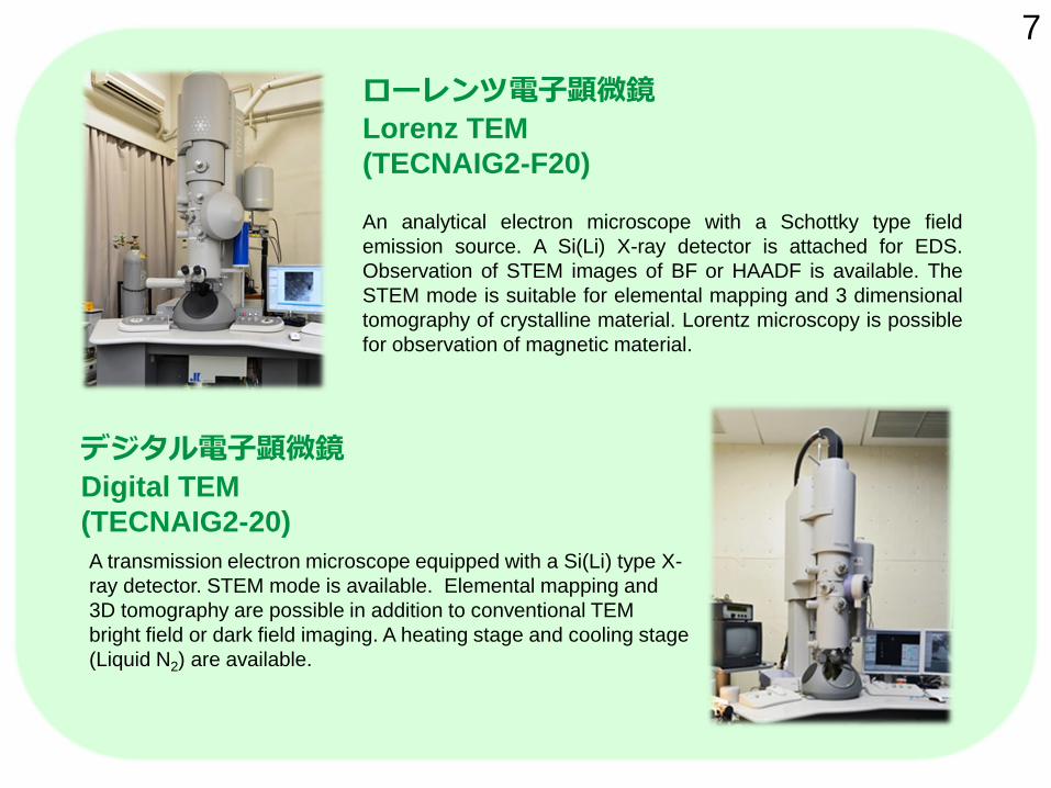
ローレンツ電子顕微鏡
Lorenz TEM
(TECNAIG2-F20)
An analytical electron microscope with a Schottky type field
emission source. A Si(Li) X-ray detector is attached for EDS.
Observation of STEM images of BF or HAADF is available. The
STEM mode is suitable for elemental mapping and 3 dimensional
tomography of crystalline material. Lorentz microscopy is possible
for observation of magnetic material.
7
デジタル電子顕微鏡
Digital TEM
(TECNAIG2-20)
A transmission electron microscope equipped with a Si(Li) type X-
ray detector. STEM mode is available. Elemental mapping and
3D tomography are possible in addition to conventional TEM
bright field or dark field imaging. A heating stage and cooling stage
(Liquid N2) are available.

マイクロカロリーメーター
高エネルギー分解能元素分析装置
Micro-Calorimeter FESEM
(SII TES+Zeiss-ULTRA55)
A micro-calorimeter X-ray detector is attached to a high resolution
Scanning Electron Microscope (SEM) along with a SDD type X-
ray detector. The micro-calorimeter uses the transition edge of
superconductor cooled below 0.1K by using a dilution refrigerator.
The energy resolution of X-rays are 10 eV at an acceleration
voltage of 6 kV. The machine is suitable for identification of
elements included in multicomponent bulk material.
8
汎用電子顕微鏡
Conventional TEM
(JEM-2000EX, JEM-2100HC)
Conventional TEMs with an acceleration
voltage of 200kV. JEM-2100HC has a 16
mega pixel CMOS camera to capture
digital TEM images.
JEM-2000EX
Conventional TEM
JEM-2100HC
High Contrast TEM

Name of equipment Performance
DITABIS micron (Imaging Plate system) TEM image readout to digital data with a high dynamic range of 5 orders.
Name of software PC Performance
Mac Tempas X Mac Simulation of HREM image and diffraction pattern
Crystal Kit Mac Drawing of crystal models
jems Win Simulation of HREM image and diffraction pattern and CBED
Crystal Maker Win Drawing of crystal models and diffraction patterns
ICDD View 2014 Win Former JCPDS, Identification of crystal structure, International Data base of crystals
Temography Win Reconstruction of 3D structure from tilted series of images
Inspect3D Win Reconstruction of 3D structure from tilted series of images
Amira Win Reconstruction of 3D structure from tilted series of images
Software for analysis of image and diffraction
TEM image recording
Name of equipment Performance
FEI Quanta 3D 200i
(Dual beam FIB)
Focused Ga ion beam milling system is combined with a scanning electron
microscope. Cutting and slicing is possible by observing the SEM image of a
sample. Pin-point sampling of he desired region is possible with this system.
Gatan PIPS, JEOL IonSlicer
Fischione TEM Mill, Nano Mill Preparation of thin TEM specimens by Ar ion milling, Acceleration voltage, Ion
beam illuminating angle. Probe sizes are different from machine to machine.
We also have a DARKROOM for film development
TEM specimen preparation equipment 9

Flow-chart of Cooperative Utilization
10

Proposal/Application of Research Project
11
○Fill up a prescribed form
and submit it to the Office
of The URC.
○The project leader should
be an academic staff who
has a budget / fund for the
payment.
○A copy of the prescribed
form is available from the
WEB site, or at the Office
of The URC.

12
Training Courses
○The URC offers training courses
for users. ○ Training courses consist of
(a) Introductory
(b) Elementary
(c) Middle
(1) HVEM, (2) Analytical TEM,
(3) High resolution TEM,
(4) Electron diffraction,
(5) Specimen preparation with FIB,
(6) TEM tomography
(7) Analytical SEM
○ Beginners should take
"Introductory" and "Elementary"
courses at least, in principle.
The training courses are offered by URC and HVEM-Forum, cooperatively. Information on the schedule of courses, etc., is available in the WEB site of The URC.
ふりがな 身 分
受講者氏名
所属部局・部門
(学生は指導教員名を
記入)
指導教員名*注1
九州大学超高圧電顕室
利用申請状況
該当の枠に✓を付けてください。
□ 利用申請済み
□ 後日利用申請予定
電話番号 所属連絡先 自宅(携帯)
ファックス
電子メール
※携帯メール不可
(受講日のお知らせは通常メールで行います。申請された方は必ずメールでの案内確認を行ってください。)
X線取扱講習 あり なし(超高圧電子顕微鏡の使用に際
しては必要となります)
受講希望日を別紙スケジュール表参照の上、第3希望までご記入下さい。
□ 第1希望
□ 第2希望
□ 第3希望
□ 第4希望以降(あればご記入下さい)
*注1 学生の方は、指導教員の承認を得た上で申請してください。
九州大学超高圧電子顕微鏡室利用者研修会参加申請書
九州大学
超高圧電子顕微鏡室講習会受付宛(E-mail添付 可)

13 Booking equipment
○ Booking reservation for equipments is
necessary. ○ Book from the request form on the WEB
site.
○ Always fill in your request date and time
up to the THIRD preference.
In case of same multiple requests or
sudden troubles of equipment, you
maybe asked to shift to another time or
day. ○ Submission of requests are due 9:00AM
of Friday of the preceding week of your
using.
○ Time table of the allocated reservations
will be noticed on the WEB site, uploaded
by the evening of Friday of the preceding
week.
○ When you have to cancel your reservation
inevitably, contact to the URC office AS
SOON AS POSSIBLE.
○ If you cancel without any notice, you will
be banned and will NEVER be able to
make further reservations.
WEB site http://www.hvem.kyushu-u.ac.jp/
Click here for booking

Equipment and Consumables Charge/Price
JPY / time-unit*
1 Old HVEM (JEM-1000) ¥ 8,400 .
2 New model of HVEM (JEM-1300NEF) ¥ 11,300 .
3 3D-tomography TEM (JEM-3200FSK) ¥ 10,000 .
4 Lorenz TEM (TECNAI-F20) ¥ 8,600 .
5 Digital TEM (TECNAI-20) ¥ 8,300 .
6 Dual-beam FIB/SEM (FEI Quanta 3D 200i) ¥ 6,200 .
7 Micro-calorimeter EDS FE-SEM
(Zeiss ULTRA55 + SII-nanotechnology) ¥ 6,800 .
8 Abberation corrected TEM (JEM-ARM200F) ¥ 9,100 .
9 Atomic Resolution Analytical TEM
(JEM-ARM200CF)
¥ 9,400 .
10 Conventional TEM (JEM-2100HC) ¥ 6,000 .
11 Conventional TEM (JEM-2000EX) ¥ 5,900 .
12 Negative films (Imaging plates) ¥ 260. / sheet
13 Liquid helium ¥ 551. / liter
14
* 1 time-unit = 4 hours
List of Charges for Use of Equipments (as July, 2015)

Submission of Records and Manuscripts
○Published papers and presentations
Every project-leader should submit the following at the end of FY.
* Brief results of the research project
* List of publications and presentations related to the use of equipments of
The URC.
○Manuscripts for Annual Reports of The URC.
>Every user and project-leader are invited to submit a
manuscript for Annual Reports of The URC. >Every research project has an obligation to submit one
manuscript at least.
* 2 pages of A4-sheet
* English is recommended.
* The dead-line of submission is the middle of May.
* The manuscript is saved in a form of electronic
document, and uploaded to the Repository of KU.
* A report based on halfway results is also acceptable.
* It needs care when the provision for intellectual
property right and/or originality of article is concerned.
15
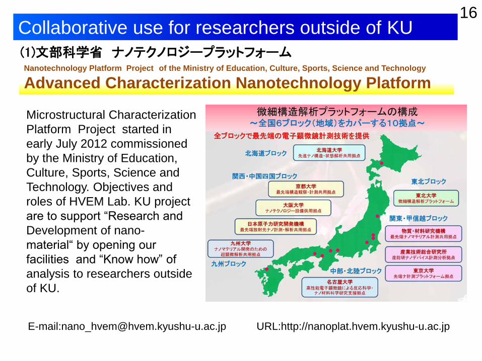
Collaborative use for researchers outside of KU
Microstructural Characterization
Platform Project started in
early July 2012 commissioned
by the Ministry of Education,
Culture, Sports, Science and
Technology. Objectives and
roles of HVEM Lab. KU project
are to support “Research and
Development of nano-
material“ by opening our
facilities and “Know how” of
analysis to researchers outside
of KU.
16
(1)文部科学省 ナノテクノロジープラットフォーム Nanotechnology Platform Project of the Ministry of Education, Culture, Sports, Science and Technology
Advanced Characterization Nanotechnology Platform
E-mail:[email protected] URL:http://nanoplat.hvem.kyushu-u.ac.jp

九州大学学術研究都市推進機構(Organization for Promotion Academic City by Kyushu University)
The Forum for High Voltage Electron Microscopy
HVEM Station Network project started in April
2011, supported by the Ministry of Education,
Culture, Sports, Science and Technology.
Objectives and roles of the project are to
establish collaboration network of the major
institutes for high voltage electron microscopy
in Japan , and to provide top-level unique
characterization. Core universities: Kyushu
Univ., Osaka Univ., Nagoya Univ. and
Hokkaido Univ.
17 (2)文部科学省 超高圧電子顕微鏡連携ステーション
Research Station for High Voltage Electron Microscopy
HVEM Forum is established in 2005 at the Organization for Promotion Academic
City by Kyushu University. The Forum aims at offering technology and
information services to researchers out side of KU. The technology services
consists of (a) Joint use of advanced instruments, (b) Assist and instruction for
observation and analysis, (c) Consulting or collaboration. Users should be a
member of the HVEM Forum.
(3)会員制研究支援組織「超高圧電子顕微鏡フォーラム」

MAP & ACCESS 18
ACCESS for Ito Campus
http://suisin.jimu.kyushu-u.ac.jp/en/info/index.html
MAP of Ito Campus
http://www.kyushu-u.ac.jp/access/map/ito/ito-e.html
ACCESS for KYUSHU UNIVERSITY
http://www.kyushu-u.ac.jp/english/university/location/location.php

●Members of The URC
Director : Prof. S. Matsumura
Chief manager:Assoc. Prof. K. Yasuda
Chair. Tech. Comm.:Prof. M. Nishida
Ito Campus : Prof. Y. Murakami
Chikushi Campus : Prof. S. Hata
Technician:H. Maeno
●Contact
TEL / FAX : 092-802-3292
E-mail: [email protected]
http://www.hvem.kyushu-u.ac.jp/
19 Staffs and Contact of the URC