paduaresearch.cab.unipd.itpaduaresearch.cab.unipd.it/8019/1/Tarzia_Vincenzo_Tesi.pdf · Management...
155
I Sede Amministrativa: Università degli Studi di Padova Dipartimento di Scienze Cardiologiche, Toraciche e Vascolari __________________________________________________________________ SCUOLA DI DOTTORATO DI RICERCA IN : Scienze Mediche, Cliniche e Sperimentali INDIRIZZO: Scienze Cardiovascolari 27° CICLO TITOLO TESI EXTRACORPOREAL MEMBRANE OXYGENATION (ECMO) IN REFRACTORY CARDIOGENIC SHOCK: IMPACT OF ACUTE VERSUS CHRONIC ETIOLOGY ON OUTCOME Direttore della Scuola : Ch.mo Prof. Gaetano Thiene Coordinatore d’indirizzo: Ch.mo Prof. Gaetano Thiene Supervisore :Ch.mo Prof. Gino Gerosa Dottorando : Dr. Vincenzo Tarzia
Transcript of paduaresearch.cab.unipd.itpaduaresearch.cab.unipd.it/8019/1/Tarzia_Vincenzo_Tesi.pdf · Management...

I
Sede Amministrativa: Università degli Studi di Padova
Dipartimento di Scienze Cardiologiche, Toraciche e Vascolari
__________________________________________________________________
SCUOLA DI DOTTORATO DI RICERCA IN : Scienze Mediche, Cliniche e Sperimentali
INDIRIZZO: Scienze Cardiovascolari
27° CICLO
TITOLO TESI
EXTRACORPOREAL MEMBRANE OXYGENATION (ECMO) IN REFRACTORY CARDIOGENIC SHOCK: IMPACT OF ACUTE VERSUS CHRONIC ETIOLOGY ON
OUTCOME
Direttore della Scuola : Ch.mo Prof. Gaetano Thiene
Coordinatore d’indirizzo: Ch.mo Prof. Gaetano Thiene
Supervisore :Ch.mo Prof. Gino Gerosa
Dottorando : Dr. Vincenzo Tarzia

II

III
Ai miei genitori

IV

V
INDEX
SUMMARY 1
RIASSUNTO 3
1. CARDIOGENIC SHOCK (CS) 6
1.1 INTRODUCTION 6
1.2 DEFINITION, DIAGNOSIS AND CAUSES OF CS 6
1.3 INCIDENCE 8
1.4 PATHOPHYSIOLOGY 9
1.5 INITIAL MANAGEMENT OF CS 13
1.6 MECHANICAL CIRCULATORY SUPPORT IN THE TREATMENT OF CS 14
1.7 INTRA-AORTIC BALLOON PUMP (IABP) 16
1.8 TANDEMHEART PERCUTANEOUS VENTRICULAR ASSIST DEVICE 19
1.9 IMPELLA RECOVER PVAD 21
1.10 CENTRIMAG VENTRICULAR ASSIST DEVICE 23
1.11 EXTRACORPOREAL MEMBRANE OXYGENATION (ECMO) 24
1.12 CONCLUSIONS 26

VI
1.13 REFERENCES 28
2. EXTRACORPOREAL MEMBRANE OXYGENATION (ECMO) 38
2.1 INTRODUCTION 38
2.2 HISTORY AND DEVELOPMENT OF OXYGENATORS 39
2.3 THE USE OF OXYGENATORS DURING THE EARLY YEARS OF OPEN-
HEART SURGERY 41
2.3.1 The Mayo-Gibbon pump-oxygenator 41
2.3.2 The Kay-Cross disc oxygenator 42
2.3.3 The DeWall bubble oxygenator 43
2.3.4Limitations Of The Performance Of Direct Contact Oxygenators 44
2.3.5 Introduction of the membrane oxygenator 46
2.3.6 Improving the gas exchange efficiency of membrane oxygenators 48
2.4 ECMO MODALITIES 50
2.4.1 VV ECMO 50
2.4.2 ECMO for Respiratory Failure 52
Acute respiratory distress syndrome 52
Hypercapnic respiratory failure 53

VII
Bridge to lung transplantation and post-transplantation primary graft dysfunction 53
2.4.3 VA ECMO 55
2.4.4 ECMO for Cardiac Failure 58
Post-operative cardiogenic shock and post-transplant primary graft failure 59
Cardiogenic shock complicating acute myocardial infarction 60
Fulminant myocarditis 61
Sepsis-associated cardiomyopathy 61
Pulmonary hypertension 61
Pulmonary embolism 62
Extracorporeal cardiopulmonary resuscitation 62
Bridge to VAD implantation or heart transplantation 64
ECMO to prevent acute right ventricular failure after LVAD implantation 65
2.5 ECMO CIRCUITS AND EQUIPMENT 66
2.5.1 Pump 66
2.5.2 Oxygenators 68
2.5.3 Cannulae and Tubing 70
2.5.4 Heat Exchangers 71

VIII
2.6 ANTICOAGULATION FOR ECMO 72
2.7 MANAGEMENT OF ECMO: Maintenance and Weaning of ECMO 75
2.8 COMPLICATIONS OF ECMO 78
2.9 OUTCOMES OF ECMO 82
2.10 CONCLUSIONS 82
2.11 REFERENCES 84
3.EXTRACORPOREAL MEMBRANE OXYGENATION (ECMO) IN
REFRACTORY CARDIOGENIC SHOCK: IMPACT OF ACUTE
VERSUS CHRONIC ETIOLOGY ON OUTCOME 105
3.1 INTRODUCTION 105
3.2 MATERIALS AND METHODS 107
3.2.1 Patients 107
3.2.2 Criteria for ECMO installation 108
3.2.3 ECMO system 109
3.2.4 ECMO placement 109
3.2.5 Anticoagulant management 110
3.2.6 Management of ECMO 110
3.2.7 Weaning trial 111

IX
3.2.8 Data Analysis 111
3.3 RESULTS 113
3.4 COMMENT 119
3.5 CONCLUSIONS 121
3.6 REFERENCES 123
DR. VINCENZO TARZIA:
CURRICULUM VITAE 126
PERSONAL CONTRIBUTION DURING THE PH.D. PERIOD 134

X

1
SUMMARY
Refractory cardiogenic shock (CS) is a condition that continues to have a very high mortality
despite advances in medical therapy. Conventional treatment typically comprises inotrope infusions,
vasopressors and intra-aortic-balloon-pump (IABP). When circulatory instability is refractory to
these treatments, mechanical circulatory support represents the only hope for survival, as indicated
by current guidelines. As most of these patients present with critical circulatory instability requiring
urgent or emergent therapy, the chosen mechanical assistance should be rapidly and easily
implanted. For this reason ExtraCorporeal Membrane Oxygenation (ECMO) represents the ideal
“bridge-to-life” and increasingly it is used to keep the patient alive while the optimal therapeutic
management is determined (bridge-to-decision). Management may then follow one of three courses:
“bridge-to-recovery”: patient recovery, and weaning from ECMO; “bridge-to-transplant”: direct
heart transplantation; “bridge-to-bridge”: placement of ventricular-assist-device or total artificial
longer-term support. There have been several large reports on the use of ECMO as a mechanical
support in post-cardiotomy patients but relatively few, mostly small case-series focusing on its role
in primary acute cardiogenic shock outside of the post-cardiotomy setting.
We present the results of our centre’s experience (Padova) in the treatment of primary acute
cardiogenic shock with the PLS-Quadrox ECMO system (Maquet) as a bridge to decision.
Furthermore, we evaluated the impact of etiology on patient outcomes by comparing acute primary
refractory CS secondary to acute myocardial infarction (AMI), myocarditis, pulmonary embolism
(PE) and post-partum cardiomyopathy (PPCM) with acute decompensation of a chronic
cardiomyopathy, including dilated cardiomyopathy (DCM), ischemic cardiomyopathy (ICM) and
grown-up-congenital-heart-diseases (GUCHD). We also analyzed whether duration and magnitude
of support may predict weaning and survival.

2
Materials and Methods. Between January 2009 and March 2013, we implanted a total of 249
ECMO; in this study we focused on 64 patients where peripheral ECMO was the treatment for
primary cardiogenic shock. Thirty-seven cases (58%) were “acute” (Group A-PCS: mostly acute
myocardial infarction, 39%), while twenty-seven (42%) had an exacerbation of “chronic” heart
failure (Group C-PCS: dilated cardiomyopathy 30%, post-ischemic cardiomyopathy 9%, congenital
3%).
Results. In group C-PCS, 23 patients were bridged to a LVAD (52%) or heart transplantation
(33%). In group A-PCS, ECMO was used as bridge-to-transplantation in 3 patients (8%), bridge-to-
bridge in 9 (24%), and bridge-to-recovery in 18 patients (49%). One patient in both groups was
bridged to conventional surgery. Recovery of cardiac function was achieved only in group A-PCS
(18 vs 0 pts, p=0.0001). Mean-flow during support ≤60% of the theoretical flow (BSA*2.4) was a
predictor of successful weaning (p=0.02). Average duration of ECMO support was 8.9 ±9 days.
Nine patients (14%) died during support; 30-day overall survival was 80% (51/64 pts); 59% of
patients were discharged, in whom survival at 48 months was 90%. Better survival was observed in
patients supported for 8 days or less (74% vs 36%, p=0.002).
Conclusions. In “chronic” heart-failure ECMO represents a bridge to VAD or heart-transplantation,
while in “acute” settings it offers a considerable chance of recovery, often representing the only
required therapy.

3
RIASSUNTO
Lo shock cardiogeno refrattario è una condizione gravata da alta mortalità nonostante i progressi
nella terapia medica. Il trattamento convenzionale comprende infusione di inotropi, vasopressori, e
contropulsazione aortica (intra-aortic-balloon-pump – IABP). Quando l’instabilità emodinamica è
refrattaria a questi trattamenti, il supporto meccanico al circolo rappresenta la sola possibilità di
sopravvivenza, come indicato dalle attuali linee guida. Tuttavia, poichè la maggior parte di questi
pazienti si presenta con severa instabilità emodinamica che richiede un intervento urgente o
emergente, l’assistenza meccanica scelta dovrebbe essere impiantabile in maniera rapida e
semplice. Per questa ragione, l’ExtraCorporeal Membrane Oxygenation (ECMO) rappresenta
l’ideale “bridge-to-life”, che sempre più viene usato per supportare le funzioni vitali in attesa che il
programma terapeutico ottimale venga stabilito (bridge-to-decision). L’iter terapeutico può poi
seguire tre diversi percorsi: “bridge-to-recovery”: il paziente recupera una funzione
cardiocircolatoria tale da permettere lo svezzamento dall’ECMO; “bridge-to-transplant”: il paziente
viene sottoposto a trapianto cardiaco; “bridge-to-bridge”: il paziente viene trattato con impianto di
un’assistenza ventricolare o di un cuore artificiale totale. Sono state riportate diverse ampie
casistiche sull’uso dell’ ECMO come supporto meccanico in pazienti con shock dopo intervento
cardiochirurgico (“post-cardiotomy”), ma relativamente poche serie, e limitate a pochi casi,
focalizzate sul ruolo dell’ECMO nello shock cardiogeno primario (non post-cardiotomico).
In questo studio si presenta l’esperienza del centro di Padova nel trattamento dello shock
cardiogeno primario con il sistema ECMO PLS-Quadrox (Maquet) come bridge-to-decision.
In particolare, la ricerca proposta si prefigge di valutare l’impatto della differente eziologia
sull'outcome dei pazienti, paragonando gli shock cardiogeni primari “acuti”, secondari ad infarto
miocardico acuto, miocardite, embolia polmonare e cardiomiopatia post-partum, con scompensi
acuti di cardiomiopatie “croniche”, includendo cardiomiopatie dilatative primitive, post-ischemiche,

4
e cardiopatie congenite dell’adulto. Si è infine analizzato se la durata e l’entità del supporto possano
predire la chance di sopravvivenza e di svezzamento.
Materiali e metodi. Tra Gennaio 2009 e Marzo 2013, sono stati impiantati con ECMO un totale di
249 pazienti, di questi 64 erano affetti da shock cardiogeno "primario" (52 uomini e 12 donne, di
50±16 anni di età) e sono stati trattati con supporto ECMO periferico. Trentasette casi (58%) sono
stati classificati come "acuti" (Gruppo A, Acuti, IMA 39%, miocardite 6%, embolia polmonare 8%,
post-partum 2%), mentre i rimanenti 27 (42%) shock erano insorti in un quadro di scompenso
cardiaco "cronico" (Gruppo B, Cronici, cardiomiopatia dilatativa primitiva 30%, cardiomiopatia
dilatativa post-ischemica 9%, patologie congenite 3%).
Risultati della ricerca. Nel gruppo con scompenso cardiaco cronico (Gruppo B), 23 pazienti sono
stati trattati con impianto o di assistenza ventricolare sinistra (52%) o trapianto cardiaco ortotopico
(33%). Nel gruppo con scompenso cardiaco acuto (Gruppo A), l' ECMO è stato usato come ponte a
trapianto in 3 pazienti (8%), come ponte ad impianto di assistenza ventricolare sinistra in 9 pazienti
(24%) e come ponte al recupero della propria funzionalità cardiaca in 18 pazienti (49%).
Un solo paziente in ogni gruppo è stato trattato con chirurgia tradizionale. Il recupero della
funzionalità cardiaca si è osservato solo all'interno del Gruppo A (18 vs. 0 pazienti, p=0,0001). E'
stato visto che mantenere un flusso medio di supporto ≤60% del flusso teorico (BSA*2,4)
costituisce un predittore positivo di svezzamento dal dispositivo (p=0,02). Globalmente, la durata
media del supporto ECMO è stata di 8,9±9 giorni. Nove pazienti (14%) sono deceduti durante il
supporto ECMO; la sopravvivenza globale a 30 giorni è stata dell' 80% (5/64 pazienti); il 59% dei
pazienti è stato dimesso dall’ ospedale e, tra questi, la sopravvivenza a 48 mesi è stata del 90%,
senza differenze significative nei due gruppi. La sopravvivenza migliore si è osservata in quei
pazienti che hanno necessitato di supporto ECMO per un periodo inferiore o uguale ad 8 giorni
(74% vs. 36%, P=0,002).

5
In conclusione nei pazienti con shock cardiogeno refrattario nell'ambito di uno scompenso cardiaco
cronico l'ECMO rappresenta un dispositivo-ponte verso l'impianto di assistenza ventricolare sinistra
o verso trapianto cardiaco. Nei pazienti con shock refrattario dovuto ad eziologia acuta, invece, tale
supporto offre sostanziali chance di recovery, costituendo spesso l'unica terapia necessaria.

6
1. CARDIOGENIC SHOCK (CS)
1.1 INTRODUCTION
Cardiogenic shock (CS) is a common endpoint of multiple disease processes that is characterized by
myocardial dysfunction, depressed cardiac output (CO) and end-organ hypoperfusion. CS is
associated with significant morbidity and mortality, and conventional medical support such as
inotropic agents or intra-aortic balloon counterpulsation is often insufficient to reverse the
hemodynamic changes seen in CS [1].
Recent research has suggested that the peripheral vasculature and neurohormonal and cytokine
systems play a role in the pathogenesis and persistence of CS.
Advances in management, including early revascularization, have led to a reduction of in-hospital
mortality of more than 10% [1-2]. A further reduction may be seen with the advancement of
mechanical circulatory support (MCS), which provides a means for patients to recover or transition
to long-term therapies for management of their underlying cardiac disease. In particular, the
development of percutaneous MCS options has facilitated rapid resuscitation of the cardiogenic
shock patient, potentially interrupting the characteristic systemic inflammatory response before it
can cause irreversible harm.
1.2 DEFINITION, DIAGNOSIS AND CAUSES OF CARDIOGENIC SHOCK
Cardiogenic shock is a state of end-organ hypoperfusion due to cardiac failure. The definition of CS
includes hemodynamic parameters: persistent hypotension (systolic blood pressure <90 mmHg or
mean arterial pressure 30 mmHg lower than baseline) with severe reduction in cardiac index (<1.8

7
l/min/m2
without support or <2.0 to 2.2 l/min/m2 with support) and adequate or elevated filling
pressure (eg, left ventricular [LV] end-diastolic pressure >18 mmHg or right ventricular [RV] end-
diastolic pressure >10 to 15 mm Hg). Hypoperfusion may be manifest clinically by cool
extremities, decreased urine output, and/or alteration in mental status. Hemodynamic abnormalities
form a spectrum that ranges from mild hypoperfusion to profound shock, and the short-term
outcome is directly related to the severity of hemodynamic derangement. In recent studies of
cardiogenic shock, eligibility criteria included systolic blood pressure <90 mmHg for >30 min or
requirement of catecholamines to maintain systolic pressure >90 mmHg, plus clinical signs of
pulmonary congestion and impaired organ perfusion with at least one of the following criteria: (I)
altered mental status; (II) cold, clammy skin and extremities; (III) oliguria with urine output <30
mL-1
; or (IV) serum lactate >2.0 mmol L-1
.
The diagnosis is usually made with the help of pulmonary artery (PA) catheterization; however,
Doppler echocardiography may also be used to confirm elevation of LV filling pressures.
Cardiogenic shock often occurs as the result of an acute event that precipitates rapid cardiovascular
collapse. Myocardial infarction (MI) with LV failure remains the most common cause of CS.
Among patients with an acute ST-elevation myocardial infarction (MI), 8% will develop
cardiogenic shock [2] typically within 24 h of the onset of symptoms [3]. In these patients,
cardiogenic shock is typically a direct consequence of regional myocardial dysfunction and
diminished contractility. Mechanical complications of MI including ventricular septal defect,
papillary muscle rupture producing acute mitral regurgitation, and free left ventricular wall rupture
can also cause cardiogenic shock. Echocardiography is the technique of choice to rule out these
entities and should be performed early unless the diagnosis is extensive anterior MI and the patient
is undergoing prompt percutaneous coronary intervention (PCI). In addition, the detection of
valvular disease before angiography may alter the revascularization approach.

8
Hemorrhage, infection, and/or bowel ischemia may contribute to shock in the setting of MI. As with
mechanical complications, a high index of suspicion is required to make these diagnoses in MI
patients, and survival may depend on timely recognition and treatment.
Any cause of acute, severe left ventricle (LV) or right ventricle (RV) dysfunction may lead to CS.
Many non-ischemic disease processes may present acutely or subacutely and result in cardiogenic
shock. Acute valvular regurgitation, regardless of cause, can rapidly progress to severe heart failure.
Several types of cardiomyopathies can present with a fulminant course, including viral myocarditis,
giant-cell myocarditis, peripartum and Takotsubo cardiomyopathy. Extracardiac disease may also
result in CS, as with a massive pulmonary embolism or pericardial tamponade. Finally, 3–4% of
patients admitted to the hospital for acute decompensation of chronic heart failure will present with
shock [4].
1.3 INCIDENCE
After decades of remarkable stability in the incidence of CS, it appears that the incidence is on the
decline in parallel with increasing rates of use of primary PCI for acute MI. CS continues to
complicate approximately 5% to 8% of STEMI and 2.5% of non-STEMI cases [5]. This translates
to 40000 to 50000 cases per year in the United States [6]. The routine use of troponin to define non-
STEMI will result in a drop in this percentage as more MIs are detected but will not alter the total
number of cases of CS.
The only way to prevent CS appears to be very early reperfusion therapy for MI. A randomized trial
of early, in-ambulance thrombolysis versus primary PCI found no CS among patients assigned to
prehospital thrombolysis [7]. Among PCI-assigned patients, just 0.5% developed CS in the group
randomized <2 hours from symptom onset. A major focus of public health campaigns is the very

9
early recognition and reperfusion of MI, which should reduce CS incidence. Risk factors for
development of CS in the context of MI include older age, anterior MI, hypertension, diabetes
mellitus, multivessel coronary artery disease, prior MI or angina, prior diagnosis of heart failure,
STEMI, and left bundle-branch block [8]. There may be clues to impending shock: heart rate is
higher and blood pressure lower on hospital presentation among patients who develop CS after
admission.
1.4 PATHOPHYSIOLOGY
Cardiogenic shock is the result of temporary or permanent derangements in the entire circulatory
system. LV pump failure is the primary insult in most forms of CS, but other parts of the circulatory
system contribute to shock with inadequate compensation or additional defects.
With the exception of acute valvular disease, CS typically occurs in the setting of pronounced
myocardial dysfunction and low CO. The reduction in MAP results in poor systemic perfusion and
end-organ ischemia. Low coronary perfusion pressure may exacerbate ischemia. Catecholamine
release attempts to compensate for the low-output state by increasing inotropy and peripheral
vasoconstriction at the cost of increasing myocardial oxygen demand. Up-regulation of pressure but
worsening congestion. There are increased cytokine levels and expression of inducible nitric oxide
synthase [2], which can exacerbate hypotension and further worsen myocardial function, causing a
deterioration of cardiovascular hemodynamics.
The degree of myocardial dysfunction that initiates CS is often, but not always, severe. LV
dysfunction in shock reflects new irreversible injury, reversible ischemia, and damage from prior
infarction. The unique position of the heart as an organ that benefits from low blood pressure via
afterload reduction and also suffers from low blood pressure via compromise of coronary flow

10
creates a situation in which changes in
hemodynamics may be simultaneously
beneficial and detrimental.
As depicted in Figure 1, a decrease in
coronary perfusion lowers cardiac output
(CO), which further decreases perfusion of
the heart and other vital organs. Coronary
flow may be additionally compromised by atherosclerosis of vessels other than the infarct artery.
Metabolic derangements occur in the remote myocardium and in the infarct region [9].
Hypoperfusion causes release of catecholamines, which increase contractility and peripheral blood
flow, but catecholamines also increase myocardial oxygen demand and have proarrhythmic and
myocardiotoxic effects. Inotropic agents and vasoconstrictors temporarily improve CO and
peripheral perfusion but do not interrupt this vicious circle. Rapid intra-aortic balloon pump (IABP)
support may temporarily relieve ischemia and support the circulation, but IABP is not definitive
therapy. Relief of coronary occlusion, best achieved through PCI or surgery, interrupts the vicious
circle and saves lives. RV dysfunction may cause or contribute to CS. Predominant RV shock
represents only 5% of cases of CS complicating MI [10]. RV failure may limit LV filling via a
decrease in CO, ventricular interdependence, or both. Treatment of patients with RV dysfunction
and shock has traditionally focused on ensuring adequate right-sided filling pressures to maintain
CO and adequate LV preload; however, patients with CS due to RV dysfunction have very high RV
end-diastolic pressure, often >20 mmHg [10]. This elevation of RV end-diastolic pressure may
result in shifting of the interventricular septum toward the LV cavity, which raises left atrial
pressure but impairs LV filling due to the mechanical effect of the septum bowing into the LV. This
alteration in geometry also impairs LV systolic function [11]. Therefore, the common practice of
aggressive fluid resuscitation for RV dysfunction in shock may be misguided. Inotropic therapy is
Figure 1: Reynolds et al.
11
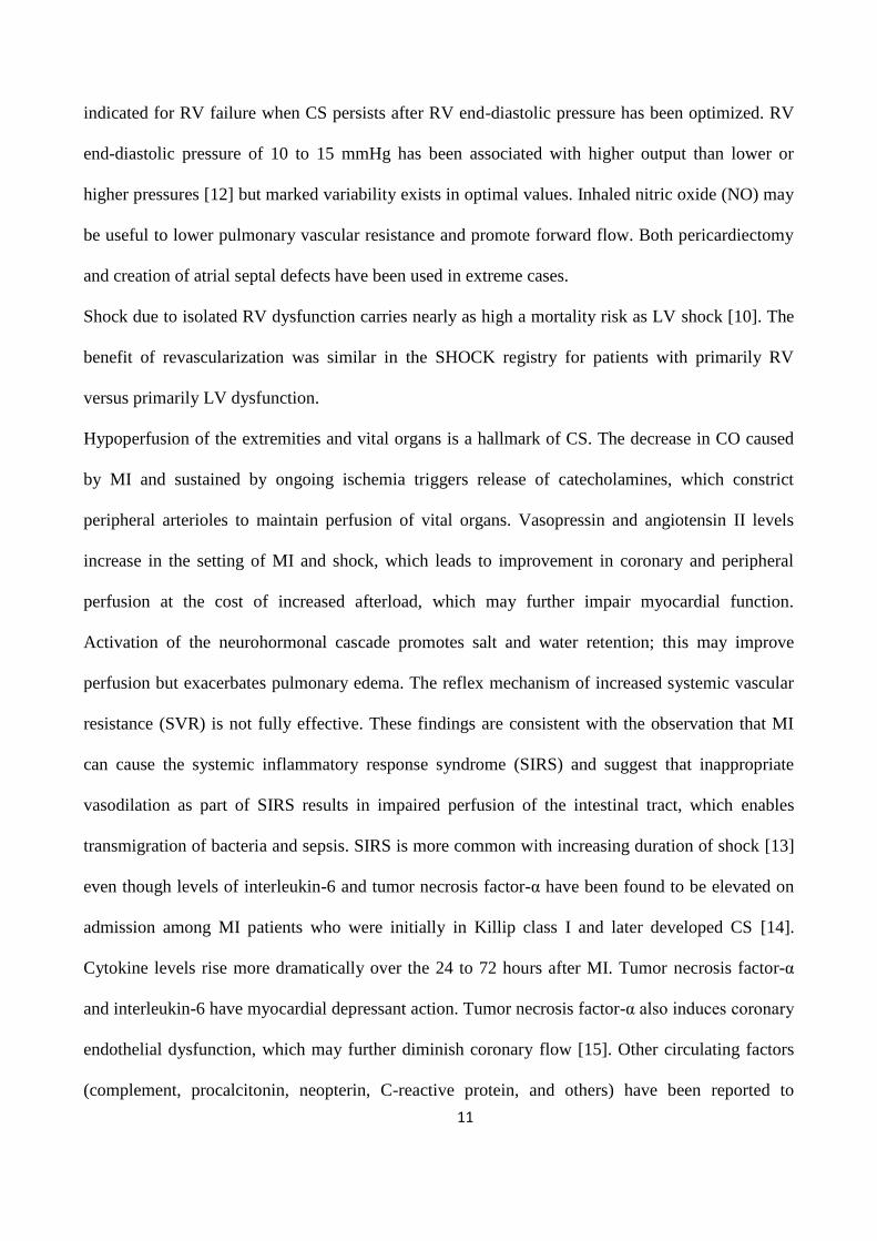
indicated for RV failure when CS persists after RV end-diastolic pressure has been optimized. RV
end-diastolic pressure of 10 to 15 mmHg has been associated with higher output than lower or
higher pressures [12] but marked variability exists in optimal values. Inhaled nitric oxide (NO) may
be useful to lower pulmonary vascular resistance and promote forward flow. Both pericardiectomy
and creation of atrial septal defects have been used in extreme cases.
Shock due to isolated RV dysfunction carries nearly as high a mortality risk as LV shock [10]. The
benefit of revascularization was similar in the SHOCK registry for patients with primarily RV
versus primarily LV dysfunction.
Hypoperfusion of the extremities and vital organs is a hallmark of CS. The decrease in CO caused
by MI and sustained by ongoing ischemia triggers release of catecholamines, which constrict
peripheral arterioles to maintain perfusion of vital organs. Vasopressin and angiotensin II levels
increase in the setting of MI and shock, which leads to improvement in coronary and peripheral
perfusion at the cost of increased afterload, which may further impair myocardial function.
Activation of the neurohormonal cascade promotes salt and water retention; this may improve
perfusion but exacerbates pulmonary edema. The reflex mechanism of increased systemic vascular
resistance (SVR) is not fully effective. These findings are consistent with the observation that MI
can cause the systemic inflammatory response syndrome (SIRS) and suggest that inappropriate
vasodilation as part of SIRS results in impaired perfusion of the intestinal tract, which enables
transmigration of bacteria and sepsis. SIRS is more common with increasing duration of shock [13]
even though levels of interleukin-6 and tumor necrosis factor-α have been found to be elevated on
admission among MI patients who were initially in Killip class I and later developed CS [14].
Cytokine levels rise more dramatically over the 24 to 72 hours after MI. Tumor necrosis factor-α
and interleukin-6 have myocardial depressant action. Tumor necrosis factor-α also induces coronary
endothelial dysfunction, which may further diminish coronary flow [15]. Other circulating factors
(complement, procalcitonin, neopterin, C-reactive protein, and others) have been reported to

12
contribute to SIRS in CS. Excess NO may also contribute to SIRS. MI is associated with increased
expression of inducible NO synthase, which leads to excess NO, which causes vasodilation,
myocardial depression, and interference with catecholamine action.
Cardiogenic shock has been divided into four stages to demonstrate severity and progression of
disease: preshock, mild shock, profound shock and severe refractory CS [16]. The progression from
mild cardiogenic shock to severe refractory cardiogenic shock reflects the severity of hemodynamic
compromise and is reflected by the number of vasoactive medications required to maintain
reasonable CO and MAP. In mild shock, the cardiovascular system may not require support or can
be easily supported with low doses of one inotrope or vasopressor. Patients with profound shock
require moderate-to-high doses of a single agent, whereas patients with severe refractory
cardiogenic shock remain hemodynamically compromised, despite high doses of multiple
vasoactive medications. Mortality increases progressively with each stage, and patients with severe
refractory cardiogenic shock generally have a very poor prognosis in the absence of MCS.
However, cardiogenic shock is not a mere decrease in cardiac contractile function, but also a
multiorgan dysfunction syndrome (MODS) resulting from peripheral hypoperfusion with
microcirculatory dysfunction, often complicated by a systemic inflammatory response syndrome
(SIRS) and sepsis [17,18-23].
Once MODS has developed, it is difficult to improve prognosis and reduce mortality by simply
increasing cardiac output with a circulatory assist device. Prevention of MODS may depend on
three critical factors:
(1) optimal timing (i.e. early initiation) of mechanical circulatory support,
(2) optimal level of mechanical circulatory support with reestablishment of adequate perfusion of
critical organs, and
(3) optimal prevention and management of potential device-related complications (i.e. device
malfunction, infection).

13
Intuitively, one would expect that haemodynamic parameters would best discriminate between
survivors and non-survivors, and at least for the calculated pressure-flow-product ‘cardiac power
output/index’, this has been demonstrated [24,25]. However, in the IABP-Shock study [20], cardiac
index itself was unrelated to patient survival beyond the first 24 h of CSMI. Likewise, biomarkers
of heart failure (e.g. BNP) were unrelated to prognosis in the first 96 h of CSMI.
On the other hand ,MODS severity (as indicated by the APACHE II or SAPS II scores) and
biomarkers of SIRS (like Interleukin 6 and receptor of advanced glycation end-products, RAGE)
can predict mortality more accurately than haemodynamic indices [26].
Although LV contractile failure and low cardiac output are the primary cause of cardiogenic shock,
improving cardiac output alone may not reverse or even halt the progression of MODS if initiated
too late. Therefore, the haemodynamic improvement of cardiac index may be a measure of technical
success of mechanical circulatory support; however, without limiting the progression of SIRS and
MODS within the first few days, these haemodynamic improvements may be futile and may not
translate into improved survival.
1.5 INITIAL MANAGEMENT OF CARDIOGENIC SHOCK
The initial management goals of cardiogenic shock include cardiovascular resuscitation and
identification of the underlying cause. Reversible cardiac causes, including arrhythmias and
conduction disturbances, should be identified and treated. If myocardial ischemia or infarction is
suspected, patients should rapidly undergo coronary angiography and either percutaneous or
surgical revascularization. In the SHOCK (One-year survival following early revascularization for
cardiogenic shock) trial, early revascularization in those presenting with cardiogenic shock reduced
1-year mortality from 66 to 53% [27]. Medical therapy of cardiogenic shock is directed at

14
normalizing hemodynamic parameters, correcting metabolic disarray and minimizing end-organ
dysfunction. Vasoactive agents (inotropes, vasopressors) are often required to augment CO but at
the expense of worsening myocardial oxygen demand, exacerbation of ischemia and potentiation of
arrhythmias. Correction of acidosis may help to prevent damage to end-organs and to promote the
effects of vasoactive agents. Those patients with continued worsening or lack of improvement of
hemodynamics despite escalation of medical therapy are considered to have severe refractory shock
and should immediately be considered for placement of MCS.
1.6 MECHANICAL CIRCULATORY SUPPORT IN THE TREATMENT OF
CARDIOGENIC SHOCK
The key concept is to quickly identify patients in need of more support than medical management
and/or an IABP can achieve, as early intervention with MCS in the patients at highest risk is most
effective when done early. MCS can interrupt the inflammatory cascade initiated by the onset of
shock and prevent progression to irreversible end-organ damage and subsequent death; however,
there remains a window of opportunity during which rescue is possible. An IABP is typically the
first line of mechanical support used due to ease of insertion and minimal risk, but it is often
insufficient in providing adequate support in patients with severe cardiogenic shock. Other options
for temporary support include the ImpellaTM
percutaneous ventricular assist device (PVAD),
TandemHeartTM
PVAD, venoarterial extracorporeal membrane oxygenation (V-A ECMO) and the
CentriMagTM
device, which can be placed surgically or percutaneously. (Figure 2) [28].

15
Figure 2: Sayer et al.
Device selection is based on a number of factors including the degree of hemodynamic support
needed, whether right ventricular failure or lung injury is present, individual patient factors (e.g.
mechanical valves, peripheral vascular disease) and the availability of interventionalists/cardiac
surgeons.
Ouweneel and Henriques [29] defined the ‘ideal device for cardiogenic shock’ as follows: ‘ . . .
during an acute critical presentation only those assist devices allowing percutaneous access are
suitable due to the invasiveness of surgical devices. The ideal device should enable both
haemodynamic support and myocardial protection. Also, a percutaneous approach is preferable to
provide for a quick and easy deployment. In addition, the ideal device should be associated with a
low complication rate, as complications may sometimes outweigh the potential beneficial effect.
Complications associated with any (percutaneous) LV assist device may include limb ischaemia,
embolisation of atherosclerotic and/or thrombotic material, stroke, infection and haemolysis’.
In line with these demands for mechanical circulatory support in CS, different technical strategies
have been developed over the past decades to improve cardiac output and unload the critically
damaged left ventricle by either afterload or pre-load reduction (i.e. pressure or volume unloading,
respectively). Additionally, circulatory support may be provided to the left ventricle alone, the right

16
ventricle alone, or to both ventricles. Biventricular assist devices may be combined with
replacement of pulmonary gas exchange (i.e. extracorporal membrane oxygenation, ECMO) or be
administered as pure right and left ventricular haemodynamic support.
Based on the different physiological concepts outlined above, we distinguish among three
categories of peripheral/percutaneous circulatory support devices in CS:
(1) mechanical LV support by LV pressure unloading [30]—the IABP;
(2) mechanical LV support by LV volume unloading [30]—the TandemHeartTM
, the Impella
Recover LP® micro-axial rotary pump;
(3) mechanical circulatory support with membrane oxygenation [30]—ECMO;
The possibilities are completed by surgically implanted mechanical support -without simultaneous
replacement of pulmonary gas exchange- combining right (RVAD) and/or left (LVAD)
paracorporeal ventricular assist device therapy.
1.7 INTRA-AORTIC BALLOON PUMP (IABP)
The IABP (Figure 3) is the most commonly used form of MCS, it’s a
balloon inserted in the descending aorta that augments coronary blood flow
by inflating during diastole, while also assisting myocardial function
through reduced afterload by deflating during systole. The ultimate effect is
limited to an increase of LV stroke volume and cardiac output by up to 1
l/min (15–30%, respectively). The haemodynamic effects of IABP in CS
[31] include:
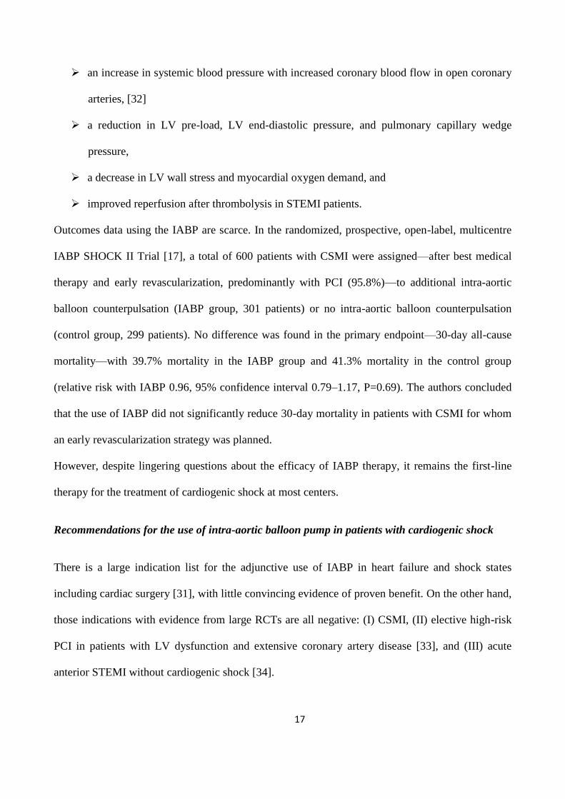
an increase in stroke volume and CO, Figure 3: Werdan et al.
17
an increase in systemic blood pressure with increased coronary blood flow in open coronary
arteries, [32]
a reduction in LV pre-load, LV end-diastolic pressure, and pulmonary capillary wedge
pressure,
a decrease in LV wall stress and myocardial oxygen demand, and
improved reperfusion after thrombolysis in STEMI patients.
Outcomes data using the IABP are scarce. In the randomized, prospective, open-label, multicentre
IABP SHOCK II Trial [17], a total of 600 patients with CSMI were assigned—after best medical
therapy and early revascularization, predominantly with PCI (95.8%)—to additional intra-aortic
balloon counterpulsation (IABP group, 301 patients) or no intra-aortic balloon counterpulsation
(control group, 299 patients). No difference was found in the primary endpoint—30-day all-cause
mortality—with 39.7% mortality in the IABP group and 41.3% mortality in the control group
(relative risk with IABP 0.96, 95% confidence interval 0.79–1.17, P=0.69). The authors concluded
that the use of IABP did not significantly reduce 30-day mortality in patients with CSMI for whom
an early revascularization strategy was planned.
However, despite lingering questions about the efficacy of IABP therapy, it remains the first-line
therapy for the treatment of cardiogenic shock at most centers.
Recommendations for the use of intra-aortic balloon pump in patients with cardiogenic shock
There is a large indication list for the adjunctive use of IABP in heart failure and shock states
including cardiac surgery [31], with little convincing evidence of proven benefit. On the other hand,
those indications with evidence from large RCTs are all negative: (I) CSMI, (II) elective high-risk
PCI in patients with LV dysfunction and extensive coronary artery disease [33], and (III) acute
anterior STEMI without cardiogenic shock [34].

18
The American College of Cardiology/American Heart Association STEMI guidelines recommend
the use of IABP as a class IIa indication for patients with CSMI [35], whereas the recent European
guidelines state that ‘intra-aortic balloon pumping may be considered (IIb/B)’ (Figure 10) [30].

19
1.8 TANDEMHEART PERCUTANEOUS VENTRICULAR ASSIST DEVICE
The TandemHeart PVAD (Cardiac Assist, Inc., Pittsburgh, Pennsylvania,
USA) (Figure 4) is an external centrifugal blood pump with percutaneous
cannulae. Oxygenated blood is aspirated from the left atrium and injected
into the lower abdominal aorta or iliac arteries via a femoral artery
cannula. The inflow cannula is placed in the left atrium via a transseptal
puncture. Pump outflow is returned to the body through a 17 French
cannula in the femoral artery (Figure 5). It typically augments CO up to
3.0–4.0 l/min. The haemodynamic effects of the TandemHeart are
superior to the IABP [35-38] leading to a greater increase in CO and MAP
and a decrease PCWP, central venous pressure, and pulmonary artery
pressure, resulting in reduced filling pressures in the left and right
ventricle, reduced cardiac workload and reduced oxygen demand
[35,39], as well as an increase
in cardiac power index.
Its use is limited by access site complications, limb ischemia
and bleeding. Implantation is more time-consuming and
requires specialized expertise, due to the need for a
transseptal puncture. The presence of a cannula in the left
atrium can be a nidus for thrombus formation. One
significant advantage of the TandemHeart is that it can be
configured to provide right ventricular support with inflow cannula placement into the right atrium
and outflow cannula placement into the main pulmonary artery [40]. The TandemHeart is FDA-
approved for up to 6h of use, but successful use has been reported for greater than 1 week [41]. In
Figure 5: Srihari et al.
Figure 4: Werdan et al.

20
the largest reported series, 117 patients with refractory cardiogenic shock were implanted with the
TandemHeart for an average of 5.8 days [42]. The population was critically ill, with a MAP of
45mmHg, cardiac index of 0.5 l/min/m2 , and lactic acid level of 24.5 mg/dl. TandemHeart support
provided rapid reversal of the hemodynamic abnormalities, increasing the MAP to 81mmHg,
cardiac index to 3.0 l/min/m2 , and decreasing the lactic acid level to 11.0 mg/dl. Although there
was no control group, the 30-day mortality of 40% was considerably better than expected outcomes
in this population. The most common complications were bleeding and sepsis.
Recommendations for the use of the TandemHeart in patients with cardiogenic shock
In the European guidelines a class IIB recommendation is given for LV assist devices in CSMI [43]
(Figure 6). The 2013 AHA/ACCGuideline for the Management of ST-Elevation Myocardial
Infarction assigns a level IIb/C indication for LV assist devices in refractory cardiogenic shock.
This includes centrifugal pumpsystems such as the TandemHeart and ECMO [35].
Figure 6: ESC GL for the management of STEMI [43]

21
1.9 IMPELLA RECOVER PERCUTANEOUS VENTRICULAR ASSIST DEVICE
The Impella Recover PVAD (AbioMed Inc, Danvers,
Massachusetts, USA) is a micro-axial rotary pump positioned across
the aortic valve to provide active support by transvalvular LV
assistance, expelling aspirated blood from the left ventricle into the
ascending aorta (Figure 7).
Two versions are currently available: the Impella Recover LP 2.5
can provide up to 2.5 L min-1
and can be inserted percutaneously.
The Impella Recover LP 5.0 can deliver up to 5.0 L min-1
but
requires surgical cutdown of the femoral or axillary artery.
The Impella 2.5 is an axial flow motor that pumps blood from the left ventricle into the ascending
aorta. The catheter is placed percutaneously through a tapered 13 or 14 French sheath and is
connected to an external power source. Flow is less robust than the Tandem-Heart, averaging less
than 2.5 l/min. However, implantation is quicker and there are fewer access site complications due
to the smaller sheath size. A comparison of the Impella 2.5 with IABP showed better initial
hemodynamic support with the Impella PVAD, but no difference in mortality or support after 6h
[44]. The primary complication of the Impella 2.5 is hemolysis, which can be severe and often
limits the duration of use. Another common issue is pump migration from its intended position,
which may lead to poor support or contribute to hemolysis.
The Impella 5.0 is a larger device, providing flows up to 5.0 l/min. Due to its size, it must be
implanted surgically, either directly into the ascending aorta or through a vascular graft to the
femoral or axillary artery. Due to the larger size of the inflow, hemolysis is a less frequent
complication.
Figure 7.

22
Several studies have demonstrated that the Impella device is safe and haemodynamically effective
in STEMI and high-risk PCI patients [29]. The unloading of the left ventricle is associated with
reduced end-diastolic wall stress and an immediate decrease in PCWP [29]. Coronary perfusion
pressure and coronary flow are reported to be increased and myocardial oxygen consumption
reduced [29].
With respect to the role of the Impella pump in cardiogenic shock and especially in CSMI, the
multicentre Impella EUROSHOCK-Registry [45] included 120 patients with CSMI receiving
temporary circulatory support with the Impella- 2.5-pLVAD. Thirty-day mortality was 64.2%. After
Impella-2.5- pLVAD-implantation, lactate levels significantly decreased from 5.8±5.0 to 2.5±2.6
mmol L-1
(p = 0.023) at 48 h.
The ISAR-SHOCK randomized trial compared the Impella 2.5 with the IABP in cardiogenic shock
patients [46] As showed in this study, CI and MAP increased more in the Impella group;
furthermore, serum lactate levels were lower in the Impella group than in the IABP group. No
differences in mortality, major bleeding, distal limb ischaemia, arrhythmias, and infections were
found.
It has been suggested that, in severe cardiogenic shock, the Impella 5.0 device may provide superior
haemodynamic support [29,47]. A lower mortality rate has been reported for Impella 5.0 in patients
with post-cardiotomy low-output syndrome with a residual CO of 1 L min-1
vs. IABP [48,49]

23
1.10 CENTRIMAG VENTRICULAR ASSIST DEVICE
The Thoratec CentriMag VAD (Thoratec Corporation, Pleasanton, California, USA) (Figure 8) is a
centrifugal pump with a magnetically levitated rotor that can provide up to 10 l/min of blood flow.
The CentriMag can be connected to many different types of circuits, including ECMO, but is
designed as an extracorporeal, surgically implanted VAD for short-term or intermediate-term
support. For left ventricular support, an inlet cannula is placed in the left ventricular apex (the left
atrium is not recommended due to the potential for thromboembolic complications) with the outlet
cannula delivering blood to the aorta. The CentriMag can also provide right ventricular support with
inflow from the right atrium and outflow into the pulmonary artery. Two CentriMags can also be
configured to provide biventricular support.
Figure 8. Kaczorowski et al.
The primary advantage of the CentriMag system is its ability to deliver high-flow rates and to
completely unload the LV. The system is relatively easy to use and has a low rate of
thromboembolism when high-flow rates are maintained The CentriMag is more durable than
PVADs and can provide effective support for weeks to months [50]. A multicenter investigation of

24
the CentriMag in 38 patients demonstrated a 47% 30-day survival. The major complications
included infection and neurological dysfunction [51]. The CentriMag can be configured to support
the right ventricle percutaneously with an inflow cannula placed in the right atrium via the femoral
vein and the outflow cannula placed in the pulmonary artery via the internal jugular vein [52].
1.11 EXTRACORPOREAL MEMBRANE OXYGENATION (ECMO)
The complete ECMO system (Figure 9)—a modified heart–lung machine—generally consists of a
centrifugal pump, a heat exchanger, and a membrane oxygenator. Venous desaturated blood is
aspirated from the right atrium into a centrifugal pump through canulla inserted into the right
atrium. The pump outflow is directed into a membrane oxygenator and is guided via an outflow
cannula into the aorta or femoral/axillary artery.
Figure 9. Westaby et al.
Veno-Arterial (V-A) ECMO can provide support for patients with lung injury as well as either
univentricular or biventricular failure. It’s has been applied in STEMI [53], myocarditis [54], post-
cardiotomy [55], interhospital transfer [56,57] and also in the cardiac catheterization laboratory in
patients who developed cardio-respiratory arrest during PCI and TAVI [58].

25
In the most commonly used percutaneous configuration, the inflow cannula is inserted into the right
atrium through either the femoral or jugular vein and the outflow cannula is placed in the lower
descending aorta via the femoral artery. Due to the large size of the arterial cannula (18 French), an
antegrade catheter is often placed in the ipsilateral femoral artery to provide adequate perfusion to
the leg. A percutaneous circuit can be established in less than 30 min, and it is feasible to put
patients on ECMO at the bedside during an emergency. When percutaneous access is not possible,
the ECMO circuit can be placed centrally, with direct cannulation of the right atrium and aorta.
Of the percutaneous MCS options, ECMO provides the most cardiac support, with the ability, based
on cannula size and position, to achieve flow of greater than 6.0 l/min. However, ECMO is resource
intensive, requiring continuous monitoring by nursing and trained perfusion staff. Complications
include limb ischemia, bleeding, stroke, and infection. Adequate levels of anticoagulation must be
maintained to prevent thromboembolic complications. In patients with pulsatility during support,
care must be taken to ensure that blood leaving the heart is adequately oxygenated, as perfusing the
coronary arteries and brain with deoxygenated blood may result in catastrophic anoxic injury.
Alternatively, with severe ventricular dysfunction, the left ventricle (LV) may not be adequately
decompressed due to return of blood to the left atrium through the bronchial circulation. Left
ventricular distension can lead to excess wall stress and may impede ventricular recovery. Several
methods of decompressing the LV have been described, including a transseptal catheter [59], a
pulmonary artery cannula [60], and minimally invasive placement of an apical vent [61]. Several
recent reports have described successful use of an Impella Recover 2.5 as a vent for the LV [62-64].
Following institution of ECMO, there is often a rapid reversal of hemodynamics with a decrease in
inotrope/vasopressor requirement, improvement in gas exchange, and reduction in markers of
endorgan failure. With meticulous care, ECMO support can be maintained for weeks.

26
Recommendations for the use of ECMO in patients with cardiogenic shock
There is a class IIb/C recommendation in the European STEMI guidelines [43] to consider an LV
assist device for circulatory support in patients with refractory cardiogenic shock (Figure 10). The
European guidelines on myocardial revascularization recommend considering— without a definite
recommendation—ECMO implantation for temporary support in CSMI patients who continue to
deteriorate due to inadequate circulatory support of the IABP. This recommendation is based on
expert consensus.
Figure 10: Werdan et al.
1.12 CONCLUSIONS
Despite optimal up-to-date therapy of CS, mortality continues to remain unacceptably high. Limited
data may support the use of levosimendan [65] but innovations in pharmacological therapy are not
forthcoming. Mild therapeutic hypothermia is promising as a potential therapeutic strategy for
CSMI [66]. It has multiple potentially beneficial effects, including the potential to improve post-
ischaemic cardiac function and haemodynamics, decrease myocardial damage, and reduce end-
organ injury from prolonged hypoperfusion. The neutral results of the IABP-SHOCK II Trial
remind us that immediate haemodynamic improvement may not automatically translate into

27
improved survival. In view of the dissociation between improvements in haemodynamic parameters
and clinical outcomes, including mortality, device therapy may be the best therapy for the future.
In fact mechanical circulatory support not only improves haemodynamics, but prevent or reduce
MODS and ultimately, mortality.
However clinical success of device therapy in CS does not depend on the mechanical qualities of
the device alone. The ease and safety of device implantation—especially under emergency
conditions and during cardiopulmonary resuscitation—will also greatly influence patient outcome.
Additionally, the rates of device-related complications such as limb ischaemia, access site bleeding,
haemolysis, and infection are still too high, and the contact of blood with these devices may
cause/worsen SIRS and MODS. Patients with CS have minimal reserve to tolerate operator error or
device complications.
Data from morbidity studies with a focus on the time course of SIRS and MODS development
indicate that haemodynamic support has limited ability to change outcome if initiated when overt
MODS has already developed. Mechanical circulatory support should not be considered the
treatment of last resort for CS, but should probably be initiated early in the disease course to
minimize the negative effects of high-dose catecholamine therapy on microcirculation and before
end-organ dysfunction with MODS.
Finally among the other mechanical circulatory support devices for CS, we believe that ECMO is
likely to have the greatest potential for wider clinical use.
Its major advantages are:
quick and easy percutaneous insertion of inflow and outflow cannulas,
full circulatory support with up to 4.0 L min-1
,
ECMO rapidly improves tissue oxygenation in situations of cardiogenic shock combined
with severe pulmonary oedema.

28
1.13 REFERENCES
1. Babaev A, Frederick PD, Pasta DJ, Every N, Sichrovsky T, Hochman JS; NRMI
Investigators. Trends in management and outcomes of patients with acute myocardial
infarction complicated by cardiogenic shock. JAMA 2005; 294:448–454.
2. Reynolds HR, Hochman JS. Cardiogenic shock: current concepts and improving outcomes.
Circulation 2008; 117:686–697.
3. Webb JG, Sleeper LA, Buller CE, Boland J, Palazzo A, Buller E, White HD, Hochman JS.
Implications of the timing of onset of cardiogenic shock after acute myocardial infarction: a
report from the SHOCK Trial Registry – should we emergently revascularize occluded
coronaries for cardiogenic shock? J Am Coll Cardiol 2000; 36 (3 Suppl A):1084–1090.
4. Adams KF Jr, Fonarow GC, Emerman CL, LeJemtel TH, Costanzo MR, Abraham WT,
Berkowitz RL, Galvao M, Horton DP; ADHERE Scientific Advisory Committee and
Investigators. Characteristics and outcomes of patients hospitalized for heart failure in the
United States: rationale, design, and preliminary observations from the first 100 000 cases in
the Acute Decompensated Heart Failure National Registry (ADHERE). Am Heart J 2005;
149:209–216.
5. Hasdai D, Harrington RA, Hochman JS, Califf RM, Battler A, Box JW, Simoons ML,
Deckers J, Topol EJ, Holmes DR Jr. Platelet glycoprotein IIb/IIIa blockade and outcome of
cardiogenic shock complicating acute coronary syndromes without persistent ST-segment
elevation. J Am Coll Cardiol. 2000;36:685– 692.
6. Thom T, Haase N, Rosamond W, Howard VJ, Rumsfeld J, Manolio T, Zheng ZJ, Flegal K,
O’Donnell C, Kittner S, Lloyd-Jones D, Goff DC Jr, Hong Y, Adams R, Friday G, Furie K,
Gorelick P, Kissela B, Marler J, Meigs J, Roger V, Sidney S, Sorlie P, Steinberger J,
Wasserthiel-Smoller S, Wilson M, Wolf P. Heart disease and stroke statistics—2006 update:
http://www.ncbi.nlm.nih.gov/pubmed/?term=LeJemtel%20TH%5BAuthor%5D&cauthor=true&cauthor_uid=15846257

29
a report from the American Heart Association Statistics Committee and Stroke Statistics
Subcommittee. Circulation. 2006;113:e85– e151.
7. Steg PG, Bonnefoy E, Chabaud S, Lapostolle F, Dubien PY, Cristofini P, Leizorovicz A,
Touboul P. Impact of time to treatment on mortality after prehospital fibrinolysis or primary
angioplasty: data from the CAPTIM randomized clinical trial. Circulation. 2003;108:2851–
2856.
8. Lindholm MG, Kober L, Boesgaard S, Torp-Pedersen C, Aldershvile J. Cardiogenic shock
complicating acute myocardial infarction: pronostic impact of early and late shock
development. Eur Heart J. 2003;24:258 –265.
9. Beyersdorf F, Buckberg GD, Acar C, Okamoto F, Sjostrand F, Young H, Bugyi HI, Allen
BS. Cardiogenic shock after acute coronary occlusion: pathogenesis, early diagnosis, and
treatment. Thorac Cardiovasc Surg. 1989;37:28 –36.
10. Jacobs AK, Leopold JA, Bates E, Mendes LA, Sleeper LA, White H, Davidoff R, Boland J,
Modur S, Forman R, Hochman JS. Cardiogenic shock caused by right ventricular infarction:
a report from the SHOCK registry. J Am Coll Cardiol. 2003;41:1273–1279.
11. Brookes C, Ravn H, White P, Moeldrup U, Oldershaw P, Redington A. Acute right
ventricular dilatation in response to ischemia significantly impairs left ventricular systolic
performance. Circulation. 1999;100:761–767.
12. Berisha S, Kastrati A, Goda A, Popa Y. Optimal value of filling pressure in the right side of
the heart in acute right ventricular infarction. Br Heart J. 1990;63:98 –102
13. Brunkhorst FM, Clark AL, Forycki ZF, Anker SD. Pyrexia, procalcitonin, immune
activation and survival in cardiogenic shock: the potential importance of bacterial
translocation. Int J Cardiol. 1999;72:3–10.
14. Theroux P, Armstrong PW, Mahaffey KW, Hochman JS, Malloy KJ, Rollins S, Nicolau JC,
Lavoie J, Luong TM, Burchenal J, Granger CB. Prognostic significance of blood markers of

30
inflammation in patients with ST-segment elevation myocardial infarction undergoing
primary angioplasty and effects of pexelizumab, a C5 inhibitor: a substudy of the COMMA
trial. Eur Heart J. 2005;26:1964 –1970.
15. Zhang C, Xu X, Potter BJ, Wang W, Kuo L, Michael L, Bagby GJ, WM. TNF-alpha
contributes to endothelial dysfunction in ischemia/reperfusion injury. Arterioscler Thromb
Vasc Biol. 2006;26:475–480.
16. Samuels LE, Kaufman MS, Thomas MP, et al. Pharmacological criteria for ventricular assist
device insertion following postcardiotomy shock: experience with the Abiomed BVS
system. J Card Surg 1999; 14:288–293.
17. Thiele H, Zeymer U, Neumann F-J, Ferenc M, Olbrich H-G, Hausleiter J, Richardt G,
Hennersdorf M, Empen K, Fuernau G, Desch S, Eitel I, Hambrecht R, Fuhrmann J, Bohm
M, Ebelt H, Schneider S, Schuler G,Werdan K, for the IABP-SHOCK II Trial Investigators.
Intraaortic balloon support for myocardial infarction with cardiogenic shock. N Engl J Med
2012;367:1287–1296.
18. Kohsaka S, Menon V, Lowe AM, Lange AM, Dzavik V, Sleeper LA, Hochman JS; SHOCK
Investigators. Systemic inflammatory response syndrome after acute myocardial infarction
complicated by cardiogenic shock. Arch Int Med 2005;165:1643–1650.
19. Kohsaka S, Menon V, Iwato K, Lowe A, Sleeper LA, Hochman JS. Microbiological profile
of septic complication in patients with cardiogenic shock following acute myocardial
infarction (from the SHOCK Study). Am J Cardiol 2007;99:802–804.
20. Prondzinsky R, Lemm H, Swyter M, Wegener N, Unverzagt S, Carter JM, Russ M, Schlitt
A, Buerke U, Christoph A, Schmidt H, Winkler M, Thiery J, Werdan K, Buerke M. Intra-
aortic balloon counterpulsation in patients with acute myocardial infarction complicated by
cardiogenic shock: the prospective, randomized IABP SHOCK Trial for attenuation of
multiorgan dysfunction syndrome. Crit Care Med 2010;38:152–160.

31
21. Prondzinsky R, Unverzagt S, Lemm H, Wegener N-A, Schlitt A, Heinroth KM, Dietz S,
Buerke U, Kellner P, Loppnow H, Fiedler MG, Thiery J, Werdan K, Buerke M. Interleukin
6, -7, and -10 predict outcome in acute myocardial infarction complicated by cardiogenic
shock. Clin Res Cardiol 2012;101:375–384.
22. Prondzinsky R, Unverzagt S, Lemm H,Wegener N, Heinroth K, Buerke U, Fiedler M,
Thiery J, Haerting J,Werdan K, Buerke M. Acute myocardial infarction and cardiogenic
shock – prognostic impact of cytokines: INF-γ, TNF-α, MIP-1β, G-CSF, and MCP-1β. Med
Klin Intensivmed Notfmed 2012;107:476–484.
23. Hochman JS. Cardiogenic shock complicating acute myocardial infarction: expanding
the paradigm. Circulation 2003;107:2998–3002.
24. Fincke R, Hochman JS, Lowe AM, Menon V, Slater JN, Webb JG, LeJemtel TH, Cotter G;
SHOCK Investigators. Cardiac power is the strongest hemodynamic correlate of mortality in
cardiogenic shock: a report from theSHOCKtrial registry. J Am Coll Cardiol 2004;44:340–
348.
25. Mendoza DD, Cooper HA, Panza JA. Cardiac power output predicts mortality across a
broad spectrum of patients with acute cardiac disease. Am Heart J 2007; 153:366–370.
26. Werdan K. Do not get in RAGE in cardiogenic shock: it is detrimental! Crit Care Med
2012;40:1669–1670.
27. Hochman JS, Sleeper LA, White HD, et al. One-year survival following early
revascularization for cardiogenic shock. JAMA 2001; 285:190–192.
28. Heart rescue: the role of mechanical circulatory support in the management of severe
refractory cardiogenic shock. Sayer GT, Baker JN, Parks KA. Curr Opin Crit Care. 2012
Oct;18(5):409-16.
29. Ouweneel DM, Henriques JPS. Percutaneous cardiac support devices for cardiogenic shock:
current indications and recommendations. Heart 2012;98:1246–1254.

32
30. Mechanical circulatory support in cardiogenic shock. Werdan K, Gielen S, Ebelt H,
Hochman JS. Eur Heart J. 2014 Jan;35(3):156-67.
31. Werdan K, Ruß M, Buerke M. The intra-aortic balloon pump. In Marco Tubaro, Nicolas
Danchin, Gerasimos Filippatos, Patrick Goldstein, Pascal Vranckx, Doron Zahger (eds). The
ESC Textbook of Intensive and Acute Cardiac Care. Oxford:Oxford University Press; 2011,
p.277–288.
32. Kern MJ, Aguirre FV, Tatineni S, Penick D, Serota H, Donohue T,Walter K. Enhanced
coronary blood flow velocity during intraaortic balloon counterpulsation in critically ill
patients. J Am Coll Cardiol 1993;21:359–368.
33. Perrera D, Stables R, Thomas M, Booth J, Pitt M, Blackman D, de Belder A, Redwood S,
for the BCIS-1 Investigators. Elective intra-aortic balloon counterpulsation during high risk
percutaneous coronary intervention – a randomized controlled trial. JAMA 2010;304:867–
874.
34. Patel MR, Smalling RW, Thiele H, Barnhart HX, Zhou Y, Chandra P, Chew D, Cohen M,
French J, Perrera D, Ohman EM. Intra-aortic balloon counterpulsation and infarct size in
patients with acute anterior myocardial infarction without shock: the CRISP-AMI
randomized trial. JAMA 2011;306:1329–1337.
35. O’Gara PT, Kushner FG, Ascheim DD, Casey DE Jr, Chung MK, de Lemos JA, Ettinger
SM, Fang JC, Fesmire FM, Franklin BA, Granger CB, Krumholz HM, Linderbaum JA,
Morrow DA, Newby LK, Ornato JP, Ou N, Radford MJ, Tamis-Holland JE, Tommaso CL,
Tracy CM,Woo YJ, Zhao DX. 2013 ACCF/AHA guideline for the management of ST-
elevation myocardial infarction: a report of the American College of Cardiology
Foundation/American Heart Association Task Force on Practice Guidelines. Circulation.
2013;127:e362–e425.

33
36. Thiele H, Sick P, Boudriot E, Diederich KW, Hambrecht R, Niebauer J, Schuler G.
Randomized comparison of intra-aortic balloon support with a percutaneous left ventricular
assist device in patients with revascularized acute myocardial infarction complicated by
cardiogenic shock. Eur Heart J 2005;26:1276–1283.
37. Kar B, Gregoric ID, Basra SS, Idelchik GM, Loyalka P. The percutaneous ventricular assist
device in severe refractory cardiogenic shock. J Am Coll Cardiol 2011;57:688–696.
38. Thiele H, Lauer B, Hambrecht R, Boudriot E, Cohen HA, Schuler G. Reversal of
cardiogenic shock by percutaneous left atrial-to-femoral arterial bypass assistance.
Circulation 2001;104:2917–2922.
39. Burkhoff D, Cohen H, Brunckhorst C, O’Neill WW; TandemHeart Investigators Group. A
randomized multicenter clinical study to evaluate the safety and efficacy of the
TandemHeart percutaneous ventricular assist device vs. conventional therapy with
intraaortic balloon pumping for treatment of cardiogenic shock. Am Heart J 2006;152:469.
40. Kapur NK, Paruchuri V, Korabathina R, et al. Effects of a percutaneous mechanical
circulatory support device for medically refractory right ventricular failure. J Heart Lung
Transplant 2011; 30:1360–1367.
41. Velez-Martinez M, Rao K, Warner J, et al. Successful use of the Tandem Heart
percutaneous ventricular assist device as a bridge to recovery for acute cellular rejection in a
cardiac transplant patient. Transplant Proc 2011; 43:3882–3884.
42. Kar B, Gregoric ID, Basra SS, et al. The percutaneous ventricular assist device in severe
refractory cardiogenic shock. J Am Coll Cardiol 2011; 57:688–696.
43. The Task Force on the management of ST-segment elevation acute myocardial infarction of
the European Society of Cardiology (ESC). ESC Guidelines for the management of acute
myocardial infarction in patients presenting with ST-segment elevation. Eur Heart J
2012;33:2569–2619.

34
44. Seyfarth M, Sibbing D, Bauer I, et al. A randomized clinical trial to evaluate the safety and
efficacy of a percutaneous left ventricular assist device versus intraaortic balloon pumping
for treatment of cardiogenic shock caused by myocardial infarction. J Am Coll Cardiol
2008; 52:1584–1588
45. Lauten A, Engstrom A, Jung C, Empen K, Erne P, Cook S, Windecker S, Bergmann M,
Klingenberg R, Lu¨scher T, Haude M, Rulands D, Butter C, Ullmann B, Hellgren L,
Modena MG, Pedrazzini G, Henriques J, Figulla H, Ferrari M. Percutaneous left ventricular
support with the Impella 2.5 assist device in acute cardiogenic shock – results of the Impella
EUROSHOCK-Registry. Circ Heart Fail 2013;61:23–30.
46. Seyfarth M, Sibbing D, Bauer I, Fro¨ hlich G, Bott-Flu¨gel L, Byrne R, Dirschinger J,
Kastrati A, Scho¨mig A. A randomized clinical trial to evaluate the safety and efficacy of a
percutaneous left ventricular assist device vs. intra-aortic balloon pumping for treatment of
cardiogenic shock caused by myocardial infarction. J Am Coll Cardiol 2008;52:1584–1588.
47. Engstro¨m AE, Cochieri R, Driessen AH, Sjauw KD, Vis MM, Baan J, de Jong M, Lagrand
WK, van der Sloot JA, Tijssen JG, de Winter RJ, de Mol BA, Piek JJ, Henriques JP. The
Impella 2.5 and 5.0 devices for ST-elevation myocardial infarction patients presenting with
severe and profound cardiogenic shock: the Academic Medical Center intensive care unit
experience. Crit Care Med 2011;39:2072–2079.
48. Siegenthaler MP, Brehm K, Strecker T, Hanke T, No¨ tzold A, Olschewski M, Weynad M,
Sievers H, Beyersdorf F. The Impella recover microaxial left ventricular assist device
reduces mortality for postcardiotomy failure: a three-center experience. J Thorac Cardiovasc
Surg 2004;127:812–822.
49. Jurmann MJ, Siniawski H, Erb M, Drews T, Hetzer R. Initial experience with miniature
axial flow ventricular assist devices for postcardiotomy heart failure. Ann Thorac Surg
2004;77:1642–1647.

35
50. Barbone A, Malyindi PG, Sorabella RA, et al. 6 months of ‘temporary’ support by
Levitronix left ventricular assist device. Artif Organs 2012; 36:639–642.
51. John R, Long JW, Massey HT, et al. Outcomes of a multicenter trial of the Levitronix
CentriMag ventricular assist system for short-term circulatory support. J Thorac Cardiovasc
Surg 2011; 141:932–939.
52. Takayama H, Naka Y, Kodali SK, et al. A novel approach to percutaneous right ventricular
mechanical support. Eur J Cardiothorac Surg 2012; 41:423–426.
53. Sheu JJ, Tsai TH, Lee FY, Fang HY, Sun CK, Leu S, Yang CH, Chen SM, Hang CL, Hsieh
YK, Chen CJ, Wu CJ, Yip HK. Early extracorporeal membrane oxygenator assisted primary
percutaneous coronary intervention improved 30-day clinical outcomes in patients with ST-
segment elevation myocardial infarction complicated with profound cardiogenic shock. Crit
Care Med 2010;38:1810–1817.
54. Asaumi Y, Yasuda S, Morii I, Kakuchi H, Otsuka Y, KawamuraA, SasakoY, NakataniT,
Nonogi H, Miyazaki S. Favourable clinicaloutcome in patients with cardiogenic shock due
to fulminant myocarditis supported by percutaneous extracorporeal membrane oxygenation.
Eur Heart J 2005;26:2185–2192.
55. Doll N, Kiali B, Borger M, Bucerius J, Kra¨mer K, Schmitt DV,Walther T, Mohr FW. Five-
year results of 219 consecutive patients treated with extracorporeal membrane oxygenation
for refractory postoperative cardiogenic shock. Ann Thorac Surg 2004; 77:151–157.
56. Arlt M, Philipp A, Voelkel S, Camboni D, Rupprecht L, Graf BM, Schmid C, Hilker M.
Hand-held minimized extracorporeal membrane oxygenation: a new bridge to recovery in
patients with out-of-centre cardiogenic shock. Eur J Cardiothorac Surg 2011;40:689–694.
57. Formica F, Avalli L, Redaelli G, Paolini G. Interhospital stabilization of adult patients with
refractory cardiogenic shock by veno-arterial extracorporeal membrane oxygenation. Int J
Cardiol 2011;147:164–165.
36
58. Arlt M, Philipp A, Voelkel S, Schopka S, Husser O, Hengstenberg C, Schmid C, Hilker M.
Early experiences with miniaturized extracorporeal life-support in the catheterization
laboratory. Eur J Cardiothorac Surg 2012;42:858–863.
59. Swartz MF, Smith F, Byrum CJ, Alfieris GM. Transseptal catheter decompression of the left
ventricle during extracorporeal membrane oxygenation. Pediatr Cardiol 2012; 33:185-187.
60. Avalli L, Maggioni E, Sangalli F, et al. Percutaneous left-heart decompression during
extracorporeal membrane oxygenation: an alternative to surgical and transeptal venting in
adult patients. ASAIO J 2011; 57:38–40.
61. Guirgis M, Kumar K, Menkis AH, Freed DH. Minimally invasive left-heart decompression
during venoarterial extracorporeal membrane oxygenation:an alternative to a percutaneous
approach. Interact Cardiovasc Thorac Surg 2010; 10:672–674.
62. Koeckert MS, Jorde UP, Naka Y, et al. Impella LP 2.5 for left ventricular unloading during
venoarterial extracorporeal membrane oxygenation support. J Card Surg 2011; 26:666–668.
63. Chaparro SV, Badheka A, Marzouka GR, et al. Combined use of impella left ventricular
assist device and extracorporeal membrane oxygenation as a bridge to recovery in fulminant
myocarditis. ASAIO J 2012; 58:285–287.
64. Jouan J, Grinda JM, Bricourt MO, et al. Successful left ventricular decompression following
peripheral extracorporeal membrane oxygenation by percutaneous placement of a micro-
axial flow pump. J Heart Lung Transplant 2010; 29:135–136.
65. Fuhrmann JT, Schmeisser A, Schulze MR, Wunderlich C, Schoen SP, Rauwolf T,
Weinbrenner C, Strasser RH. Levosimendan is superior to enoximone in refractory
cardiogenic shock complicating acute myocardial infarction. Crit Care Med 2008;36: 2257–
2266. Editorial: 2450–2451; Comments: 2009:37:1181–1182.

37
66. Stegman BM, Newby LK, Hochman JS, Ohman EM. Post-myocardial infarction cardiogenic
shock is a systemic illness in need of systemic treatment. J Am Coll Cardiol 2012;59:644–
647.

38
2.EXTRACORPOREAL MEMBRANE OXYGENATION (ECMO)
2.1 INTRODUCTION
Since the first successful application of the heart–lung machine in 1953 by John Gibbon [1], great
efforts havebeen made to modify the bypass techniques and devices in order to allow prolonged
extracorporealcirculation in the intensive care unit (ICU), commonly referred to as extracorporeal
membrane oxygenation(ECMO). It is instituted for the management of life threatening pulmonary
or cardiac failure (or both), when no other form of treatment has been or is likely to be successful.
ECMO uses classic cardiopulmonary bypass technology to support circulation. It provides
continuous, non-pulsatile cardiac output and extracorporeal oxygenation [2] and it is used as
temporary support, usually awaiting recovery of organs.
At the time, Gibbon’s invention was revolutionary and propelled the field of cardiac surgery.
Although Gibbon’s creation allowed for the care of the cardiac patient in the operating room, its
applicability outside the operating theater was largely unimaginable at the time. Eventually, the
need to provide mechanical circulatory support (MCS) expanded outside the operating room,
resulting in the development of extracorporeal membrane oxygenator (ECMO) technology.
However, innovations in the MCS field progressed, leading to the development of smaller
implantable pumps compact enough to be inserted through a peripheralvessel but powerful enough
to maintain end-organ perfusion.

39
2.2 HISTORY AND DEVELOPMENT OF OXYGENATORS
Extracorporeal oxygenators are artificial devices that substitute for anatomical lungs by delivering
oxygen to, and extracting carbon dioxide from, blood. They were first conceptualised by the
English scientist and philosopher Robert Hooke (1635–1703) who demonstrated experimentally in
1667 that inflation and deflation of the lungs of an animal was not mandatory for the oxygenation of
the blood flowing through them. He also speculated as to ‘whether suffering the blood to circulate
through a vessel, so that it may be openly exposed to the fresh air [might] not suffice for the life of
an animal’ without using the lungs for oxygenation [3,4].The artificial oxygenation and perfusion of
individual organs was an objective of early 19th century physiologists. Julien-Jean Cesar le Gallois
failed in his attempts to perfuse isolated decapitated rabbits by the injection of arterial blood in 1812
because of coagulation [5] but, following the description of the method of defibrinating blood by
Prevost and Dumas in 1821 [6], Lobell [7] successfully perfused an isolated kidney by injecting
arterial blood in 1849. In 1858, Brown-Sequard perfused the head of a dog with moderate success
by injection, and showed that 5 min of ischaemia of the brain resulted in death of the organ [8,9].
The first ‘direct contact’ artificial oxygenation of blood in an extracorporeal circulation was
achieved in 1869 byLudwig and Schmidt [10] by shaking together defibrinated blood with air in a
balloon. Further development of‘direct-contact’, ‘three-dimensional’ extracorporeal oxygenation of
blood was the perfusion of an isolated kidney in 1882 by Schroder of Strasburg, using the first
simple‘bubble oxygenator’ [4,8,11,12] and, from the same laboratory in 1882, Frey and Gruber
described the first‘two-dimensional’, direct-contact extracorporeal oxygenator that exposed a thin
film of blood to air in an inclined cylinder which was rotated at a frequency of 30 min-1
by an
electric motor [4,8,12,13].

40
Several bubble and surface type oxygenators were developed in the first two decades of the 20th
century [12]. For example, Hooker described in 1910 a bubble oxygenator [12,14] and in 1915 a
film oxygenator in which blood flowed over a single rotating disc to be oxygenated [12,13].
Richards and Drinker described anoxygenator in 1915 that incorporated a perforated silk screen
through which the blood flowed [12,16].
The problem of reliably preventing coagulation in perfusion was solved by the 1916 discovery of
heparin by Jay Maclean. Maclean, a medical student working in the laboratory of W. H. Howell at
the John Hopkins University at Baltimore, demonstrated that a phosphatide (cuorin) extracted from
canine heart muscle prevented coagulation of the blood [4,8,17]. Subsequently, it was discovered
that the active substance could also be extracted from dog liver in reasonable quantities and it was
given the name ‘heparin’ [8,18]. The discovery of the anticoagulant property of heparin paved the
way to the development of whole body perfusion in animals and, subsequently, extracorporeal
oxygenators for use in human cardiac surgery [4,8].
The first whole body extracorporeal perfusion with isolation of the heart was, in fact, demonstrated
in a canine model by the Russian scientists Brukhonenko and Tchetchuline in 1929 [4,19–21]. They
used the quiescent isolated lung as an oxygenator in a remarkable series of perfusion experiments,
first with the isolated head and then using the whole body of the animal. Many workers described
ingenious oxygenating systems for isolated organs between 1920 and 1950. Von Schroder of
Strasburg, for instance, built the first bubble oxygenator in 1882 [11], in which air was introduced
into a venous reservoir and the subsequent increase in pressure in the reservoir forced oxygenated
blood into an arterial reservoir, which then perfused an isolated organ. VonEuler and Heymans [22]
in 1932 developed the opposite and novel approach of introducing ‘atomised’ blood into an
air⁄oxygen environment. However, there were three important devices that were to be ultimately
developed into apparatus for clinical open heart surgery in man:the film oxygenator developed by

41
Gibbon between 1937 and 1953 [4,12,23–27]; the rotating disc oxygenator described in 1948 by
Bjork [4,12,28]; and the improved all-glass bubble oxygenator described in 1952 by Clarke, Gollan
and Gupta [4,29].
2.3 THE USE OF OXYGENATORS DURING THE EARLY YEARS OF OPEN-HEART
SURGERY
2.3.1 The Mayo-Gibbon pump-oxygenator
The painstaking work of Gibbon in designing experimental film oxygenators that had extended over
two decades [23–27] was crowned with success when he carried out the first successful human
intracardiac operation under direct vision using a mechanical extracorporeal pump-oxygenator on
18th May 1953 [4,12,24,27].Gibbon’s original objective was to develop an apparatus capable of
suspending the natural circulation during Trendelenburg’s emergency operation for pulmonary
artery embolectomy [23,24]. However, the first human operation was in fact closure of an atrial
septal defect [27]. Gibbon’s early experimental oxygenator filmed blood over the inner surface of a
rotating cylinder in an oxygen atmosphere [4,23–25]. However, this could not be enlarged to
perfuse animals larger than a cat [30]. Thus, in Gibbon’s later animal experiments and the first
human operation, he used a stationary screen oxygenator that he had developed [26,27]. This
consisted of a series of six to eight wire mesh screens arranged vertically and in parallel in a plastic
container down which the blood flowed, forming a stable film that was exposed to a flow of oxygen
[12]. Each of the screens was 60 cm high and had a width of 10 cm. Kirklin et al. [31,32], at the
Mayo Clinic in Rochester Minnesota, further developed the Gibbon-type stationary screen
oxygenator into the Mayo-Gibbon pump-oxygenator apparatus after careful animal
experimentation. This was a sophisticated commercially available unit [8]. Kirklin et al. began their

42
pioneering series of human intracardiac operations in March 1955 using the Mayo-Gibbon pump-
oxygenator and were very successful [33]. A number of cardiac surgery units worldwide obtained
and used this apparatus [4,33,35]. The results were satisfactory but the apparatus was bulky and
cumbersome, quite complicated to sterilise and operate, prone to the problem of blood streaming
(resulting in diminishing blood surface area for gas exchange), and also required a large blood and
saline priming volume [35].
2.3.2 The Kay-Cross disc oxygenator
The Bjork rotating disc film oxygenator (1948) [4,28] employed in animal studies was modified for
clinical use by Melrose in 1953, by Kay and Cross and their colleagues in 1956 [36,37], by Osborn,
Bramson and Gerbode in 1960 [38] and by other workers. The Melrose model described in 1953
after comprehensive animal studies was used clinically very successfully for open heart surgery for
human patients at the Royal Postgraduate Hospital in London and other cardiac units [4,39]. It was
an ingenious drum and disc oxygenator. It gave good service in the early days of clinical
cardiopulmonary perfusion, but it was cumbersome and required considerable time and expertise to
service and assemble. Later, simpler versions with Teflon®-coated stainless steel discs in a silicone-
coated Pyrex®chamber were preferred [8,12,24,36–38]. Some disc oxygenators with presterilised
disposable plastic discs became available later [8].The need for blood conservation required the
filming discs to be placed close to their disc enclosure. This led to risks of foaming and haemolysis
when the discs spun, which limited their clinical applicability [24,35]. Nevertheless, rotating disk
oxygenators continued to be favoured by many clinicians in the 1960s and 1970s despite the
dominance of the practical disposable single use disposable bubble oxygenators until single-use
membrane oxygenators became generally available. This was in part due to their perception that the
Kay-Cross oxygenator caused less blood damage than the bubble oxygenator in longer surgical
cases [12,40].

43
2.3.3 The DeWall bubble oxygenator
Studies of the function of the version of the bubble oxygenator devised by Clarke, Gollan and
Gupta were important for the subsequent design of the DeWall bubble oxygenator [41] that was
used by Lillehei and his team in their continuing pioneer work in intracardiac surgery [4,8,41–43].
Clarke, Gollan and Gupta [29] reported in 1950 that although small bubbles with their large surface
area to volume ratio favoured oxygen uptake, they were less buoyant. This means that smaller
bubbles are less likely to rise spontaneously to the surface and are more likely to remain in
suspension – airembolism is therefore more likely. An optimum balance has therefore to be
obtained. This optimum is believed to exist if the bubbles are between 2 mm and 7 mm in diameter
[44]. Alternatively, a mixture of small and big bubbles may be used [43]. Furthermore, since carbon
dioxide removal occurs by diffusion, the partial pressure of the gas vented from the oxygenator
cannot exceed its partial pressure in blood, which is normally 4.5 kPa, in contrast to the 13.3 kPa of
oxygen [35]; increasing the gas exchange area is of limited benefit. Carbon dioxide removal is
therefore limited by the rate of fresh gas flow necessary to maintain an optimum carbon dioxide
partial pressure differential.The commercially available DeWall oxygenator [12] had a vertical
oxygenating column through which oxygen bubbled upwards at a high gas flow rate. The resulting
foamy blood subsequently entered a defoaming chamber, in which silicone-coated surfaces
decreased the surface tension of the bubbles, causing the smaller bubbles to coalesce into larger
bubbles. These larger bubbles were then eliminated in a helical tubular reservoir in which the
bubbles floated upwards while the blood was pumped downwards. In March 1954, after careful
animal research, Lillehei and his team of the University of Minneapolis, Minnesota,began to
conduct an interesting and relatively successful series of intracardiac operations for the closure of
atrioventricular defects in children using the arterialblood of an adult in a controlled cross-
circulation technique [45]. Lillehei et al. then began to use the DeWall bubble oxygenator clinically

44
in May 1955 [42,46]. DeWall type bubble oxygenators gained widespread acceptance, being used in
an estimated 90% of open heart operations worldwide in 1976 [46]. This was because of their many
advantages: they were highly efficient because of the large cumulative surface area of the oxygen
bubbles; they had a simple design without moving parts other than the mechanical pumps that drove
the circulation; the components of the circuit were easily sterilized and they were disposable
[4,8,40,47]. Their popularity was cemented with the advent of single-use, relatively inexpensive,
presterilised and prepacked plastic versions[4,47–50]. A further advantage was that the priming
volume required for these disposable devices was so small that a saline prime would often suffice
without the addition of donor blood. Some bubble oxygenators were fitted with integral heat
exchangers and had plastic venous reservoirs that allowed direct observation of the changes of
blood volume in the circuit [35]. The DeWall oxygenator is a ‘sequential bubble oxygenator’, i.e.
the components (bubbler, defoamer, reservoir and pump) are arranged linearly in series. Other
variants have been designed that are ‘concentric bubble oxygenators’, in which for the sake of
compactness the components are arranged concentrically,and also ‘foam oxygenators’, in which gas
exchange is achieved when blood films down a column in a counter-current system [51]. Foam
oxygenators consequently share some functional properties with both filmand bubble oxygenators.
2.3.4 Limitations Of The Performance Of Direct Contact Extracorporeal Oxygenators
The length of time for which either bubble or film direct contact extracorporeal oxygenators could
be used without causing serious complications did not extend muchbeyond 4 h [8,47]. The principal
limiting factor was damage to blood constituents due to the direct contact of blood with air surfaces
and to contact with the plastic and metal constituents of the pump oxygenator circuit. Blood trauma
included damage and destruction of red blood cells and platelets, coagulation disorders and protein
denaturation. Prolonged extracorporeal perfusion could also result in vascular problems, including
diffuse capillary leakage, poor peripheral perfusion, acidosis and progressive organ failure [52].

45
These disadvantages of direct contact extracorporeal oxygenators could be accepted and usually
counteracted for the relatively short duration of intracardiac surgery.Many cardiac surgery units
used profound hypothermia, cooling the body with the aid of a heat exchanger to a nasopharyngeal,
i.e. brain, temperature of 10–12 °C. This allowed the perfusion to be turned off completely for up to
1 h while prolonged intracardiac surgery was performed [8,47]. Some units adopted the Drew
method of profound hypothermia [53,54]. This technique eliminated the use of an extracorporeal
oxygenator completely. The patient’s own lungs were employed as oxygenators during cooling by
bypassing both the left and right sides of the heart with separate simultaneous perfusions
[8,47,53,54]. The Mayo-Gibbon type screen oxygenators, the disposable versions of the DeWall-
type bubble oxygenator, and the Kay-Cross disc oxygenator and its modifications remained in use
throughout the 1960s and 1970s. The disposable bubble oxygenators became very popular because
of their convenience in use and low prime volume but there was a general view that the rotating disc
oxygenators were somewhat better for longer cases [4,12,40,47]. There are very few papers
attempting to make direct comparisons of the clinical performance of the various forms of direct
contact extracorporeal oxygenators. However, in 1961 Gerbode et al. gave their reasonsfor
marginally preferring a rotating disc over a bubble oxygenator [55]. Engell et al., writing in the
same year, also gave their reasons for marginally preferring the disposable bubble oxygenator over
a stationary screen oxygenator [56].The limit on the length of time that direct-contact extracorporeal
oxygenators could be used made them unsuitable for use in the longer term for the therapeutic
support of adults and infants suffering from respiratory distress syndrome (RDS). One thing was
certain: clinicians concerned with cardiopulmonary perfusion in the 1950s, 1960s and 1970s
followed the lengthy gestation of membrane oxygenators very closely, and looked forward with
keen anticipation to the advent of a practical, disposable version.
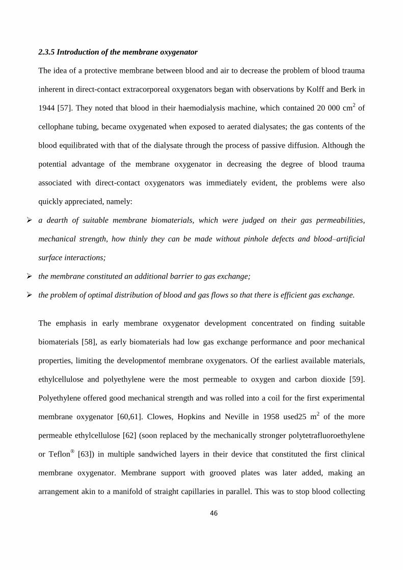
46
2.3.5 Introduction of the membrane oxygenator
The idea of a protective membrane between blood and air to decrease the problem of blood trauma
inherent in direct-contact extracorporeal oxygenators began with observations by Kolff and Berk in
1944 [57]. They noted that blood in their haemodialysis machine, which contained 20 000 cm2 of
cellophane tubing, became oxygenated when exposed to aerated dialysates; the gas contents of the
blood equilibrated with that of the dialysate through the process of passive diffusion. Although the
potential advantage of the membrane oxygenator in decreasing the degree of blood trauma
associated with direct-contact oxygenators was immediately evident, the problems were also
quickly appreciated, namely:
a dearth of suitable membrane biomaterials, which were judged on their gas permeabilities,
mechanical strength, how thinly they can be made without pinhole defects and blood–artificial
surface interactions;
the membrane constituted an additional barrier to gas exchange;
the problem of optimal distribution of blood and gas flows so that there is efficient gas exchange.
The emphasis in early membrane oxygenator development concentrated on finding suitable
biomaterials [58], as early biomaterials had low gas exchange performance and poor mechanical
properties, limiting the developmentof membrane oxygenators. Of the earliest available materials,
ethylcellulose and polyethylene were the most permeable to oxygen and carbon dioxide [59].
Polyethylene offered good mechanical strength and was rolled into a coil for the first experimental
membrane oxygenator [60,61]. Clowes, Hopkins and Neville in 1958 used25 m2 of the more
permeable ethylcellulose [62] (soon replaced by the mechanically stronger polytetrafluoroethylene
or Teflon® [63]) in multiple sandwiched layers in their device that constituted the first clinical
membrane oxygenator. Membrane support with grooved plates was later added, making an
arrangement akin to a manifold of straight capillaries in parallel. This was to stop blood collecting

47
unpredictably in thick rivulets, a problem similar to that of blood streaming in film oxygenators.
One disadvantage of hydrophilic membrane oxygenators is their tendency to leak plasma in a
manner akin to tents leaking water when wetted on the inside. This severely shortened the duration
of the use of the membrane lung. To prevent this, membranes made of hydrophobic polymers [58]
were used. These materials were initially derived from packaging materials used inthe capacitor
industry, such as polytetrafluoroethylene (Teflon®). With hydrophobic membranes, Melrose [64]
realised in 1958 that it is carbon dioxide removal that was the limitation, as carbon dioxide
solubility inhydrophobic solids is much less than its solubility inhydrophilic solids. To emphasise
the importance of carbon dioxide transfer, the term ‘membrane lung’ was coined. This problem was
partly solved by the use of silicone as the hydrophobic material in membrane lungs. Although
silicone had been discovered in 1947 and had played an important role as a defoaming agent in
direct contact devices, its excellent gas exchange properties [65] were not discovered until 1957.
Silicone had a permeability (the product of diffusivity and solubility) for oxygen from four to five
times higher than this for carbon dioxide, these figures being some 40 times higher than those for
Teflon®. Early silicone membranes had a number of problems. They had low mechanical strength
and were not free of pinholes when made into a thin film. Solutions were found to these problems.
In 1959, Thomas designed the first silicone membrane lung using a thin continuous silicone
membrane on a fabric support, which was made by dip-coating a nylon mesh, and the pairing of two
sheets of ultrathin membranes together so as to decrease the risk of leakages through random
pinhole defects. The definitive solution belonged to Burns [66] who, in1959, developed a new
process at Hammersmith Hospital in London for the low-cost production of thin films of silicone
membranes that were virtually free of pinholes and had greatly increased strength. Modern versions
of the silicone membrane oxygenators are still in use and are marketed as long-term extracorporeal
oxygenators [67].
48
2.3.6 Improving the gas exchange efficiency of membrane oxygenators
With sufficiently thin membranes, the gas exchange bottleneck was now recognised as having
shifted from the membrane to gas diffusion through blood. This was a result of the thickness of the
blood layer. Marx et al. [68] determined that oxygen transfer into a blood film is proportional to the
square of the thickness of the blood film and the diffusion resistance of the boundary layer. This
latter factor requires elaboration. In alveolar capillaries, the blood channels are one cell thick,
resulting in efficient gas exchange. In artificial oxygenators, the blood channels are much thicker.
With laminar flow, blood flows in orderly layers through these channels. The boundary layer, i.e.
the layer that is adjacent to the gas exchange surface, quickly equilibrates with the venting gas.
However, this equilibration is slow to spread through the other layers because of the low diffusivity
of gas through blood, with consequent deterioration of gas exchange efficiency. Subsequent
development therefore concentrated on minimising these two bottlenecks. The first silicone
capillary oxygenator, comprising capillaries that were 100–500 µm in diameter, was designed by
Bodell et al.[69] in 1963. The small diameter of the hollow fibres decreases the distance of the
blood layers from the gas exchange surface in two dimensions. Capillary oxygenators also have the
additional advantage of being better able to control the volumes of both gas and blood
compartments. Configurations with blood inside the fibres and oxygen outside and the reverse
configuration of blood outside the fibres and oxygen inside were tried. Wilsonet al. [70] showed
that the preferred configuration used blood flowing inside the fibres. However, the opposite is the
case with current microporous hollow-fibreoxygenators, as is described later. The second problem,
i.e. the diffusion resistance of the boundary layer, was addressed by the induction of secondary
flows in blood. Secondary flows are fluid flows that are additional to the mainstream. They may be
used to disrupt laminar flow and impart convective mass transfer to the mass of the fluid. Mixing
and therefore gas exchange performance are thus increased. Passive internal secondary flows are

49
induced by forcing blood to ‘eddy’ around passive obstacles, e.g. forcing blood to move in curved
paths around a helically wound tube [71] or by the insertion of surface elements into blood passages
[72]. Active secondary flows are induced by using energetic mechanisms to disrupt laminar flow
more effectively, resulting in greater gas exchange efficiency. Mechanisms devisedfor this include:
periodic deformation of the membrane through cyclical oxygen pressure to produce microscopic
foci of mixing [73];
a rocking membrane envelope [74];
pneumatic pulsation of the membrane [74];
arotating membrane-bound disk [74];
moving rod massage [75];
the toroidal membrane oxygenator [76,77];
the annular membrane oxygenator by Gaylor et al. [78];
vortex-shedding designs [79,80].
The measures employed were very successful in enhancing gas mass transfer. Kolobow’s device
[73] infact exceeded 200 ml oxygen.min-1
.m2, the limit for the thickness of the silicone membrane
used. This was because the membrane inverted and stretched into the gas side, increasing the
surface area when hypobaric pressure was applied. Kolobow’s device was mass-manufactured and
marketed by SciMed, then AveCor and now MedTronic, becoming the only solid silicone
membrane device consistently available for long-term life support during the past few decades [81].
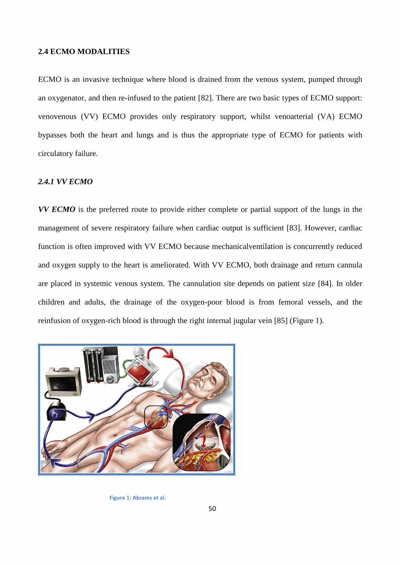
50
2.4 ECMO MODALITIES
ECMO is an invasive technique where blood is drained from the venous system, pumped through
an oxygenator, and then re-infused to the patient [82]. There are two basic types of ECMO support:
venovenous (VV) ECMO provides only respiratory support, whilst venoarterial (VA) ECMO
bypasses both the heart and lungs and is thus the appropriate type of ECMO for patients with
circulatory failure.
2.4.1 VV ECMO
VV ECMO is the preferred route to provide either complete or partial support of the lungs in the
management of severe respiratory failure when cardiac output is sufficient [83]. However, cardiac
function is often improved with VV ECMO because mechanicalventilation is concurrently reduced
and oxygen supply to the heart is ameliorated. With VV ECMO, both drainage and return cannula
are placed in systemic venous system. The cannulation site depends on patient size [84]. In older
children and adults, the drainage of the oxygen-poor blood is from femoral vessels, and the
reinfusion of oxygen-rich blood is through the right internal jugular vein [85] (Figure 1).
Figure 1: Abrams et al.

51
VV ECMO can also be delivered using a single double-lumen cannula inserted through the internal
jugular vein into the right atrium, although cannula size constraints have previously restricted this
technique to infants [86] (Figure 2).
Figure 2: Abrams et al.
One hundred percent oxygenated blood from the ECMO return cannula mixes with systemic venous
blood, which than traverses the pulmonary system. Theoretically, only oxygen-depleted systemic
venous blood enters the ECMO drainage cannula. However, depending on the positions of the
inserted cannula, a variable proportion of oxygenated blood from the return cannula enters the
drainage cannula, known as recirculation. Mixed venous oxygen saturation (SvO2) is determined by
the relative contributions of the ECMO return cannula (oxygenated blood) to pulmonary blood flow
and the systemic venous return (deoxygenated blood). Recirculation reduces the delivery of
oxygenated blood to the pulmonary artery, and thus reduces the SvO2. SaO2 is determined by
ECMO flow, patient’s systemic venous return, the degree of recirculation, the SvO2 venous blood

52
and pulmonary function. Of these parameters, ECMO circuit flow is the most important and easily
manipulated determinant of SaO2 in the absence of significant recirculation [87].
2.4.2 ECMO for Respiratory Failure
Acute respiratory distress syndrome.The most extensively studied respiratory indication for ECMO
is acute respiratory distress syndrome (ARDS) [88]. In circumstances in which invasive mechanical
ventilation is necessary to support gas exchange, positive-pressure ventilation may potentiate lung
injury [89]. The only ventilation strategy proved to reduce mortality in ARDS is a volume and
pressure-limited ventilation strategy [90]. ECMO has he potential to improve outcomes in patients
with ARDS by providing adequate oxygenation while facilitating lung protective ventilation by
correcting unsustainable levels of hypercapnia and respiratory acidosis that may accompany low–
tidal volume ventilation [91]. Potential indications for ECMO in the setting of ARDS have been
proposed [88].
Early randomized trials were unsuccessful in demonstrating a survival benefit from ECMO in
patients with severe forms of ARDS. More recently, the impact of modern extracorporeal support
on survival in patients with severe ARDS was evaluated in the Conventional Ventilation or ECMO
for Severe Adult Respiratory Failure trial, in which 180 subjects with severe, potentially reversible
respiratory failure were randomized to conventional mechanical ventilation or referral to a
specialized center for consideration of ECMO (92). Compared with the conventionally managed
group, those referred for consideration of ECMO had a significantly lower rate of death or severe
disability at 6 months (37% vs. 53%; relative risk: 0.69; p = 0.03).
Other nonrandomized observational studies, particularly during the influenza A (H1N1) pandemic
in 2009, have shown conflicting results of the impact of ECMO on survival in severe ARDS [93].

53
Hypercapnic respiratory failure. With an improved risk benefit ratio, there is great potential to use
ECCO2R to manage hypercapnic respiratory failure, thereby minimizing or even eliminating the
need for a ventilator. In chronic obstructive pulmonary disease, invasive mechanical ventilation is
associated with multiple complications, including dynamic hyperinflation and elevations in intrinsic
positive end-expiratory pressure, ventilator-associated pneumonia, and impaired delivery of
aerosolized medications [94,95]. Several small case series have demonstrated the feasibility of
avoidance of or rapid weaning from invasive mechanical ventilation, with ECCO2R used to manage
gas exchange [96–98]. With correction of hypercapnia and respiratory acidosis, dyspnea and work
of breathing rapidly improve, facilitating early mobilization [98]. Although early mobilization has
been described with invasive mechanical ventilation [99], it is more likely to be successful with the
substitution of ECCO2R for mechanical ventilation because of the significant improvement in
dyspnea that is seen with ECCO2R compared with mechanical ventilation in the hypercapnic
population [98]. The risks of ECCO2R must be weighed against the benefit of minimizing invasive
mechanical ventilation, and additional studies are required to define the ideal patient population and
the economic impact of such a strategy before it can be recommended for clinical use. Similarly,
ECCO2R may be considered in other forms of hypercapnic respiratory failure, including refractory
status asthmaticus, in which the ability to avoid invasive mechanical ventilation altogether is
potentially advantageous (100,101).
Bridge to lung transplantation and post-transplantation primary graft dysfunction. Although
ECMO has traditionally been considered a relative contraindication to lung transplantation because
of poor perioperative outcomes [102], more recent studies have reported excellent post
transplantation survival, especially at centers with more extensive experience [103,104]. With the
potential for ECMO to provide sufficient gas exchange to supplant the ventilator, a nonintubated
ECMO strategy may be considered for some transplantation candidates who would otherwise be

54
ventilator dependent, a population with poor outcomes related to ventilator-associated
complications [105]. Outcomes may be further optimized when such a strategy is combined with
active physical therapy and should be considered in patients who would otherwise be inactivated
from the transplantation list because of deconditioning. This is particularly true for those patients in
whom ECMO or ECCO2R alleviates dyspnea sufficiently to permit rehabilitation [104,106,107]. A
major limitation to the use of ECMO for end-stage respiratory failure is the lack of a destination
device therapy, with ECMO remaining an intervention for which an intensive care unit is required.
In severe cases of primary graft dysfunction, a form of acute lung injury that is the leading cause of
early death after lung transplantation [108], ECMO may be used to support gas exchange while the
allograft recovers. Studies have described similar survival in cases of ECMO-supported severe
primary graft dysfunction compared with those with less severe primary graft dysfunction without
ECMO support, particularly when instituted early [109]. However, ECMO has not been shown to
affect long-term allograft function.

55
2.4.3 VA ECMO
VA ECMO is essentially a modification of the cardiopulmonary bypass circuit which is used
routinely in cardiac surgery. In VA ECMO, oxygen-depleted blood drains into the circuit from
venous system and oxygenated blood is pumped back into the systemic side, which both augments
gas exchange and supports cardiac function. Cannulation for VA ECMO can be established
centrally or peripherally. With central cannulation (Figure 3), blood is drained directly from the
right atrium and return to the proximal of ascending aorta.
Figure 3: Marasco et al.

56
Figure 4: Marasco et al.
With peripheral cannulation (Figure 4), blood is drained from the proximal great veins (via a
femoralor jugular vein) using a surgically cut-down or modified Seldinger technique, and returned
to the aorta via cannulation of a carotid, axillary or femoral artery (Figure 5).

57
Figure 5: Abrams et al.
Systemic arterial blood flow is the total of the ECMO circuit flow and any ejection from the left
ventricle. Systemic blood pressure is determined by total arterial blood flow and the vascular tone.
When left ventricle (LV) cannot contribute to the systemic arterial blood flow, the patient’s
systemic arterial oxygen saturation (SaO2) is determined entirely by the oxygen saturation of blood
in the ECMO return cannula, which is 100%. However, if there is a contribution from the LV, SaO2
depends on the relative flow and oxygen saturation of blood from both the ECMO circuit and blood
ejected by LV. This detail becomes important if you place the ECMO return cannula via femoral
artery in a patient suffering from a severely compromised lung function. In this condition, coronary
arteries, cerebral blood vessels and upper limbs are under threat of hypoxaemia because proximal
branches of the aorta receive predominantly deoxygenated blood ejected from the left heart. Even
though there is a presence of sufficient LV function, this situation persists as long as pulmonary
function is good or the return cannula is placed centrally [87].

58
ECMO remains a short-term support device, essentially the circuit is smaller than a standard
cardiopulmonary bypass circuit, transportable and closed to the atmosphere. The cannulae are also
specifically designed for ECMO. The duration of support with ECMO has greatly increased with
improving oxygenators and medical management,and whereas support was previously in the order
of days, patients can now be maintained on ECMO for weeks. In most patients the duration of
support required is approximately1 week. Most commonly, it is instituted in an emergency or urgent
situation after failure of other treatment modalities.
2.4.4 ECMO for Cardiac Failure
In terms of cardiac failure, the most common indications for ECMO are post-cardiotomy (that is
unable to get the patient off cardiopulmonary bypass following cardiac surgery) [110], post-heart
transplant (usually due to primary graft failure) [111] and severe cardiac failure due to almost any
other cause (e.g. decompensated cardiomyopathy, myocarditis, acute coronary syndrome with
cardiogenic shock, profound cardiac depression due to drug overdose or sepsis).
In making the decision to institute ECMO, several considerations must be weighed. Most
importantly, one must consider the likelihood of organ recovery. If the organ failure is thought to be
reversible with therapy and rest on ECMO, then institution of this mechanical support is
appropriate. If recovery is thought not to be a possibility in that particular patient, then other factors
must be taken into account. With regards to cardiac failure, if recovery is not expected, what other
options are there available for this patient? Consideration must be given to the patient’s eligibility
for transplantation, other mechanical assist devices to bridge the patient to transplant or whether it is
appropriate for a definitive mechanical assist device to be inserted as destination therapy. Obviously
implantation of ECMO in an 80 year old with no hope of native organ recovery is a futile exercise
as there is no endpoint for this patient’s treatment. Such implementation of ECMO could be seen as

59
unethical in that it puts a patient (and their family) through a futile exercise, falsely raises their
hopes, utilizes resources better used on someone with achance of survival and blocks an intensive
care bed. Other contraindications to the institution of ECMO include disseminated malignancy,
advanced age, graft vs.host disease, known severe brain injury, unwitnessed cardiac arrest or
cardiac arrest of prolonged duration. There may be other technical contraindications to consider
such as aortic dissection or aortic incompetence (discussed below).
If ECMO has been instituted for cardiac support, thereare several other end points to their recovery
pathway. ECMO can either be weaned if the heart has recovered, a more permanent mechanical
device can be inserted orthe patient may receive a heart transplant. The more permanent mechanical
devices which can be implanted allow the patient to ambulate, rehabilitate, and be discharged home
with the device. The aim then is to either wait for further cardiac recovery to allow explant of the
device or to list the patient for transplantation. Patients can be supported by a VAD (Ventricular
Assist Device) at home for months or even a year prior to receiving a heart transplant. More
recently, fully implantable mechanical assist devices are being used as destination therapy.
Destination therapy is considered in patients who have end stage heart failure which has not
responded to any other form of treatment, and who are not suitable for transplantation. This is
usually due to advanced age (>65) or other comorbidities. Several technical considerations need to
be taken into account also before instituting ECMO. Aortic incompetence is a relative
contraindication, particularly if severe. In such situations consideration of aortic valve replacement
would need to be made. Lesser degrees of aortic incompetence may be managed with a left
ventricular vent. Failure to insert a vent leads to ventricular distension, compromised
subendocardial blood flow and would impact on recovery and the ability to wean ECMO.
Post-operative cardiogenic shock and post-transplant primary graft failure. Post-cardiotomy
cardiogenic shock is an uncommon but highly lethal complication of cardiac surgery. ECMO may

60
be considered as temporary support post-operatively, particularly when unable to wean from
cardiopulmonary bypass in the operating room [112]. Mortality in patients requiring this level of
support remains high [113]. Primary graft failure (PGF) is a well-recognized complication of heart
transplantation associated with a high mortality, for which ECMO has been used as temporary
support [114,115]. As expected, overall survival for patients with PGF requiring ECMO is worse
than in those who do not develop PGF. However, patients with ECMO-supported PGF who survive
beyond the early post-transplantation period have comparable long-term survival with non-PGF
transplant recipients [115,116].
Cardiogenic shock complicating acute myocardial infarction. There are no randomized controlled
trials comparing ECMO with other mechanical support systems in myocardial infarction–associated
cardiogenic shock, but several nonrandomized studies suggest a survival advantage from the early
use of ECMO in such circumstances (117,118). In an observational study of patients with ST-
segment elevation myocardial infarction–related cardiogenic shock undergoing percutaneous
coronary intervention (PCI) with and without ECMO support, those receiving ECMO had
significantly lower 30-day mortality (39.1% vs. 72%;p = 0.008) (54). Interpretation of these data is
limited by the fact that cohorts were enrolled over different time frames (1993 to 2002 for the non-
ECMO cohort vs. 2002 to 2009 for the ECMO cohort), potentially leading to discrepancies in
treatment between groups, especially given that coronary stents were unavailable at the study center
before 1998. Higher rates of Thrombolysis In Myocardial Infarction flowgrade 3 achieved in the
ECMO group may reflect improved hemodynamic stability in the catheterization laboratory or,
alternatively, may be a consequence of improved PCI technique over time. Ultimately, randomized
controlled trials are needed to determine the true benefit, if any, of ECMO in myocardial infarction–
associated cardiogenic shock.
61
Fulminant myocarditis. ECMO use has been investigated as a modality to support non ischemic
cardiogenic shock, including fulminant myocarditis [119-122]. Patients with fulminant myocarditis
who are successfully bridged with ECMO to recovery may have long-term prognoses comparable
with those of hemodynamically stable patients with acute myocarditis [119]. In 1 cohort of patients
who received either a biventricular assist device (n = 6) or ECMO (n = 35) for fulminant
myocarditis with refractory cardiogenic shock, overall intensive care unit survival was 68%, with
higher severity of illness and elevated cardiac biomarkers serving as independent predictors of
mortality and an inability to wean from ECMO (121). ECMO may be as efficacious as a ventricular
assist device (VAD) while having the advantage of being less invasive. In a study comparing
ECMO with biventricular assist devices for fulminant myocarditis, those receiving ECMO had
comparable rates of weaning from device therapy and survival to hospital discharge without the
need for transplantation (83% vs. 80%) and more rapid improvement in renal and hepatic laboratory
profiles, despite having a higher severity of illness and worse left ventricular function before device
implantation [122].
Sepsis-associated cardiomyopathy. Myocardial depression is a well-recognized consequence of
severe septic shock [123]. There are emerging data suggesting that ECMO may have a role in
supporting patients who develop refractory cardiac failure in this setting [124,125]. Larger studies
are needed to determine whether the benefit of ECMO outweighs the risk, especially in cases in
which septic shock is complicated by marked disturbances in coagulation.
Pulmonary hypertension. ECMO is an emerging management option in patients with
decompensated pulmonary hypertension with concomitant right ventricular failure, particularly
when there is an acutely reversible process, medical management has not been optimized, or lung
transplantation is a consideration [126]. ECMO for this indication typically requires a femoral
venoarterial configuration to bypass the high resistance of the pulmonary vasculature and

62
decompress the right ventricle. However, 3 configurations have been used to avoid femoral
cannulation. Internal jugular venous drainage may be combined with subclavian arterial reinfusion.
In patients with pre-existing intra-atrial defects, a dual-lumen cannula maybe oriented with the
reinfusion jet directed across the defect (rather than across the tricuspid valve), effectively
providing an oxygenated right-to-left shunt while decompressing the right ventricle [127,128].
Additionally, arteriovenous ECMO can be inserted between the main pulmonary artery and the left
atrium, though this typically requires a sternotomy [129]. When ECMO is initiated as a bridge to
recovery, pulmonary vasodilators may be optimized while the underlying acute process is treated
[126]. When the goal of ECMO is to bridge to transplantation, pulmonary vasodilators may be
down-titrated to preferentially shunt blood through the extracorporeal circuit and away from the
high-resistance pulmonary vasculature, thereby optimizing systemic oxygenation [130]. Reducing
the dosage of intravenous pulmonary vasodilators will also minimize the degree of systemic
vasodilation that may occur as the medications pass through the ECMO circuit and into the arterial
circulation.
Pulmonary embolism. Massive pulmonary embolism may likewise benefit from ECMO. A
retrospective single-center review of ECMO for massive pulmonary embolism, including patients in
active cardiac arrest, demonstrated 62% overall survival when combined with anticoagulation or
surgical embolectomy [131]. The combination of ECMO, thrombolysis, and catheter-directed
thrombectomy or embolus fragmentation has also been reported, with 30-day survival of 70%
[132].
Extracorporeal cardiopulmonary resuscitation. “Extracorporeal cardiopulmonary resuscitation”
(ECPR) is the term used to describe the institution of extracorporeal support to restore circulation
during cardiac arrest. Although there are no randomized controlled trials investigating the efficacy
of ECPR for cardiac arrest, its use has been steadily increasing [133–136). In a prospective,

63
observational study of witnessed in-hospital cardiac arrests, propensity analysis matching 46
subjects who received conventional cardiopulmonary resuscitation (CPR) with 46 subjects who
received ECPR demonstrated significantly higher survival at discharge in the ECPR group (32.6%
vs. 17.4%; p < 0.0001) and at 1 year (hazard ratio: 0.53; p = 0.006) (69), with a trend toward
improved neurological outcomes. In multivariate analysis, an initial rhythm of ventricular
fibrillation or ventricular tachycardia and use of ECPR were positively associated with survival to
discharge. A more recent propensity analysis of patients who experienced in-hospital cardiac arrest
demonstrated significantly higher 2-year survival with minimal neurological impairment in those
treated with ECPR compared with conventional CPR (20% vs. 5% p = 0.002). Independent
predictors associated with minimal neurological impairment included age <65 years, CPR duration
<35 min, and subsequent cardiovascular intervention [135]. Regarding out-of-hospital cardiac
arrests, a recent matched propensity analysis demonstrated significantly higher neurologically intact
survival at 3 months in those who received ECPR compared with conventional CPR (29.2% vs.
8.3%; p = 0.018) (72).With the ability of ECMO to both maintain systemic circulation during
cardiac arrest and increase coronary perfusion pressure, the combination of ECPR and intra-arrest
PCI may greatly improve the likelihood of successful defibrillation and recovery in cardiac arrest
due to an acute coronary syndrome. A multicenter nonrandomized study demonstrated the
feasibility of combining ECMO and emergency coronary angiography in 81 subjects, 61 of whom
received intra-arrest PCI [137]. Compared with those who received ECMO and coronary
angiography without PCI, those receiving PCI achieved higher rates of resumption of spontaneous
beating (100% vs. 60%; p < 0.001), weaning from ECMO (59% vs. 28%; p = 0.009), 30-day
survival (36% vs. 12%; p = 0.03), and favorable neurological outcomes(33% vs. 4%; p = 0.005). In-
hospital (vs. out-of-hospital) cardiac arrest and shorter duration from collapse to initiation of ECMO
were correlated with 30-day survival. Although observational trials suggest a possible survival
advantage of ECPR over conventional CPR, overall survival remains relatively low. More research

64
is needed to define the patient population that would derive the greatest benefit from extracorporeal
resuscitation, with an emphasis on survival with minimal neurological impairment. The 2010
American Heart Association guidelines for cardiopulmonary resuscitation and emergency
cardiovascular care do not recommend the routine use of ECPR for cardiac arrest. However, ECPR
may be considered when the time without spontaneous circulation is short, resuscitation attempts
are adequate, and the cause of cardiac arrest is potentially reversible [138]. The guidelines
emphasize that ECPR use should be restricted to centers at which it is readily available and that its
initiation and management require highly trained personnel and specialized equipment.
Bridge to VAD implantation or heart transplantation. VADs may be used as a bridging therapy to
heart transplantationin appropriately selected patients with severe cardiac dysfunction [139], with
the advantage of being able to function as destination devices if transplantation is not feasible.
ECMO has also been reported as a bridging therapy to heart transplantation or VAD implantation or
as a bridge to decision when prognosis is uncertain [140–142]. However,the duration of support that
can be provided is shorter than for VADs, making transplantation or transition to VAD of greater
urgency, and patients receiving ECMO support must remain within an intensive care unit [142].
Success of ECMO bridging varies greatly and depends in large part on pre-ECMO patient
characteristics and organ availability in the cases in which transplantation is the goal. In a
retrospective review of 70 patients in whom ECMO was used as a bridgeto heart transplantation, 31
(44%) were bridged to either heart transplantation (n = 15) or VAD implantation (n = 16), though
only 11 (73%) and 8 (50%) of the heart transplantand VAD recipients, respectively, survived to
hospital discharge, highlighting the limitations of bridging therapy in this highly morbid patient
population [140]. Age > 50 years, CPR before ECMO initiation, and high sequential organ failure
assessment score were independent predictors of unsuccessful bridging. Pre-implantation CPR as a
predictor of poor outcomes was corroborated in a more recent single-center study of 90 patients

65
who received mechanical circulatory support (VAD or ECMO) for refractory cardiogenic shock
[143]. Forty-nine percent received short-term VAD support as a bridge to decision, and 51%
received ECMO when neurological status was uncertain or there was complete hemodynamic
collapse or severe coagulopathy. Overall survival was 49%, with 26% of patients transitioned to
implantable VADs, 18% recovering sufficient native cardiac function, and 11% bridged to
transplantation. CPR at the time of implantation was an independent predictor of in-hospital
mortality (odds ratio: 5.79; p = 0.022). These studies highlight the need for careful consideration of
relative and absolute contraindications to mechanical circulatory support. In particular, ECMO for
cardiogenic shock superimposed on chronic cardiomyopathymay be associated with particularly
poor outcomes [144].
ECMO to prevent acute right ventricular failure after LVAD implantation. Both femoral
venoarterial ECMO and percutaneous venous-to-pulmonary arterial ECMO have been successfully
used to provide right ventricular support in patients with biventricular dysfunction undergoing
LVAD implantation [145,146]. In this setting, ECMO can allow time for the already compromised
right ventricle to get accustomed to the increasing preload,thereby avoiding distension and right
ventricular failure leading to poor filling of the LVAD.

66
2.5 ECMO CIRCUITS AND EQUIPMENT
Basically, an ECMO circuit consists of the drainage and return cannulae, tubing, a driving force
(pump) and a gas/heat exchanger. Apart from differences in the cannulae, similar circuits are used
for VV and VA ECMO.
2.5.1 Pump
Most ECMO circuits use two-types of pumps: roller and centrifugal. The literature is inconsistent
with regard to biocompatibility and complication rates comparing these two pump principles, with a
slight tendency in favour of the roller pump [146–148]. Centrifugal pumps consist of a vaned
impeller or nested, smooth plastic cones, which when rotated rapidly up to 3000 revolutions/min,
propel blood by centrifugal force [149]. Centrifugal blood pumps generate up to 900 mmHg of
forward pressure, but only 400–500 mmHg of negative pressure, and therefore less cavitation and
fewer gaseous microemboli. Blood flow is preload and afterload dependent. Thus, there is no fixed
relationship between pump speed and blood flow, necessitating the presence of a flowmeter within
the circuit. They can pump small amounts of air, but become “deprimed” if more than 30–50 ml of
air enters the blood chamber. If hypovolaemia occurs, the inlet pressure becomes more negative and
pump speed remains constant, however the blood flow rate is decreased. During VA ECMO, a
change in systemic vascular resistance changes the relationship between the circuit flow and the
speed of the pump. Similarly, pump failure during VA ECMO can result in a reverse of the flow in
the circuit [87]. Roller pumps consist of a length of 1/4- to 5/8-inch (internal diameter) tubing,
which is compressed by two rollers 180° apart inside a curved raceway. Rollers mounted on a
rotating arm progressively compress a segment of tubing pushing blood ahead of the roller and flow
rate depends on the diameter of the tubing rate of rotation, the length of the compression raceway,
and completeness of compression. A roller pump is usually used in conjunction with a blood-filled
67
bladder sited between the drainage cannula and the pump to allow continuous pumping despite the
alterations in the patient’s intravascular effective volume. If hypovolaemia occurs, pump speed and
therefore blood flow decrease. Roller pumps are afterload independent. During VA ECMO, a
significant change in systemic vascular resistance does not influence blood pumping. Since the
driving force of drainage into the bladder is gravity, roller pumps have to be kept below the level of
patient, which becomes crucial when transporting patients on ECMO. With roller pumps, there is a
direct relationship between pump rate and blood flow; thus, a flowmeter is not required. Roller
pumps are cheaper, reliable, safe and have small priming volumes, but can produce high negative
pressures and micro-particles shed from compressed tubing (spallation)[150]. Roller pumps are
vulnerable to careless operation,back-flow when not in use if rollers are not sufficiently occlusive,
excessive pressure with rupture of connections if the return tubing becomes obstructed, tears in
tubing, and changing roller compression settings during the operation. Roller pumps, but not
centrifugal pumps, are used for sucker systems and for delivering cardioplegic solutions.
Centrifugal pumps produce pulseless blood flow and standard roller pumps produce a sine wave
pulse around 5 mmHg. The return tubing cannula dampens the pulse of pulsatile pumps, and is
difficult to generate pulse-pressures above 20 mmHg within the body during full CPB [151].
The two principles can so far be looked at as being more or less equivalent, even for long-term
extracorporeal support. Some centres use two pumps in parallel, thus backing up their unit in case
of pump or oxygenator malfunction.
A nonocclusive pump avoids stress to blood components while abolishing negative pressures acting
on the drained blood. However, in relation to the pump rate and the patient’s intravascular volume,
various amounts of blood might be pumped at a time. Using a servo-controlled mechanism is
another way to prevent excessive negative pressure. Servo-controlled pumping allows setting of a
calculated maximum negative pressure and slows down the rate of the pump when you are close to

68
the limit [86]. An exciting development was the discovery of intubing pumps which are the
miniaturised forms of high-end pumps that are able to pump blood flows between 0 and 10 L/min.
These pumps require very small amounts of priming volumes and can be integrated at any location
of the ECMO. Inline pumps can support arteriovenous circuits at any time,drive VV orVA circuits;
and can be placed at the bedside.
2.5.2 Oxygenators
Membrane oxygenators mimic the human lung by interspersing a thin membrane of either
microporous polypropylene hollow fibre (0.3- to 0.8-μm pores) or non-microporous silicone rubber
between the gas and blood phases. Membrane oxygenators are safer than bubble oxygenators
because they produce less particulate and gaseous microemboli, are less reactive to blood elements,
and allow superior control of blood gases [152]. Considering microporous membrane oxygenators,
while preventing gas from entering blood, plasma-filled pores also facilitate transfer of both oxygen
and carbon dioxide. In order to obtain a sufficient amount of oxygen, blood has to be spread as a
thin film over a large area with high differential gas pressures because oxygen is poorly diffusible in
plasma whereas carbon dioxide is highly diffusible in plasma so it can easily be removed from the
blood. Areas of turbulence and secondary flow increase diffusion of oxygen within blood.
Considering non-microporous membrane oxygenators, O2 and CO2 diffuse across thin silicone
membranes, which are made into envelopes and wound around a spool to produce a spiral coil
oxygenator. Gas passes through the envelope and blood passes between the coil windings.
Polypropylene hollow-fibre oxygenators are the standard oxygenators used during CPB. Their
priming volumes are small, gas-transfer rates are high and resistance is low so they are at least
theoretically superior to silicone membrane oxygenators [153]. However, in the past series, it was
discovered that over time, the micropores become permeable to fluid causing plasma to leak into the

69
gas phase and out the exhalation port resulting in deteriorating gas exchange and developing
coagulopathy. Because of protein leakage that deteriorates membrane function, these spiral coil
oxygenators are preferred over hollow fibre microporous oxygenators for the prolonged perfusions
(>2–3 days) [154].
Peek et al. compared silicone membrane oxygenators with polypropylene hollow-fibre oxygenators
and concluded that, silicone oxygenators have excellent biocompatibility and durability but provide
less efficient gas exchange, are bulky, are difficult to prime and have high resistance to blood flow
[155].
In 2002, Wiesenack et al. tested a new membrane oxygenator featuring a very thin (0.05μm), solid
membrane on the blood side of a highly porous support matrix. This membrane reduced the risk of
gas emboli and plasma leakage during prolonged CPB, but impaired transfer of volatile anaesthetics
[156].
Haworth directed the oxygenator development to the construction of smaller devices with low-
priming volumes, high gas transfer rates, and a lower resistance against the streaming blood [153].
Another very interesting development was the introduction of small silicone rubber hollow fibre
oxygenators by Motomura et al., reaching gas transfer rates very similar to those of conventional
hollow fibre oxygenators [157].
In the early 2000s, a new generation of oxygenator was introduced into clinical practice, containing
non-microporous hollow fibres constructed of polymethylpentene (PMP). PMP oxygenators
combine the durability of silicone membranes with the ease of use and efficient gas exchange of
hollow-fibre construction. In 2005, Khoshbin et al. [155], suggested that, compared with silicone
membrane oxygenators, PMP oxygenators are associated with reduced red cell and platelet
70
transfusion and improved gas change. Compared with polypropylene hollow-fibre oxygenators,
PMP oxygenators have improved durability and reduced requirement for blood product transfusion.
2.5.3 Cannulae and Tubing
The tip of the drainage cannula is usually the narrowest part of the perfusion system and
inappropriate catheters can produce high pressure differentials and turbulence. To put it simply, the
larger the diameter of the cannula, the better the flow. Usually for the adult patients, the drainage
cannula should be 23F to 25F, and the return cannula should be 17F to 21F. The various
components of the heart–lung machine are connected by polyvinyl tubing and fluted polycarbonate
connectors. Medical grade polyvinyl chloride tubing is universally used because it’s flexible,
compatible with blood, inert, nontoxic, smooth, transparent, and resistant to kinking and collapse,
and can be heat sterilised. Bioactive coating of artificial surfaces to decrease the necessity of
systemic anticoagulation is another issue that has an important effect on the rate of complications
during ECMO procedures [159]. Heparin can be attached to blood surfaces of all components of the
extracorporeal circuit by ionic or covalent bonds. There are many reports that have studied the use
of heparin-coated circuits but the effectiveness of them remains controversial because studies are
contaminated by patient selection, reduced doses of systemic heparin, and washing or discarding
field-aspirated blood [160]. Little is known about the use of heparin-coated ECMO units in patients
with heparin/platelet factor4 (HPF 4) antibodies, very probably responsible for the development of
heparin induced thrombocytopaenia type II (HIT II) [86]. Koster et al., suggested that heparin
immobilised on artificial surfaces neither induces HPF 4 antibody formation nor supports its
persistence [161]. On the contrary, Cruz et al. pointed out a relation between HIT II development
and thromboembolic complications in patients with heparin-coated cardiac stents [162]. In general,
heparin as a substance acting late in the coagulation cascade seems not to be the ideal drug to be
used for the purpose of biocompatibility.

71
Direct contact between blood and the ECMO circuit leads to activation of inflammatory mediators,
particularly the complement activation pathway [163]. Although a majority of studies indicate that
heparin coatings reduce concentrations of C3a and C5b-9 [164,165], the inflammatory response to
extracorporeal circulation is not reduced and the evidence for clinical benefit is not convincing
[166].
2.5.4 Heat Exchangers
Heat exchangers control body temperature by changing the temperature of blood passing through
the perfusion circuit. Hypothermia is preferred in cardiovascular surgery in order to reduce oxygen
demand. Gases are more soluble in cold blood so rapid rewarming of cold blood may cause
formation of bubbles therefore gas emboli [167]. Most membrane oxygenator circuits incorporate a
heat exchanger upstream to the oxygenator to minimize bubble emboli. Blood is not heated over
40°C to prevent denaturation of plasma proteins, and temperature difference within the body and
perfusion circuit are limited to 5–10°C to prevent bubble emboli.

72
2.6 ANTICOAGULATION FOR ECMO
The goal of anticoagulation for ECMO is to adjust such an equilibrium between attenuating platelet
and thrombin activation to prevent life-threatening thrombosis and providing sufficient clotting to
prevent excessive bleeding. Furthermore, an accurate device that has an ability to assess the degree
of anticoagulation is not clinically available. Unidentified macroscopic clots cause a variety of
thromboembolic events in patients considered adequately anticoagulated [168]. It seems to be that
the effectiveness of anticoagulation worsens with the duration of ECMO. Monitoring for
anticoagulation is the heart beat of ECMO management. The most popular test to monitor
anticoagulation is the activated clotting time (ACT) and ACT still remains the predominant test to
manage heparin anticoagulation during ECMO. It measures the integrity of the intrinsic coagulation
and common pathways. However, the ACT’scapability to correctly measure the level of
anticoagulation has been questioned [169]. The concerns about ACT’s accuracy are: test results are
affected by coagulopathy, maturity of coagulation system, platelet dysfunction, degree of
hypothermia, antithrombin level, age, haemodilution, sample size, and temperature [170,171].
Ongoing synthesis of thrombin is another problem especially in children and may result in
inadequate anticoagulation despite high levels of ACT [171–173]. The popular range for the ACT
with heparin during ECMOhas been 180–220 s [174]. Baird et al. analysed 604 consecutive
paediatric ECMO patients retrospectively for factors that affected outcome [170]. The mean ACT
for all patients was 227 ± 50 s but the range of 158–620 s was very broad. They suggested that
increased heparin dosing increases survival. The measurement of heparin concentration is an option
to manage anticoagulation. Laboratory derived heparin concentration is the gold standard but is not
easily or quickly obtained for patients on. The Hepcon (MedtronicPerfusion Systems, Minneapolis,
MN) is able to provide heparin concentrations that have relatively good correlation to the laboratory
73
derived anti-Xa plasma heparin measurements [175] and has demonstrated superior results
compared with ACT [174]. More importantly, unlike the ACT, use of heparin concentrations have
the benefit of being less sensitive to changes in the patient’s platelet and clotting factor levels.
However, studies of heparin concentration monitoring for anticoagulation during ECMO are few
compared to the ACT so target heparin levels have not been determined. Several centres used
viscoelastic tests, such as thromboelastogram (TEG/ROTEM) [176]. The TEG/ROTEM is a device
that measures the viscoelastic properties of the blood to examine the whole clotting system instead
of isolated parts. It provides ongoing coagulation profiles looking at not only the initiation of
clotting but also the strength and dissolution of the clot as in the case of fibrinolysis. The activated
partial thromboplastin time (APTT) is universally recognised as a standard monitor for heparin
therapy except when high heparin dosing is required as in CPB. In situations that do not require
high heparin dosing, such as ECMO, the APTT is a valuable tool to assess anticoagulation. In a
comparison between laboratory APTT and ACT, theACT was found to correlate poorly with the
APTT [168]. Furthermore, in very ill patients requiring continuous heparin infusion, ACT could not
delineate between low and moderate levels of anticoagulation compared with the APTT[177]. The
APTT that will prevent thrombus extension with heparin has been reported as 1.5 times baseline
APTT [178]. This APTT corresponds to a heparin level of 0.2–0.3 u/mL and does not correlate
moderately well with heparin concentrations [179] .Heparin-continues to be the first choice for
ECMO because it is rapidly acting, easily reversible, cheap, easily available, and well tolerated by
paediatric and adult patients. Biologic activity varies between 30 min and six hours depending on
the systemic heparin concentration. Heparin dosing for adults and paediatric patients is different for
several reasons. The larger blood volume/weight ratios in neonates compared with adults require
greater heparin dosing [168]. The more rapid metabolic rates with neonates and infants may allow
greater excretion by the kidneys and therefore affects heparin dosing. Differences between adults
and paediatric patients may also derive from the differences in thrombin generation. Traditionally

74
heparin dosing ranges between 20 and 70 U/kg/h for ECMO. Most agree that the APTT should be
1.5–2.5 times the control. The most recent studies of heparin concentrations suggest a value 0.3–0.7
u/mL[179]. Heparin dosing for an effective anticoagulation is closely connected with antithrombin
concentration. Reduced heparin responsiveness is called heparin resistance and for effective
inhibition of thrombin, adequate levels of antithrombin are required. It is clear that prolonged
durations of ECMO will lead to consumption of antithrombin and the patient may not be capable of
maintaining the level. Koster et al., used bivalirudin, a direct thrombin inhibitor, in a patient with
myocardial failure after repair of acute type A aortic dissection who developed acute heparin-
induced thrombocytopaenia during ECMO and achieved successful results in terms of postoperative
blood loss and transfusion requirements [180].

75
2.7 MANAGEMENT OF ECMO: Maintenance and Weaning of ECMO
Achieving the suggested range of anticoagulation and cannulation, ECMO is commenced by
unclamping the circuit and slowly increasing flows to the target range. Standard initial settings and
goals for ECMO are specified in literature and listed in Figure 6 [87,181]
Figure 6: Gokhan Lafc et al.
The basic function of VA-ECMO in supplying mechanical circulatory support is to drain blood
from the venous circulation, oxygenate it and then return it to the arterial circulation at physiologic
perfusion pressures. Although ECMO does a very good job of unloading the right ventricle, it does
not do as good a job of unloading the left ventricle, even though left ventricular preload is
significantly reduced by the diminished return from the lungs. For this reason, attempts to improve
left ventricular contractility, reducing left ventricular distension and clot formation are of utmost
importance. Such measures should include inotropic support and may include intra-aortic-balloon
pumping (IABP). Another option in central ECMO is to insert a left ventricular vent to empty the
ventricle, splicing this line into the venous line of the ECMO circuit. In peripheral ECMO however,

76
the use of an IABP can be detrimental as the peripheral arterial flow from the femoral arterial
cannula will be competing with the inflated state of the IABP. ECMO flow can be very volume
dependent and will drop with hypovolaemia, cannula malposition, pneumothorax and pericardial
tamponade. This usually manifests as ‘kicking’ or ‘chatter’ of the venous tubing as well as a drop in
output. Management includes a volume challenge, exclusion of intra-abdominal distension or
compartment syndrome, cardiac tamponade or pneumothorax. If this does not work, a slight
reduction in flows may be helpful or there may be a need to insert another venous cannula. Further,
centrifugal pumps in contrast to roller pumps are very afterload dependent and therefore
hypertension is another variable that can reduce flows and should be avoided. Whilst on ECMO, the
aim is to support and rest the heart and/or lungs. From a respiratory point of view, hypoxia is treated
by increasing the flow rate and FiO2 of the ECMO circuit, not by altering the FiO2 and PEEP on the
ventilator. Attempts should be made to wean the FiO2 on the ventilator and maintain a PEEP level
of 5–10 cmH2O. A protective lung ventilation strategy with low plateau pressures and low tidal
volumes should be aimed for as well as low respiratory rates, unless trying to wean off the ECMO
circuit. PCO2 control should be via the ECMO fresh gas flow to the oxygenator, not by altering the
respiratory rate on theventilator. From a cardiac point of view the aim should be to minimise the use
of inotropes and thus rest the heart. However, often a low-dose inotrope infusion is maintained to
ensure some contractility and adequate emptying of the left ventricle. It is also important to ensure
the patient is not hypovolaemic. There are no standardised methods or techniques with regards to
weaning ECMO. With regards to VV ECMO, the actual ECMO flows do not need to be altered to
assess native respiratory function—this is done by altering gas flow through the ECMO circuit. The
patient may be weanable if gas exchange is able to be maintained with a low FiO2 on the
oxygenator (e.g. <30%) and low fresh gas flow rates into the circuit (e.g. <2 L/min) provided that
the respiratory rate and PEEP set on the ventilator are not too high (e.g. <25 breaths/min and
<15cmH2O, respectively).With regards to VA ECMO, factors indicating cardiac recovery and thus

77
a potentially weanable patient include; an increasing blood pressure which may need vasodilators,
return of pulsatility or increasing pulsatility on the arterial pressure waveform, falling pO2 by a right
radial arterial line indicating more blood is being pumped through the heart which may be less well
oxygenated, and falling central venous and/or pulmonarypressures. Assessment of native cardiac
function is performedby reducing flows in the ECMO circuit which requires changes to the
ventilator and oxygenator gas flow settings, and an increased dose of heparin due to the increased
risk of stasis and thrombosis at low ECMO flows. A trans oesophageal echocardiogram (TOE) is
also useful to quantify degree of cardiac recovery and assessing response to reduced flow rates. It
would be reasonable to reduce pump flows in 0.5 L decrements to 2 L/min over 36–48 h watching
the above variables. Below this flow rate, major concern would exist with clot formation in the
circuit. The remainder of the wean generally takes place in the operating room under TOE
observation and then the patient can be decannulated in a surgical setting.It is important to note that
cardiac outputs from a pulmonary artery catheter are inaccurate in patients on ECMO because most
of the circulating blood volume is bypassing the pulmonary circulation (and passing through the
ECMO circuit) [182].

78
2.8 COMPLICATIONS OF ECMO
Not surprisingly, ECMO does have an attendant myriad of possible complications. Complications
of ECMO are classified as mechanical or patient related (Figure 7).
Figure 7: Gokhan Lafc et al.
With regards to circuit complications, gas embolism and catastrophic blood loss (tubing rupture,
disconnection) are acutely life-threatening. With centrifugal pumps, a large negative pressure (up to
100 mmHg) is generated between the drainage cannula and the pump head [87]. Mostly air enters
from this part of the circuit leading to massive gas embolism. The second cause of gas embolism is
cavitation. Loss or reduction of circuit flow is a frequent complication usually caused by
hypovolaemia. The other causes are cardiac tamponade, tension pneumothorax, and cannula
malposition.

79
With regards to patient complications, haemorrhage, particularly from cannulation and surgical
sites is the most frequent complication, which can rapidly become life-threatening. Less common,
but potentially more serious, is gastrointestinal, tracheostomy, and intracranial bleeding.
Haemorrhage appears to be out of proportion to the degree of coagulopathy and patient platelet
count. ECMO is well known to cause coagulopathy. Causes of coagulopathy include heparin,
thrombocytopaenia, fibrinolysis, uraemia and hepatic dysfunction [87]. Continuous activation of the
contact and fibrinolytic systems by the circuit as well as consumption and dilution of factors occur
within minutes of initiation of ECMO [163]. The duration of ECMO is very important by means of
haematologic complications.
Haemolysis is another well-recognised complication of ECMO with an incidence between 5% and
8% [183].Thromboembolism, caused by thrombus formation within the extracorporeal circuit is an
infrequent but serious complication, and is more likely to occur with VA than VV ECMO, because,
with the latter, the oxygenated blood is returned to the systemic arterial circulation. In addition,
aortic thrombosis has been described when blood flows retrograde towards the heart (oxygenated
blood is returned to the systemic circulation via femoral arterial access in peripheral ECMO), and
stagnates there in the setting of low left ventricular output [184]. Local complications, particularly
at the site of peripheral insertion of VA-ECMO can occur, of which the most concerning is leg
ischaemia. A down-flow cannula into the superficial femoral artery can be inserted during
percutaneous cannulation of the common femoral artery. In open procedures, a Dacron graft can be
sutured onto the common femoral artery and the cannula inserted into the graft without
compromising the artery [185]. Fasciotomy for severe leg ischaemia can be required in 6% of the
patients [186]. Another patient complication is infection. Infection occurs more frequently in
ECMO patients than other surgical intensive care unit patients. However, sites of infection
(bloodstream, lower respiratory tract, urinary tract, wound) and causative microorganisms (gram-

80
negative bacilli and staphylococci) are similar [187]. Patients who suffer from post-cardiotomy
cardiogenic shock are more vulnerable to nosocomial infections than other ECMO patients [188].
Haemodynamic instability can be seen in both ECMO modalities with different causes. VA ECMO
totally supports the circulation, so hypotension during VA ECMO implies reduced vascular tone.
VV ECMO does not have any direct effect on haemodynamics, so hypotension during VV ECMO
may be caused by reduced vascular tone, reduced preload, or cardiac dysfunction. Severe sepsis can
lead to marked hypotension and venous desaturation, despite full VA ECMO support. LV distention
may occur during VA ECMO, particularly in patients with mitral and aortic valve regurgitation. The
complication is often identified by the presence of alveolar oedema on the chest radiograph or
oedema fluid frothing up the endotracheal tube shortly after the institution of ECMO. Increasing
pump flows reduces pulmonary blood flow and may ameliorate this problem. Failing this, the left
heart must be vented, either surgically or in the catheter laboratory by percutaneous atrial
septostomy [189]. Fatal arrhythmias can be seen due to electrolyte imbalance or hypoxaemia.
Hypoxaemia is another patient complication. VA ECMO, usually provides adequate SaO2. But, the
presence of significant left ventricular ejection and impaired lung function can result in upper-body
hypoxaemia. This situation most commonly occurs when VA ECMO is inappropriately used for
treating isolated respiratory failure or when VA ECMO has been used appropriately for cardio-
respiratory failure and cardiac function has recovered. The most common cause of hypoxaemia
during VV ECMO is inadequate circuit flow. The other causes include sepsis, inadequate sedation,
iatrogenic overheating, overfeeding, seizures, or recirculation. The most common neurologic
complication associated with ECMO is intracranial haemorrhage. The reported incidence varies
between 1.6% and 18.9% [190-193]. The other neurologic complications are ischaemic stroke and
seizures [193]. Intracranial haemorrhage and/or ischaemic stroke occur due to systemic
heparinization, thrombocytopaenia, the ligation of carotid artery and jugular vein, coagulopathy or
systemic hypertension. Renal failure is another complication that is not uncommon. Oliguria is

81
frequently seen in the early stages of ECMO. Acute tubular necrosis may require haemofiltration or
haemodialysis.

82
2.9 OUTCOMES OF ECMO
The results of ECMO support are fairly consistently related to the indication for institution of such
therapy.
The Extracorporeal Life Support Organization (ELSO) established in 1989 and located at the
University of Michigan, maintains a registry of all known cases in which ECMO was used. Over
250 international centres have contributed data to the registry. Currently there are over 65,000 cases
in the Registry including over 34,000 newborns, 16,000 children and 14,000 adults. In the adult
population (>18 years), 7,008 cases of ECMO support for respiratory failure were reported of which
57% survived to discharge. 5,603 patients were supported for cardiac failure of which only 41%
survived to discharge. A separate category of patients requiring urgent deployment of ECMO for
established or impending cardiopulmonary arrest are presented in the report. Of the 639 cases
reported 28% survived to discharge (Figure 8).
Unfortunately, good quality randomised controlled trials of ECMO outcomes in the adult
population are lacking.
2.10 CONCLUSIONS
Although controversial, ECMO may be of benefit in selected adult patients with cardiopulmonary
failure. Due to its complexity, patients requiring ECMO are best served in centers which use this
technique regularly. However, all intensivists should be familiar with the principles and methods of
ECMO both to optimize its use and also to facilitate education for staff, patients, and families.

83
Figure 8: ELSO REGISTRY

84
2.11 REFERENCES
1. Daily PO, Johnston GG, Simmons CJ, Moser KM. Surgical management of chronic pulmonary
embolism: surgical treatment and late results. J Thorac Cardiovasc Surg 1980;79(April
(4)):523–31.
2. Koutouzis M, Kolsrud O, Albertsson P, Matejka G, Grip L, KjellmanU. Percutaneous coronary
intervention facilitated by extracorporealmembrane oxygenation support in a patient with
cardiogenic shock.Hellenic J Cardiol 2010;51(May–June (3)):271–4.
3. Hooke R. An account of an experiment made by R Hooke of preserving animals alive by
blowing air through their lungs with bellows. Philosophical Transactions of the Royal Society
1667; 2: 539.
4. Rendell-Baker L. History of thoracic anaesthesia. In: Mushin WW, ed. Thoracic Anaesthesia,
Chapter 20, Oxford: Blackwell Scientific Publications, 1963: 598–61.
5. LeGallois JJC. Expe´riences sur le principe de la vie. Notamment sur Celui des Mouvemens du
Coeur, et Sur le Sie´ge de ce Principe; Suivies du rapport fait a´ la premie´re classe de l’Institt
surcelles relatives aux movemens du Coeur. Paris: D’Hautel, 1812.
6. Pre´vost JL, Dumas JB. Examen du sang et de son actiondans les divers phe´nome`nes de la
vie. Annales de Chimie1821; 18: 280–96.
7. Lobell CE. De conditionibus quibus secretiones in glandulisperficiuntur, Diss Marburgi
Cattorum: typ Elwerti, 1849.
8. Wylie WD, Churchill-Davidson HC. A Practice of Anaesthesia,3rd edn. London: Lloyd-Luke,
1972: 691–715.
9. Brown-Sequard E. Du sang rouge et du sang noir, et deleurs principaux elements gazeuse,
l’oxygene et l’acidecarbonique. Journal of Anatomie (Paris) 1858; 1:95.

85
10. Ludwig C, Schmidt A. Das Verhalten der Gase, Welche mit dem Blut durch die reizbaren
Saugethiermuskelz stromen. Leipzig Berichte 1868; 20: 12–72.
11. von Schroder W. Uber die Bildungstatte des Harnstoffs. Archiv Fur Experimentelle Pathologie
und Pharmakologie 1882; 15: 364–402.
12. Hewitt RL, Creech O Jr. History of the pump oxygenator. Archives of Surgery 1966; 93: 680–
96.
13. von Frey M, Gruber M. Studies on metabolism of isolated organs. A respiration-apparatus for
isolated organs. Untersuchungen uber den stoffwechsel Isolierter organe. Ein respirations-
apparat fur isolierte organe [in German]. Virchows Archiv fur Physiologie 1885; 9: 519–32.
14. Hooker DR. A Study of the isolated kidney: the influence of pulse pressure upon renal
function. The American Journal of Physiology 1910; 27: 24–44.
15. Hooker DR. The perfusion of the mammalian medulla. The effect of calcium and of potassium
on the respiratoryand cardiac centers. American Journal of Physiology 1915; 38:200.
16. Richards AN, Drinker CK. An apparatus for the perfusion of isolated organs. Journal of
Pharmacology and Experimental Therapeutics 1915; 7: 467–83.
17. McLean J. The thromboplastin action of cephalin. American Journal of Physiology 1916; 41:
250–7.
18. Howell WH, Holt E. Two new factors in blood coagulation heparin and pro-antithrombin.
American Journal of Physiology 1918; 47: 328–41.
19. Probert WR, Melrose DG. An early Russian heart-lung machine. British Medical Journal of
1960; 1: 1047–8.
20. Brukhonenko S. Circulation artificielle du sang dans l’ organisme entire d’un chin avec Coeur
exclu. Journal of de Physiologie et de Pathologie Generale 1929; 27: 251–72.
21. Brukhonenko S, Tchetchuline S. Experiences avec la tete isolee du chien. Journal of de
Physiologie et de Pathologie Generale 1929; 27: 31–79.

86
22. Von Euler US, Heymans C. An oxygenator for perfusion experiments. Journal of Physiology
1932; 74: 2–3.
23. Gibbon JH Jr. Artificial maintenance of circulation during experimental occlusion of the
pulmonary artery. Archives of Surgery 1937; 34: 1105–31.
24. Galletti PM, Mora CT. Cardiopulmonary bypass: the historical foundation, the future promise.
In: Mora CT, ed. Cardiopulmonary Bypass: Principles and Techniques of Extracorporeal
Circulation, Chapter 1. New York: Springer, 1995.
25. Gibbon JH Jr. An oxygenator with large surface area to Volume ratio. Journal of Laboratory
and Clinical Medicine 1939; 24: 1192–8.
26. Miller BJ, Gibbon JH, Fineburg C. An improved mechanical heart and lung apparatus; its use
during open cardiotomy in experimental animals. Medical Clinics of North
America 1953; 1: 1603–24.
27. Gibbon JH Jr. Application of a mechanical heart and lung apparatus to cardiac surgery.
Minnesota Medicine 1954; 37:171–85.
28. Bjork VO. Brain perfusions in dogs with artificially oxygenated blood. Acta Chirurgica
Scandinavica; 137: 1948; 96:1–5.
29. Clarke LC Jr, Gollan F, Gupta VB. The oxygenation of blood by gas dispersion. Science 1950;
111 (2874): 85–7.
30. Bjork VO, Sternlieb JJ, Davenport C. From the spinning disc to the membrane oxygenator for
open-heart surgery. Scandinavian Journal of Thoracic and Cardiovascular Surgery 1985; 19:
207–16.
31. Jones RE, Donald DE, Swan JC, Harshbarger HG, Kirklin JW, Wood EH. Apparatus of the
Gibbon type formechanical bypass of the heart and lungs; preliminaryreport. Proceedings of the
Staff Meetings of the Mayo Clinic1955; 30: 105–13.

87
32. Kirklin JW, Theye RA, Patrick RT. The StationaryVertical Screen Oxygenator. In: Allen JG,
ed. ExtracorporealCirculation. Thesis. Springfield, Illinois: Charles C Thomas, 1958:57–66.
33. Kirklin JW, Donald DE, Harshbarger HG, et al. Studies in extracorporeal circulation. I.
Application of Gibbon-typepump-oxygenator to human intracardiac surgery: 40 cases.Annals
of Surgery 1956; 144: 2–8.
34. Moffit EA, Patrick RT, Swan HJC, Donald DE. A study of blood flow, venous blood oxygen
saturation, blood pressure and peripheral resistance during total body perfusion. Anesthesiology
1959; 20: 18–26.
35. Galletti PM, Colton CK. Artificial lungs and blood-gas exchange devices. In: Bronzion JD, eds.
The Biomedical Engineering Handbook, Chapter 125. Boca Raton: CRC Press, 1995.
36. Berne RM, Cross FS, Hirose Y, Jones RD, Kay EB, Zimmerman HA. Certain clinical aspects
of the use of a pump-oxygenator. Journal of the American Medical Association
1956; 162: 639–41.
37. Cross ES, Berne RM, Hirose Y. Evaluation of a rotating disc type oxygenator. Proceedings of
the Society for Experimental Biology and Medicine 1956; 93: 210–4.
38. Osborn JJ, Bramson ML, Gerbode F. A rotating disc bloodoxygenator and integral heat
exchanger of improvedinherent efficiency. Journal of Thoracic Surgery 1960; 39:427–37.
39. Melrose DG. Mechanical heart-lung for use in man. BritishMedical Journal 1953; 2: 57–62.
40. Bartlett RH, Harken DE. Instrumentation for cardiopulmonary bypass – past, present and
future. Medical Instrumentation1976; 10: 119–24.
41. DeWall RA, Gott VL, Lillehei CW, Read RC, Varco RL, Warden HE, Ziegler NR. A simple,
expendable, artificialoxygenator for open heart surgery. Surgical Clinics of North
America 1956; 36: 1025–34.
42. Lillehei CW, DeWall RA, Read C, Warden HE, Varco R.Direct vision intracardiac surgery in
man using a simpledisposable artificial oxygenator. Diseases of the Chest 1956;29: 1–8.

88
43. Lillehei CW, DeWall RA. Design and clinical applicationof the helix reservoir pump-
oxygenator system for extracorporeal circulation. Postgraduate Medicine 1958; 23: 561–73.
44. Nunn JF. Nunn’s Applied Respiratory Physiology, 4th edn.Oxford: Butterworth Heinemann,
1993.
45. Lillehei CW, Cohen M, Warden HE, Varco RL. Thedirect vision correction of congenital
abnormalities bycontrolled cross circulation; results in thirty-two patientswith ventricular septal
defects, tetralogy of Fallot, and atrioventricularis communis defects. Surgery 1955; 38: 11–29.
46. Lillehei CW. History of the development of extracorporealcirculation. In: Arensman RM,
Cornish JD, eds, ExtracorporealLife Support. Boston: Blackwell Scientific Publications,1993:
9–30.
47. Hurt R. The technique and scope of open-heart surgery.Postgraduate Medical Journal 1967; 43:
668–74.
48. Gott VL, DeWall RA, Paneth M, et al. A self-containeddisposable oxygenator of plastic sheet
for intracardiacsurgery. Thorax 1957; 12: 1–9.
49. Rygg IH, Kyvsgaard E. A disposable polyethyleneoxygenator system applied in a heart-lung
machine. ActaChirurgica Scandinavica 1956; 112: 433–7.
50. Rygg IH, Kyvsgaard E. Further development of the heartlung machine with Rygg Kyvsgaard
plastic bag oxygenator.Minerva Chirurgica 1958; 13: 1402–4.
51. Galletti PM, Brecher GA. Heart-lung bypass: principles andtechniques of extracorporeal
circulation. Thesis. NewYork: Grune & Stratton, 1962.
52. Bartlett RH. The development of prolonged extracorporealcirculation. In: Arensman RM,
Cornish JD, eds,Extracorporeal Life Support. Boston: Blackwell ScientificPublications, 1993:
31–41.
53. Drew CE, Anderson IM. Profound hypothermia incardiac surgery, report of three cases. Lancet
1959; i: 748–50.

89
54. Boulton TB. Profound hypothermia by the Drew technique.International Anesthesiology
Clinics 1967; 5: 381–410.
55. Gerbode F, Osborn J, Melrose DG, Perkins HA, Norman A, Baer DM. Extracorporeal
circulation in intracardiacsurgery. A comparison between two heart-lung machines.Lancet
1958; ii: 284–6.
56. Engell HC, Rygg E, Arnfred E, Frederiksen T, PoulsenT.Clinical comparison between a
stationary-screen oxygenatorand a bubble-oxygenator in total body perfusion.Acta Chirurgica
Scandinavica 1961; 122: 243–51.
57. Kolff WJ, Berk HT. Artificial kidney: dialyzer with greatarea. Acta Medica Scandinavica 1944;
117: 121–34.
58. Aebischer P, Goddard M, Galletti PM. Materials andmaterials technologies for artificial organs.
In: MaterialsScience and Technology – a Comprehensive Treatment, Vol. 14,Medical and
Dental Materials, Chapter 4. In: Cahn RW,Haasen P, Kramer EJ Williams DF, eds. Cambridge:
VCHWeinheim, 1992.
59. Clowes GHA Jr, Hopkins AL, Kolobow T. Oxygen diffusionthrough plastic films. Transactions
– American Societyfor Artificial Internal Organs 1955; 1: 23–4.
60. Kolff WJ, Baltzer R. The artificial coil lung. Transactions –American Society for Artificial
Internal Organs 1955; 1: 39–42.
61. Kolff WJ, Effler DB, Groves LK, Peereboom G, MoracaPP. Disposable membrane oxygenator
(heart-lungmachine) and its use in experimental surgery. ClevelandClinic Quarterly 1956; 23:
69–97.
62. Clowes GH Jr, Hopkins AL, Neville WE. An artificial lungdependent upon diffusion of oxygen
and carbon dioxidethrough plastic membranes. Journal of Thoracic Surgery 1956;32: 630–7.

90
63. Clowes GHA Jr, Neville WE. Further development of ablood oxygenator dependent upon the
diffusion of gasesthrough plastic membranes. Transactions – American Societyfor Artificial
Internal Organs 1957; 3: 52–8.
64. Melrose. DG, Bramson ML, Osborn JJ, Gerbode F. Themembrane oxygenator; some aspects of
oxygen and carbondioxide transport across polyethylene film. Lancet 1958; i:1050–1.
65. Kammermeyer K. Silicone rubber as a selective barrier.Industrial Engineering Chemicals 1957;
49: 1685–6.
66. Burns N. Production of a silicone rubber film for themembrane lung. Biomedical Engineering
1969; 4: 356–9.
67. Iwahashi H, Yuri K, Nose Y. Development of the oxygenator: past, present, and future. Journal
of Artificial Organs 2004; 7: 111–20.
68. Marx TI, Snyder WE, St John AD, Moeller CE. Diffusionof oxygen into a film of whole blood.
Journal of AppliedPhysiology 1960; 15: 1123–9.
69. Bodell BR, Head JM, Head LR, Formolo AJ, Head JR.A capillary membrane oxygenator.
Journal of Thoracic andCardiovascular Surgery 1963; 46: 639–50.
70. Wilson R, Shepley DJ, Llewellyn-Thomas E. A membraneoxygenator with a low priming
volume for extra-corporealcircuit. Canadian Journal of Surgery 1965; 8: 309–11.
71. Dorson W Jr, Baker E, Hull H. A shell and tubeoxygenator. Transactions – American Society
for ArtificialInternal Organs 1968; 14: 242–9.
72. Peirce EC 2nd, Dibelius WR. The membrane lung: studieswith a new high permeability co-
polymer membrane.Transactions – American Society for Artificial Internal Organs
1968; 14: 220–6.
73. Kolobow T, Bowman RL. Construction and evaluation of an alveolar membrane artificial
heart-lung. Transactions –American Society for Artificial Internal Organs 1963; 9: 238–43.
74. Drinker PA. Progress in membrane oxygenator designs.Anesthesiology 1972; 37: 242–60.

91
75. Frantz SL, Chopra P, Goldenberg AL, Brown L, MillerFM, Dennis C. A membrane combined
oxygenator andpump – principles. Transactions – American Society for Artificial
Internal Organs 1968; 14: 233–5.
76. Bartlett RH, Isherwood J, Moss RA, Olszewski WL, PoletH, Drinker PA. A toroidal flow
membrane oxygenator:four day partial bypass in dogs. Surgical Forum 1969; 20:152–3.
77. Bartlett RH, Drinker PA, Burns NE, Fong SW, Hyans T.The toroidal membrane oxygenator:
design, performance,and prolonged bypass testing of a clinical model. Transactions– American
Society for Artificial Internal Organs 1972; 18:369–74.
78. Gaylor JD, Murphy JF, Caprini JA, Zukerman L, MockrosLF. Gas transfer and thrombogenesis
in an annular membraneoxygenator with active blood mixing. Transactions–American Society
for Artificial Internal Organs 1973; 19:516–24.
79. Bellhouse BJ, Bellhouse FH, Curl CM, et al. A highefficiency membrane oxygenator and
pulsatile pumpingsystem, and its application to animal trials. Transactions –American Society
for Artificial Internal Organs 1973; 19: 72–9.
80. Dorrington KL, Gardaz JP, Bellhouse BJ, Sykes MK.Extracorporeal oxygen and CO2 transfer
of a polypropylenedimpled membrane lung with variable secondaryflows: partial bypass in the
dog. Journal of BiomedicalEngineering 1986; 8: 36–42.
81. Bartlett RH, Commentary on ‘Kolobow T, Bowman RL.Construction and evaluation of an
alveolar membrane artificialheart-lung. Transactions – American Society for Artificial
InternalOrgans 1963; 9: 238–42.
82. Skinner SC, Hirschl RB, Bartlett RH. Extracorporeal life support. Semin Pediatr Surg 2006
Nov;15(4):242–50.
83. Pettignano R, Fortenberry JD, Heard ML, Labuz MD, Kesser KC, Tanner AJ, et al. Primary use
of the venovenous approach for extracorporeal membrane oxygenation in pediatric acute
respiratory failure. Pediatr Crit Care Med 2003 Jul;4(3):291–8.

92
84. Betit P. Extracorporeal membrane oxygenation: quo vadis? Respir Care 2009 Jul;54(7):948-57.
85. Rich PB, Awad SS, Crotti S, Hirschl RB, Bartlett RH, Schreiner RJ. A prospective comparison
of atrio-femoral and femoro-atrial flow in adult venovenous extracorporeal life support. J
Thorac Cardiovasc Surg 1998 Oct;116(4):628–32.
86. Mielck F, Quintel M. Extracorporeal membrane oxygenation. Curr Opin Crit Care 2005
Feb;11(1):87–93.
87. Sidebotham D, McGeorge A, McGuinness S, Edwards M, Willcox T, Beca J. Extracorporeal
membrane oxygenation for treating severe cardiac and respiratory failure in adults: part 2-
technical considerations. J Cardiothorac Vasc Anesth 2010 Feb;24(1):164–72.
88. Brodie D, Bacchetta M. Extracorporeal membrane oxygenation for ARDS in adults. N Engl J
Med 2011;365:1905–14.
89. International Consensus Conferences in Intensive Care Medicine: ventilator-associated lung
injury in ARDS. Am J Respir Crit CareMed 1999;160:2118–24.
90. The Acute Respiratory Distress Syndrome Network. Ventilation withlower tidal volumes as
compared with traditional tidal volumes foracute lung injury and the acute respiratory distress
syndrome. N Engl JMed 2000;342:1301–8.
91. Combes A, Brechot N, Luyt CE, Schmidt M. What is the niche for extracorporeal membrane
oxygenation in severe acute respiratorydistress syndrome? Curr Opin Crit Care 2012;18:527–
32.
92. Peek GJ, Mugford M, Tiruvoipati R, et al. Efficacy and economicassessment of conventional
ventilatory support versus extracorporealmembrane oxygenation for severe adult respiratory
failure CESAR: amulticentre randomised controlled trial. Lancet 2009;374:1351–63.
93. Combes A, Pellegrino V. Extracorporeal membrane oxygenation for 2009 influenza A H1N1-
associated acute respiratory distress syndrome. Semin Respir Crit Care Med 2011;32:188–94.

93
94. Ai-Ping C, Lee KH, Lim TK. In-hospital and 5-year mortality ofpatients treated in the ICU for
acute exacerbation of COPD: aretrospective study. Chest 2005;128:518–24.
95. Bekaert M, Timsit JF, Vansteelandt S, et al. Attributable mortality of ventilator-associated
pneumonia: a reappraisal using causal analysis.Am J Respir Crit Care Med 2011;184:1133-9.
96. Kluge S, Braune SA, Engel M, et al. Avoiding invasive mechanicalventilation by
extracorporeal carbon dioxide removal in patients failingnoninvasive ventilation. Intensive
Care Med 2012;38:1632–9.
97. Burki NK, Mani RK, Herth FJ, et al. A novel extracorporeal CO2removal system: results of a
pilot study of hypercapnic respiratoryfailure in patients with COPD. Chest 2013;143:678-86.
98. Abrams DC, Brenner K, Burkart KM, et al. Pilot study of extracorporealcarbon dioxide
removal to facilitate extubation and ambulationin exacerbations of chronic obstructive
pulmonary disease. Ann AmThorac Soc 2013;10:307–14.
99. Schweickert WD, Pohlman MC, Pohlman AS, et al. Earlyphysical and occupational therapy in
mechanically ventilated,critically ill patients: a randomised controlled trial. Lancet
2009;373:1874–82.
100. Brenner K, Abrams D, Agerstrand C, Brodie D. Extracorporeal carbon dioxide removal for
refractory status asthmaticus: experience indistinct exacerbation phenotypes. Perfusion
2014;29:26–8.
101. Mikkelsen ME, Woo YJ, Sager JS, Fuchs BD, Christie JD. Outcomesusing extracorporeal
life support for adult respiratory failure due tostatus asthmaticus. ASAIO J 2009;55:47–52.
102. Maurer JR, Frost AE, Estenne M, Higenbottam T, Glanville AR, forThe International
Society for Heart and Lung Transplantation, theAmerican Thoracic Society, the American
Society of TransplantPhysicians, the European Respiratory Society. International guidelinesfor
the selection of lung transplant candidates. Transplantation 1998;66:951–6.

94
103. George TJ, Beaty CA, Kilic A, Shah PD, Merlo CA, Shah AS.Outcomes and temporal
trends among high-risk patients after lungtransplantation in the United States. J Heart Lung
Transplant 2012;31:1182–91.
104. Javidfar J, Brodie D, Iribarne A, et al. Extracorporeal membraneoxygenation as a bridge to
lung transplantation and recovery. J ThoracCardiovasc Surg 2012;144:716–21.
105. Mason DP, Thuita L, Nowicki ER, Murthy SC, Pettersson GB, Blackstone EH. Should lung
transplantation be performed forpatients on mechanical respiratory support? The US
experience.J Thorac Cardiovasc Surg 2010;139:765–73.
106. Fuehner T, Kuehn C, Hadem J, et al. Extracorporeal membraneoxygenation in awake
patients as bridge to lung transplantation. Am JRespir Crit Care Med 2012;185:7638.
107. Hoopes CW, Kukreja J, Golden J, Davenport DL, Diaz-Guzman E, Zwischenberger JB.
Extracorporeal membrane oxygenation as abridge to pulmonary transplantation. J Thorac
Cardiovasc Surg 2013;145:862–7.
108. Christie JD, Edwards LB, Kucheryavaya AY, et al. The Registry ofthe International Society
for Heart and Lung Transplantation: 29th
adult lung and heart-lung transplant reportd2012. J
Heart LungTransplant 2012;31:1073–86.
109. Hartwig MG, Appel JZ III, Cantu E III, et al. Improved resultstreating lung allograft failure
with venovenous extracorporeal membraneoxygenation. Ann Thorac Surg 2005;80:1872–9.
110. Marasco SF, Esmore DS, Negri J, Rowland M, Newcomb A, Rosenfeldt F, Bailey M,
Richardson M. Early institution of mechanical support improves outcomes in primary cardiac
allograft failure. J Heart Lung Transplant 2005;24(12):2037–42.
111. Fiser S, Tribble CG, Kaza AK, Long SM, Zacour RK, Kern JA, Kron IL. When to
discontinue extracorporeal membrane oxygenation for postcardiotomy support. Ann Thorac
Surg 2001;71:210–4.

95
112. Doll N, Kiaii B, Borger M, et al. Five-year results of 219 consecutive patients treated with
extracorporeal membrane oxygenation for refractory postoperative cardiogenic shock. Ann
Thorac Surg 2004;77: 151–7.
113. Rastan AJ, Dege A, Mohr M, et al. Early and late outcomes of 517 consecutive adult
patients treated with extracorporeal membrane oxygenation for refractory postcardiotomy
cardiogenic shock. J Thorac Cardiovasc Surg 2010;139:302–11.
114. D’Alessandro C, Aubert S, Golmard JL, et al. Extra-corporeal membrane oxygenation
temporary support for early graft failure after cardiac transplantation. Eur J Cardiothorac Surg
2010;37:343–9.
115. D’Alessandro C, Golmard JL, Barreda E, et al. Predictive risk factors for primary graft
failure requiring temporary extra-corporeal membrane oxygenation support after cardiac
transplantation in adults. Eur J Cardiothorac Surg 2011;40:962–9.
116. Marasco SF, Vale M, Pellegrino V, et al. Extracorporeal membrane oxygenation in primary
graft failure after heart transplantation. Ann Thorac Surg 2010;90:1541–6.
117. Sakamoto S, Taniguchi N, Nakajima S, Takahashi A. Extracorporeal life support for
cardiogenic shock or cardiac arrest due to acute coronary syndrome. Ann Thorac Surg
2012;94:1–7.
118. Sheu JJ, Tsai TH, Lee FY, et al. Early extracorporeal membrane oxygenator-assisted
primary percutaneous coronary intervention improved 30-day clinical outcomes in patients with
ST-segment elevation myocardial infarction complicated with profound cardiogenic shock. Crit
Care Med 2010;38:1810–7.
119. Asaumi Y, Yasuda S, Morii I, et al. Favourable clinical outcome inpatients with cardiogenic
shock due to fulminant myocarditis supportedby percutaneous extracorporeal membrane
oxygenation. EurHeart J 2005;26:2185–92.

96
120. Fayssoil A, Nardi O, Orlikowski D, Combes A, Chastre J, Annane D. Percutaneous
extracorporeal membrane oxygenation for cardiogenic shock due to acute fulminant
myocarditis. Ann Thorac Surg 2010;89:614–6.
121. Mirabel M, Luyt CE, Leprince P, et al. Outcomes, long-term qualityof life, and psychologic
assessment of fulminant myocarditis patientsrescued by mechanical circulatory support. Crit
Care Med 2011;39:1029–35.
122. Pages ON, Aubert S, Combes A, et al. Paracorporeal pulsatilebiventricular assist device
versus extracorporal membrane oxygenationextracorporallife support in adult fulminant
myocarditis. J ThoracCardiovasc Surg 2009;137:194–7.
123. Court O, Kumar A, Parrillo JE, Kumar A. Clinical review: myocardialdepression in sepsis
and septic shock. Crit Care 2002;6:500–8.
124. Brechot N, Luyt CE, Schmidt M, et al. Venoarterial extracorporealmembrane oxygenation
support for refractory cardiovascular dysfunctionduring severe bacterial septic shock. Crit Care
Med 2013;41:1616–26.
125. Huang CT, Tsai YJ, Tsai PR, Ko WJ. Extracorporeal membraneoxygenation resuscitation in
adult patients with refractory septicshock. J Thorac Cardiovasc Surg 2013;146:1041–6.
126. Abrams DC, Brodie D, Rosenzweig EB, Burkart KM,Agerstrand CL, Bacchetta MD. Upper-
body extracorporeal membraneoxygenation as a strategy in decompensated pulmonary
arterialhypertension. Pulm Circ 2013;3:432–5.
127. Javidfar J, Brodie D, Sonett J, Bacchetta M. Venovenous extracorporeal membrane
oxygenation using a single cannula in patients with pulmonary hypertension and atrial septal
defects. J Thorac Cardiovasc Surg 2012;143:982–4.
128. Camboni D, Akay B, Sassalos P, et al. Use of venovenous extracorporealmembrane
oxygenation and an atrial septostomy for pulmonaryand right ventricular failure. Ann Thorac
Surg 2011;91:144–9.

97
129. Strueber M, Hoeper MM, Fischer S, et al. Bridge to thoracic organtransplantation in patients
with pulmonary arterial hypertension usinga pumpless lung assist device. Am J Transplant
2009;9:853–7.
130. Rosenzweig E, Brodie D, Abrams D, Agerstrand C, Bacchetta M.Extracorporeal membrane
oxygenation as a novel bridging strategyfor acute right heart failure in group 1 PAH. ASAIO J
2014;60:129–33.
131. Maggio P, Hemmila M, Haft J, Bartlett R. Extracorporeal life supportfor massive pulmonary
embolism. J Trauma 2007;62:570–6.
132. Munakata R, Yamamoto T, Hosokawa Y, et al. Massive pulmonaryembolism requiring
extracorporeal life support treated with catheterbasedinterventions. International heart journal
2012;53:370–4.
133. Chen YS, Lin JW, Yu HY, et al. Cardiopulmonary resuscitation withassisted extracorporeal
life-support versus conventional cardiopulmonaryresuscitation in adults with in-hospital cardiac
arrest: an observationalstudy and propensity analysis. Lancet 2008;372:554–61.
134. Shin TG, Choi JH, Jo IJ, et al. Extracorporeal cardiopulmonary resuscitationin patients with
inhospital cardiac arrest: a comparison withconventional cardiopulmonary resuscitation. Crit
CareMed 2011;39:1–7.
135. Shin TG, Jo IJ, Sim MS, et al. Two-year survival and neurologicaloutcome of in-hospital
cardiac arrest patients rescued by extracorporealcardiopulmonary resuscitation. Int J Cardiol
2013;168:3424–30.
136. Maekawa K, Tanno K, Hase M, Mori K, Asai Y. Extracorporealcardiopulmonary
resuscitation for patients with out-of-hospital cardiacarrest of cardiac origin: a propensity-
matched study and predictoranalysis. Crit Care Med 2013;41:1186–96.

98
137. Kagawa E, Dote K, Kato M, et al. Should we emergently revascularizeoccluded coronaries
for cardiac arrest? Rapid-response extracorporealmembrane oxygenation and intra-arrest
percutaneous coronary intervention.Circulation 2012;126:1605–13.
138. Cave DM, Gazmuri RJ, Otto CW, et al. Part 7: CPR techniques anddevices: 2010 American
Heart Association guidelines for cardiopulmonaryresuscitation and emergency cardiovascular
care. Circulation2010;122 Suppl 3:S720–8.
139. McMurray JJ, Adamopoulos S, Anker SD, et al. ESC guidelines for the diagnosis and
treatment of acute and chronic heart failure 2012: the Task Force for the Diagnosis and
Treatment of Acute and Chronic Heart Failure 2012 of the European Society of Cardiology.
Developed in collaboration with the Heart Failure Association HFA of the ESC. Eur Heart J
2012;33:1787–847.
140. Chung JC, Tsai PR, Chou NK, Chi NH, Wang SS, Ko WJ. Extracorporeal membrane
oxygenation bridge to adult heart transplantation. Clin Transplant 2010;24:375–80.
141. Barth E, Durand M, Heylbroeck C, et al. Extracorporeal life support as a bridge to high-
urgency heart transplantation. Clin Transplant 2012;26:484–8.
142. Russo CF, Cannata A, Lanfranconi M, et al. Veno-arterial extracorporeal membrane
oxygenation using Levitronix centrifugal pump as bridge to decision for refractory cardiogenic
shock. J Thorac Cardiovasc Surg 2010;140:1416–21.
143. Takayama H, Truby L, Koekort M, et al. Clinical outcome of mechanical circulatory support
for refractory cardiogenic shock in the current era. J Heart Lung Transplant 2013;32:106–11.
144. Bermudez CA, Rocha RV, Toyoda Y, et al. Extracorporeal membrane oxygenation for
advanced refractory shock in acute and chronic cardiomyopathy. Ann Thorac Surg
2011;92:2125–31.
145. Lebreton G, Nicolescu M, Leger P, Leprince P. Implantation of left ventricular support
under extracorporeal membrane oxygenation. Eur J Cardiothorac Surg 2011;40:e165–7.

99
146. Takayama H, Naka Y, Kodali SK, et al. A novel approach to percutaneous right-ventricular
mechanical support. Eur J Cardiothorac Surg 2012;41:423–6.
147. Moen O, Fosse E, Bra°ten J, Andersson C, Høga°sen K, Mollnes TE, et al. Differences in
blood activation related to roller/centrifugal pumps and heparin-coated/uncoated surfaces in a
cardiopulmonary bypass model circuit. Perfusion 1996 Mar;11(2):113–23.
148. Baufreton C, Intrator L, Jansen PG, te Velthuis H, Le Besnerais P, Vonk A, et al.
Inflammatory response to cardiopulmonary bypass using roller or centrifugal pumps. Ann
Thorac Surg 1999 Apr;67(4):972–7.
149. Leschinsky BM, Itkin GP, Zimin NK. Centrifugal blood pumps – a brief analysis:
development of new designs. Perfusion 1991;6(2):115–21.
150. Uretzky G, Landsburg G, Cohn D. Analysis ofmicroembolic particles originating in
extracorporeal circuits. Perfusion 1987;2–9.
151. Wright G. Mechanical simulation of cardiac function by means of pulsatile blood pumps. J
Cardiothorac Vasc Anesth 1997 May;11(3):299–309.
152. Blauth CI, Smith PL, Arnold JV, Jagoe JR, Wootton R, Taylor KM. Influence of oxygenator
type on the prevalence and extent of microembolic retinal ischemia during cardiopulmonary
bypass, assessment by digital image analysis. J Thorac Cardiovasc Surg 1990 Jan;99(1):61– 9.
153. Haworth WS. The development of the modern oxygenator. Ann Thorac Surg
2003;76(December (6)):S2216–9.
154. Thiara AP, Hoel TN, Kristiansen F, Karlsen HM, Fiane AE, Svennevig JL. Evaluation of
oxygenators and centrifugal pumps for long-term pediatric extracorporeal membrane
oxygenation. Perfusion 2007;22:323–6.
155. Peek GJ, Killer HM, Reeves R, Sosnowski AW, Firmin RK. Early experience with a
polymethyl pentene oxygenator for adult extracorporeal life support. ASAIO J 2002;48:480–2.

100
156. Wiesenack C, Wiesner G, Keyl C, Gruber M, Philipp A, Ritzka M, et al. In vivo uptake and
elimination of isoflurane by different membrane oxygenators during cardiopulmonary bypass.
Anesthesiology 2002;97(July (1)):133–8.
157. Motomura T, Maeda T, Kawahito S, Matsui T, Ichikawa S, Ishitoya H, et al. Development
of silicone rubber hollow fiber membrane oxygenator for ECMO. Artif Organs
2003;27(November (11)):1050–3.
158. Khoshbin E, Roberts N, Harvey C, Machin D, Killer H, Peek GJ, et al. Poly-methyl pentene
oxygenators have improved gas exchange capability and reduced transfusion requirements in
adult extracorporeal membrane oxygenation. ASAIO J 2005;51:281–7.
159. Palatianos GM, Foroulis CN, Vassili MI, Astras G, Triantafillou K, Papadakis E, et al. A
prospective, double-blind study on the efficacy of the bioline surface-heparinized
extracorporeal perfusion circuit. Ann Thorac Surg 2003;76(July (1)):129–35.
160. Edmunds Jr LH, Stenach N. The blood-surface interface. In: Gravlee GP, Davis RF, Kurusz
M, Utley Jr, editors. Cardiopulmonary bypass: principles and practice. 2nd ed., Media, PA:
Williams & Wilkins; 2000p. 149.
161. Koster A, Sa¨nger S, Hansen R, Sodian R, Mertzlufft F, Harke C, et al. Prevalence and
persistence of heparin/platelet factor 4 antibodies in patients with heparin coated and noncoated
ventricular assist devices. ASAIO J 2000;46(May–June (3)):319–22.
162. Cruz D, Karlsberg R, Takano Y, Vora D, Tobis J. Subacute stent thrombosis associated with
a heparin-coated stent and heparininduced thrombocytopenia. Catheter Cardiovasc Interv
2003;58(January (1)):80–3.
163. Plotz FB, van Oeveren W, Bartlett RH, Wildevuur CR. Blood activation during neonatal
extracorporeal life support. J Thorac Cardiovasc Surg 1993;105(May (5)):823–32.

101
164. Moen O, Høga°sen K, Fosse E, Dregelid E, Brockmeier V, Venge P, et al. Attenuation of
changes in leukocyte surface markers and complement activation with heparin-coated
cardiopulmonary bypass. Ann Thorac Surg 1997;63(January (1)):105–11.
165. Videm V, Mollnes TE, Fosse E, Mohr B, Bergh K, Hagve TA, et al. Heparin-coated
cardiopulmonary bypass equipment, I. Biocompatibility markers and development of
complications in a high-risk population. J Thorac Cardiovasc Surg 1999;117(April (4)):794
802.
166. Ranucci M, Mazzucco A, Pessotto R, Grillone G, Casati V, Porreca L, et al. Heparin coated
circuits for high-risk patients: a multicenter, prospective, randomized trial. Ann Thorac Surg
1999;67(April (4)):994–1000.
167. Geissler HJ, Allen SJ, Mehlhorn U, Davis KL, de Vivie ER, Kurusz M, et al. Cooling
gradients and formation of gaseous microemboli with cardiopulmonary bypass: an
echocardiographic study. Ann Thorac Surg 1997;64(July (1)):100–4.
168. Oliver WC. Anticoagulation and coagulation management for ECMO. Semin Cardiothorac
Vasc Anesth 2009;13(September (3)):154–75.
169. Despotis GJ, Joist JH, Hogue Jr CW, Alsoufiev A, Joiner-Maier D, Santoro SA, et al. More
effective suppression of hemostatic system activation in patients undergoing cardiac surgery by
heparin dosing based on heparin blood concentrations rather than ACT. Thromb Haemost
1996;76(December (6)):902–8.
170. Baird CW, Zurakowski D, Robinson B, Gandhi S, Burdis-Koch L, Tamblyn J, et al.
Anticoagulation and pediatric extracorporeal membrane oxygenation: impact of activated
clotting time and heparin dose on survival. Ann Thorac Surg 2007;83(March (3)):912–9.
171. Martindale SJ, Shayevitz JR, D‘Errico C. The activated coagulation time: suitability for
monitoring heparin effect and neutralization during pediatric cardiac surgery. J Cardiothorac
Vasc Anesth 1996;10(June (4)):458–63.

102
172. Guzzetta NA, Bajaj T, Fazlollah T, SzlamF, Wilson E, Kaiser A, et al. Acomparison of
heparin management strategies in infants undergoing cardiopulmonary bypass. Anesth Analg
2008;106(February (2)):419–25.
173. Codispoti M, Mankad PS. Management of anticoagulation and its reversal during paediatric
cardiopulmonary bypass: a review of current UK practice. Perfusion 2000;15(June (3)):191–
201.
174. Lawson DS, Walczak R, Lawson AF, Shearer IR, Ing R, Schulman S, et al. North American
neonatal extracorporeal membrane oxygenation (ECMO) devices: 2002 survey results. J Extra
Corpor Technol 2004;36 (March (1)):16–21.
175. Despotis GJ, Summerfield AL, Joist JH, Goodnough LT, Santoro SA, Spitznagel E, et al.
Comparison of activated coagulation time and whole blood heparin measurements with
laboratory plasma anti-Xa heparin concentration in patients having cardiac operations. J Thorac
Cardiovasc Surg 1994;108(December (6)):1076–82.
176. Agati S, Ciccarello G, Salvo D, Turla G, Undar A, Mignosa C. Use of a novel
anticoagulation strategy during ECMO in a pediatric population: single-center experience.
ASAIO J 2006;52(September–October (5)):513–6.
177. De Waele JJ, Van Cauwenberghe S, Hoste E, Benoit D, Colardyn F. The use of the activated
clotting time for monitoring heparin therapy in critically ill patients. Intensive Care Med
2003;29(February (2)):325–8.
178. Hirsh J, Raschke R, Warkentin TE, Dalen JE, Deykin D, Poller L. Heparin: mechanism of
action, pharmacokinetics, dosing considerations, monitoring, efficacy, and safety. Chest
1995;108(October (4 Suppl.)):258S–75S.
179. Muntean W. Coagulation and anticoagulation in extracorporeal membrane oxygenation.
Artif Organs 1999;23(November (11)):979–83.

103
180. Koster A, Weng Y, Bo¨ttcher W, Gromann T, Kuppe H, Hetzer R. Successful use of
bivalirudin as anticoagulant for ECMO in a patient with acute HIT. Ann Thorac Surg
2007;83(May (5)):1865–7.
181. Use of Extracorporeal Membrane Oxygenation in Adults. Lafç G, Budak AB, Yener AÜ,
Cicek OF. Heart Lung Circ. 2014 Jan;23(1):10-23.
182. Review of ECMO (extra corporeal membraneoxygenation) support in critically ill adult
patients. Marasco SF, Lukas G, McDonald M, McMillan J, Ihle B. Heart Lung Circ. 2008;17
Suppl 4:S41-7
183. Conrad SA, Rycus PT, Dalton H. Extracorporeal life support registry report 2004. ASAIO J
2005;51(January–February (1)):4–10.
184. Spina R, Forrest AP, Adams MR, Wilson MK, Ng MK, Vallely MP. Veno-arterial
extracorporeal membrane oxygenation for high-risk cardiac catheterisation procedures. Heart
Lung Circ 2010;19(December (12)):736–41.
185. Marasco SF, Lukas G, McDonald M, McMillan J, Ihle B. Review of ECMO (extra corporeal
membrane oxygenation) support in critically ill adult patients. Heart Lung Circ 2008;17(Suppl.
4):S41–7.
186. Doll N, Kiaii B, Borger M, Bucerius J, Kra¨mer K, Schmitt DV, et al. Five-year results of
219 consecutive patients treated with extracorporeal membrane oxygenation for refractory
postoperative cardiogenic shock. Ann Thorac Surg 2004;77(January (1)):151–7.
187. Burket JS, Bartlett RH, Vander Hyde K, Chenoweth CE. Nosocomial infections in adult
patients undergoing extracorporeal membrane oxygenation. Clin Infect Dis 1999;28(April
(4)):828–33.
188. O‘Neill JM, Schutze GE, Heulitt MJ, Simpson PM, Taylor BJ. Nosocomial infections during
extracorporeal membrane oxygenation. Intensive Care Med 2001;27(August (8)):1247–53.

104
189. Camboni D, Akay B, Sassalos P, Toomasian JM, Haft JW, Bartlett RH, et al. Use of
venovenous extracorporeal membrane oxygenation and an atrial septostomy for pulmonary and
right ventricular failure. Ann Thorac Surg 2011;91(January (1)):144–9.
190. Hemmila MR, Rowe SA, Boules TN, Miskulin J, McGillicuddy JW, Schuerer DJ, et al.
Extracorporeal life support for severe acute respiratory distress syndrome in adults. Ann Surg
2004;240(October (4)): 595–605.
191. Kasirajan V, Smedira NG, McCarthy JF, Casselman F, Boparai N, McCarthy PM. Risk
factors for intracranial hemorrhage in adults on extracorporeal membrane oxygenation. Eur J
Cardiothorac Surg 1999;15(April (4)):508–14.
192. Mols G, Loop T, Geiger K, Farthmann E, Benzing A. Extracorporeal membrane
oxygenation: a ten-year experience. Am J Surg 2000; 180(August (2)):144–54.
193. Smedira NG, Moazami N, Golding CM, McCarthy PM, Apperson- Hansen C, Blackstone
EH, et al. Clinical experience with 202 adults receiving extracorporeal membrane oxygenation
for cardiac failure: survival at five years. J Thorac Cardiovasc Surg 2001;122(July (1)):92–102.

105
3. EXTRACORPOREAL MEMBRANE OXYGENATION (ECMO) IN REFRACTORY
CARDIOGENIC SHOCK: IMPACT OF ACUTE VERSUS CHRONIC ETIOLOGY ON
OUTCOME
3.1 INTRODUCTION
Acute cardiogenic shock is a condition that continues to have a very high mortality despite
advances in medical therapy [1-3]. Conventional treatment typically comprises inotrope infusions,
vasopressors and intra-aortic-balloon-pump (IABP) [1, 4]. When circulatory instability is refractory
to these treatments, mechanical circulatory support represents the only hope for survival, as
indicated by current guidelines [3, 4]. As most of these patients present with critical circulatory
instability requiring urgent or emergent therapy, the chosen mechanical assistance should be rapidly
and easily implanted. For this reason ExtraCorporeal Membrane Oxygenation (ECMO) represents
the ideal “bridge-to-life” and increasingly it is used to keep the patient alive while the optimal
therapeutic management is determined (bridge-to-decision). Management may then follow one of
three courses: “bridge-to-recovery”: patient recovery, and weaning from ECMO; “bridge-to-
transplant”: direct orthotopic heart transplantation; “bridge-to-bridge”: placement of ventricular-
assist-device or total artificial longer-term support [5, 6] There have been several large reports on
the use of ECMO as a mechanical support in post-cardiotomy patients [3,7-9] but relatively few,
mostly small case-series focusing on its role in primary acute cardiogenic shock outside of the post-
cardiotomy setting [10-13].
We present the results of our centre’s experience (Padova) in the treatment of primary acute
cardiogenic shock with the PLS-Quadrox ECMO system (Maquet) as a bridge to decision.

106
Furthermore, we evaluated the impact of etiology on patient outcomes by comparing acute primary
refractory CS secondary to acute myocardial infarction (AMI), myocarditis, pulmonary embolism
(PE) and post-partum cardiomyopathy (PPCM) with acute decompensation of a chronic
cardiomyopathy, including dilated cardiomyopathy (DCM), ischemic cardiomyopathy (ICM) and
grown-up-congenital-heart-diseases (GUCHD). We also analyzed whether duration and magnitude
of support may predict weaning and survival.

107
3.2 MATERIALS AND METHODS
3.2.1 Patients
Between January 2009 and March 2013, a total of 249 patients were treated with ECMO in our
center. We prospectively analyzed 64 patients implanted with the PLSTM
ECMO system for
treatment of cardiogenic shock refractory to maximal inotropic and vasopressor therapy and IABP,
excluding postcardiotomy, pediatric and respiratory supports. We divided patients in two groups
according to etiology: 37 were assigned to the “Acute” group (A-PCS), where the primary cause
was an acute event in a previously healthy heart, including 26 AMI, 4 myocarditis, 6 PE, 1 post-
partum cardiomyopathy; 27 to the “Chronic” group (C-PCS), in whom the etiology was an acute
deterioration of a chronic cardiomyopathy, including 20 DCM, 5 ICM, 2 GUCHD (Table 1).
Written informed consent was obtained for all patients.
Table 1. Etiologies leading to ECMO implantation, in the overall population, A-PCS group,
and C-PCS group.
Etiology Overall (n=64) A-PCS (n=37) C-PCS (n=27)
AMI 26 (41%) 26 (70%) 0
Myocarditis 4 (6%) 4 (11%) 0
Pulmonary embolism 6 (9%) 6 (16%) 0
Post-partum CM 1 (2%) 1 (3%) 0
DCM 20 (31%) 0 20 (74%)
ICM 5 (8%) 0 5 (19%)
Congenital 2 (3%) 0 2 (7%)
AMI: acute myocardial infarction; CM: cardiomyopathy; DCM: dilated CM; ICM: ischemic CM.

108
3.2.2 Criteria for ECMO installation
Patients who met the criteria of profound CS due to pump failure were candidates for ECMO.
Profound shock was defined as systolic blood pressure less than 75 mm Hg, cardiac index ≤ 1.8
L/minute/m2, with left ventricular end-diastolic pressure >20 mmHg, despite receiving multiple
high-dose intravenous inotropic agents (dopamine ≥10 µg/kg/minute, dobutamine ≥10
µg/kg/minute, epinephrine ≥0,1 µg/kg/minute, norepinephrine ≥0,1 µg/kg/minute) and/or IABP,
associated with clinical signs of pulmonary congestion, and impaired end-organ perfusion (renal,
respiratory and hepatic failure and altered mental status) [14]. The diagnosis of impaired end-organ
function required at least one of the following: altered mental status; cold, clammy skin and
extremities; oliguria with urine output of less than 30 ml per hour; respiratory failure was defined as
PaO2 <60 mmHg and/or PaCO2 >45 mmHg (type I-II) or need for mechanical ventilation in the
setting of cardiogenic shock (type IV); serum lactate level higher than 2.0 mmol per liter; bilirubin
or transaminases > three times the upper normal limit set by the local laboratory; multi-organ-
failure (MOF) was defined as failure of two or more organs, in addition to cardiac dysfunction.
[4,15] The only absolute contraindication to ECMO was the presence of severe neurologic
involvement after arrest (significant anisocoria and signs of decerebration or focality); age > 75
years and severe peripheral vascular disease (PVD) were considered relative contraindications. [6]

109
3.2.3 ECMO system
The system used in our institute was the PLSTM
ECMO (Maquet, Cardiopulmonary AG, Hirrlingen,
Germany) [7,16]. This system has a portable, “all-in-one” design including oxygenator (Quadrox
DTM
), centrifugal pump (RotaflowTM
), and heparin-coated tubes as well as an optional heat
exchanger, with specific features to minimise thrombotic risk [7,16]. Furthermore it has obtained
CE approval for 14 days of support.
3.2.4 ECMO placement
In our institute, when feasible, we opted to implant ECMO with the patient awake and breathing
independently, with local anaesthetic, at the bedside. Cannulation was performed using a
percutaneous veno-arterial Seldinger technique, with venous drainage cannula placed in the femoral
vein (18F to 28F) and arterial return cannula placed in the femoral artery (18F to 22F). When this
strategy was unsuccessful or contra-indicated (eg. significant peripheral vascular disease, small
femoral vessels) the patient was taken to theatre, sedated and intubated. At the surgeon’s discretion,
a choice between one of the following cannulation techniques was made: subclavian or inguinal
dissection and anastomosis of an 6 to 10 mm Dacron vascular graft onto the subclavian or femoral
artery for cannulation, or, in a single case, sternotomy and central cannulation (venous cannula in
right atrium and arterial cannula in ascending aorta). Limb ischemia subsequent to percutaneous
femoral arterial cannulation was dealt with distal cannulation using a smaller cannula (either
percutaneous or surgical) or by shifting the cannulation site.

110
3.2.5 Anticoagulant management
Before placement of cannulae, a heparin bolus of 70 U/Kg (usually 5000 units) was administered to
obtain an ACT of 180 s. We then performed aPTT, INR and antithrombin assays 4 times per day
and platelet counts, fibrinogen and d-dimer assays once daily. Patients were kept anticoagulated
with heparin maintaining and aPTT in the range of 50 – 60 sec. When d-dimer was elevated or there
were haemorrhagic or thrombotic complications, a thromboelastometry was carried out and targeted
therapy, based on these results, was commenced.
3.2.6 Management of ECMO
ECMO is intended as a bridge-to-life, and at first all patients need full-flow support (patient’s
estimated required cardiac output (CO), calculated as BSA*2.4 l/min). In this phase stabilization is
obtained, with cardiac function totally replaced by ECMO, which guarantees circulatory support
and organ perfusion.
After this phase, some pulsatility may be observed, and this would be a sign of an initial recovery of
cardiac function. In this second phase, we maintain inotropic support and IABP when already
present prior to ECMO implantation; we wake the patient to guarantee sympathetic tone; we
perform weaning from mechanical ventilation, achieving extubation as soon as possible, to reduce
pulmonary resistance; we start to reduce support, monitoring pulsatility and organ perfusion
(lactate, urine output, central venous pressure, pulmonary capillary wedge pressure). We maintain
inotrope therapy, to help the native heart to open the aortic valve and empty the left ventricle (LV),
thus eliminating the need for a vent. If the patient remains stable, the third phase is commenced,
which consists of a weaning protocol.

111
3.2.7 Weaning trial
After obtaining hemodynamic stabilization and improvement of organ function (either neurologic,
respiratory, renal and hepatic), ECMO support was progressively decreased to 1 L per minute.
Standard management involved serial echocardiograms during this phase, but we mostly relied on
persistence of satisfactory hemodynamics and organ function on low-to-medium dose inotropic
support, to assess the feasibility of weaning from ECMO. In detail, we performed
echocardiographic evaluation of ventricular function (LV EF >35%, good RV contractility) and
volume (absence of excessive ventricular distension, or severe tricuspid regurgitation), as well as
clinical parameters such as normal systemic pressure (systolic >85mmHg) and central venous
pressure, normal blood lactate level and urine output.
3.2.8 Data Analysis
Continuous variables are expressed as means ±SD, median (range). Categorical variables are
summarized by reporting absolute frequency distribution and percentage, and were compared using
the χ2-test or Fisher’s exact test as appropriate. Student t test (for unpaired data) or the Mann-
Whitney test were used to compare continuous variables, as appropriate (normal distribution was
assessed by the Shapiro-Wilk normality test). Overall survival and survival after discharge were
estimated by the Kaplan-Meier method. Statistical findings were considered significant if the
critical level was less than 5% (p<.05). Statistical analysis was performed using STATA software
(release 10.0 for Windows; Stata Corporation LP, College Station, Tex).

112
Table 2. Preoperative characteristics.
Preoperative
characteristics
Overall
(n=64)
A-PCS
(n=37)
C-PCS
(n=27)
p
(A vs C)
Age (y) 50 ± 16 52 ± 15 48 ± 17 0.38
Sex (female) 12 (19%) 10 (27%) 2 (7%) 0.004
BSA (m2) 1.8 ± 0.2 1.9 ± 0.2 1.8 ± 0.1 0.001
Malignant arrythmia 29 (45%) 18 (49%) 11 (41%) 0.04
CPR within 72h 32 (50%) 26 (70%) 6 (22%) <0.0001
N. of inotropes 1.9 ± 1 1.7 ± 1 2.1 ± 1.1 0.009
IABP 25 (39%) 19 (51%) 6 (22%) 0.001
Respiratory failure 55 (86%) 32 (86%) 23 (85%) 0.04
Mechanical
ventilation
46 (72%) 29 (78%) 17 (63%) 0.01
Renal failure 32 (50%) 14 (38%) 18 (67%) 0.001
CVVH 8 (13%) 4 (11%) 4 (15%) 0.71
Hepatic failure 20 (31%) 7 (19%) 13 (48%) 0.0007
MOF 24 (38%) 12 (32%) 12 (44%) 0.32
APACHE IV score -
predicted mortality
61% ± 16% 58% ± 16% 65% ± 17% 0.38

113
3.3 RESULTS
Preoperative characteristics, implantation technique, mechanical support data and outcomes are
reported in Tables 2, 3, 4 and 5 respectively. Mean age was 50 ± 16 years with no differences
between A-PCS and C-PCS groups. In A-PCS group there was a higher incidence of malignant
arrhythmias (A-PCS: 49% vs C-PCS: 41%), CPR either in the previous 72 hours (A-PCS: 70% vs
C-PCS: 22%) or during ECMO implantation (A-PCS: 38% vs C-PCS: 11%), IABP (A-PCS: 51%
vs C-PCS: 22%), and mechanical ventilation (A-PCS: 78% vs C-PCS: 63%). In C-PCS group we
observed a greater rate of end-organ dysfunction, demonstrated by the higher number of inotropes
(C-PCS 2.1 vs A-PCS 1.7), renal (C-PCS 67% vs A-PCS 38%) and hepatic failure (C-PCS 48% vs
A-PCS 19%). The Acute Physiology and Chronic Health Evaluation (APACHE) IV score indicated
a 61% ±16% predicted mortality, with no differences between the two groups (Table 2).
Table 3. Implantation technique.
Implantation technique Overall (n=64) A-PCS (n=37) C-PCS (n=27) P (A vs C)
Bedside 31 (48%) 21 (57%) 10 (37%) 0.11
Salvage CPR 17 (27%) 14 (38%) 3 (11%) 0.02
Peripheral F-F 51 (80%) 32 (86%) 19 (70%) 0.11
Peripheral S-F 12 (19%) 4 (11%) 8 (30%) 0.1
Upgrade F-F to S-F 9 (14%) 4 (11%) 5 (19%) 0.48
Central 1 (1%) 1 (3%) 0 0.04
BSA: body surface area; CPR: cardio-pulmonary-resuscitation; CVVH: continuous-veno-venous
hemofiltration; F-F: femoro-femoral; IABP: intra-aortic-balloon-counterpulsation; MOF: multi-
organ-failure; S-F: subclavian-femoral.

114
V-A ECMO was implanted peripherally in
all but one case. Fifty-one patients were
treated with F-F cannulation, and 12 with S-
F ECMO. Nine patients required shifting of
cannulation site from femoral to subclavian
artery, because of limb ischemia or issues in
femoral cannulation (Table 3).
Mean duration of ECMO support was 9±9
days (range, 1–46), with no differences
between the two groups. Mean percentage of
theoretical flow was 61 ±15%. Patients in C-
PCS group required higher support compared
with A-PCS (67% vs 57%) to achieve
adequate organ perfusion, as confirmed by
the absence of difference in serum lactates
(expressed as the average of the peak lactate
on a daily basis). As expected, given the
greater frequency of AMI in the A-PCS
group, peak troponin was significantly
greater in this group (Table 4). The outcomes
of ECMO support differed significantly
between the two groups. In C-PCS group, 23 patients were bridged to either a left-ventricular-assist-
device (52%) or heart-transplantation (33%). Conversely, in A-PCS group ECMO was used as
bridge-to-transplantation in 3 patients (8%), bridge-to-bridge in 9 patients (24%), and bridge-to-
Figure 1: Analysis of support duration vs: (A) mortality on
ECMO (p=0.06); (B) 30-day mortality (p=0.05)]; (C)
survival to discharge (p=0.002).

115
recovery in 18 patients (49%). One patient in both groups was bridged to conventional surgery.
Recovery of cardiac function was achieved only in A-PCS group (18 vs 0 pts, p=0.0001). In terms
of mortality during ECMO support (16% in A-PCS vs 11% in C-PCS), hospital discharge (59% in
A-PCS vs 56% in C-PCS) and survival at 4-years follow-up (49% in group A-PCS vs 44% in C-
PCS), the two groups were comparable; only 30-day mortality differed between the groups being
significantly greater in the C-PCS group (26% in C-PCS vs 16% in A-PCS, p=0.02). (Table 5).
Complications during support were similar in the two groups (Table 6).
Table 4. Details of ECMO support.
Parameter
Overall
(n=64)
A-PCS (n=37)
C-PCS
(n=27)
p (A vs C)
Duration (days)* 7 (2-11.5) 7 (2.75-10.25) 7 (2-12.75) 0.86
Flow (% of theoretical CO) 61 ±15 57 ±13 67 ±15 0.004
N. of inotropes (mean) 2.6 ±1.1 2.2 ±1.1 3.1 ±0.8 <0.001
Serum lactates (mmol/L)*
2.55
(1.7-3.6)
2.3
(1.55-3.25)
2.85
(1.9-4.3)
0.14
TnI peak (μg/L)*
3.75
(0.33-100)
53
(2.2-231.9)
0.47
(0.15-1.98)
0.0007
Legend: CO: cardiac output . *median (IQR)

116
Table 5. Outcomes of ECMO support.
Outcome
Overall
(n=64)
A-PCS
(n=37)
C-PCS (n=27) p (A vs C)
BTD 9 (14%) 6 (16%) 3 (11%) 0.72
BTR 18 (28%) 18 (49%) 0 <0.0001
BTS 2 (3%) 1 (3%) 1 (4%) 0.04
BTB 23 (36%) 9 (24%) 14 (52%) 0.001
BTT 12 (19%) 3 (8%) 9 (33%) 0.001
Mortality in ECMO 9 (14%) 6 (16%) 3 (11%) 0.72
Mortality 30-day 13 (20%) 6 (16%) 7 (26%) 0.02
Discharged from hospital 37 (58%) 22 (59%) 15 (56%) 0.75
Legend: BTD: bridge-to-decision; BTR: bridge-to-recovery; BTS: bridge-to-conventional surgery;
BTB: bridge-to-bridge; BTT: bridge-to-transplant.
Analysis of support duration revealed that duration tends to affect mortality on ECMO and also at
30 days [Figure 1A (p=0.06); figure 1B (p=0.05)], and is a significant predictor of hospital
discharge (Figure 1C, p=0.002). We have also shown that a support duration of less than 8 days
predicts improved outcomes [mortality in ECMO (p=0.05), 30-day mortality (p=0.01), hospital
discharge (p=0.002)]. Analysis of flow rates has demonstrated that in the A-PCS group, the required
percent of support is a significant predictor of recovery [Figure 2A (p=0.03)]. In fact, we have
shown that flow rates less than 60% correlate with probability of recovery (p=0.02). A subgroup of
particular interest is represented by patients suffering AMI, in whom this phenomenon is
particularly accentuated [Figure 2B (p=0.01)] (Figure 2).

117
Table 6. Complications during ECMO support.
Complications during
ECMO
Overall
(n=64)
A-PCS
(n=37)
C-PCS
(n=27)
p (A vs C)
Neurological 12 (19%) 8 (22%) 4 (15%) 0.49
Limb ischemia 9 (14%) 6 (16%) 3 (11%) 0.72
Leg amputation 0 0 0 1.00
Bleeding 13 (20%) 8 (22%) 5 (19%) 0.76
Hemolysis 1 (2%) 1 (3%) 0 1.00
Renal failure 25 (39%) 12 (32%) 13 (48%) 0.20
CVVH 22 (34%) 10 (27%) 12 (44%) 0.14
Oxygenator change 14 (22%) 5 (14%) 9 (33%) 0.06
Malfunction 4 (6%) 2 (5%) 2 (7%) 1.00
ARDS/Pulmonary congestion 5 (8%) 1 (3%) 4 (15%) 0.15
Sepsis 9 (14%) 3 (8%) 6 (22%) 0.15
MOF post 14 (22%) 8 (22%) 6 (22%) 0.95
Legend: ARDS: acute respiratory distress syndrome; CVVH: continuous veno-venous
hemofiltration; MOF: multi-organ-failure.

118
Overall survival did not differ between the
two groups (A-PCS 73% vs C-PCS 70% at 1
month, A-PCS 57% vs C-PCS 56% at 6
months, A-PCS 51% vs C-PCS 55% at 1 and
4 years) (Figure 3A). The overall actuarial
survival of the cohort after discharge was
89% at 1-year and 86% at 4-year follow-up
(Figure 3B). A result of particular interest
was the survival of those patient who
recovered cardiac function, which was 83%
at 4-years follow-up (Figure 3C).
Figure 2: Analysis of mean flow vs. (A) recovery in A-
PCS group (p=0.03); (B) recovery in AMI group (p=0.01).
Figure 3: Kaplan-Meier estimates of actuarial survival
in different populations: (A) overall; (B) discharged
patients; (C) bridge-to-recovery subgroup after
discharge.

119
3.4 COMMENT
The aim of this study was to evaluate the impact of etiology – in particular acute vs chronic – on the
outcome of patients in acute cardiogenic shock treated with ECMO.
The outcomes assessed were death during ECMO support, 30-day mortality, hospital discharge,
survival at follow-up, both overall and in the two aetiology groups. The two groups were
comparable in terms of their pre-implantation condition and post-implantation complications.
Differences between the groups could be attributed to the differing etiology of cardiogenic shock: in
the chronic group, shock represents the end-stage of a progressive decline in cardiac function with
chronic structural damage, a picture not seen in group A-PCS, where the most common cause of
cardiogenic shock was AMI. In the analysis of primary end points the only difference between the
groups was the greater 30-day mortality in the chronic group when compared to the acute group.
This data is consistent with the recent series published by Bermudez et al [6], but differs regarding
long-term survival. Indeed in our series the two groups were comparable in terms of mortality
during support, hospital discharge and even survival at follow-up. When considering all patients,
the outcomes are in line with the existing literature, as is overall survival [13,18]. In light of these
results it is possible to say that the use of ECMO allows survival to be maximized in patients who
are otherwise untreatable and face an extremely severe prognosis, independent of etiology of the
shock and hence also their study group. Indeed, in our experience, unlike Bermudez et al [6],
patients in a chronic setting can also benefit from ECMO as a bridge to candidacy for VAD or
transplant.
As secondary end points we have looked at the proportion of patients who had recovery of cardiac
function (bridge-to-recovery) to determine if the duration and magnitude of support are predictors
of recovery and survival.

120
In keeping with the markedly different profiles of the two groups, was the fact that the recovery
occurred only within the acute group. This difference was highly significant and reinforced the idea
that cardiogenic shock, in the context of chronic progressive cardiac failure, represents the terminal
stage of the disease which is not possible to remedy without replacing the heart’s pump function. As
a result, in group C-PCS, ECMO acted essentially as either a bridge to VAD or bridge-to-
transplant. Conversely in group A-PCS, ECMO support temporarily rests the damaged myocardium
allowing it to recover functionality [19]. In patients with refractory shock secondary to an acute
aetiology ECMO offers a substantial chance of recovery, often representing, after failed PCI, the
only therapy required.
In patients with the best outcomes in terms of mortality during support and at 30 days, the duration
of ECMO was significantly inferior; the same relationship was seen in hospital discharge:
specifically a duration of support of less than 8 days is a significant predictor of improved chance of
survival and hospital discharge. Potentially the shorter duration of support reflects, at least partly, a
less substantial systemic compromise.
Another part of this study is related to the management of the flow rates. In our experience, after the
first phase of “resuscitation” at full-flow, we aimed to reduce the support, whenever possible.
Specific strategies make this possible. Firstly, the use of ECMO reduces venous return and hence
the preload of the LV, thus reducing the work it is required to do. Secondly, ECMO provides most
of the splanchnic circulation, while not increasing the afterload on the left ventricle as much as full
support would. Finally, with the help of inotropes, the native heart is often able to open the aortic
valve and emptying the left ventricle (LV), thus eliminating the need for a vent, and also providing
most of the coronary and cerebral perfusion. Within the acute group, analyses of flow rates
demonstrate that the maintenance of support at <60% of theoretical requirement was significantly
associated with recovery of cardiac function, likely reflecting a less severe initial myocardial

121
condition and a greater degree of reversibility. Of particular interest within the acute group is the
subgroup of patients with AMI, both for its epidemiological significance and its high frequency in
the acute group (n=26). In this subgroup the proportion of patients who recover cardiac function is
very promising, totaling 42%. These results are comparable to existing literature and in particular to
studies reporting specifically the results of patients with cardiogenic shock secondary to AMI [18].
It is reasonable to propose that in this group ECMO helps to contain the damage caused by
ischemia-reperfusion injury and evolution of the infarcted area by directly reducing the afterload
against which the heart has to work and hence reducing the oxygen demand of the myocardium
[19]. In this subgroup it was also seen that the degree of ECMO support was a predictor of cardiac
function recovery: patients who experienced recovery had mean flows of 55% of theoretical
predicted requirement.
When analysing all patients as a single cohort, an interesting finding was survival both short and
long term. In our study 30-day survival was around 80% and discharge from hospital was around
60%. A particularly encouraging result was the 89% survival at 2-years follow-up seen in patients
discharged from hospital. The subgroup with shock due to AMI was consistent with these findings,
but peculiar in so far as ECMO was the only necessary treatment after failed PCI for 42% of the
patients, and provided a long-term survival in the order of 80%. These results would support the use
of ECMO in PCS, where treatment with IABP was recently shown to confer no benefit in terms of
mortality [20].
3.5 CONCLUSIONS
ECMO is intended as a bridge-to-life and should be considered as a first line of support in patients
with refractory cardiogenic shock. In the setting of a chronic decline in cardiac function, ECMO

122
represents a bridge to implantation of VAD or orthotopic heart transplant. In patients with
refractory shock due to acute aetiologies, ECMO offers a substantial chance of recovery, often
representing, after failed PCI, the only necessary treatment. In our experience the magnitude and
duration of the ECMO support significantly impacts the chance of weaning and also of survival,
reflecting a less severe initial myocardial condition and a greater degree of reversibility.
Maintaining flows at around 60% of the theoretical requirement and minimising the duration of
support appears to be the strategy which offers the best chance of survival.

123
3.6 REFERENCES
1. Goldstein D, Neragi-Miandoab S. Mechanical bridge to decision: what are the options for the
management of acute refractory cardiogenic shock? Curr Heart Fail Rep 2011;8:51-58.
2. Koerner MM, Jahanyar J. Assist devices for circulatory support in therapy-refractory acute heart
failure. Curr Opin Cardiol 2008;23:399-406.
3. Russo CF, Cannata A, Lanfranconi M et al. Veno-arterial extracorporeal membrane oxygenation
using Levitronix centrifugal pump as bridge to decision for refractory cardiogenic shock. J Thorac
Cardiovasc Surg 2010;140:1416-1421.
4. McMurray JJ, Adamopoulos S, Anker SD et al. ESC Guidelines for the diagnosis and treatment
of acute and chronic heart failure 2012. Eur Heart J 2012;33:1787–1847.
5. Marasco SF, Lukas G, McDonald M, McMillan J, Ihle B. Review of ECMO (extra corporeal
membrane oxygenation) support in critically ill adult patients. Heart Lung Circ 2008;17:S41-47.
6. Bermudez CA, Rocha RV, Toyoda Y et al. Extracorporeal Membrane Oxygenation for Advanced
Refractory Shock in Acute and Chronic Cardiomyopathy. Ann Thorac Surg 2011;92:2125–2131.
7. Formica F, Avalli L, Martino A et al. Extracorporeal membrane oxygenation with a poly-
methylpentene oxygenator (Quadrox D). The experience of a single Italian centre in adult patients
with refractory cardiogenic shock. ASAIO J 2008;54:89-94.
8. Rastan AJ, Dege A, Mohr M et al. Early and late outcomes of 517 consecutive adult patients
treated with extracorporeal membrane oxygenation for refractory postcardiotomy cardiogenic
shock. J Thorac Cardiovasc Surg 2010;139:302-311.

124
9. Hei F, Lou S, Li J et al. Five-year results of 121 consecutive patients treated with extracorporeal
membrane oxygenation at Fu Wai hospital. Artif Organs 2011;35:572-578.
10. Arlt M, Philipp A, Voelkel S et al. Hand-held minimised extracorporeal membrane
oxygenation: a new bridge to recovery in patients with out-of-centre cardiogenic shock. Eur J
Cardiothorac Surg 2011;40:689-694.
11. Tang GH, Malekan R, Kai M, Lansman SL, Spielvogel D. Peripheral venoarterial
extracorporeal membrane oxygenation improves survival in myocardial infarction with cardiogenic
shock. J Thorac Cardiovasc Surg. 2013;145:e32-3. doi: 10.1016/j.jtcvs.2012.12.038.
12. Moraca RJ, Wanamaker KM, Bailey SH et al. Salvage Peripheral Extracorporeal Membrane
Oxygenation Using Cobe Revolution® Centrifugal Pump as a Bridge to Decision for Acute
Refractory Cardiogenic Shock. J Card Surg 2012;27:521-527.
13. Chen YS, Yu HY, Huang SC et al. Experience and result of extracorporeal membrane
oxygenation in treating fulminant myocarditis with shock: what mechanical support should be
considered first? J Heart Lung Transplant 2005;24:81-87.
14. Tsao NW, Shih CM, Yeh JS et al. Extracorporeal membrane oxygenation-assisted primary
percutaneous coronary intervention may improve survival of patients with acute myocardial
infarction complicated by profound cardiogenic shock. J Crit Care 2012;27:530e1-11.
15. Extracorporeal Life Support Organization (ELSO) guidelines for cardiopulmonary
extracorporeal life support, Version 1:1. April 2009 Ann Arbor, MI www.elso.med.umich.edu
16. Arlt M, Philipp A, Voelkel S et al. Extracorporeal membrane oxygenation in severe trauma
patients with bleeding shock. Resuscitation 2010;81:804-809.

125
17. Hoefer D, Ruttmann E, Poelzl G et al. Outcome evaluation of the bridge-to-bridge concept in
patients with cardiogenic shock. Ann Thorac Surg 2006;82:28-33.
18. Wu MY, Lee MY, Lin CC, Chang YS, Tsai FC, Lin PJ. Resuscitation of non-postcardiotomy
cardiogenic shock or cardiac arrest with extracorporeal life support: the role of bridging to
intervention. Resuscitation. 2012;83:976-81.
19. Schopka S, Philipp A, Lunz D et al. Single-center experience with extracorporeal life support in
103 nonpostcardiotomy patients. Artif Organs 2013;37:150–156.
20. Thiele H, Zeymer U, Neumann FJ et al. Intraaortic balloon support for myocardial infarction
with cardiogenic shock. N Engl J Med 2012;4:367:1287
http://www.ncbi.nlm.nih.gov/pubmed?term=lee%2520my%255bauthor%255d&cauthor=true&cauthor_uid=22269099
http://www.ncbi.nlm.nih.gov/pubmed?term=lin%2520cc%255bauthor%255d&cauthor=true&cauthor_uid=22269099
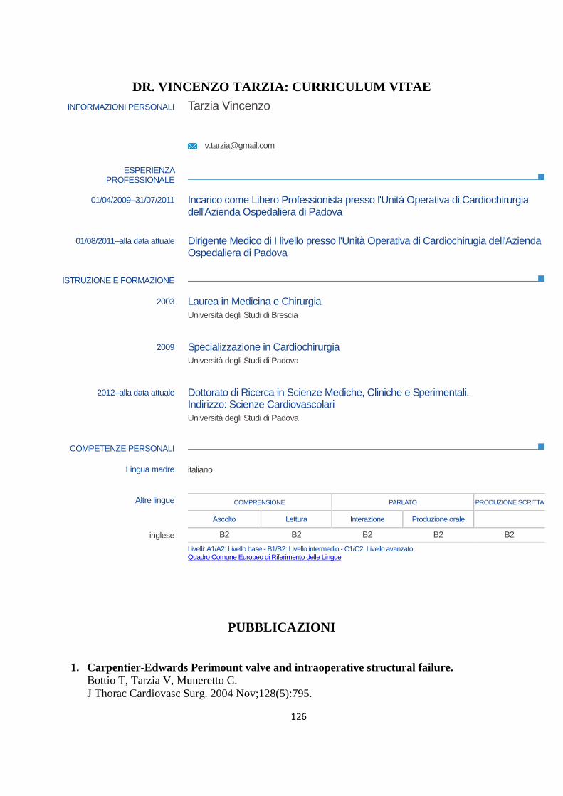
126
DR. VINCENZO TARZIA: CURRICULUM VITAE
ESPERIENZA PROFESSIONALE
ISTRUZIONE E FORMAZIONE
COMPETENZE PERSONALI
PUBBLICAZIONI
1. Carpentier-Edwards Perimount valve and intraoperative structural failure.
Bottio T, Tarzia V, Muneretto C.
J Thorac Cardiovasc Surg. 2004 Nov;128(5):795.
INFORMAZIONI PERSONALI Tarzia Vincenzo
01/04/2009–31/07/2011 Incarico come Libero Professionista presso l'Unità Operativa di Cardiochirurgia dell'Azienda Ospedaliera di Padova
01/08/2011–alla data attuale Dirigente Medico di I livello presso l'Unità Operativa di Cardiochirugia dell'Azienda Ospedaliera di Padova
2003 Laurea in Medicina e Chirurgia
Università degli Studi di Brescia
2009 Specializzazione in Cardiochirurgia
Università degli Studi di Padova
2012–alla data attuale Dottorato di Ricerca in Scienze Mediche, Cliniche e Sperimentali. Indirizzo: Scienze Cardiovascolari
Università degli Studi di Padova
Lingua madre italiano
Altre lingue COMPRENSIONE PARLATO PRODUZIONE SCRITTA
Ascolto Lettura Interazione Produzione orale
inglese B2 B2 B2 B2 B2
Livelli: A1/A2: Livello base - B1/B2: Livello intermedio - C1/C2: Livello avanzato Quadro Comune Europeo di Riferimento delle Lingue

127
2. Parasternal wire technique and sternal dehiscence.
Bottio T, Tarzia V, Muneretto C.
Ann Thorac Surg. 2005 Mar;79(3):1096-7.
3. Small aortic annulus: the hydrodynamic performances of 5 commercially available
tissue valves.
Gerosa G, Tarzia V, Rizzoli G, Bottio T.
J Thorac Cardiovasc Surg. 2006 May;131(5):1058-64.
4. Commissural dehiscence: a rare and peculiar cause of porcine valve structural
deterioration.
Bottio T, Valente M, Rizzoli G, Tarzia V, Bisleri G, Pettenazzo E, Gerosa G, Thiene G.
J Thorac Cardiovasc Surg. 2006 Nov;132(5):1017-22.
5. Small aortic annulus: the hydrodynamic performances of 5 commercially available
tissue valves.
Gerosa G, Tarzia V, Rizzoli G, Bottio T.
J Thorac Cardiovasc Surg. 2006 Dec;132(6):1499-500; author reply 1500-1.
6. Small aortic annulus: the hydrodynamic performances of 5 commercially available
tissue valves.
Gerosa G, Tarzia V, Rizzoli G, Bottio T.
J Thorac Cardiovasc Surg. 2007 Mar;133(3):846-7; author reply 847-9.
7. Temporary coronary artery occlusion during off-pump surgery and endothelial vessel
dysfunction: is it still an unresolved mystery?
Bottio T, Tarzia V, Gerosa G.
J Thorac Cardiovasc Surg. 2007 May;133(5):1397.
8. Extended (31 years) durability of a Starr-Edwards prosthesis in mitral position.
Tarzia V, Bottio T, Testolin L, Gerosa G.
Interact Cardiovasc Thorac Surg. 2007 Aug;6(4):570-1.
9. In-vitro testing of three totally supra-annular bileaflet mechanical valves:
hydrodynamics in the Sheffield pulse duplicator.
Bottio T, Tarzia V, Rizzoli G, Gerosa G.
J Heart Valve Dis. 2008 Mar;17(2):222-6.

128
10. Valve prostheses evaluation: it is a complex scenario and not only a matter of gradient.
Bottio T, Tarzia V, Rizzoli G, Gerosa G.
Ann Thorac Surg. 2008 Aug;86(2):691.
11. The changing spectrum of bioprostheses hydrodynamic performance: considerations
on in-vitro tests.
Bottio T, Tarzia V, Rizzoli G, Gerosa G.
Interact Cardiovasc Thorac Surg. 2008 Oct;7(5):750-4.
12. Aortic valve stenosis management: old strategies and future directions.
Bottio T, Tarzia V, Rizzoli G, Gerosa G.
Eur Heart J. 2008 Oct 2.
13. Arterial switch operation, aortic root dilatation, and long-term valve competence. Bottio T, Thiene G, Tarzia V, Rizzoli G, Gerosa G.
Ann Thorac Surg. 2008 Dec;86(6):2025-6.
14. Total arterial revascularization, conventional coronary artery bypass surgery, and age
cut-off for the loss of benefit from bilateral internal thoracic artery grafting.
Bottio T, Tarzia V, Rizzoli G, Gerosa G.
Eur J Cardiothorac Surg. 2009 Jan;35(1):191.
15. Application of wavelet analysis to the phonocardiographic signal of mechanical heart
valve closing sounds.
Bagno A, Anzil F, Tarzia V, Pengo V, Ruggeri A, Gerosa G.
Int J Artif Organs. 2009 Mar;32(3):166-172.
16. Is the analysis over the time domain or over the frequency domain significant for the
detection of bileaflet mechanical heart valve dysfunction? Bagno A, Anzil F, Buselli R, Tarzia V, Bottio T, Gerosa G, Pengo V.
Ann Thorac Surg. 2009 Mar;87(3):986-7; author reply 987-8.
17. PCI versus CABG for multivessel coronary disease in diabetics.
Tarantini G, Ramondo A, Napodano M, Favaretto E, Gardin A, Bilato C, Nesseris G, Tarzia
V, Cademartiri F, Gerosa G, Iliceto S.
Catheter Cardiovasc Interv. 2009 Jan 1;73(1):50-8.
18. Application of wavelet analysis to the phonocardiographic signal of mechanical heart
valve closing sounds.
Bagno A, Anzil F, Tarzia V, Pengo V, Ruggeri A, Gerosa G.
Int J Artif Organs. 2009 Mar;32(3):166-72.
19. Application of wavelet analysis to the phonocardiographic signal of mechanical heart
valve closing sounds.phonocardiographic analysis.
Bagno A, Anzil F, Buselli R, Pesavento E, Tarzia V, Pengo V, Bottio T, Gerosa G.
J Artif Organs. 2009;12(3):172-81.

129
20. Valve surgery in octogenarians: does it prolong life?
Rizzoli G, Bejko J, Bottio T, Tarzia V, Gerosa G.
Eur J Cardiothorac Surg. 2010 May;37(5):1047-55.
21. The changing hydrodynamic performance of the decellularized intact porcine aortic
root: considerations on in-vitro testing.
Bottio T, Tarzia V, Dal Lin C, Buratto E, Rizzoli G, Spina M, Gandaglia A, Naso F, Gerosa
G.
J Heart Valve Dis. 2010 Jul;19(4):485-91.
22. Comparative classification of thrombotic formations on bileaflet mechanical heart
valves by phonographic analysis.
Romata C, Susin FM, Cambi A, Tarzia V, Pengo V, Gerosa G, Bagno A.
J Artif Organs. 2011 Jun;14(2):100-11.
23. Thrombectomy for massive bioprosthetic valve thrombosis.
Tarzia V, Dal Lin C, Bottio T, Gerosa G.
Eur J Cardiothorac Surg. 2011 Dec;40(6):1540.
24. Peripheral adaptation mechanisms in physical training and cardiac rehabilitation: the
case of a patient supported by a CardioWest total artificial heart.
Bellotto F, Compostella L, Agostoni P, Torregrossa G, Setzu T, Gambino A, Russo N,
Feltrin G, Tarzia V, Gerosa G.
J Card Fail. 2011 Aug;17(8):670-5.
25. In-vitro detection of thrombotic formation on bileaflet mechanical heart valves.
Susin FM, Tarzia V, Bottio T, Pengo V, Bagno A, Gerosa G.
J Heart Valve Dis. 2011 Jul;20(4):378-86.
26. In vitro comparison of different mechanical prostheses suitable for replacement of the
systemic atrioventricular valve in children.
Bottio T, Dal Lin C, Lika A, Rizzoli G, Tarzia V, Buratto E, Gerosa G.
J Thorac Cardiovasc Surg. 2012 Mar;143(3):558-68.
27. Freedom solo stentless aortic valve: quantitative and qualitative assessment of
thrombocytopenia.
Tarzia V, Bottio T, Buratto E, Spiezia L, Simioni P, Gerosa G.
Ann Thorac Surg. 2011 Nov;92(5):1935.
28. The hazard of comparing apples and oranges: the proper indication for the use of
recombinant activated clotting factor VII in cardiac surgery.
Tarzia V, Bottio T, Buratto E, Spiezia L, Simioni P, Gerosa G.
J Thorac Cardiovasc Surg. 2011 Dec;142(6):1588-9.
29. Occult gastrointestinal bleeding in patients with a left ventricular assist device axial
flow pump: diagnostic tools and therapeutic algorithm.
Tarzia V, Dal Lin C, Bottio T, Benvenuti S, Chilovi F, Gerosa G.
J Thorac Cardiovasc Surg. 2012 Apr;143(4).

130
30. Carpentier-Edwards Magna ease versus Magna valves: a comparison of in-vitro valve
hydrodynamic performance.
Dal Lin C, Bottio T, Buratto E, Tarzia V, Rizzoli G, Savona V, Gerosa G.
J Heart Valve Dis. 2012 Jan;21(1):112-7.
31. Development of artificial neural network-based algorithms for the classification of
bileaflet mechanical heart valve sounds.
Bagno A, Licciardello C, Tarzia V, Bottio T, Pengo V, Gerosa G.
Int J Artif Organs. 2012 Apr 13;35(4):279-287.
32. Aortic valve calcium scoring is a predictor of paravalvular aortic regurgitation after
transcatheter aortic valve implantation.
Colli A, Gallo M, Bernabeu E, D'Onofrio A, Tarzia V, Gerosa G.
Ann Cardiothorac Surg. 2012 Jul;1(2):156-9.
33. Descending aorta-to-coronary artery bypass graft imaging by means of multislice
computed tomography.
Tarzia V, Dal Lin C, Bottio T, Testolin L, De Biasio V, Gerosa G.
Tex Heart Inst J. 2012;39(4):585
34. Nitinol flexigrip sternal closure system and chest wound infections: insight from a
comparative analysis of complications and costs.
Bejko J, Tarzia V, De Franceschi M, Bianco R, Castoro M, Bottio T, Gerosa G.
Ann Thorac Surg. 2012 Dec;94(6):1848-53.
35. The impact of transcatheter aortic valve implantation on patients' profiles and
outcomes of aortic valve surgery programmes: a multi-institutional appraisal.
D'Onofrio A, Alfieri OR, Cioni M, Alamanni F, Fusari M, Tarzia V, Rizzoli G, Gerosa G.
Interact Cardiovasc Thorac Surg. 2013 May;16(5):608-11.
36. Aortic valve hydrodynamics: considerations on the absence of sinuses of Valsalva.
Bottio T, Buratto E, Dal Lin C, Lika A, Tarzia V, Rizzoli G, Gerosa G.
J Heart Valve Dis. 2012 Nov;21(6):718-23.
37. Cardiac autonomic dysfunction in the early phase after left ventricular assist device
implant: Implications for surgery and follow-up.
Compostella L, Russo N, Setzu T, Tursi V, Bottio T, Tarzia V, Compostella C, Covolo E,
Livi U, Gerosa G, Sani G, Bellotto F.
Int J Artif Organs. 2013 Jun 25;36(6):410-8.
38. First quantification of alpha-Gal epitope in current glutaraldehyde-fixed heart valve
bioprostheses.
Naso F, Gandaglia A, Bottio T, Tarzia V, Nottle MB, d'Apice AJ, Cowan PJ, Cozzi E, Galli
C, Lagutina I, Lazzari G, Iop L, Spina M, Gerosa G.
Xenotransplantation. 2013 Jul-Aug;20(4):252-61.

131
39. Less invasive surgical and perfusion technique for implantation of the Jarvik 2000 left
ventricular assist device.
Gerosa G, Gallo M, Tarzia V, Di Gregorio G, Zanella F, Bottio T.
Ann Thorac Surg. 2013 Aug;96(2):712-4.
40. Ultrasound phonocardiography for detecting thrombotic formations on bileaflet
mechanical heart valves.
Melan G, Bellato A, Susin FM, Bottio T, Tarzia V, Pengo V, Gerosa G, Bagno A.
J Heart Valve Dis. 2013 Nov;22(6):828-36.
41. Ventricular assist devices.
Tarantini G, Fraccaro C, Napodano M, Buja P, Tarzia V, Isabella G, Gerosa G, Iliceto S.
Minerva Cardioangiol. 2013 Dec;61(6):691-700. Review.
42. HeartWare Ventricular Assist Device as Bridge to Transplant in Children and
Adolescents.
Padalino MA, Bottio T, Tarzia V, Bortolussi G, Cerutti A, Vida VL, Gerosa G, Stellin G.
Artif Organs. 2014 May;38(5):418-22.
43. Less-invasive off-pump ventricular assist device implantation in regional paravertebral
analgesia. Bottio T, Bejko J, Falasco G, Bortolussi G, Gallo M, Tarzia V, Gerosa G.J
Artif Organs. 2014 Sep;17(3):275-7.
44. Impact of vacuum-assisted closure therapy on outcomes of sternal wound dehiscence. Tarzia V, Carrozzini M, Bortolussi G, Buratto E, Bejko J, Comisso M, Mescola V, Penzo V,
Guarino M, De Franceschi M, Pagnin C, Castoro M, Guglielmi C, Testolin L, Bottio T,
Gerosa G.
Interact Cardiovasc Thorac Surg. 2014 Jul;19(1):70-5.
45. HeartWare LVAD implantation in a patient with a rare ARVD: Carvajal syndrome.
Bottio T, Bejko J, Tarzia V, Gerosa G.
Int J Artif Organs. 2014 Jul 31;37(7):563-6.
46. The Danger of Using a Sledgehammer to Crack a Nut: ROTEM-Guided
Administration of Recombinant Activated Factor VII in a Patient With Refractory
Bleeding Post-Ventricular Assist Device Implantation.
Tarzia V, Buratto E, Bortolussi G, Paolini C, Bejko J, Bottio T, Gerosa G.
Artif Organs. 2014 Jul 28.
47. Results with Syncardia Total Artificial Heart Beyond One Year.
Torregrossa G, Morshuis MJ, Varghese R, Hosseinian L, Vida V, Tarzia V, Loforte A,
Duveau D, Arabia FA, Leprince P, Kasirajan V, Beyersdorf F, Musumeci F, Hetzer R,
Krabatsch T, Gummert J, Copeland JG, Gerosa G.
ASAIO J. 2014 Aug 25.

132
48. Colchicine for prevention of postpericardiotomy syndrome and postoperative atrial
fibrillation: the COPPS-2 randomized clinical trial.
Imazio M, Brucato A, Ferrazzi P, Pullara A, Adler Y, Barosi A, Caforio AL, Cemin R,
Chirillo F, Comoglio C, Cugola D, Cumetti D, Dyrda O, Ferrua S, Finkelstein Y, Flocco R,
Gandino A, Hoit B, Innocente F, Maestroni S, Musumeci F, Oh J, Pergolini A, Polizzi V,
Ristic A, Simon C, Spodick DH, Tarzia V, Trimboli S, Valenti A, Belli R, Gaita F; COPPS-
2 Investigators.
JAMA. 2014 Sep 10;312(10):1016-23.
49. Bilateral mini-thoracotomy off-pump Jarvik 2000 implantation in regional asymmetric
paravertebral analgesia. Bottio T, Bejko J, Guariento A, Tarzia V, Pittarello D, Gerosa G.
J Cardiovasc Med (Hagerstown). 2014 Oct 20.
50. Cellular, molecular, genomic changes occurring in the heart under mechanical
circulatory support. Michele Gallo, Vincenzo Tarzia, Laura Iop, Jonida Bejko, Giacomo Bortolussi, Roberto
Bianco, Tomaso Bottio, Gino Gerosa.
Ann Cardiothorac Surg. 2014 Sep;3(5).
51. Clinical psychological and neuropsychological issues with left ventricular assist devices
(LVADs). Daniela Mapelli, Annachiara Cavazzana, Chiara Cavalli, Tomaso Bottio, Vincenzo Tarzia,
Gino Gerosa, Bianca Rosa Volpe.
Ann Cardiothorac Surg. 2014 Sep;3(5).
52. The Jarvik-2000 ventricular assist device implantation: how we do it.
Fabio Zucchetta, Vincenzo Tarzia, Tomaso Bottio, Gino Gerosa.
Ann Cardiothorac Surg. 2014 Sep;3(5).
53. Implantation of the HeartWare HVAD: from full sternotomy to less invasive
techniques. Vincenzo Tarzia, Edward Buratto, Michele Gallo, Giacomo Bortolussi, Jonida
Bejko, Roberto Bianco, Tomaso Bottio, Gino Gerosa.
Ann Cardiothorac Surg. 2014 Sep;3(5).
54. Jarvik 2000: evolution of surgical implantation from conventional to minimally
invasive technique. Vincenzo Tarzia, Edward Buratto, Carlo Dal Lin, Michele Gallo, Giacomo Bortolussi,
Jonida Bejko, Tomaso Bottio, Gino Gerosa.
Ann Cardiothorac Surg. 2014 Nov;3(6):621-3.
55. Surgical implantation of the CardioWest Total Artificial Heart. Vincenzo Tarzia, Edward Buratto, Michele Gallo, Giacomo Bortolussi, Jonida Bejko, Carlo
Dal Lin, Gianluca Torregrossa, Roberto Bianco, Tomaso Bottio, Gino Gerosa.
Ann Cardiothorac Surg. 2014 Nov;3(6):624-5.

133
56. Haemorrhage and thrombosis with different LVAD technologies: a matter of flow? Vincenzo Tarzia, Edward Buratto, Giacomo Bortolussi, Michele Gallo, Jonida Bejko,
Roberto Bianco, Tomaso Bottio, Gino Gerosa.
Ann Cardiothorac Surg. 2014 Nov;3(6):582-4.
57. Nitinol flexigrip sternal closure system and standard sternal steel wiring: insight from
a matched comparative analysis.
Bejko J, Bottio T, Tarzia V, De Franceschi M, Bianco R, Gallo M, Castoro M, Bortolussi G,
Gerosa G.
J Cardiovasc Med (Hagerstown). 2015 Feb;16(2):134-8

134
DR. VINCENZO TARZIA: PERSONAL CONTRIBUTION DURING THE
PH.D. PERIOD (2012-2014)
ARTICOLI (I anno 2012)
1) Nitinol Flexigrip Sternal Closure System and Chest Wound Infections: Insight
From a Comparative Analysis of Complications and Costs.
Bejko J, Tarzia V, Franceschi MD, Bianco R, Castoro M, Bottio T, Gerosa G.
Ann Thorac Surg. 2012 Oct 25.
2) Descending aorta-to-coronary artery bypass graft imaging by means of
multislice computed tomography.
Tarzia V, Dal Lin C, Bottio T, Testolin L, De Biasio V, Gerosa G.
Tex Heart Inst J. 2012;39(4):585.
3) Development of artificial neural network-based algorithms for the classification
of bileaflet mechanical heart valve sounds.
Bagno A, Licciardello C, Tarzia V, Bottio T, Pengo V, Gerosa G.
Int J Artif Organs. 2012 Apr 13;35(4):279-287.
4) Carpentier-Edwards Magna ease versus Magna valves: a comparison of in-vitro
valve hydrodynamic performance.
Dal Lin C, Bottio T, Buratto E, Tarzia V, Rizzoli G, Savona V, Gerosa G.
J Heart Valve Dis. 2012 Jan;21(1):112-7.
5) Occult gastrointestinal bleeding in patients with a left ventricular assist device
axial flow pump: diagnostic tools and therapeutic algorithm.
Tarzia V, Dal Lin C, Bottio T, Benvenuti S, Chilovi F, Gerosa G.
J Thorac Cardiovasc Surg. 2012 Apr;143(4):e28-31.
6) In vitro comparison of different mechanical prostheses suitable for replacement
of the systemic atrioventricular valve in children.
Bottio T, Dal Lin C, Lika A, Rizzoli G, Tarzia V, Buratto E, Gerosa G.
J Thorac Cardiovasc Surg. 2012 Mar;143(3):558-68.
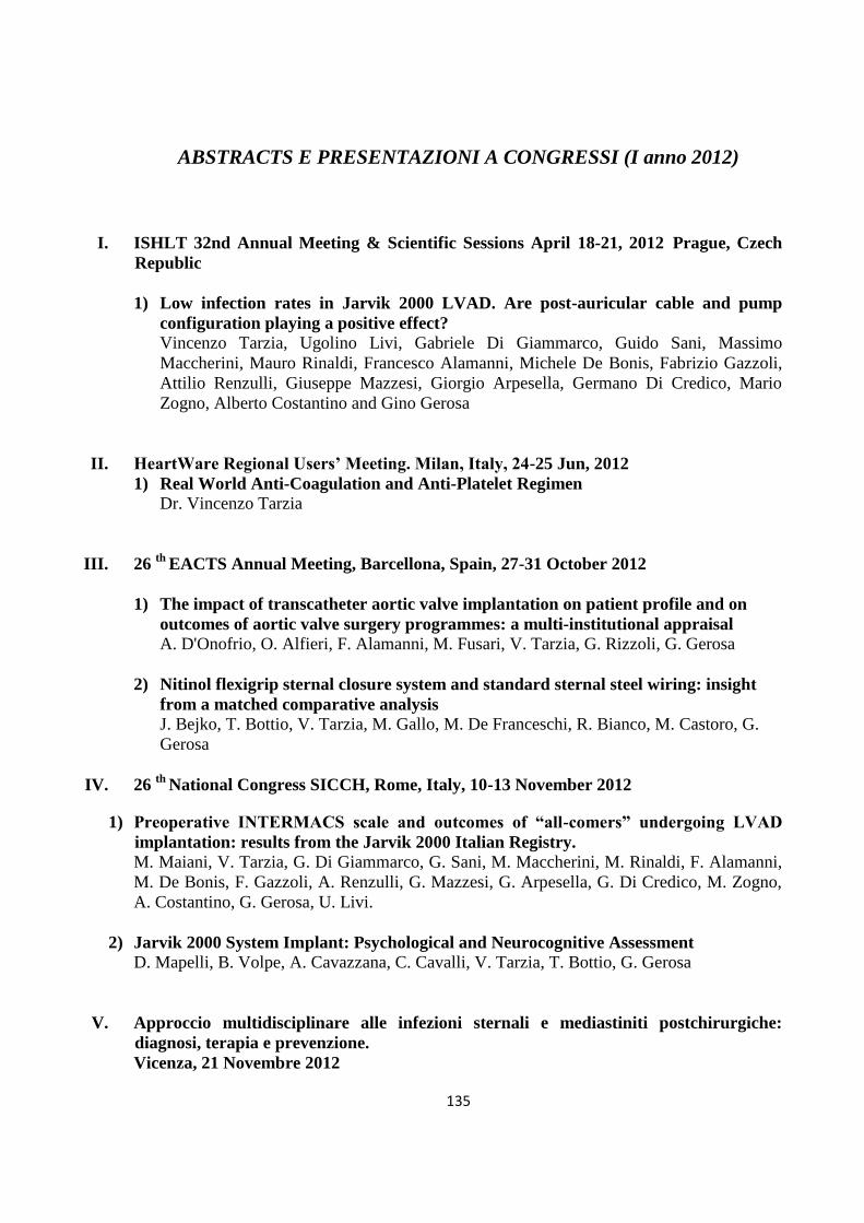
135
ABSTRACTS E PRESENTAZIONI A CONGRESSI (I anno 2012)
I. ISHLT 32nd Annual Meeting & Scientific Sessions April 18-21, 2012 Prague, Czech
Republic
1) Low infection rates in Jarvik 2000 LVAD. Are post-auricular cable and pump
configuration playing a positive effect?
Vincenzo Tarzia, Ugolino Livi, Gabriele Di Giammarco, Guido Sani, Massimo
Maccherini, Mauro Rinaldi, Francesco Alamanni, Michele De Bonis, Fabrizio Gazzoli,
Attilio Renzulli, Giuseppe Mazzesi, Giorgio Arpesella, Germano Di Credico, Mario
Zogno, Alberto Costantino and Gino Gerosa
II. HeartWare Regional Users’ Meeting. Milan, Italy, 24-25 Jun, 2012
1) Real World Anti-Coagulation and Anti-Platelet Regimen
Dr. Vincenzo Tarzia
III. 26 th
EACTS Annual Meeting, Barcellona, Spain, 27-31 October 2012
1) The impact of transcatheter aortic valve implantation on patient profile and on
outcomes of aortic valve surgery programmes: a multi-institutional appraisal
A. D'Onofrio, O. Alfieri, F. Alamanni, M. Fusari, V. Tarzia, G. Rizzoli, G. Gerosa
2) Nitinol flexigrip sternal closure system and standard sternal steel wiring: insight
from a matched comparative analysis
J. Bejko, T. Bottio, V. Tarzia, M. Gallo, M. De Franceschi, R. Bianco, M. Castoro, G.
Gerosa
IV. 26 th
National Congress SICCH, Rome, Italy, 10-13 November 2012
1) Preoperative INTERMACS scale and outcomes of “all-comers” undergoing LVAD
implantation: results from the Jarvik 2000 Italian Registry.
M. Maiani, V. Tarzia, G. Di Giammarco, G. Sani, M. Maccherini, M. Rinaldi, F. Alamanni,
M. De Bonis, F. Gazzoli, A. Renzulli, G. Mazzesi, G. Arpesella, G. Di Credico, M. Zogno,
A. Costantino, G. Gerosa, U. Livi.
2) Jarvik 2000 System Implant: Psychological and Neurocognitive Assessment
D. Mapelli, B. Volpe, A. Cavazzana, C. Cavalli, V. Tarzia, T. Bottio, G. Gerosa
V. Approccio multidisciplinare alle infezioni sternali e mediastiniti postchirurgiche:
diagnosi, terapia e prevenzione.
Vicenza, 21 Novembre 2012

136
1) Il sistema VAC nella esperienza della cardiochirurgia di Padova
Dr. Vincenzo Tarz
CAPITOLI DI LIBRI (I anno 2012)
I. Dawn and Evolution of Cardiac Procedures: Research Avenues in Cardiac Surgery and
Interventional Cardiology
1)Surgical treatment of atrial fibrillation
Gino Gerosa, Carlo Dal Lin, Vincenzo Tarzia
II. Trattamento chirurgico della patologia dell'aorta toracica. Piccin
1) Chirurgia dell’ arco aortico : Tecniche di protezione cerebrale
Vincenzo Tarzia, Carlo Dal Lin, Gino Gerosa

137
ARTICOLI (II anno 2013)
1. Ultrasound phonocardiography for detecting thrombotic formations on bileaflet
mechanical heart valves. Melan G, Bellato A, Susin FM, Bottio T, Tarzia V, Pengo V,
Gerosa G, Bagno A. J Heart Valve Dis. 2013 Nov;22(6):828-36.
2. Less invasive surgical and perfusion technique for implantation of the Jarvik 2000 left
ventricular assist device. Gerosa G, Gallo M, Tarzia V, Di Gregorio G, Zanella F, Bottio T.
Ann Thorac Surg. 2013 Aug;96(2):712-4. doi: 10.1016/j.athoracsur.2013.01.086.
3. First quantification of alpha-Gal epitope in current glutaraldehyde-fixed heart valve
bioprostheses. Naso F, Gandaglia A, Bottio T, Tarzia V, Nottle MB, d'Apice AJ, Cowan PJ,
Cozzi E, Galli C, Lagutina I, Lazzari G, Iop L, Spina M, Gerosa G. Xenotransplantation.
2013 Jul-Aug;20(4):252-61.
4. Cardiac autonomic dysfunction in the early phase after left ventricular assist device
implant: Implications for surgery and follow-up. Compostella L, Russo N, Setzu T, Tursi
V, Bottio T, Tarzia V, Compostella C, Covolo E, Livi U, Gerosa G, Sani G, Bellotto F. Int J
Artif Organs. 2013 Jun 25;36(6):410-8.
5. The impact of transcatheter aortic valve implantation on patients' profiles and
outcomes of aortic valve surgery programmes: a multi-institutional appraisal. D'Onofrio A, Alfieri OR, Cioni M, Alamanni F, Fusari M, Tarzia V, Rizzoli G, Gerosa G.
Interact Cardiovasc Thorac Surg. 2013 May;16(5):608-11.

138
ABSTRACTS E PRESENTAZIONI A CONGRESSI (II Anno 2013)
I. Patologia dell’ aorta toracica: Trattamento chirurgico ed endovascolare,
Camisano Vicentino (VI), 2 Febbraio 2013.
1) Trattamento chirurgico della patologia dell’ aorta toracica: tecniche di protezione
cerebrale. V. Tarzia, G. Gerosa
II. ISHLT 33rd Annual Meeting & Scientific Sessions April 24-27, 2013
Montréal, Québec, Canada
1) Long Term Results with Total Artificial Heart: Is It Prime Time for Destination
Therapy? G. Torregrossa, G. Gerosa, V. Tarzia, V. Vida, D. Duveau, F. Arabia, P. Leprince, V. Kasirajan, F.
Beyersdof, A. Loforte, F. Musumeci, R. Hetzer, T. Krabatsch, J. Gummert, M. Morshuis, J.
Copeland
2) Age Is No Boundary to Long Term Survival on Permanent MCS: A Multicentre
Experience. V. Tarzia, T. Bottio, U. Livi, M. Maiani, G. Di Giammarco, G. Sani, M. Maccherini, M. Rinaldi, F.
Alamanni, M. De Bonis, F. Gazzoli, A. Renzulli, G. Arpesella, G. Gerosa
3) Different Impact on the Coagulation System of Two Continuous Flow LVADs: Axial
Versus Centrifugal Flow. V. Tarzia, F. Vasques, G. Bortolussi, J. Bejko, M. Gallo, M. Carrozzini, M. Comisso, E. Buratto, M.
De Franceschi, E. Campello, L. Spiezia, P. Simioni, T. Bottio, G. Gerosa
III. 27 th
EACTS Annual Meeting, Vienna, Austria, 5-9 October 2013
1) Efficacy and safety of paravertebral block analgesia versus general anaesthesia for
ventricular assist device implantation: a single-centre experience. T. Bottio, J. Bejko, G. Bortolussi, M. Comisso, M. Carrozzini, R. Bianco, V. Tarzia, G. Gerosa
2) Impact of vacuum-assisted closure therapy on outcome of sternal wound dehiscence
V. Tarzia, M. Carrozzini, G. Bortolussi, J. Bejko, M. Comisso, M. De Franceschi, T. Bottio, G.
Gerosa
3) Extracorporeal membrane oxygenation in primary cardiogenic shock: the impact of
acute versus chronic aetiology on outcome V. Tarzia, G. Bortolussi, R. Bianco, A. Marzari, L. Cacciavillani, S. Iliceto, T. Bottio, G. Gerosa

139
IV. First International Padua Meeting on Severe Bleeding Management,
Padua, Italy, 12 November 2013
1) From theory to practice: what would you have done?
Clinical Cases in Cardiac Surgery V. Tarzia
CAPITOLI DI LIBRI (II Anno 2013 )
The Globesity Challenge to General Surgery: A Guide to Strategy and
Techniques.
Cardio-thoracic and Vascular
Gino Gerosa, Marco Schiavon, Giuseppe Marulli, Vincenzo Tarzia, Federico Rea

140
ARTICOLI (III anno 2014)
58. HeartWare ventricular assist device as bridge to transplant in children and
adolescents. Padalino MA, Bottio T, Tarzia V, Bortolussi G, Cerutti A, Vida VL, Gerosa G,
Stellin G. Artif Organs. 2014 May;38(5):418-22
59. Less-invasive off-pump ventricular assist device implantation in regional paravertebral
analgesia. Bottio T, Bejko J, Falasco G, Bortolussi G, Gallo M, Tarzia V, Gerosa G.J Artif
Organs. 2014 Sep;17(3):275-7.
60. Impact of vacuum-assisted closure therapy on outcomes of sternal wound dehiscence. Tarzia V, Carrozzini M, Bortolussi G, Buratto E, Bejko J, Comisso M, Mescola V, Penzo V,
Guarino M, De Franceschi M, Pagnin C, Castoro M, Guglielmi C, Testolin L, Bottio T,
Gerosa G. Interact Cardiovasc Thorac Surg. 2014 Jul;19(1):70-5.
61. HeartWare LVAD implantation in a patient with a rare ARVD: Carvajal syndrome.
Bottio T, Bejko J, Tarzia V, Gerosa G. Int J Artif Organs. 2014 Jul 31;37(7):563-6.
62. The Danger of Using a Sledgehammer to Crack a Nut: ROTEM-Guided
Administration of Recombinant Activated Factor VII in a Patient With Refractory
Bleeding Post-Ventricular Assist Device Implantation. Tarzia V, Buratto E, Bortolussi G,
Paolini C, Bejko J, Bottio T, Gerosa G. Artif Organs. 2014 Jul 28.
63. Results with Syncardia Total Artificial Heart Beyond One Year. Torregrossa G,
Morshuis MJ, Varghese R, Hosseinian L, Vida V, Tarzia V, Loforte A, Duveau D, Arabia
FA, Leprince P, Kasirajan V, Beyersdorf F, Musumeci F, Hetzer R, Krabatsch T, Gummert
J, Copeland JG, Gerosa G. ASAIO J. 2014 Aug 25.
64. Colchicine for prevention of postpericardiotomy syndrome and postoperative atrial
fibrillation: the COPPS-2 randomized clinical trial. Imazio M, Brucato A, Ferrazzi P,
Pullara A, Adler Y, Barosi A, Caforio AL, Cemin R, Chirillo F, Comoglio C, Cugola D,
Cumetti D, Dyrda O, Ferrua S, Finkelstein Y, Flocco R, Gandino A, Hoit B, Innocente F,
Maestroni S, Musumeci F, Oh J, Pergolini A, Polizzi V, Ristic A, Simon C, Spodick DH,
Tarzia V, Trimboli S, Valenti A, Belli R, Gaita F; COPPS-2 Investigators. JAMA. 2014 Sep
10;312(10):1016-23.
65. Bilateral mini-thoracotomy off-pump Jarvik 2000 implantation in regional asymmetric
paravertebral analgesia. Bottio T, Bejko J, Guariento A, Tarzia V, Pittarello D, Gerosa G.
J Cardiovasc Med (Hagerstown). 2014 Oct 20.
66. Cellular, molecular, genomic changes occurring in the heart under mechanical
circulatory support. Michele Gallo, Vincenzo Tarzia, Laura Iop, Jonida Bejko, Giacomo
Bortolussi, Roberto Bianco, Tomaso Bottio, Gino Gerosa. Ann Cardiothorac Surg. 2014
Sep;3(5).

141
67. Clinical psychological and neuropsychological issues with left ventricular assist devices
(LVADs). Daniela Mapelli, Annachiara Cavazzana, Chiara Cavalli, Tomaso Bottio,
Vincenzo Tarzia, Gino Gerosa, Bianca Rosa Volpe. Ann Cardiothorac Surg. 2014 Sep;3(5).
68. The Jarvik-2000 ventricular assist device implantation: how we do it. Fabio Zucchetta,
Vincenzo Tarzia, Tomaso Bottio, Gino Gerosa. Ann Cardiothorac Surg. 2014 Sep;3(5).
69. Implantation of the HeartWare HVAD: from full sternotomy to less invasive
techniques. Vincenzo Tarzia, Edward Buratto, Michele Gallo, Giacomo Bortolussi, Jonida
Bejko, Roberto Bianco, Tomaso Bottio, Gino Gerosa. Ann Cardiothorac Surg. 2014
Sep;3(5).
70. Jarvik 2000: evolution of surgical implantation from conventional to minimally
invasive technique. Vincenzo Tarzia, Edward Buratto, Carlo Dal Lin, Michele Gallo,
Giacomo Bortolussi, Jonida Bejko, Tomaso Bottio, Gino Gerosa. Ann Cardiothorac Surg.
2014 Nov;3(6):621-3.
71. Surgical implantation of the CardioWest Total Artificial Heart. Vincenzo Tarzia,
Edward Buratto, Michele Gallo, Giacomo Bortolussi, Jonida Bejko, Carlo Dal Lin, Gianluca
Torregrossa, Roberto Bianco, Tomaso Bottio, Gino Gerosa. Ann Cardiothorac Surg. 2014
Nov;3(6):624-5.
72. Haemorrhage and thrombosis with different LVAD technologies: a matter of flow?
Vincenzo Tarzia, Edward Buratto, Giacomo Bortolussi, Michele Gallo, Jonida Bejko,
Roberto Bianco, Tomaso Bottio, Gino Gerosa. Ann Cardiothorac Surg. 2014 Nov;3(6):582-
4.
73. Nitinol flexigrip sternal closure system and standard sternal steel wiring: insight from
a matched comparative analysis.
Bejko J, Bottio T, Tarzia V, De Franceschi M, Bianco R, Gallo M, Castoro M, Bortolussi G,
Gerosa G.
J Cardiovasc Med (Hagerstown). 2015 Feb;16(2):134-8

142
ABSTRACTS E PRESENTAZIONI A CONGRESSI (III Anno 2014)
I. ISHLT 34th Annual Meeting & Scientific Sessions April 10-13, 2014 San
Diego, USA
1) From Bench To Bedside: Can the Improvements in LVAD Design Mitigate Adverse
Events and Increase Survival Rate?; V. Tarzia, G. Di Giammarco, M. Maccherini, T. Bottio, V. Tursi, M. Maiani, S. Bernazzali, M.
Foschi, S. M. Diso, U. Livi, G. Sani, G. Gerosa.
2) Efficacy and Safety of Paravertebral Block Analgesia Versus General Anaesthesia for
Ventricular Assist Device Implantation: A Single-Centre Experience J. Bejko, T. Bottio, G. Bortolussi, M. Carrozzini, V. Tarzia, D. Pittarello, G. Di Gregorio, G. Gerosa.
3) Fortuity or Causality: Relation Between Outflow Graft Site of Anastomosis on Aorta
and Cerebral Ischemic Events in LVAD Implantation J. Bejko, T. Bottio, G. Bortolussi, V. Tarzia, R. Bianco, G. Rizzoli, G. Gerosa.
II. 5th Jarvik 2000 LVAD Users Meeting. Petriolo September 27th, 2014
1) When anticoagulation gets tough: case reports & strategy
V. Tarzia
2) Telemedicine “TELEMACO” project V. Tarzia
III. 28 th
EACTS Annual Meeting, Milan, Italy, 11-15 October 2014
1) Are all continuous flow left ventricular assist devices equal on platelet activation and
inflammatory response? A comparison of centrifugal versus axial flow pump. V. Tarzia, G. Bortolussi, V. Penzo, J. Bejko, M. Gallo, R. Bianco, T. Bottio, G. Gerosa.
2) Minimally invasive surgical and anaesthetic approach for ventricular assist device
implantation: A single-centre experience. T. Bottio, J. Bejko, G. Bortolussi, R. Bianco, G. Falasco, D. Pittarello, V. Tarzia, G. Gerosa.

143
IV. ATBV 2014 Congresso Nazionale. Hot-Topics in tema di terapia
antitrombotica. Le sfide in tema di terapia antitrombotica. Milano 24-25
Ottobre 2014
1) La trombosi…altrove. La chirurgia cardiaca.
V. Tarzia
V. SICCH. 27th
National Congress, Rome. November, 28-30th
2014.
1) Outcome of sternal wound dehiscences in diabetic patients: Can vac therapy play a
role? V. Tarzia
2) Impact of different LVAD technologies on the coagulation system: Axial versus
Centrifugal flow. V. Tarzia

144
CAPITOLI DI LIBRI (III Anno 2014 )
I. Chirurgia per le professioni sanitarie (Mario Lise) (Piccin)
Cuore e Pericardio
Vincenzo Tarzia, Michele Gallo, Gino Gerosa
II. Trattato di Cardiochirurgia (Luigi Chiariello)
Trattamento chirurgico dello scompenso cardiaco refrattario a terapia medica
Vincenzo Tarzia, Gino Gerosa
III. Manuale di Malattie Cardiovascolari (Società Italiana di Cardiologia)
VAD e Trapianto Cardiaco
Gino Gerosa, Vincenzo Tarzia
PROGETTI DI RICERCA SANITARIA FINALIZZATA 2014
Regione Veneto
I. Applicazione della sanità digitale in Cardiologia
Responsabile scientifico:Vincenzo Tarzia
EACTS QUIP ADULT CARDIAC DATABASE
V. Tarzia, G. Gerosa

145