Á John O’Keefe - Weizmann
75
Transcript of Á John O’Keefe - Weizmann


11 Á John O’Keefe
Hippocampal Neurophysiologyin the Behaving Animal
475
Á11.1 Overview
This chapter addresses a central question in the study of thehippocampus: What information is represented in hippocam-pal electrical activity? When asking this question, we areimmediately confronted with one of the central problems inneuroscience, the nature of how information is coded in thenervous system. Historically, there have been two answers tothis question: On the one hand is the view that the nervoussystem is composed of a large set of individual computing ele-ments, the neurons, and that these neurons interact with eachother by passing discrete bundles of information along theiraxons. An alternative view is that the system is organized onmore holistic principles, with large numbers of cells acting inconcert perhaps reflected in synchronous neuronal firing or inrhythmical electroencephalographic (EEG) activity.
The general position taken in this chapter is that the hip-pocampus uses both strategies. There is ample evidence thatindividual hippocampal pyramidal cells code for specific loca-tions in an environment by a marked increase in firing rate;but, equally, that part of the code for location involves the tim-ing of cell firing relative to a clock wave, represented by theEEG theta wave and associated interneurons, and that groupsof pyramidal cells can act cooperatively in an ensemble fash-ion. On this view, each pyramidal cell acts as an oscillator, anda sophisticated set of mechanisms exists for producing theseoscillations. Each cell has the biophysical machinery to oscil-late in isolation from other cells or external inputs. These indi-vidual oscillators are stabilized and synchronized by a set ofinhibitory feedback circuits primarily involving inhibitoryinterneurons. Neurons in the septum and brain stem providethe neuromodulatory inputs necessary for hippocampalpyramidal cells to enter the oscillatory state and may also pro-vide a driving signal that sets the overall frequency of theoscillations.
One powerful approach to the study of the functions of abrain region is to correlate the electrical activity of its neuronswith some aspect of observed behavior or inferred cognition.This approach reveals what information is available to thatpart of the brain and when it is available. Furthermore, it issometimes possible to record both the inputs and the outputsof an area, and under these conditions it may be possible tocompute what transfer function is being performed.
Several patterns of electrical activity have been recordedfrom the hippocampus, and they have been correlated withbehavioral or psychological states. During the mid-1960s,Vanderwolf placed a relatively large electrode into the hip-pocampus of the freely moving rat and recorded the EEGactivity during a wide range of behaviors (Vanderwolf, 1969).He identified three distinct states: the rhythmical theta state,the large irregular amplitude activity (LIA) state, and thesmall irregular amplitude activity (SIA) state. Theta, in turn,could be classified into two subtypes on the basis of behav-ioral correlate and pharmacological sensitivity. In this chapter,these two types are called a-theta (for arousal/attention theta),which is sensitive to anticholinergic drugs such as atropine,and t-theta (for translation movement theta), which may beserotoninergic and glutamatergic.
The behavioral/psychological correlates of the two types oftheta activity have been characterized best in rats: In this ani-mal, the atropine-resistant component or t-theta occurs dur-ing a class of movements that may be loosely characterized asthose that normally change the spatial relation between theanimal’s head and the environment. The correlates of theatropine-sensitive theta, or a-theta, are less well defined andare best summed up as psychological states such as arousal,attention, or intention to move. The behavioral correlates ofLIA are those that do not change the animal’s location in theenvironment: quiet sitting, eating, drinking, and grooming inthe absence of postural shifts. The SIA state occurs duringbehavioral transitions, often when the animal awakens from

slow-wave or rapid-eye-movement (REM) sleep or when itabruptly stops running. It has not received much attention,and its behavioral correlates and physiological function areless well understood.
The rhythmical theta state reflects the synchronous mem-brane oscillations of large numbers of pyramidal cells in theCA1 field and dentate gyrus that are locked into synchrony bythe inhibitory interneuronal network. One of its functions isto provide a clock signal against which the action potentials ofthe individual pyramidal cells can be timed. Another functionmight be to set up the optimal circumstances for the induc-tion of long-term potentiation (LTP) (see Chapter 10). Thenonrhythmical LIA state has a more random, broader spec-trum especially in the lower-frequency range and may repre-sent an inactive, relaxed state of the same network when it isnot being driven. Alternatively, it may be an active state in itsown right in which memories previously encoded in the hip-pocampus are strengthened and/or transferred to otherregions of the brain. LIA is characterized by sharp waves ofabout 100 ms duration that occur randomly, with an aver-age interval of 1 second. Associated with them are higher-frequency “ripple” oscillations of 100 to 200 Hz, which alsomay reflect the operation of the inhibitory network. In addi-tion to oscillations in the theta and ripple bands, oscillationsat intermediate frequencies (beta: 12–30 Hz; gamma: 30–100Hz) have been recorded in association with various aspects ofolfactory behavior.
Placing a microelectrode rather than a gross electrode intothe CA1 hippocampal cell layer reveals a much richer and sur-prising set of behavioral correlates correlations between phys-iology and behavior. On the basis of the EEG recordings onemight expect to see cell-firing patterns that are correlated withthe animal’s movements, and indeed this is observed.However, it is not the whole story. Ranck (1973) placed micro-electrodes into the pyramidal cell layers of CA3 and CA1 andfound two classes of cellular response. One type, which hecalled the theta cell, fired at frequencies ranging from 10 Hz(when the animal sat quietly or engaged in other “LIA” behav-iors) to as high as 100 Hz (as it ran around the environmentor engaged in other “theta” behaviors). Furthermore, theyburst at the same frequency and showed consistent correla-tions with different phases of the various EEG waves. Here, atthe level of the single cell, was one clear correlate of the EEGpatterns, banishing forever any lingering skepticism abouttheir functional significance. The second class of cell, the com-plex spike cell, had a much lower baseline firing rate, andmany were effectively “silent” for long periods of time. Theirdefining characteristic is the occasional short burst of actionpotentials with successively decreasing amplitudes. Theirmajor behavioral correlate was first identified by O’Keefe andDostrovsky (1971) as the animal’s location. They reportedthat these place cells were typically silent as the rat movedaround the environment until it entered a small patch of theenvironment when the cell began to fire (the place field). It isnow recognized that, within the place field, these cells also fire
in a rhythmical bursting pattern during the EEG theta state.Unlike the theta cells, however, the frequency of bursts isslightly higher than the gross EEG theta, causing each succes-sive burst to precess to earlier phases of the theta cycle as theanimal moves through the place field. This temporal codeworks together with the overall rate code to identify the ani-mal’s location. In addition, variations in firing rate can signalaspects of behavior that occur in the place field or the presence(or absence) of objects encountered there. The same cellsoften fire in different environments, but the preferred loca-tions are unrelated if the environments are sufficiently dis-similar to each other. One notable feature of these place cellsis that in unconstrained open fields—environments in whichthe animal is free to move in all directions—the cells fire in theplace field irrespective of the direction in which the animal isfacing. In environments that constrain the animal’s behavior,the cells become directionally sensitive and may be said to rep-resent the successive locations along a path. In addition to theanimal’s location, some pyramidal cells signal the presence orabsence of particular objects within the place field or the per-formance of particular behaviors there.
In addition to representing locations and features of theenvironment, the place cells have been shown to learn aboutnew environments or changes in a familiar environment. Forexample, place cells initially treat similar environments asidentical but can learn to differentiate between them withrepeated exposure.
Place cells are typically recorded from the hippocampusproper but have also been recorded from other parts of thehippocampal formation, namely the subiculum, presubicu-lum, parasubiculum, and entorhinal cortex as well as the hip-pocampus. The properties of cells in these various regionsvary, and it is still not clear how this diverse population ofplace cells is organized into a functional network or whichfunctions are performed by each region.
Two other major classes of spatial cell have been found inthe hippocampal formation: the head direction (HD) cell andthe grid cell. The HD cell is sensitive to the orientation of therat’s head with respect to the environmental frame, irrespectiveof the animal’s location in that environment. These cells havebeen found in several regions, most notably the anterior thal-amus and dorsal presubiculum. Different cells have differentpreferred directional orientations. The animal’s orientation isgiven partly by environmental cues and partly by interoceptivecues derived from vestibular and/or proprioceptive inputs. Inaddition to directly controlling behavior based on environ-mental directions, these cells may provide directional informa-tion to the place cells. The third major class of spatial cell,the grid cell, provides a metric for marking off distances in theenvironment. These cells have been found in layers 2/3 of themedial entorhinal cortex, which sends a major projection tothe hippocampus proper, and in the lower layers, which receiveinputs from the hippocampus. Each of these cells lays a grid-like pattern of firing on top of every environment the animalencounters. The orientation and spacing of the grid varies in a
476 The Hippocampus Book
systematic fashion from cell to cell and appears to depend oninformation generated by the animal’s self motion.
Place cells have also been described in primates includinghumans, as have a variety of other spatial cells: HD cells andspatial view cells, which respond when the animal looks at aparticular location.
Nonspatial behavioral correlates of hippocampal complexspike cells have also been reported in rodents and primates.Simple sensory stimuli, such as tones or somatosensory stim-uli, appear to be relatively ineffective in the untrained animal,although there are several reports that pyramidal cells respondto these stimuli following classic conditioning or discrimina-tion learning tasks. Furthermore, increased firing in complex-spike cells has been reported to correlate with different aspectsof behavior in approach tasks, such as whether the animal isapproaching an area containing cues to be recognized or dis-criminated or containing a reward. Some authors have arguedthat these findings support the idea that the hippocampus isinvolved in many types of relational processing in addition tothose in the spatial domain. As we shall see, it is sometimesdifficult to decide whether the firing of a hippocampal cell ina particular task is spatial or nonspatial. Whether these non-spatial correlates can eventually be explained within a spatialframework or alternatively, signal the need for an extension ofthe functions attributed to the hippocampus into nonspatialdomains is discussed.
Á11.2 Hippocampal ElectroencephalogramCan Be Classified into Distinct Patterns,with Each Providing Information Aboutan Aspect of Hippocampal Function
If an electrode is placed in the hippocampus and electricalactivity in the frequency range 1 to 200 Hz is recorded as theanimal goes about its daily business, distinct patterns of elec-trical activity are seen that vary as a function of state of alert-ness, sensory stimulation, behavior, and anatomical location.These patterns of electrical activity are known collectively asthe “electroencephalogram,” or EEG. The EEG reflects theactivity of large numbers of neurons and probably includescontributions from action potentials in disparate cell types,excitatory and inhibitory synaptic potentials, and dendriticand glial slow potentials. As such, it can provide a measure ofinformation about the overall function of a brain region butnot at the same level of precision as the activity of single units.It is probably most useful for signaling when large numbers ofneurons are acting together synchronously. Because this is animportant mode of operation of cortical areas such as the hip-pocampus, it follows that different EEG patterns can serve asa bridge between behavior on the one hand and single or mul-tiple unit activity on the other. As we shall see in this chapter,there are several types of hippocampal EEG pattern, and eachtype provides information about a different aspect of hip-
pocampal function, although none tells the whole story on itsown. Theta and LIA have been studied most and so receive themost attention here.
11.2.1 Hippocampal EEG Can Be Classifiedinto Four Types of Rhythmical and TwoTypes of Nonrhythmical Activity
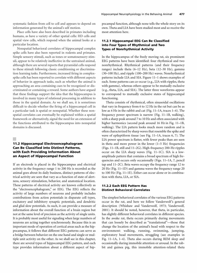
In the hippocampus of the freely moving rat, six prominentEEG patterns have been identified: four rhythmical and twononrhythmical. Rhythmical patterns (and their frequencyranges) include theta (6–12 Hz), beta (12–30 Hz), gamma(30–100 Hz), and ripple (100–200 Hz) waves. Nonrhythmicalpatterns include LIA and SIA. Figure 11–1 shows examples ofeach. Some patterns can co-occur (e.g., LIA with ripples, thetawith gamma), whereas others appear to be mutually exclusive(e.g., theta, LIA, and SIA). The latter three waveforms appearto correspond to mutually exclusive states of hippocampalfunctioning.
Theta consists of rhythmical, often sinusoidal oscillationsthat vary in frequency from 6 to 12 Hz in the rat but can be aslow as 4 Hz in the rabbit and cat (Fig. 11–1A, traces 1–4). Thefrequency power spectrum is narrow (Fig. 11–1B, walking),with a sharp peak around 7 to 10 Hz and often associated withhigher harmonics (second peak around 16 Hz in Fig. 11–1B,walking). The LIA pattern looks much more random and isoften characterized by sharp waves that resemble the spike andwave of epileptiform tissue (see Fig. 11–1A, traces 6, 7). TheLIA power spectrum is flatter, with fewer peaks than are seenin theta and more power in the lower (1–5 Hz) frequencies(Figs. 11–1B, still and 11–2G). High-frequency 200-Hz ripplesoccur on the LIA sharp waves (Fig. 11–1C). SIA is a low-amplitude pattern that contains a broad spectrum of high fre-quencies and occurs only occasionally (Figs. 11–1A, 7, penciltap and 11–2C). Beta waves occupy the frequency range 12 to20 Hz (Fig. 11–1D) and gamma waves the frequency range 20to 100 Hz (Fig. 11–1E). Either can occur alone or in combina-tion with theta, LIA, or SIA.
11.2.2 Each EEG Pattern HasDistinct Behavioral Correlates
The simplest behavioral correlates of the various EEG patternsoccur in the rat, and here we follow Vanderwolf ’s generaldescription (Whishaw and Vanderwolf, 1973; Vanderwolf,2001). It should be noted, however, that theta, in particular,has slightly different behavioral correlates in different species.In the awake rat, theta occurs primarily during movementsthat can loosely be described as “translational”—those thatchange the location of the animal’s head with respect to theenvironment: walking, running, swimming, jumping,exploratory head movements, struggling (see examples inFig. 11–1A, 1–4). Theta also occurs during REM sleep andoccasionally during immobile attention or arousal. In the rab-bit and guinea pig, this immobile attention-related theta
Hippocampal Neurophysiology in the Behaving Animal 477

478 The Hippocampus Book
A
22 inch jump
placed in box
movement movement
11 inch jump
picked up placed in water swim climb out
sitting still teeth chatter head turn sitting still
sleep
sleep pencil tap
1.5
PowerLog
0 50 100Frequency (c/s)
Still
Walk
1 Hz to 10 kHz
500 Hz to 10 kHz
100 to 400 Hz
1 to 50 Hz
100 ms
1 s 500 mV
Mucosa
Movement
L Dentate
R Dentate
30-80 Hz
P
5.0 s
s
OB
H
D
10-50 Hz
M
S
1
2
3
4
5
6
7
1
2
3
4
1
2
3
4
5
1
2
3
456
B
C
D E
4.5
Figure 11–1. Hippocampal electroencephalography (EEG) patterns.A. Theta during rapid-eye-movement (REM) sleep (trace 1), jump-ing (2, 3), and swimming (4). Large irregular amplitude activity(LIA) during quiet sitting (5) and slow-wave sleep (6, 7). Note thelarge amplitude sharp waves especially prominent during slow-wavesleep. Small irregular amplitude activity (SIA) during brief arousalfrom slow-wave sleep after a pencil tap (7). (Source: Whishaw andVanderwolf, 1973.) B. Frequency power spectrum during walkingand standing still. Note the peaks around 8 and 16 Hz and the gen-erally higher amplitude in the beta and gamma bands during walk-ing. (Source: Leung, 1992.) C. LIA ripples and sharp waves in
broad-band (trace 1) and filtered (2–4) recordings. Ripples (3) andsharp waves (4) are accompanied by bursts of action potentials (2)in hippocampal interneurons (small spikes) and principal cells(large spikes). (Source: Buzsaki et al., 1992.) D. Beta activity in theolfactory bulb (OB) and dentate gyrus (D) during sniffing toluene(thickening of s in trace 6). Note the absence of beta in the hip-pocampus (2, H) and the absence of gross movement (5, M). E.Gamma activity (3, 4) in the dentate during sniffing (s). Trace5 shows the increase in breathing recorded during sniffing.(Source: Vanderwalf, 2001.)

occurs more often. In the cat, it is related to active exploratoryeye movements.
LIA occurs during behaviors that do not change the ani-mal’s location, such as sitting quietly, eating, drinking, groom-ing, and during slow wave sleep (SWS) (Fig. 11–1A, 5–7). SIAis seen infrequently: in the awake rat when ongoing move-ment is abruptly halted after a long train of theta, during sud-den, motionless transitions from rest/sleep to alertness (Fig.11–1A, 7, pencil tap), and during periods of SWS after REMepisodes. Much less is known about the full range of behav-ioral correlates of beta and gamma, but they both have beenshown to be associated with different types of olfactory input.Gamma occurs when the animal sniffs at a wide range ofodors (Fig. 11–1E), whereas beta waves have been observedonly during the sniffing of odors associated with predators(Fig. 11–1D).
Á11.3 Hippocampal Theta Activity
11.3.1 Hippocampal Theta Activity:Historical Overview
Theta activity and its behavioral correlates have been studiedextensively ever since its discovery in the rabbit hippocampusby Jung and Kornmuller in 1938 (Jung and Kornmuller,1938). The initial skepticism about the possibility that suchregular large amplitude waves could be generated in the brainwas overcome when Green and Arduini (1954) repeated theobservations during the 1950s and showed that the thetapattern correlated inversely with neocortical desynchroniza-tion, suggesting that it represented the hippocampal arousalpattern. Later, attempts were made to correlate the theta rhy-
Hippocampal Neurophysiology in the Behaving Animal 479
A
B
C
E
F
Movement
Immobile
Procaine
Carbachol + Immobile
Atropine + Movement
D Carbachol + Movement
10
20
30
0
20
30
10
0 255 10 20 3015
30
00 5 15 2510 20 30
10
0
10
20
30
0 5 10 15 20 3025
G Atropine + Immobile
0
20
30
po
wer
(db
er(d
b)
50 10 15 20 25 30
frequency (hz)
6.6
6.5
8.6
10
Figure 11–2. EEG patterns and frequency power spectra duringmovement and immobility in the rat. A. Theta during spontaneousmovement. B. LIA during quiet immobility. C. All theta is abolishedby a procaine injection into the medial septum. D.Theta duringmovement following intraseptal injection of the cholinomimeticcarbachol. E. a-Theta during immobility following intraseptal injec-tion of the cholinomimetic carbachol has a lower frequency than
in D. F. t-Theta during movement is not blocked by an intraseptalinjection of the anticholinergic atropine. G. LIA during immobilityfollowing intraseptal atropine. All EEG traces are 3 s long except C,which is 2 seconds. Arrows in D, E, and F indicate peak theta fre-quency in hertz. (Source: After Lawson and Bland, 1993, with per-mission.)

thm with aspects of learning: Adey et al. (1960) in the UnitedStates reported frequency shifts during simple discriminationlearning, and Grastyan et al. (1959) in Hungary found high-frequency theta during orienting behavior early in auditory-reward conditioning but a desynchronized SIA pattern later,after the animal had learned to approach the reward. Studiesby Gray (1971) in Britain suggested that particular frequenciesof hippocampal theta activity (ca. 7.7 Hz) in the rat may alsooccur in association with reactions to nonreward.
These differences of opinion from different laboratoriesled to speculation that the behavioral correlate of theta variedfrom species to species and perhaps from task to task. Somemeasure of order was brought to the field by Vanderwolf ’scareful observations of the strictly behavioral correlates ofhippocampal EEG patterns (Vanderwolf, 1969). He made theimportant point that changes of the kind seen by Adey andGrastyan that appeared to be correlated with different phasesof learning were also correlated with specific changes inbehavior at the same time. The changes in theta frequencyseen by Adey, for example, tended to be correlated withchanges in motor behavior during the course of learning.Vanderwolf argued that it was the strictly behavioral correlateof these EEG patterns that was primary. In various rodentspecies, he and his colleagues observed that theta activity wasassociated with what he termed “voluntary movements,”whereas LIA occurred during stereotyped “fixed-action pat-terns.” His subsequent observation that there were two typesof theta, each with different behavioral correlates, clarifiedmatters further (Kramis et al., 1975). One type is assoc-iated with arousal or attention, whether in association withmovement or immobility, and the other with a class of move-ment alone. These two types are expressed to different degreesin different species and depend on different neuromodula-tory inputs. The changes in hippocampal EEG during classicconditioning in the rabbit are examined in more detail in thesection on nonspatial learning and memory (see Section11.11).
11.3.2 Hippocampal Theta Activity IsComprised of Two Components, a-Thetaand t-Theta, Which Can Be Distinguishedon the Basis of Behavioral Correlatesand Pharmacology
In addition to differences in their behavioral correlates, thetwo components of theta recorded in freely moving animalsalso differ in their pharmacology. One is affected by drugs thatact at cholinergic synapses, such as the antagonists atropineand scopolamine, the agonist carbachol, and the anticholin-esterase, eserine. Vanderwolf and colleagues (Kramis et al.,1975) called this component atropine-sensitive theta, but werefer to it here by the more psychological name, arousal- orattentional-theta (a-theta for short). The second componentof theta is correlated with translational movements and isunaffected by cholinergic drugs. There is some evidence thatit is dependent on the transmitters serotonin and glutamate. Itis called translation-movement theta (t-theta for short).
Theta is not uniformly distributed in the hippocampal for-mation but varies in both phase and amplitude in differentparts. Whereas synchronous, large-amplitude theta waves areprominent in the dentate gyrus and CA1 areas of the hip-pocampus, most mapping studies report the absence of thetain area CA3. Despite this, CA3 cells can show good phase cor-relates to the EEG recorded elsewhere (e.g., in CA1 or the den-tate gyrus). The implication is that the presence or absence oftheta waves in the EEG is not a simple function of the under-lying single-cell activity. The presence of theta must depend, atleast in part, on other factors, such as the anatomical arrange-ment of the cells and the phase relations of their activity (seeBuzsaki, 2002 for a recent review of the cellular basis of theta).
11.3.3 Both Types of Theta Activity AreDependent on the Medial Septal/DBB but Onlyt-Theta Is Dependent on the Entorhinal Cortex
As discussed in Chapter 3, the major cholinergic input to thehippocampal formation arises from the medial septal nucleusand the nucleus of the diagonal band of Broca. The relativecontributions of the medial septal nucleus and entorhinal cor-tex to the overall hippocampal theta pattern have been dis-sected using a combination of lesions and pharmacologicalmanipulations (Bland and Oddie, 2001; Buzsaki, 2002).Lesions of the medial septal nucleus and associated diagonalband eliminate both types of theta activity. Injection of vari-ous drugs into the septum reveals differences in the pharma-cological basis of a- and t-theta (Lawson and Bland, 1993).Inactivation by intraseptal injections of the local anestheticprocaine or the �-aminobutyric acid (GABA)ergic drug mus-cimol eliminates all theta from the hippocampus (Fig. 11–2C). Intraseptal injection of cholinergic antagonists (such asatropine) also block both thetas if the animal sits quietly (Fig.11–2G) but leave movement-related t-theta intact (Fig.11–2F). Conversely, injections of cholinergic agonists such ascarbachol produce a state of continuous theta in the hip-pocampus regardless of whether the animal is moving (Fig.11–2D) or sitting quietly (Fig. 11–2E). The frequency of the a-theta that occurs during immobility is about 2 Hz lower thanwhen the animal is moving (compare power spectra in Fig.11–2, D and E).
Lesions of the entorhinal cortex, in contrast, eliminate t-theta but leave a-theta intact (Kramis et al., 1975). Followingsuch lesions, injections of atropine eliminate the remainingtheta. Chronic injections of para-chlorophenylalanine (PCPA)or reserpine, both of which reduce the levels of serotonin inthe brain, appear to eliminate t-theta. Because the N-methyl-D-aspartate (NMDA) receptor blocker ketamine also elimi-nates t-theta and results in a depth profile similar to urethane,it is likely that the glutamatergic afferents from the entorhinalcortex to the distal dendrites of CA1 and CA3 are responsiblefor this subcomponent of theta (Buzsaki, 2002).
The simplest explanation of these findings is that t-thetadepends on fibers that pass through, or synapse in, the medialseptum on their way to the entorhinal cortex with onwardconnection to the hippocampus. On the other hand, a-theta
480 The Hippocampus Book

requires the integrity of the direct cholinergic projection fromthe medial septal-diagonal band of Broca to the hippocampusperhaps through the activation of networks of inhibitoryinterneurons (see Chapter 8).
11.3.4 t-Theta Occurs DuringMovement Through Space
Following Vanderwolf ’s observational studies, the tight cou-pling between t-theta and certain types of movement wasstrengthened by the results of “theta-conditioning” experi-ments by Black (1975). The idea was to reward animals forproducing short trains of theta activity under two trainingconditions: In one they were allowed to move, but in the otherthey were forced to hold still. Training to produce theta withfrequencies greater than 7 Hz (t- theta) was successful onlywhen movement was allowed. Thus, movement is necessaryfor t-theta, but what aspect of such movement is being sig-naled by theta activity?
Vanderwolf ’s original suggestion that theta was correlatedwith some aspect of voluntary behavior is not operationalenough because it is not clear whether, in the case of animals,a movement can ever be said to be truly volitional.Considering the movements during which theta occurs mostreadily—walking, running, swimming, jumping—the likelycommon factor is translation through space. The fact thatsmall head movements during exploratory sniffing are alsoaccompanied by theta suggests that the crucial factor is thetranslation of the head through space or, more specifically,the generation of motor outputs that would normally result intranslation of the head through space. Changes in the fre-quency of theta activity are correlated with either the animal’sspeed of movement through the environment (Rivas et al.,1996; Slawinska and Kasicki, 1998) or the rapidity with whicha movement is initiated. The latter has been shown in experi-ments in which rats have been trained to jump up onto a ledgeor to initiate running in a runway (Whishaw and Vanderwolf,1973).
The behavioral correlate of theta amplitude is less clear.Although Vanderwolf and colleagues failed to find a positivecorrelation between amplitude and speed, Rivas et al. (1996)reported a positive correlation between both the frequencyand the amplitude of theta and the speed of movement in theguinea pig when movements are initiated in simple runways.On the other hand, there is no correlation in jumping experi-ments between amplitude and speed of movement. It seemsthat there are many factors contributing to the amplitude oftheta, such that a consistent correlate of amplitude is notalways seen.
11.3.5 a-Theta Occurs During Arousaland/or Attention as well as Movement
a-Theta may correlate with psychological states such asarousal or attention. It rarely occurs in isolation in the rat butis much more common in rabbits, guinea pigs, and cats. Inrats, it occurs naturally only when the animal freezes follow-
ing a noxious stimulus, in response to an aversive conditionedstimulus, or when the animal is immobile but preparingto move (such as just before a jump). It also co-occurs witht-theta during movement. In rabbits and guinea pigs, it isreadily produced in immobile animals by innocuous visual,auditory, or tactile stimuli. Repeated stimulation, however,leads to habituation of this response. Sainsbury and colleagues(1987a,b) have shown that the ability of a stimulus to elicithippocampal a-theta is dependent on the preexisting level ofarousal. A relatively neutral stimulus such as a tone, whichordinarily has little effect, readily elicits a-theta if the animalhas previously been “sensitized” with an arousing stimulussuch as the sound of an owl or the sight of a predator. Onepossibility is that a-theta represents a subthreshold activationof the motor system. Experiments in support of this idea havebeen carried out by Sinnamon and his colleagues (Sinnamon,2000; Sinnamon et al., 2000). They recorded theta activity inurethane-anesthetized rats before, during, and after hind-limbstepping movements elicited by electrical stimulation of thehypothalamus or pharmacological block of the midbrainraphe nucleus. Both manipulations elicited low-frequencypremovement (presumptive a- type) theta activity as well asthe higher-frequency (presumptive t- type) theta, whichaccompanied the hind-limb stepping movements. It might bethat a- theta reflects the activation of movement programs inthe absence of the movement itself. Alternatively or in addi-tion, it might reflect the organization of sensory inputs asreflected in the correlation of single-unit activity in differentsensory nuclei with hippocampal theta (see Section 11.3.7).
11.3.6 Theta and Sleep
Theta occurs during the REM phase of sleep; LIA and SIAoccur during slow-wave sleep; and SIA bursts frequently comeat the termination of an REM episode. Pharmacological stud-ies have shown that the theta recorded during the actual eyemovements of the REM phase of sleep is unaffected by cholin-ergic drugs and therefore resembles t-theta, and that outsideof these episodes is a-theta.
11.3.7 Theta Activity in Nonhippocampal Areas
In the rat, the theta system is centred on the hippocampal for-mation, taken here to include the septum, subicular area, andentorhinal cortex (Alonso and Garcia-Austt, 1987a,b;Brankack et al., 1993). However there are also reports of EEGand cellular activity phase-locked to theta in the cingulate cor-tex (Leung and Borst, 1987), prefrontal cortex (Hyman et al.,2005; Jones and Wilson, 2005; Siapas et al., 2005), perirhinalcortex (Muir and Bilkey, 1998), posterior hypothalamusincluding the mammillary bodies (Kirk and McNaughton,1991; Bland et al., 1995; Slawinska and Kasicki, 1995; Kocsisand Vertes, 1997), brain stem reticular formation (Nunez etal., 1991), amygdala (Paré and Gaudreau, 1996; Seidenbecheret al., 2003), and superior (Natsume et al., 1999) and inferior(Pedemonte et al., 1996) colliculi. Some of these areas, suchas the posterior hypothalamus and the brain stem reticular
Hippocampal Neurophysiology in the Behaving Animal 481
formation, are involved in the circuitry that generates thehippocampal theta rhythm and might be expected to showactivity synchronized with theta, whereas others are part ofthe sensory systems (the colliculi) or the limbic system (amyg-dala and cingulate cortex). These latter correlations must rep-resent a fairly widespread function for theta, such as thesynchronization or binding together of neurons in many sen-sory, motor, and emotional/motivational centers as well asthose involved in spatial perception and memory.
11.3.8 Does the Hippocampal EEG in Monkeysand Humans Have a Theta Mode?
Although theta patterns are readily observed in cats, dogs, androdents, it has been difficult to establish whether a clear rhyth-mic theta pattern can been recorded from either monkey orhuman hippocampus. There have been hints over the yearsthat it might exist. For example, Stewart and Fox (1991)reported a theta-like pattern (7–9 Hz) in the hippocampalEEG of urethane-anesthetized monkeys. In an earlier study,Watanabe and Niki (1985) reported the existence of therhythmical theta-like firing patterns in monkey hippocampalcells. Similarly, rare experiments using depth electrodes in thehuman hippocampus have observed activity at theta frequen-cies, although their behavioral correlates were not clear(Halgren et al., 1978; Arnolds et al., 1980). This paucity of datahas led some authors to suggest that theta may not exist inmonkeys and primates or may not have the same behavioralcorrelates as in other mammals. Some reasons for the failureto record prominent theta patterns in the primate EEG areexamined in the next section.
First, the existence of an oscillatory theta pattern in theEEG depends not only on the rhythmical firing of cells butalso on the correct cytoarchitectonic orientation of pyramidalcells to create the appropriate electrical dipole. As was seen inthe section on theta activity in field CA3, cells can burst witha theta pattern in the absence of pronounced theta waves inthe EEG. Different neuroarchitecture could account for atheta system in primates in the absence of rodent-like thetapatterns in the gross EEG.
A second problem is that most monkey and human record-ing is done while the subject is immobile, a condition that isnot conducive to recording t-theta. Thirdly, most recordingsin humans have been from the scalp, and it is possible that theskull and scalp are acting as filters, effectively screening out thetheta patterns. Some evidence to support the last two possibil-ities comes from recent findings (Kahana et al., 1999) thattheta patterns could be recorded from electrodes placed onthe surface of the human neocortex while subjects navigatedthrough a virtual reality maze. Theta activity was recordedfrom several cortical regions, but temporal lobe theta showedthe best correlation with maze difficulty. More theta activitywas seen during traverses through more difficult 12-choicemazes than through simpler 6-choice mazes. The same grouphas used depth as well as subdural electrodes in humans per-forming a virtual taxi driver task (Caplan et al., 2003) to show
that theta oscillations in humans are found during virtualmovement, exploratory search, and goal-seeking. One differ-ence from the rat is that human theta bursts tend to be ofshorter duration. These kinds of studies confirm the suspicionthat the difficulty recording primate theta has more to do withinappropriate behavioral paradigms and recording techniquesthan with the absence of a theta system per se.
There has also been recent interest in the related field offrequency analysis of EEGs recorded from scalp electrodes.For example, increases in the power present in the EEG attheta frequencies have been shown to be related to the suc-cessful encoding of new information (for a review seeKlimesch, 1999). However, these studies typically do notdemonstrate the peak in the power spectrum at theta fre-quencies or the long continuous records of trains of thetaactivity shown by Kahana and colleagues in their virtual real-ity study. Localizing the source of theta in scalp-recordedEEGs is even more problematical than with subdural elec-trodes; one experiment where it was attempted implicated theanterior cingulate rather than the hippocampus (in a taskshowing increased theta with increased working memoryload) (Gevins et al., 1997). Spectral peaks at theta frequencieshave been found in experiments using magnetoencephalogra-phy and have been interpreted as consistent with a generatornear the hippocampus (Tesche and Karhu, 2000), althoughthis technique also suffers from problems of accurate sourcelocalization. Nevertheless, these findings, and particularly thesubdural recordings of Kahana et al., clearly show that thetaactivity exists in the human brain and tempt one to speculatethat it might be related to the hippocampal system. There is amore extensive discussion of recording of the EEG and evokedpotentials from the human brain in Chapter 12.
11.3.9 Functions of Theta
Work on the rat hippocampus suggests three possible func-tions for theta. First, it acts as a global synchronizing mecha-nism, essentially locking the entire hippocampal formationinto one global processing mode and organizing the activity ineach hippocampal region with respect to the others.Simultaneous recordings of the EEG in different hippocampallocations have shown that theta activity at comparable loca-tions (e.g., in the CA1 pyramidal layer) is in synchrony andcoherent across large areas of the hippocampal formation(Mitchell and Ranck, 1980; Fox et al., 1986; Bullock et al.,1990). This means that if two cells have firing patterns that aresystematically related to the local theta cycle, they have sys-tematic temporal relations to each other, even if they arelocated far apart in the hippocampus. Although the thetarhythm is centered on the hippocampal formation, sensoryand motivational areas are also brought under its sway. Webegin to see here evidence for a widespread system of oscilla-tions that organizes the activity of many disparate brain areas.
A second function of the theta oscillations is to provide aperiodic clocking system for the timing of hippocampalspikes. As set out in greater detail in Section 11.7.9, the phase
482 The Hippocampus Book

relation of each pyramidal cell measured against the concur-rent theta activity is not constant but can vary from one cycleto the next (O’Keefe and Recce, 1993; Skaggs et al., 1996). As arat runs through the firing field of a spatially coded pyramidalcell (the place field), the cell fires bursts of spikes at an inter-burst frequency slightly higher than that of the concomitantEEG theta. This leads to a precession of the phase of firing toearlier points on each successive cycle. Over the course of thefive to seven theta cycles that comprise the typical place field,the phase of the EEG at which the cell fires may precessthrough a full 360�, although smaller amounts of precessionare also seen. Furthermore, the phase of firing is highly corre-lated with the animal’s location within the place field (more sothan with the duration spent in the field). Thus, temporalvariation in spike firing conveys information about the ani-mal’s spatial location. An analysis of this phenomenon (Jensenand Lisman, 2000) shows that the temporal information pro-vided by the phase precession can improve localization of theanimal’s position by more than 40% compared to thatobtained by the use of firing rates alone.
A third function for theta is to provide temporal controlover long-term potentiation (LTP) induction and, by infer-ence, the storage and retrieval of information from the hip-pocampus. As noted in Chapter 10, theta-burst electricalstimulation of hippocampal afferents is an effective way toinduce LTP. Furthermore, there is some evidence that volleysarriving at different phases of the ongoing theta are differen-tially effective (Pavlides et al., 1988; Huerta and Lisman, 1995;Holscher et al., 1997; Hyman et al., 2003). Inputs arriving atthe positive phase of CA1 theta result in synaptic potentiation,whereas those arriving at the negative phase yield depotentia-tion or depression. On the basis of this and other evidence,Hasselmo (2005) proposed that the various phases of thetaoscillation represent different modes of operation. Specifi-cally, the peak of the CA1 theta is the period during whichencoding of new information entering the hippocampus fromthe entorhinal cortex takes place, and the trough is the periodduring which retrieval of information from the hippocampusto the entorhinal cortex occurs.
Á11.4 Non-theta EEG Patterns in theHippocampal EEG: LIA, SIA,Ripples, Beta, and Gamma
11.4.1 Sharp Waves, Ripples, andSingle Units During Large Irregular Activity
During LIA, large sharp waves occur in the hippocampal EEG(Fig. 11–1A, traces 5–7 and Fig.11–1C, trace 4). In CA1, thesharp waves occur most frequently during slow wave sleep andquiet sitting, less frequently during eating and drinking, andleast frequently during grooming. They appear to occur dur-ing periods of low arousal and are often, but not always, inhib-ited by arousing stimuli. They may represent a resting state
of the hippocampus as a whole or the absence of some aspectof hippocampal function such as the theta state; functionally,it has been suggested that they represent a neural correlate ofmemory consolidation. Each sharp wave lasts 50 to 100 msand has a maximum amplitude in the stratum radiatum thatcan be as large as 1 mV or more. Their resemblance to theinterictal spike and wave complex of epileptogenic cortexmay give some clues to the peculiar susceptibility of the hip-pocampus to seizure activity (Bragin, 1999; Draguhn et al.,2000; Buzaki and Draguhn, 2004). They occur more or lesssynchronously over large areas of the CA1 field of the dorsalhippocampus: Recordings at different points along the septo-temporal axis of the hippocampus have shown that they arein phase over the entire extent (Buzsaki et al., 1992; Chrobakand Buzsaki, 1996). They reverse polarity in the pyramidalcell layer and reach their maximum amplitude several hun-dred microns into the stratum radiatum (O’Keefe and Nadel,1978, pp. 150–153). Buzsaki and colleagues have suggestedthat sharp waves originate in the CA3 field and that thesharp waves recorded in CA1 are the summated extracellularexcitatory postsynaptic potentials (EPSPs) of the Schaffer col-laterals that result from synchronous firing of the CA3 pyram-idal cells (Csicsvari et al., 2000). Direct stimulation of thesefibres results in evoked potentials with similar shape anddepth profiles.
Around the time of the negative peak of the sharp wave,there is a high-frequency oscillation of between 120 and 200Hz whose peak amplitude occurs in the CA1 pyramidal celllayer (O’Keefe and Nadel, 1978, pp. 150–153; Buzsaki et al.,1992) (Fig. 11–1C). During these “ripples” there are synchro-nous bursts in almost all theta interneurons and in about 1 in10 of the complex-spike pyramidal cells. Intracellular record-ings from the soma of pyramidal cells during ripples revealsintracellular oscillations that mirror the extracellular pattern.Hyperpolarization results in a reduction of ripple amplitudeat 70 mV, with a reversal at more negative potentials. Thissequence of events suggests that the sharp wave itself reflectsthe synchronous barrage of afferent activity from CA3 cellsonto the apical dendrites of CA1 cells. The rapid activation ofinhibitory interneurons causes synchronized hyperpolarizinginhibitory postsynaptic potentials (IPSPs) in the somata ofpyramidal cells.
Are sharp waves and ripples confined to the hippocampusproper, or do they spread to other structures? Recordings inthe subiculum and the deep layers of the entorhinal cortexshow that sharp waves also occur in these structures at aboutthe same time as those in hippocampus. The sharp wave in lay-ers V and VI of the entorhinal cortex, which receive inputsfrom the hippocampus, followed those in the hippocampus by5 to 30 ms (Chrobak and Buzsaki, 1996). In contrast, the pres-ence of hippocampal sharp waves is not reflected in the activ-ity of the upper layers (II and III) of the entorhinal cortex,which project to the dentate gyrus, hippocampus, and subicu-lum. Recordings from the dentate gyrus show that the granulecells are also influenced by the sharp-wave activity of CA3. Theanatomical basis for this retrograde effect is not entirely clear.
Hippocampal Neurophysiology in the Behaving Animal 483

There are projections from CA3 into the polymorph layer, andthese fibers may be ending on mossy cells, which in turn proj-ect to the dentate granule cells. In the ventral tip of the hip-pocampus, there appear to be some CA3 pyramidal cells thatproject directly into the molecular layer of the dentate gyrus.
11.4.2 Dentate EEG Spikes During LIA
Sharp-wave activity during LIA also occurs in the granule celllayers of the dentate gyrus. It is also associated with a high-frequency ripple, although the frequency is not as high as thatof the CA1 ripple. Most granule cells do not fire during den-tate sharp waves, but intracellular recordings have shown thatthey are nevertheless depolarized at this time. Surprisingly,the overall effect of dentate spikes on CA3 cells is inhibitory.This presumably reflects the heavy innervation of interneu-rons by the mossy fibers (Acsady et al., 1998) (see Chapter 5,Section 5.7).
11.4.3 Pharmacology of LIA
Little is known about the pharmacology of LIA, the sharpwaves, or the ripples. Ripples are eliminated under halothaneanesthesia, and their frequency is reduced under urethane andketamine to around 100 Hz (Ylinen et al., 1995). In the mouse,sharp waves and ripples can be elicited by application of KClto the dendrites of pyramidal cells following pharmacologicalblock of GABAA receptors, making it unlikely that they aredue to activity in networks of the inhibitory interneurons(Nimmrich et al., 2005). Synchronization between ripplesappears to depend on gap junctions. Consistent with the ideathat the LIA state is a passive absence of theta, procaine ormuscimol suppression of the medial septum, which inhibitstheta activity, has only a small effect on the power of hip-pocampal LIA (Bland et al., 1996). Recordings of medial sep-tal and diagonal band cells during LIA show that the firingrates of most of these cells are reduced relative to theta,strengthening this suggestion. On the other hand, cholinergicactivation of the septum completely suppresses hippocampalLIA, replacing it with low-frequency theta even in the immo-bile animal (Fig. 11–2E). Theta appears to be the active statedriven from the medial septum, and LIA is the passive statethat occurs in its absence.
11.4.4 Behavioral Correlatesand Functions of LIA
In their discussion of hippocampal sharp waves and ripplesduring LIA, O’Keefe and Nadel (1978, pp. 150–153) suggestedthat one way to think about this EEG state was as an absenceof the theta state—it is the hippocampus in a non-theta idlingmode. This suggestion was based partly on the observationthat there was always a period of at least a few secondsbetween the onset of an LIA-associated pattern of behavior(such as immobility) and the onset of the sharp wave and rip-ples. They also argued that the nearly synchronous bursts in a
sizable percentage of the pyramidal cells, when compared withtheir highly differentiated patterns of activity during the thetastate as the animal moved around an environment, would beunlikely to convey much information. Perhaps most tellingly,it was found that lesions of the fornix decreased neither thefrequency nor the amplitude of ripples and sharp waves; ifanything, they increased them—again suggesting release ofthe hippocampus from an activated theta state. In this view,the LIA state is the passive activity of a system with extensivepositive and negative feedback loops and other oscillatorymechanisms.
An alternative hypothesis is that LIA is an active, ratherthan an idling, state of the hippocampus whose function is tostrengthen synaptic modifications that have occurred duringthe immediately prior periods (Buzsaki, 1989). Pointing to thesimilarity between the synchronous volleys of afferent activityimpinging on the dendrites of CA1 dendrites during LIA andthe type of tetanic stimulation known to cause LTP, Buzsakisuggested that synaptic potentiation occurs naturally duringLIA. He went on to propose that this potentiation acts as aboost to synapses that had been only weakly modified duringthe previous theta behaviors and perhaps plays a role in amemory consolidation process. Evidence in support of thisidea comes from experiments by Skaggs and McNaughton(1996). They looked at pairs of CA1 pyramidal cells with over-lapping place fields in freely moving rats that were runningaround a small triangular runway. When identified cells wererecorded before and after the animal had run around forextended periods, they observed during LIA an increase in thecross-correlation between cells that had been simultaneouslyactive during the previous period. That is, cells with firingfields that were close together in the environment were morelikely to fire close together in time during an ensuing periodof slow-wave sleep than cells with more distant fields. Thesmall number of collateral fibers between CA1 pyramidal cellssuggests that this effect might be due to an increase in the effi-cacy of the common input from CA3 or the entorhinal cortexonto these cells, rather than to the direct connections betweenthem. In the consolidation view of LIA, the uncoupling of thehippocampus from the septum during LIA is merely a neces-sary condition for hippocampal consolidation to occur.
A related idea is that LIA is a period involving transfer ofinformation from hippocampus to neocortex. Support forthis view comes from experiments (Siapas and Wilson, 1998)that showed a correlation between hippocampal sharp wavesand neocortical spindle waves during slow-wave sleep. Thehypothesis that information might be transferred from thehippocampus to the neocortex as a result of LIA-associatedsharp waves is consonant with evidence from behavioralexperiments addressing the question of long-term storage ofmemory “traces” in or outside of the hippocampus. Chapter12 presents evidence that lesions to the hippocampus in ani-mals and humans can sometimes cause temporally gradedamnesia. One interpretation of such a gradient is that mem-ory traces are stored only in the hippocampus for a shortperiod before being sent to the neocortex for permanent stor-
484 The Hippocampus Book

age (see Chapter 13, Section 13.3 for a more extensive discus-sion of consolidation). After this period, damage to the hip-pocampus would no longer result in memory loss. Althoughthe sharp waves may be part of such a mechanism, there issome evidence against this general idea. For example, Leonardand colleagues (1987) have shown that LTP cannot be inducedin the hippocampus during slow-wave sleep. If this findingcan be generalized to other parts of the brain, it would greatlyreduce the attractiveness of the hypothesis that sharp wavescould serve as a basis for consolidation because no new LTP-based learning could take place. There is clearly much more tounderstand about ripple/sharp-wave states. A possible role inmemory formation may not be restricted within the confinesof the currently dominant theory of intrahippocampal con-solidation or hippocampo-neocortical trace transfer duringsleep. For example, ripple/sharp-wave events may have apurely intrahippocampal housekeeping function. To take justone possibility, synaptic renormalization or overall gain con-trol processes might occur during LIA.
11.4.5 Small Irregular Activity
Small irregular activity (SIA) is characterized by low-amplitude irregular activity in the hippocampus and desyn-chronization in the neocortical EEG. In 1967, Pickenhainand Klingberg reported low-amplitude irregular activity inthe hippocampus of rats during transitions to alertness whereno orienting movements were made, such as when a clickawakened them from sleep. Vanderwolf and Whishaw(Whishaw and Vanderwolf, 1971) noted a similar pattern dur-ing transitions to alertness but added the observation thatSIA, as they called it, occurred when rats abruptly halt volun-tary movement.
Jarosiewicz and colleagues (2002) have extended the char-acterization of SIA to include periods during sleep. Sleep SIAbursts occur repeatedly during all periods of slow-wave sleepand after nearly every REM episode. Each burst typically lastsa few seconds, with a range from 200 ms to many seconds. Abrief tone presented in sleep routinely elicited an increase inelectromyographic (EMG) and neocortical arousal accompa-nied by hippocampal SIA, suggesting that it is a state interme-diary between LIA and theta (Jarosiewicz and Skaggs, 2004b).Sleep SIA is characterized by the cessation of firing in mostpyramidal cells. The 3% to 5% that continue to fire do soactively and repeatedly over successive bursts. These place cellsare probably continuing to represent the location where therat fell asleep because rotating the platform and the sleepingrat away from a given cell’s place field location relative to thetesting laboratory did not have an effect on SIA firing(Jarosiewicz and Skaggs, 2004a).
11.4.6 Beta/Gamma Activity in the Hippocampus
In addition to the low-frequency EEG activity seen duringtheta and LIA, and the higher frequency ripple activity char-acteristic of sharp waves and dentate spikes, intermediate fre-
quency 10- to 100-Hz waves have also been described. Thisband is further divided into beta (10–20 Hz) (Leung, 1992)(Fig. 11–1D) and gamma (20–100 Hz) (Fig. 11–1E) activity.Gamma waves were first described in the cat amygdala byLesse in 1955 and have since been reported in widespreadbrain regions of animals and humans (Singer and Gray, 1995).It has been suggested that gamma synchrony between variousregions of the neocortex binds together the simple elements ofa complex representation. In the hippocampal formation, theyoccur most clearly in the entorhinal cortex and dentate buthave also been reported in the CA fields (Csicsvari et al.,2003). They may be related to 40-Hz oscillations that havebeen reported in the olfactory (Freeman, 1975) and visual(Singer and Gray, 1995) systems.
Gamma activity is slightly depressed in the rat by both sep-tal lesions and cholinergic antagonists (Leung, 1985). In rab-bits, drugs that stimulate a-theta during immobility (e.g.,physostigmine) increase the amount of this intermediate fre-quency. However, this increase does not seem to occur inimmobile rats. Seizures cause a dramatic increase in theamount of beta/gamma activity, an effect blocked by cholin-ergic antagonists and unaffected by the animal’s ongoingbehavior (Leung, 1992). The extent to which this beta/gammaactivity is a reflection of the underlying behavior of neural ele-ments (principal cells or interneurons) is unclear, although itmay be a correlate of the activity of hilar theta cells.
11.4.7 Olfactory Stimulation Can ElicitHippocampal Gamma and Beta Waves
In the rat hippocampal formation, both beta and gammawaves occur preferentially during olfaction. The dentategamma waves appear to occur during the sniffing of odors ingeneral but do not occur during odorless sniffing or othersensory stimulation (Vanderwolf, 2001) (Fig. 11–1E). Thebehavioral correlates of the CA1/CA3 gamma have not beenestablished. In general, gamma and theta occur independ-ently, but under some (undefined) circumstances theybecome synchronized, with the gamma occurring preferen-tially at the positive peak of the dentate theta waves. Thedentate, but not the CA1, gamma is dependent on the per-forant path input from the entorhinal cortex, as lesions of theentorhinal cortex abolish the dentate gyrus gamma butenhance the CA1/CA3 gamma (Bragin et al., 1995). Dentategamma waves may be part of the mechanism for synchroniz-ing the olfactory inputs arriving via the entorhinal cortexwith the hippocampal theta. Beta waves have a more restrictedolfactory correlate than gamma waves. They occur in the den-tate gyrus in response to olfactory inputs that signal, or mimicthose that signal, the presence of predators (Fig. 11–1D)(Vanderwolf, 2001): compounds found in the anal scent glandsecretions of weasels and foxes, most organic solvents includ-ing toluene and xylene, and phytochemicals derived fromplants such as eucalyptol and salicylaldehyde. In general, theseodors also elicit a fear response and behavioral avoidance.Other strong smells, such as ammonia, cadaverine, or
Hippocampal Neurophysiology in the Behaving Animal 485

putrescine, which are either approached or not avoided, donot elicit dentate beta waves. Vanderwolf has argued thatthese olfactory correlates of gamma and beta waves in thehippocampus suggest a primary olfactory function for thisstructure, but it is more likely that, like the theta waves,they represent one aspect of a more complex overall func-tion, such as cognitive mapping or associative memory for-mation.
Á11.5 Single-cell Recording in the HippocampalFormation Reveals Two Major Classes ofUnits: Principal Cells and Theta Cells
Although EEG recordings provide some information aboutthe circumstances under which large numbers of neurons in abrain region become synchronously active, it is generallyaccepted that a complete understanding of function can begained only by looking at the behavioral correlates of singleunits. One reason for this is that although neighboring neu-rons often share common functional properties they may notalways respond to the same specific stimulus or behavior. Aswe shall see, this is particularly true of the hippocampus,where neighboring pyramidal cells share the property of rep-resenting places in an environment. However, because differ-ent cells become active in different parts of an environment,this property is not revealed in the hippocampal EEG.
The recording of individual neuronal responses in the hip-pocampus of the awake freely moving rat began during theearly 1970s with the work of Ranck (1973) and O’Keefe(O’Keefe and Dostrovsky, 1971). They both encouraged theiranimals to move around the environment, engaging in every-
day tasks such as eating, drinking, sleeping, and searching forfood and water. This emphasis on naturalistic behavioral cor-relates led to several important discoveries. The first was thatthey noticed that there were two major classes of cells thatcould be distinguished by differences in their anatomical andphysiological properties (e.g., firing rates, action potentialwidth, and relative locations in the hippocampus). Rancktermed these two classes complex-spike and theta cells, andthese terms are still in general usage. We describe their prop-erties in a subsequent section. Perhaps more importantly, theydiscovered that the firing patterns of the two cell types hadrepeatable behavioral correlates. Ranck had trained his ani-mals to approach one location to obtain food and another toget water, and emphasized the relation of the complex-spikecell firing pattern to the behavioral approach to reward.O’Keefe was more impressed by the spatial correlate andnamed the cells place cells. It is now widely accepted that thelocation of the animal in a familiar environment is the majordeterminant of when such cells fire.
The second class of neurons, the theta cells, has less specificbehavioral correlates. As the name implies, their behavioralcorrelates are closely related to those of the gross EEG wavesand in particular theta. They tend to change rate during hip-pocampal EEG theta, and many display strong phase lockingto the individual theta waves.
Extracellular recordings in the freely moving rat enablecomplex-spike and theta cells to be distinguished on the basisof differences in their wave shapes, firing rates, and otherproperties (Fig. 11–3). Complex-spike place cells have a muchbroader action potential than theta cells (Fig. 11–3B–D), oftendisplay a complex-spike burst pattern in which the later spikesin a burst are smaller in amplitude and of longer durationthan the first (Fig. 11–3A), and have a lower spontaneous
486 The Hippocampus Book
Figure 11–3. Complex-spike cells and theta cells have differentphysiological properties. A. Theta cells have a steady firing rate andconstant size amplitude spikes, whereas C-S cells sometimes emit acomplex-spike burst in which the later action potentials in the burstare lower in amplitude and broader. B–D. Action potentials of C-Scells (pyr) are broader and have a larger initial hump than those of
theta cells (int). E. C-S cells have a narrower range of interspikeintervals (ISI) F. Three-dimensional plot of firing rate againstmean ISI and spike asymmetry (a-b in D) separates the overallpopulation into two clusters. (Source: A,B. After Christian andDeadwyler, 1986; C–F. After Csicsvari et al., 1998 with permission.)

background firing rate (generally about 1 Hz) (Fig. 11–3F). Incontrast, theta cells have a much higher firing rate (10–100Hz) (Fig. 11–3F) with all action potentials being of the sameamplitude (Fig. 11–3A) and of shorter duration (Fig. 11–3B-D). An important caveat is that in the rabbit this separation ofspikes into two nonoverlapping classes is less clear as thereappears to be a large subclass of complex-spike cells withspontaneous firing rates as high as 6 Hz.
It is highly likely that in the rat the complex spike cells arepyramidal cells and the theta cells are one or more types ofinterneuron. Intracellular staining of neurons that displaycomplex spikes in brain slices reveals they have the morphol-ogy of pyramidal cells, whereas those without complex spikesare interneurons. Sometimes it is possible to activate complexspike cells antidromically from electrodes placed in hip-pocampal outflow pathways, but the theta cells can only bedriven orthodromically (Berger and Thompson, 1978; Foxand Ranck, 1981; Christian and Deadwyler, 1986). Anotherstrong piece of evidence in support of the idea that complex-spike cells are pyramidal cells and theta cells are interneuronscomes from the temporal relation between their firing pat-terns. Most pyramidal cells innervate neighboring interneu-rons via an axon collateral, which should give rise to anincreased probability of firing in the theta cell shortly afteran action potential in the pyramidal cell. Just such a relation
has been reported. Some theta cells tend to fire a few millisec-onds after a complex-spike cell recorded on the same tetrode(see Box 11–1), and this is reflected in a short latency cross-correlation between their spike trains (Csicsvari et al., 1998)(Fig. 11–4B-D and Box 11–2).
The same generalizations can be made for rabbits.However, there appears to be an additional group of interme-diate cells that exhibit complex spikes and that can beantidromically activated by stimulation of projection path-ways; but they have a resting firing rate that is intermediatebetween the theta and complex-spike cell firing rates shown inrats (Berger et al., 1983).
11.5.1 Distinctive Spatial Cells—Complex-spikePlace Cells, Head-direction Cells, and GridCells—Are Found in Various Regions of theHippocampal Formation
Discovery of the place cells led to the development of the cog-nitive map theory of hippocampal function by O’Keefe andNadel (1978), which has guided much of the subsequentresearch and theorizing on hippocampal function (seeChapter 13, Section 13.4). Equally important for our under-standing of the spatial functions of the hippocampal forma-tion was the discovery of two other classes of spatial cell: In
Hippocampal Neurophysiology in the Behaving Animal 487
Box 11–1Microelectrode Recording Technique
Microelectrodes placed in the extracellular space in the vicinity of a neuron detect the currentflow associated with action potentials. Experience has shown that relatively large electrodeswith flat tips are preferable for recording in chronic animals because they do not puncture thecells and therefore do less damage during small movements of the electrode relative to thebrain. One problem with the use of single electrodes in a structure such as the hippocampus isthat it is difficult to isolate single units on the basis of action potential amplitude and shape.This is because of the close packing of the identically sized and shaped cells. Action potentialsfrom all cells on a sphere with the electrode at the center are identical. There is a danger thatall such spikes are considered to have come from one neuron. Template-matching algorithms,which are so useful when different cell types are in close proximity, offer no way out of thisambiguity and also have difficulty coping with the fact that a single pyramidal cell sometimesfires simple spikes and sometimes complex spike bursts in which the later action potentials inthe burst have reduced amplitude and broader waveforms than the initial spike (Fig. 11–3A).One solution, introduced by McNaughton et al. (1983b), is to use multiple electrodes whosetips are close enough to sense the action potentials from a group of neurons but, being spaced ashort distance apart, cannot both be at the center of a notional sphere. Each electrode tip is aslightly different distance from a given cell and, consequently, records its action potential witha slightly different amplitude and shape. These sometimes subtle differences can be used todistinguish a multiunit recording from several electrodes into spikes emanating from differentcells. The principle is analogous to a “stereophonic” recording in which two or more micro-phones are used to capture the sound of an orchestra; hence, the earliest electrodes of thistype used two wires and were called “stereotrodes.” In general, n � 1 electrodes are necessaryto identify uniquely the action potential from a neuron in n-space. In the hippocampus, “trio-trodes” with three tips oriented in the plane of the quasi-two-dimensional pyramidal cell layerwould probably suffice; but in practice, “tetrodes” are commonly used to ensure adequateisolation (O’Keefe and Recce, 1993; Wilson and McNaughton, 1993). Figure 11–4A shows theprofile of action potentials of complex-spike and theta cells on the four electrodes of a tetrode.

488 The Hippocampus Book
0.2ms
intpyr
1
2
3
4
B
2
40.2ms
A
pyr int
-50 500
-50 0 50ms
C
D
Box 11–2Auto-correlation and Cross-correlation Functions
Correlation techniques are used to investigate the temporal relations of spike occurrences tothemselves (auto-correlation) (Fig. 11–3 E), to the occurrence of spikes in other neurons(cross-correlation) (Fig. 11–4C,D), and to other brain events (e.g., theta waves) (Fig. 11–5) orto sensory and motor events in the environment (e.g., peristimulus event histogram) (see Fig.11–17, later). The auto-correlation function is useful for revealing repetitive or rhythmical pat-terns of firing. A graph is constructed in which the x-axis represents time intervals before andafter each spike event, and the y-axis represents the probability that the cell will fire during eachinterval before or after that spike event. A period of 50 ms is a useful period of time for lookingat the complex spike properties of principal cells in which the cell fires repetitively within 10ms (Fig. 11–3A,E); 500 ms is a useful length of time for demonstrating the rhythmical patternof theta cell firing. The cross-correlation function reveals the tendency of two cells to fire with aparticular temporal relation to each other. Here one cell is chosen as the target, and its spikesfix the zero point on the time axis. The probability of the other cell firing at times earlier andlater is calculated and displayed as a histogram. This type of analysis is useful for identifyingpotential synaptic relations between cells or the existence of common inputs to the cells. Thecross-correlogram between a complex-spike cell and a theta cell is shown in Figure 11–4C,D.The peak at 2 ms suggests that there is a short-latency excitatory synaptic connection from thepyramidal cell to the theta cell. The probability associated with this peak gives some indicationof the strength of this connection. The correlation between hippocampal units and the phase ofthe global EEG theta signal is useful for showing the temporal relations of different types ofunit to the EEG and to each other (Fig. 11–5). A final use of correlation techniques is to lookfor a relation of spike firing to a sensory event or motor action in the external world or toanother brain event. Illustrated in Figures 11–17 to 11–19 are peristimulus histograms of hip-pocampal unit responses to auditory stimuli as a result of conditioning experiments and inFigure 11–20 (see later) the increase in activity during different aspects of the behavioral learn-ing paradigm.
Figure 11–4. Hippocampal interneurons sometimes fire a few mil-liseconds after neighboring pyramidal cells. A. Waveforms of apyramidal (pyr) and an interneuron (int) recorded on the sametetrode (traces 1–4). B. Multiple sweeps triggered on the pyramidalcell show that the interneuron often fired at a short but variablelatency shortly after, suggesting a monosynaptic coupling betweenthe two. C. Cross-correlogram between the firing trains of the two
cells in B shows the interneuron fires at a peak latency of 2 ms fol-lowing the spike in the pyramidal cell. D. Controlling for the repeti-tive rhythmical pattern of firing within each of the spike trains doesnot eliminate the short-term causal relation between the two.Horizontal line in D indicates a significance level above which thecorrelation is highly significant. (Source: After Csicsvari et al., 1998,with permission.)

1984, Ranck described the head direction units, and in 2005Hafting and colleagues discovered the entorhinal grid cells.The availability of directional and distance information to themapping system was a prediction of the theory, and the dis-covery of these cell types in the hippocampal formation pro-vides strong support for it.
Head direction cells were first discovered in the dorsal pre-subiculum (or postsubiculum) (Ranck, 1984; Taube et al.,1990a). The activity of these cells complements that of theplace cells: They do not take into account the animal’s locationbut signal the direction in which it is pointing relative to theenvironmental frame. They are rarely found in the hippocam-pus proper but mainly in another part of the hippocampal for-mation, the dorsal presubiculum, which has strong anatomicalconnections to the hippocampus via its projections to theentorhinal cortex, perhaps to the grid cells located there. Thebest estimate is that about 25% of the cells in this area are sen-sitive to head direction, whereas other cells there have angularhead velocity, running speed, locational, and both directionaland locational correlates (Sharp, 1996; Cacucci et al., 2004).They are discussed in Section 11.9.
The grid cells are found in the medial entorhinal cortex.Their firing maps lay down a regular pattern of locationsacross all environments the animal encounters. They are wellsuited to provide the self-motion or idiothetic information forthe construction of place cells, as well as the distances anddirections between them. They are considered in greater detailin Section 11.7.7.
Not everyone who has recorded from principal cells in thehippocampus agrees with this spatially conditioned way oflooking at the data. For example, there have been reports oftemporal correlates between pyramidal cell firing with eyelidresponses during nictitating membrane conditioning andsensory, motor, and task-related correlates during olfactorylearning and memory tasks. These reports have suggesteda function for the hippocampus broader than spatial memoryand navigation, perhaps as a storage device for nonspatialas well as spatial relations. Several authors, most notablyEichenbaum and Cohen (Cohen and Eichenbaum, 1993;Eichenbaum and Cohen, 2001), have returned to the origi-nal Ranck description of these cells as having behavioralapproach as well as spatial and relational correlates. The next
Hippocampal Neurophysiology in the Behaving Animal 489
0 360 7200.00
0.16
firin
gpr
obab
ility
theta phase
0.0
0.6
firin
gpr
obab
ility
0.00
0.20
firin
gpr
obab
ility
0.00
0.04
0.08fir
ing
prob
abilit
y
normalized time
0.00
0.15
0.30
firin
gpr
obab
ility
0.0
0.4
1.0
firin
gpr
obab
ility
0.00
0.05
0.10
firin
gpr
obab
ility
0.00
0.04
firin
gpr
obab
ility
-1 0 1
O-LMCells
Axo-axonicCells
PV Basket Cells
Cells
0 360 720
thetaripple
PyramidalCells
Figure 11–5. Phase relations of pyramidal cells and various classes of interneurons for theta(top left) and sharp-wave ripples (top right). Note that the various interneurons have phase rela-tions different from those of theta activity. Note also that pyramidal cells and basket cells fireduring the ripples, axo-axonic cells fire slightly before, and both they and the O-LM cells aresilent during the ripples. (Source: After Klausberger et al., 2003.)

sections focus on the extensive literature on place cells, thetacells, and on other spatially related cells in particular, head-direction cells, and grid cells. Work on nonspatial correlates isdescribed in later sections (see Section 11.11).
Á11.6 Theta Cells
The second major class of cells in the rodent hippocampal for-mation is the theta cell. These cells are distinguishable from thecomplex-spike cells by having a briefer action potential, ahigher firing rate, and a different anatomical location (Fig.11–3). They also have a different relation to the hippocampalEEG. Given the predominance of theta EEG activity duringcertain behaviors, it is not surprising to find that many cells inthe hippocampus have a rhythmical pattern of firing that isrelated to theta. Theta cells were originally defined by theirstrong, consistent phase correlation to the EEG theta patternand by their increased firing rate during theta, irrespective ofthe animal’s location. Subsequently, the class was broadened toinclude cells that decrease their firing rates during theta. Incontrast, the timing of complex-spike action potentials relativeto the theta waves is more complex and changes as the animalmoves through the place field (see Section 11.7.9). In the restof this section, the relation of theta cells to the EEG, their phar-macology, and their correlation with behavior are described.
11.6.1 Theta Cells Fire with aConsistent Phase Relation to EEG Theta
The defining feature of theta cells is their close relation to thehippocampal EEG. Bland and his colleagues (Colom andBland, 1987) have identified four classes of theta cell, theta-onand theta-off cells, each of which is subdivided into phasicand tonic subtypes. Theta-on cells increase their firing ratesduring theta activity, whereas theta-off cells decrease theiractivity. Phasic cells have strong constant phase relations totheta; tonic ones do not. The theta-off cells are found muchless frequently than theta-on cells and are particularly rare inthe freely moving rat.
In the urethane-anesthetized rat, both CA1 and dentatetheta cells fire close to the negative peak of the dentate theta.In contrast, in the awake animal, maximal firing is foundmuch closer to the positive peak of the dentate theta (Fox etal., 1986). In addition, there is a broad range of preferredphases in the various cells pointing to a heterogeneous popu-lation of theta cells. It is likely that more than one class ofinterneuron is involved, corresponding to the different classesof hippocampal interneuron identified in Chapter 8. Differenttypes of interneuron have different phase correlates to thetaand the LIA sharp waves. For example, Klausberger (2003)reported that the basket cells, which have their cell body in thepyramidal cell layer and inhibit the perisomatic region of thepyramidal cells, preferentially fire on the positive/negativepart of the theta wave and on each wave of the sharp-wave rip-
ple oscillation; oriens/lacunosum-moleculare cells, with theirsomata in the stratum oriens and their axons targeted on thedistal dendrites of the pyramidal cells, fire on the negativephase of the theta wave and are silent during sharp waves; axo-axonic cells, which target the axon hillock of the pyramidalcells, fire on the positive theta wave and are also silent on thesharp waves (Fig. 11–5).
In the rabbit, the same cells take part in both a-theta and t-theta. Typically, the firing rate during a-theta is lower thanduring t-theta even for the same frequency of theta. Withineach type of theta, the firing rates of the theta cells vary as afunction of the frequency of theta. In Section 11.7.9, we dis-cuss in greater detail the temporal relation between the com-plex-spike and theta cells and the EEG theta.
11.6.2 Pharmacology of Theta Cells
As we have seen in Chapters 3 and 8, interneurons in the hip-pocampus receive both GABAergic and cholinergic inputsfrom the septal nuclei, GABAergic and endorphin peptidergicinputs from other inhibitory interneurons, and glutamatergicinputs from the pyramidal cells. Finally, they receive cate-cholaminergic inputs from the brain stem and might beexpected to respond to transmitters such as noradrenaline(norepinephrine). Firing rates of theta cells recorded in theurethane-anesthetized rat decreased by up to 75% followingiontophoretic application of atropine or scopolamine (Stewartet al., 1992). There was, however, no change in the phase rela-tion between the remaining spikes and the EEG theta. This ismarkedly different from the effect of the same drugs on com-plex-spike cells, which did not change their rate of firing butaltered their patterns of firing from the usual complex-spikeburst pattern to more continuous firing. This suggests that thecholinergic input to the hippocampus from the medial septalnucleus makes direct contact with the theta cells and increasestheir firing rate but has no influence on their theta burst modeof firing. It further suggests that the pattern of activity in com-plex spike cells, in contrast to the rate, has only a modest influ-ence on the pattern of firing of the theta cell interneurons.Opiates have a different effect: They directly inhibit theinterneurons and indirectly disinhibit the pyramidal cells,increasing their discharge rate firing (Pang and Rose, 1989).Finally norepinephrine has an effect that is broadly oppositeto that of the opiates, exciting interneurons and inhibiting theprincipal cells in both the CA1 field (Pang and Rose, 1987)and the dentate gyrus (Rose and Pang, 1989).
11.6.3 Hippocampal Theta Cells HaveBehavioral Correlates Similar toThose of the Hippocampal EEG
In the rat, theta-on cells increase their firing rates during theEEG theta and begin to fire in a bursting pattern in phase withthe theta rhythm. This normally occurs during behaviors suchas walking and swimming, which change the animal’s locationin an environment. During LIA behaviors (e.g., slow-wave
490 The Hippocampus Book

sleep, quiet sitting, eating, drinking, grooming), theta cell fir-ing is lower in frequency and more random in pattern.Interestingly, low-frequency theta mode firing occurs duringthe postural shifts of grooming, such as changes from face-washing to flank-grooming.
Theta-off cells have the opposite behavioral correlates,increasing their firing rate during LIA and decreasing theirrates during theta. There are differences between the behav-ioral correlates of theta in the rat and rabbit (see above), andsimilar differences are found in the correlates of theta cells inthese two animals. For example, a-theta in the rabbit occursmuch more readily in response to arousing stimuli in theabsence of movement. It is no surprise, then, that the thetacells also fire during arousing stimuli as well as during move-ments such as walking and hopping. As we shall see in the sec-tion on conditioned responses in single hippocampal units(11.11.5), this a-theta related firing may help explain theinvolvement of the hippocampus in the timing of suchresponses to the (arousing) conditioned stimulus.
An important pointer to one function of the theta cellscomes from an interesting observation by Nitz andMcNaughton (2004). They found that a large number of CA1theta cells turn off during the first exposure to a novel envi-ronment, whereas the dentate interneurons increased firingrates. Perhaps the CA1 interneurons are part of a mechanismfor identifying familiar and novel environments and forcontrolling the processes of learning that occur in CA1pyramidal cells when the animal is confronted with environ-mental novelty.
Á11.7 Complex-spike Cellsand Spatial Processing
The primary correlate of hippocampal complex-spike cell fir-ing is the animal’s location in an environment. For this reasonthey have been called place cells (Fig. 11–21A, see color insert).This section begins with a description of the properties ofplace cells, followed by the factors that control the location,size, and shape of place fields. Both exteroceptive sensory cuesand internal proprioceptive/vestibular cues play a role indetermining place field structure and location. The frame ofreference for the spatial coordinates of place fields can varydepending on the environment. It is known that the spatialcode is conveyed by the temporal firing pattern of the com-plex-spike cells as well as by their absolute rate of firing. Theanimal’s location in an environment is not given by the activ-ity of a single place cell but by the pattern of firing across alarge number of such cells. It follows, therefore, that it isimportant to look at the network properties of place cells.Place cells have been found in areas outside of the hippocam-pus, in particular in the subiculum and entorhinal cortex; andthe properties of these cells and how they differ from those inhippocampus are discussed. The final section examines thedata suggesting that place cell firing is controlled by activity in
brain regions outside the hippocampal formation and in par-ticular by cells in the septal region and in the thalamus.
11.7.1 Place Cells Signal the Animal’sLocation in an Environment
Place cells were discovered by O’Keefe and Dostrovsky in 1971(O’Keefe and Dostrovsky, 1971). After recording from rat CA1complex-spike cells during various behavioral tasks and inresponse to various types of sensory stimulation, they noticedthat the activity of some cells was more closely related to theanimal’s location than to any aspect of the task in which it wasengaged. O’Keefe (1976) confirmed and extended these obser-vations by recording from rats as they ran between the arms ofa three-arm maze to obtain different rewards. He christenedthese cells place cells and called the location where each cellfired its place field. There were several aspects of the firing pat-terns of these cells that suggested they were signaling theabstract concept of place rather than acting as simple sensorycells. First, it was not possible to identify any single sensorystimulus that reliably controlled the cell’s activity. Second, afterthe rat had some experience with running on the maze in thedark as well as in the light, many of the complex-spike cellscontinued to fire in the appropriate location with the lightsout. Third, on the broad-armed maze used by O’Keefe, manycells fired equally well as the animal faced in any direction.Finally, place field firing did not seem to depend on the ani-mal’s motivation or incentive for visiting a location. For exam-ple, interchanging the food and water rewards at the ends ofthe different arms of the maze had no effect on place cell firing.Thus, complex-spike cells did not appear to be tuned to spe-cific sensory stimuli, they tolerated radical changes in lighting,and they were omnidirectional and uninfluenced by reward.The notion that something more abstract was being signaled—such as location—seemed an appropriate conclusion.
Some skepticism greeted the first qualitative reports of theproperties of these cells. However, the introduction of photo-graphic methods and, later, the development of computa-tional methods of obtaining objective data graduallyconvinced the most ardent skeptics (Box 11–3 and Fig. 11–6).Over the years, improvements in single-cell isolation (see Box11–2), coupled to ever more sophisticated behavioral proce-dures and unusual bits of apparatus (e.g., “morph” boxes withwalls that can be reconfigured), have helped unravel many ofthe properties and determinants of place cells.
O’Keefe (1976) noted that although many complex-spikecells could be classified as simple place cells, others had morecomplex properties. For these cells, the firing rate was depend-ent on factors in addition to location. For example, some cellsincreased their firing rates if the animal experienced an objectin their place field or engaged in a particular type of behaviorthere (e.g., running or sniffing). The clearest example of thistype of cell was one that fired maximally when the animalwent to a specific location on the maze and either failed tofind something that had been there often before or foundsomething new. These complex spatial cells were called mis-
Hippocampal Neurophysiology in the Behaving Animal 491

492 The Hippocampus Book
7.2
Box 11–3Quantitative Recording of Place Fields
An objective description of the firing field of a place cell isachieved by constructing a map of firing frequencies acrossthe surface of an environment (see Fig. 11–6). The locationof a small light fixed to the animal’s head is monitored by anoverhead television camera, and the animal’s position isrecorded throughout the trial. The environment is broken upinto a set of squares, and two independent measures arecomputed for each square: the amount of time the animalspent there and the number of times the cell fired while theanimal occupied that square. A firing rate per square can becalculated by dividing the number of spikes in each square bythe amount of time the animal spent there. Spatial firingrates are often portrayed as false color or grayscale maps inwhich different colors or shades of gray represent differentfiring rates. In some laboratories, the data are spatially aver-aged and smoothed before this step. The colors or grayscalesallocated to the different firing rates are usually adjusted rela-tive to each cell’s peak firing rate (auto-scaling) to give com-parable field pictures from high- and low-firing cells. Whenfields are created for the same cell across different conditions,it is often useful to fix all of the color map levels to the firstone to facilitate comparisons. In some experiments, two dif-ferently colored lights or infrared lights of different intensi-ties are fixed at different positions on the animal’s head sothe computer monitoring the animal’s movements can com-pute the instantaneous heading direction as well as location.The best estimates of spatial firing are achieved during tasksin which the animal visits each section of the environmentfor an equal amount of time.
Figure 11–6. Firing field of a CA1 place cell. A. Raw data from a ratforaging for food in a square box for 10 minutes. Gray line tracesthe animal’s path through the environment; black boxes show firingof place cell. B. Place firing field as a grayscale map where darkercolors represent higher firing rates. Inset number in white gives thepeak firing rate. (Source: After Wills et al., 2005, with permission.)
place cells. Whether the firing of these misplace cells wasdue to the absence of the expected object or reward or to themyostatial (exploratory) sniffing elicited by the mismatchwas not clear. Ranck (1973) noted similar behavioral corre-lates of hippocampal complex-spike cells but did not identifytheir spatial properties. He labeled one class “approach-consummate” cells because they fired as the animal ran towardthe food or the water reward. Another class he called“approach-consummate mismatch” cells because they firedmaximally when the animal sniffed around the reward loca-tion when the reward was withheld. It now seems likely thatmany and perhaps all of Ranck’s first class of cells are synony-mous with O’Keefe’s category of place cells and his secondclass synonymous with the misplace cells. Eichenbaum and hiscolleagues have recorded cells during odor discriminationtasks with properties similar to those described by Ranck and
questioned whether all of these cells are best described as hav-ing spatial fields. This work is described in more detail later(see Section 11.11).
11.7.2 Basic Properties of Place Fields
The size and shape of place fields vary with the shape and per-haps also the size of the testing enclosure as well as with thepart of the hippocampus from which the recordings are taken.In the dorsal hippocampus, fields on small, open platformshave a tendency to be located along the edges or just off them(seen when the animal peers over the edge), whereas those inclosed cylinders or rectangular boxes tend to be equally dis-tributed around the environment (Muller et al., 1987) (Fig.11–7). On mazes, fields may be located anywhere, with no ten-dency for them to cluster in particular arms or in particularparts of the maze such as at choice points (O’Keefe, 1976;Olton et al., 1978; McNaughton et al., 1983a; O’Keefe andSpeakman, 1987). There are, however, occasional reports thatprovide exceptions to this generalization: Hetherington andShapiro (1997) reported that fields tended to cluster closer towalls than in the center of rectangular boxes, and Hollup et al.(2001) found a greater representation of fields in the region ofthe goal in an annular-shaped swimming pool.
A
B

Hippocampal Neurophysiology in the Behaving Animal 493
Place fields of 32 simultaneously recorded complex-spike cells
Figure 11–7. Firing fields of 32 place cells simultaneously recordedwhile a rat foraged for food in a 62-cm2 box. The place field mapsare arranged topographically so fields in the northwest of the boxare located at the upper left, fields in the southwest are located at
the lower left, and so on. In reality there is no topographical relationbetween the location of cells in the hippocampus and the locationof their fields in an environment. (Source: After Lever et al., 2002,with permission.)

One distinctive feature of the representation of any partic-ular environment by the hippocampal place cells is theabsence of topographical mapping of function onto anatomy,such as is seen in the neocortex. That is, place cells locatednext to each other in the hippocampus are no more likely tohave fields located next to each other than those far away(O’Keefe et al., 1998). In contrast, there have been somereports that neighboring cells have fields closer than expectedby chance (Eichenbaum et al., 1989) or that cells in the hip-pocampus are functionally organized in bands separated by300 to 400 �m along the long axis (Hampson et al., 1999). Adefinitive resolution of the question seems to have been givenby Redish and colleagues (2001). Using tetrodes to ensure ade-quate spike isolation, they collected a large number of record-ings of pairs of complex-spike cells during tasks in which theanimals ran along circular or linear tracks. They compared theplace fields of cells recorded from the same tetrode (see Box11–2) and therefore anatomically neighboring each other,with those of cells from different tetrodes and therefore moredistant from each other. They found no tendency for the placefields of neighboring cells to be closer to each other thanwould be expected for cells located at any two points in thehippocampus. They also recorded the activation of the imme-diate early gene Arc, which identifies active cells, as the animalexplored two different environments. In neither case did theyfind any evidence for clustering of cells with similar spatial ortemporal correlates. One is tempted to speculate that theremust exist some mechanism for preventing neighboring cellswith their overlapping inputs from adopting similar placefields in any environment. Perhaps one function of theinhibitory interneuronal networks is to allow some pyramidalcells to capture a territory in an environment and to excludetheir neighbors from firing in that region.
Place cells rarely have more than one field in a single envi-ronment (typical testing environments are less than 1 m2).The best current estimate is that cells with double fields com-prise no more than about 5% to 10% of the total. The sizes ofplace fields in the dorsal hippocampus vary considerably. Tosome extent, field size is a function of the threshold usedfor separating signal from noise and the location of therecording site along the dorsal septo-temporal axis of the hip-pocampus. Using a 1-Hz threshold as the minimum ratewithin a place field, field sizes in a cylinder of 76 cm radiusranged from a minimum of 4% of the surface area to a maxi-mum of 62%, with a median size of 18% (Muller et al., 1987).(These data were obtained with single-electrode recordingtechniques; it is likely that the actual place field sizes are evensmaller.) As can be seen from Figure 11–7, there appears to bea slight tendency for the fields in the center of the environ-ment to be larger than those toward the periphery, but this hasnot been quantified.
The average size of the place field varies with locationalong the long axis of the hippocampus. As the recording elec-trode is moved to more ventral portions of the hippocampus,the size of place fields expands. Fields in the middle region ofthe hippocampus are almost twice as large as those in the sep-tal hippocampus (Jung et al., 1994). The most ventral regions
of the hippocampus were not extensively explored in thesestudies, but a small number of cells recorded there (Maurer etal., 2005) appear to have fields twice the size of those in themiddle region. It seems reasonable to assume, therefore, thatfield sizes increase as one goes from the dorsal to the ventralhippocampus; and some fields in the most ventral hippocam-pus might be very large, covering areas the size of the usuallaboratory testing enclosures or even larger. This wide rangeof field sizes has important implications for analysis of thesensory correlates of hippocampal cells and for analysis of thedifferences in behavioral functions of the dorsal versus ventralhippocampus. Some authors have claimed that hippocampalcomplex-spike cells respond to specific sensory inputs (e.g., anodor) in the absence of a spatial correlate because the cells didnot fire at higher rates in one part of the testing apparatusthan in any other (e.g., Wood et al., 1999). Clearly, however, ifsome spatial firing fields can be as large as or even larger thanthe size of the apparatus, this conclusion is not warranted. Atthe very least, it is necessary to test sensory responses in twoenclosures located in different laboratories. Similarly, whentrying to discern the differences in function between the dor-sal and ventral hippocampi, it is important to take this aver-age field size difference into account. Fields that cover anentire testing enclosure might be described as spatial contextneurons and in conditioning paradigms, for example, mighthave the primary function of distinguishing between two test-ing boxes rather than identifying different locations within thesame testing box.
How quickly are place fields formed, and how do theydevelop? The answers to these questions can provide someindication of whether some or all of the representation of anenvironment is prefigured and what rules govern the plastic-ity involved in the initial development of the spatial represen-tation. Several studies have provided reasonably goodanswers. On a small sample of 15 place cells, Hill (1978) foundthat 11 had fully fledged fields on the first entry into the rele-vant part of the environment. On a larger sample of cells andusing tetrode recordings, Wilson and McNaughton (1993)allowed their rats to explore one-half of an open field boxuntil it was familiar; they then removed a partition wall of thebox, giving access to the whole. They reported that all placefields had stabilized within 10 to 15 minutes of entry into thisnew part of the enclosure. Frank et al. (2004) looked at thedevelopment of fields on a three-arm maze when one of theusual arms was closed and a new one with a different angularorientation was opened. They found that about one-quarterof the new fields on the novel arm developed rapidly withinthe first 2 minutes and that most fields had stabilized after 5 to6 minutes of experience with this arm. Interestingly, cells thathad less than 4 minutes of novel experience on the first dayshowed considerable plasticity on the second day, whereasthose with more than 4 minutes’ experience were much morestable. The firing in some fields strengthened in the initiallocation, that of others weakened, and some shifted to a newlocation altogether. Still others were initially silent on the arm,but after several minutes of exposure to the new arm turnedon from this zero baseline and began to fire. The authors
494 The Hippocampus Book

pointed out that the latter phenomenon is incompatible witha role for postsynaptic spike timing-dependent plasticity inthe recorded cells as the mechanism underlying place fielddevelopment. This mechanism suggests that presynapticactivity coincident with postsynaptic spiking strengthens theactive synapses, and the absence of postsynaptic cell firingprior to the development of the full-blown place field rules itout. Of course, it could be that the changes are taking place inan earlier part of the circuit with postsynaptic cells that areinitially active. Our own studies (Lever, Cacucci, Burton,O’Keefe, unpublished) have examined CA1 place field dynam-ics during the first exposure to an entirely novel environment:a 60-cm sided square box located in a curtained environmentof which the animal had no prior experience. In agreementwith Frank and colleagues, we found that place field establish-ment was rapid and could occur within 2 to 3 minutes. Mostof the CA1 cells that developed fields during the first exposureto an environment maintained these fields, but some stoppedfiring or changed field location. This is reminiscent of theprocess of place field development upon exposure to a newpart of a familiar environment, described above. Setting up arepresentation of a new environment or part of an otherwisefamiliar environment seems to involve a competitive compe-tition among a group of neighboring cells perhaps mediatedby the inhibitory interneuronal networks. Initially, a smallnumber of seed cells have place fields from the moment theanimal enters the environment, although the field firing ratemay increase or decrease with continued experience. Thesecells prevent neighbors from firing in their territories throughactivation of inhibitory interneurons. Other cells gradually fillin the unclaimed territory until the entire space is represented,at which point no other cells are allowed to develop placefields in that environment and the representation stabilizes.Both Wilson and McNaughton (1993) and Frank and col-leagues (2004) noted that some theta cells showed a marked
reduction in firing rate during the first entry into the novelenvironment, although according to Frank et al. this lastedonly for the first minute of exploration. In a subsequent sec-tion, evidence is presented in support of O’Keefe’s (O’Keefe,1976) original idea that place cells could be activated in theplace field by sensory cues experienced there or by path inte-gration signals that compute the distance and direction fromother locations. McNaughton et al. (1996) has speculated thatwhen an animal first enters a novel environment the firing ofplace cells is driven by the path integration inputs and thatonly subsequently are the environmental stimuli experiencedin each location associated with these place fields. A plausiblealternative is that the small number of seed cells are driven bythe appropriate combination of environmental cues from thefirst exposure (perhaps including the animal’s own urine),and the rest of the cells are subsequently co-opted by the pathintegration system as the animal moves around the environ-ment relative to these known locations. Further experimenta-tion is needed to distinguish between these alternatives.
11.7.3 Place Fields are Nondirectional inUnrestricted Open-field Environmentsbut Directional When Behavior IsRestricted to Routes
Place field firing sometimes depends on the direction in whichthe animal faces and sometimes it does not. In open-fieldenvironments, the place fields are nondirectional (Muller etal., 1994) (Figs. 11–8A; see Fig. 11–21A, color insert); on radialarm mazes (McNaughton et al., 1983a; Muller et al., 1994;Markus et al., 1995) and narrow linear tracks (O’Keefe andRecce, 1993; Gothard et al., 1996a), they are highly directional(Fig. 11–8B). Whereas in the first situation each cell may besaid to represent a location, in the second it might more prop-erly be described as representing a serial position along a path.
Hippocampal Neurophysiology in the Behaving Animal 495
all
cell 1
cell 2
cell 3
eastward runs westward runs
Figure 11–8. Place fields are nondirectional in open-field environ-ments but directional on linear tracks. A. Central panel shows thefiring field of a place cell in a 78 cm diameter cylinder regardless ofthe direction in which the rat is facing. The four outer panels showthe field when the animal is facing in the four cardinal directions:northward at the top, eastward to the right, and so on. (Peak ratesare shown in black: 12 center, 10 north, 12 east, 9 south, 9 west.) B.
Firing fields of three cells recorded simultaneously as the rat runsback and forth on a 1.5-m linear track for food reward at each end.Cells 1 and 2 fired as the animal ran from left to right but not in theopposite direction; cell 3 fired during right to left runs. (Peak ratesshown in black: 9 top, 10 middle, 7 bottom.) (Source: Courtesy ofO’Keefe.)

The difference between the two situations appears to be due,in part, to the constraints that the shape of the testing appara-tus places on the animal’s behavior. It is not a matter of sam-pling from two cell populations because the same cells canhave directional or nondirectional fields in different testingapparatuses or under different behavioral constraints (Mulleret al., 1994; Markus et al., 1995). The most important variablesappear to be whether the animal can turn around in any givenlocation and if it approaches the same location from manydirections. If it can, cells are relatively nondirectional.However, if the animal always runs through the same locationalong the same one or two paths, place cells tend to be direc-tional. Note that one can induce narrow and stereotypicalpathtaking in open fields by reward shaping. Markus et al.(1995) originally trained rats to perform random foraging inan open cylinder and then retrained them to take direct pathsin the same cylinder among four reward locations in eitherclockwise or anticlockwise sequences. The proportion of placefields defined as directional doubled from about 20% in therandom foraging condition to 40% in the direct path condi-tion. An important study has reexamined directionality inCA1 cells (Battaglia et al., 2004). These authors trained rats torun on narrow linear and circular tracks of the kind thatwould normally induce a high proportion of unidirectio-nal place fields. Their finding across experiments was thatplacing various proximal multimodal cues along the trackssignificantly increased the proportion of bidirectional placefields. This is puzzling because there are usually no distinctivelocal cues in open fields where the cells are typically nondirec-tional. The dynamics of (non)directionality need furtherstudy; there are no published reports examining directionalityon first exposures in linear tracks. Does omnidirectionalityappear as soon as a place field develops, or do the fields startout with a directional bias and only lose this directionalitywith further experience of the environment (Kali and Dayan,2000)?
The existence of both directional and nondirectionalmodes of spatial representation raises the question of whetherthe hippocampus is providing fundamentally different typesof information in these two situations. Specifically, does thespatial localization system, of which the place cells are a part,“know” that the rat is in the same location when, say, travelingeast as when traveling west? Perhaps it is only acting to itsfullest capacity when the cells are omnidirectional and isshowing limited functionality when they are unidirectional.Against this is the recent demonstration (Rosenzweig et al.,2003) of a good correlation between the degree to which placecells recorded on a linear track used a particular referenceframe and the animal’s ability to localize an unmarked loca-tion defined within that framework. Place cells were recordingon a linear track where locations could be identified in one oftwo conflicting frameworks: in the framework of (1) a goalbox that changed location along the track from trial to trial or(2) stable room cues. The target location remained fixed rela-tive to the stable room cues but moved from trial to trial rela-tive to the moving goal box. The authors showed that thehigher the percentage of place cells that related to the frame-
work of the stable room cues in each rat’s hippocampus, themore likely the animal was to be successful at the task. Therelation of place fields to different environmental referenceframes is considered at greater length in a subsequent section.
One major difference between directional and nondirec-tional field firing is the influence on the firing rate within thefield of the overall trajectory of the path in the former but not,so far as is known, in the latter. During behavioral tasks thatpromote directional firing and require the rat to traverse thesame part of the apparatus as part of two different paths, placecells fire differently in the place field on the two paths (Franket al., 2000; Wood et al., 2000; Ferbinteanu and Shapiro, 2003).For example, Wood and colleagues (2000) trained animals ona continuous alternation task in a T maze that had been fittedwith return tracks from each goal to the start of the stem sothe animal could run in a continuous figure-of-eight path byturning left or right alternatively at the choice point of the Tjunction. Activity in two-thirds of the place fields in the stemof the T varied markedly depending on whether the animalentered the stem following a return from the left-hand orright-hand goal. In the Wood et al. study, it was not possibleto distinguish between firing owing to the previous part of thepath (retrospective coding) and the future part of the path(prospective coding). Two of the three studies (Frank et al.,2000; Ferbinteanu and Shapiro, 2003), however, did allow forthis distinction and reported that, in addition to the retro-spective effect of the previous path on place cell firing, thesubsequent path (i.e., the upcoming turn to be made at the Tjunction) also had an effect on firing in the stem (a prospec-tive effect), although the effect was seen in only a few hip-pocampal cells in the Frank et al. study and was much moreprominent in cells in the entorhinal cortex.
These effects appear to be due to the animal repeatedlyrunning in a continuous trajectory in the same directionthrough the entire path and thus activating a fixed sequence ofplace cells. These prospective and retrospective effects havenot been reported in nondirectional fields in open-field envi-ronments, suggesting they have something to do with thestereotyped ballistic nature of the paths taken on linear tracks.In the one study in which the effect was not seen, the animalswere trained to alternate on a Y maze with broad arms (Lenck-Santini et al., 2001a). They had to return to the start arm fol-lowing each visit to a goal arm, resulting in a start arm → leftgoal → start arm → right goal → start arm, etc. alternationpattern. This behavioral pattern would activate the same cellsin a different order on successive runs, and the wide armswould allow the rat to take slightly differing paths on eachrun. Interestingly, one observation in the Ferbineatu andShapiro study suggests that the effect is not due solely to theactivation of the immediately prior place cell in the sequenceor the execution of the immediately preceding turn. On errortrials in which the animal first visited an incorrect arm beforeentering the correct goal arm for that trial, about one-half ofthe cells with place fields in the goal arm continued to fire cor-rectly. This shows that small deviations from the standardpath through the environment do not disrupt the effect. Theauthors suggested that it is the overall journey from one place
496 The Hippocampus Book

to another that is important, not the actual sequence of loca-tions traversed. Buzsaki (2005) has speculated that the firingof any given place cell may be influenced by place cells fromearly stages in the path and not just by the immediately pre-ceding cell in the sequence. At this stage, it is not clear howmuch these effects are due to the beginning and end points ofthe path and how much to particular motor behaviors, such asbody turns made during the path. One possibility is that thefiring of many cells on these tracks is driven by the path inte-gration system (see Section 11.7.7), and activation of themotor, proprioceptive, and vestibular systems at the turn intothe stem of the T maze marks an important location on thepath and is carried over to the rest of the path. This wouldexplain some of the retrospective effects but would notexplain the prospective effects unless there is activation of theturning machinery some period of time prior to reaching theturn itself.
These studies have been interpreted by their authors aspointing to a place cell basis for the episodic functions of thehippocampus (see Chapter 13, Section 13.4). O’Keefe andNadel (1978, 1979) originally suggested that the spatial func-tions of the rat hippocampus could be elaborated into a spa-tiotemporal episodic memory in humans by adding atemporal time-stamp signal, which would allow each set ofevents occurring in the same location to be distinguishedfrom each other on the basis of their time of occurrence. Thepresent results, however, do not provide evidence for thisunique time-stamp signal, which would enable different runsto be identified as independent episodes. Rather, it seems to bemore appropriately interpreted as evidence of the organiza-tion of place cells into integrated, ordered sequences, as mightbe expected for the representation of paths.
To conclude this section, it is clear that the place cells canbe directional or nondirectional depending on the specific cir-cumstances in which they are recorded. It is important toemphasize that the nondirectional mode of place cell activityrules out the possibility that they are simply responding to aparticular configuration of distal sensory cues. They cannotbe responsive just to a particular view or scene in front of theanimal (sometimes called the “local view”), as this wouldchange radically as a function of the heading direction.Moreover, as these experiments routinely control for localsensory inputs such as intramaze smells and textures, nondi-rectionality is evidence in favor of the idea that these cells cansignal something quite abstract.
11.7.4 What Proportion of Complex-spikeCells Are Place Cells?
To estimate the proportion of complex-spike cells that areplace cells, it is necessary to record from the same cells in sev-eral environments. This is because most place cells do not havea place field in every environment. For example, early studies(O’Keefe and Conway, 1978; Kubie and Ranck, 1983) reportedthat whereas some complex-spike cells had fields in two ormore recording environments others had fields in one but notthe other. A second problem is that many complex-spike cells
are silent most of the time, making it difficult to estimate thepopulation number. Thompson and Best (1989) tried to get anaccurate estimate of the population by recording during slow-wave sleep or while the rat was under light barbiturate anes-thesia. Both procedures are known to enhance thespontaneous firing rates of complex-spike cells and may there-fore provide a better estimate of the total population under therecording electrode than can be obtained during the active,awake state. They also recorded complex spikes in three envi-ronments: an elevated eight-arm radial maze, an enclosedcylinder, and an enclosed rectangular environment. They con-cluded that slightly more than a third of the cells that could beidentified under barbiturate anesthesia had place fields in atleast one of the three environments. Almost all of the othercells recorded had very low spontaneous rates in all environ-ments (and were thus termed “silent cells”), with many firingno spikes for the entire period of testing. Only 14% of cells hadfields in two environments, and only 1% had fields in all threeenvironments. That is, many cells with fields in one environ-ment acted like silent cells in the other environments. On thebasis of the physiological similarities between the place cellsand the silent cells, and the distribution of fields across thethree boxes, Thompson and Best suggested that if enoughenvironments were tested every complex-spike cell would havea field in at least one of them. In several studies, McNaughtonand his colleagues (Wilson and McNaughton, 1993a; Gothardet al., 1996a) have sampled large groups of complex-spike cellsand find that 30% to 70% have place fields in a given environ-ment. Their group (Guzowski et al., 1999) has used the imme-diate early gene Arc to label all of the CA1 cells active in twogroups of animals, each exploring a different environment. Inone group 44% of CA1 cells were active and in the other group45%—in broad agreement with the results from single-unitrecordings. In animals that were allowed to explore both boxesand in which double staining was carried out, 22% of the cellswere active in one environment, 23% in the other, and 16% inboth. These studies make it clear that a large proportion ofcomplex-spike cells have a place field in some environments,consistent with the view that spatial representation is one ofthe primary functions of the hippocampus.
Is there any relation between the size or shape of the placefields that a cell displays in one environment and thoseobtained in another? The answer depends on the similaritybetween the environments and the amount of experiencethe animal has had with them. When the environments aresufficiently different, such as the eight-arm radial maze with-out walls and a small enclosed rectangular box used in thestudy of Thompson and Best (1989), and the animal has hadconsiderable experience in both, the fields are very different.In contrast, when both environments have walls and theanimal is inexperienced, most fields are initially similar inshape and location despite differences of shape or color be-tween boxes. With experience, the place cells begin to differ-entiate between the two environments, and after a period oftime most cells fire differently in the two boxes (Bostock et al.,1991; Lever et al., 2002; Wills et al., 2005). This phenomenonis called remapping and is a good example of a type of learn-
Hippocampal Neurophysiology in the Behaving Animal 497

ing that is reflected in hippocampal cell firing. Most of theremapping that occurred between square and circular envi-ronments involves the cessation of firing in one of the twoenvironments; more rarely, there is a shift in field locationbetween the two (Lever et al., 2002). One important factorinfluencing whether a complex-spike cell has a place field inany given environment might be the overall level of inhibitionexperienced by that cell in that environment. Thompson andBest found higher background rates in environments in whichthe cell had a place field than in environments in which it didnot. A reasonable speculation, therefore, is that silent cells arepotential place cells whose level of inhibition is high in the“silent” environment perhaps due to enhanced excitatoryinputs to its inhibitory interneurons from place cells withmaintained fields in that environment.
11.7.5 Frame of Reference of Place Fields
If place cells identify an animal’s location, they must do soultimately on the basis of sensory information. In this section,this issue is addressed from the perspective of frames of refer-ence. In the next, the role of specific sensory modalities isexamined. The framework to which place fields are referencedmight be egocentric or allocentric. An egocentric frameworkis one that is anchored to the animal’s body (or some part ofit, such as the trunk, head, or eye) and that travels with theanimal as it moves through the environment. An allocentricframework, on the other hand, is one that is fixed to some partof the environment. It is therefore a framework in which theanimal’s position changes as it traverses the environment. Thefact that complex-spike cells recorded in an open field envi-ronment fire in their place field irrespective of the direction inwhich the animal is facing appears to rule out a purely ego-centric reference framework. On the other hand, the fact thatcomplex-spike cells can sometimes be directional (e.g., on lin-ear tracks) shows that under some circumstances they can beegocentric or that the inputs to these cells may be coded inegocentric frameworks.
In open fields, where place cells appear to be anchoredwithin an allocentric reference frame, it has been shown thatthis frame can be a reference to the room, the testing box, orthe maze in which the animal has been placed. When record-ing takes place on symmetrical elevated mazes or open fieldswith low side-walls, the room generally provides the overallframework. This is readily revealed by rotating the testingapparatus. The usual result is that place fields follow cueslocated outside the apparatus (O’Keefe, 1976; Olton et al.,1978) (see Fig. 11–12B, later). This is not to say that the intra-apparatus, or what are commonly referred to as “intramaze,”cues are without influence. They ordinarily may contribute toplace field size or location but be overshadowed in importanceby more distal, or “extramaze,” cues when the two types areput in opposition. Maze rotation, however, sometimes givesrise to locational ambiguity. A cell may develop two placefields immediately after the rotation, but they quickly resolveinto one field with repeated experience of the new configura-tion (O’Keefe, 1976; Thompson and Best, 1989).
In boxes with high walls and limited views of the room,place cells often use the box itself as the frame of reference.Rotation of the box or rotation of a “cue-card” suspendedprominently on the interior wall of the box, generally causesequal rotation of the place fields (Kubie and Ranck, 1983;Muller and Kubie, 1987). On multi-arm mazes with highwalls, the fields sometimes take their reference from specificarms and not from the entire maze. Under these circum-stances, interchanging arms can result in the fields following aspecific arm of the maze irrespective of its allocentric location(Shapiro et al., 1997). In this situation, the cells do not fire allover the surface of the arm but maintain a localized firing fieldrelative to the “frame of the arm” and can thus still be said tobe spatially coded. It should not be thought, however, thatthere is a sharp distinction between extramaze and intramazecues and frameworks or that any given cell is forced to choosebetween them. In an experiment described in more detaillater, O’Keefe and Burgess (1996) found that although mostplace fields were controlled by either the animal’s location rel-ative to the walls of the testing box or relative to the laboratoryframe, some were responsive to combinations of one wall ofthe testing box and an extramaze cue, such as the wall of thetesting room. Bures and colleagues (Rossier et al., 2000) devel-oped an elegant behavioral paradigm for dissociating intra-maze from extramaze frames of reference. Rats are trained toavoid a prohibited area of a static circular platform with agood view of the surrounding laboratory. Under these cir-cumstances, the prohibited area can be defined with referenceto the extramaze laboratory reference frame or the intramazeframe of the platform itself. Recording from place cells withthe platform rotating slowly showed that the firing fields ofsome are fixed to the extramaze room cues, whereas those ofothers remain with the rotating intramaze cues. Turning offthe lights increased the number of intramaze fields. A largeproportion of place fields disintegrated when the two framesof reference were dissociated, suggesting that informationfrom both was required.
In a subsequent experiment, Zinyuk et al. (2000) demon-strated that the proportion of cells related to the extramazeframe, the intramaze frame, or both depended on whether theanimal was trained to navigate within the environment. Theanimals in the navigation group were trained to use the extra-maze environmental cues to go to an unmarked location inthe static open-field box to receive food pellets. Each entryinto this target zone triggered the release of a food reward intoa random location for which the rats had to forage. When theintra- and extramaze frames of reference were dissociated byrotating the box at 1 rpm, 38% of the place fields stayed withthe extramaze framework, 9% with the rotating intramazeframework, and 31% with the conjunction of both. Cells inthis last category fired only at that point of the cycle when thetwo frameworks coincided. The firing fields of only one-fifthof the cells were disrupted during rotation. In contrast, whenanother group of animals carried out a random foraging taskin the same environment without the navigational compo-nent, almost three-fifths of the cells were disrupted by rotat-ing the platform. Of the small number of intact fields, 21%
498 The Hippocampus Book

stayed with the extramaze environment framework, 14% withthe rotating intramaze framework, and 7% with both. Thisstudy makes several important points. First, it confirms thatplace cells can be anchored by the extramaze environmentalcues, the intramaze box cues, or both, in agreement with pre-vious findings. Second, it shows that training the animals topay attention to the extramaze cues increases the proportionof place cells that use these cues, alone or in combination,from less than one-third to more than two- thirds. Third,somewhat surprisingly, training to attend to the extramazecues markedly increases the proportion of cells responsive tothe conjunction of the relevant extramaze cues and the irrele-vant intramaze framework. It appears that in the absence ofexplicit training to attend to the spatial aspects of the envi-ronment many place cells take their inputs from combinationsof a small number of weak distal and local intra-maze cueswhich are easily disrupted by the rotation. In animals trainedto attend to the distal cues the cells incorporate much morerobust information especially about the extra-maze cues andare less easily disrupted when these cues are placed intoconflict. Support for this interpretation comes from experi-ments in the same testing paradigm using blockade of onehippocampus by injection of tetrodotoxin. This has no effecton the ability of rats to avoid a place successfully on the basisof either extramaze or intramaze cues alone but blocks theirability to avoid a place on the basis of extramaze cues inthe face of conflicting intramaze cues (Wesierska et al., 2005).The effects of unilateral blockade are probably due to the dis-ruptive effect of the procedure on the other intact hippo-campus (Olypher et al., 2006). These results should serveas a warning that some of the conclusions drawn from studieson animals foraging in the open fields in the absence ofan explicit requirement to encode the spatial aspects ofthe environment might not be providing an accurate pic-ture of the full range of the spatial capacities of the hip-pocampus.
Interestingly, this group has also shown that training ratsto use room-based but not arena-based cues to navigateincreases the amount of total variance in place cell firing thatis accounted for by position (Olypher et al., 2002).Accordingly, they speculated that some of the variance inplace cell firing not accounted for by position (Fenton andMuller, 1998) is attributable to attentional switches betweenalternative spatial reference frames. Their analysis may pro-vide an interesting window into ostensibly unstable coding ofplace by hippocampal cells in some experiments.
11.7.6 Place Fields Can Be Controlledby Exteroceptive Sensory Cues
What sensory information does a place cell use to determinewhere it fires? O’Keefe (1976) suggested that two sources ofinformation were available.
Each place cell receives two different inputs, one con-veying information about the large number of envi-ronmental stimuli or events, and the other from a
navigational system which calculates where an animalis in an environment independently of the stimuliimpinging on it at that moment. The input from thenavigational system gates the environmental input,allowing only those stimuli occurring when the animalis in a particular place to excite a particular cell. Onepossible basis for the navigational system relies on thefact that information about changes in position anddirection in space could be calculated from the ani-mal’s movement. When the animal had located itselfin an environment (using environmental stimuli),the hippocampus could calculate subsequent posi-tions in that environment on the basis of how far,and in what direction the animal had moved in theinterim. . . . In addition to information about distancetraversed, a navigational system would need to knowabout changes in direction of movement either rela-tive to some environmental landmark or withinthe animal’s own egocentric space. . . . (O’Keefe, 1976,pp. 107–108)
Both hypotheses have received experimental support: Placefields can be controlled by exteroceptive information from theenvironment or by movement-generated proprioceptive andvestibular stimuli. This section looks at the external sensoryinformation arising from the environment and asks: Are somesensory modalities privileged over others? Which cues deter-mine the angular orientation of a field in a symmetricallyshaped environment, and do these differ from those that con-trol its distance from the borders of the environment? The fol-lowing section examines the role of internal cues generated bythe animal’s own behavior in providing estimates of distanceand direction traveled.
We begin by asking: Are some sensory modalities privi-leged over others? There are two ways to identify the role ofsensory inputs in controlling place fields. The first follows thepioneering methods of Honzik (1936) in his study of the sen-sory control of maze learning. Here, a specific sensory modal-ity is eliminated by lesion or occlusion and the consequencesexamined. The second approach is to construct artificial envi-ronments in which there are a few controlled cues that can beexperimentally manipulated. Various manipulations havebeen explored: Cues have been rotated; the distance betweenthem changed; the geometry of the environment altered; asso-ciative value or cue meaning taken into account. The resultsshow that many place cells receive information from morethan one sensory input; they can use subsets of this total inputto identify correctly the preferred place; and geometricalinformation about distance to features of the environment isparticularly important.
A simple manipulation to test the role of vision is to turnout the room lights (O’Keefe, 1976; Quirk et al., 1990; Markuset al., 1994). Some cells are unaffected, but others show a moreinteresting response. For example, the field may disappear onthe first visit to the preferred area but return on the second orthird visit. In a cylindrical open-field apparatus, place fieldstend to remain intact if the animal is in the environment when
Hippocampal Neurophysiology in the Behaving Animal 499

the lights are extinguished. However, if the animal is removedfrom the environment and then put back into it in the dark,about half of the fields change (Quirk et al., 1990). New fieldsmay appear, and they are maintained even if the lights are nowturned on. In contrast, there are many fewer cells that retainsimilar fields in the light and the dark on the radial arm maze.Furthermore, of the cells firing in both light and dark, the fir-ing in the light was more reliable. Such observations offer yetanother indication that place cells act in distinct ways in dif-fering environments. It appears that, in a cylindrical arena orrectangle, once a pattern of place fields is set up for a particu-lar environment it is relatively stable to alterations of extero-ceptive cues—unless the animal is removed from theenvironment. In contrast, place cells are more sensory-boundon radial arm mazes and in other situations that restrict ananimal’s movements to a linear path.
Eliminating sensory modalities by occlusion or usinglesions indicates that there are still place cells after the elimi-nation of olfactory, visual, or both visual and auditory inputs.Save and colleagues (1998) found that rats blind from birthhave completely normal place fields when tested as adults. Theonly discernible difference was a decreased firing rate of placecells compared to controls. Hill and Best’s study (1981) of ani-mals deprived of both vision and audition during adulthoodrevealed they also had apparently normal place fields.However, rotating the arms of the maze showed that, on aver-age, these animals were much more “bound” to the local cuesof an arm. Further experiments on the small group of cellsthat had fields anchored to the allocentric room frame of ref-erence showed that rapid rotation of the animal for 20 to 30seconds prior to running on the maze caused their fields alsoto come under the control of the intramaze cues. On theassumption that the rotation primarily affected the vestibularsystem, these results suggest that, in the absence of distalvisual and auditory cues, the angular orientation of place cellscan be controlled by olfactory/tactile cues on the maze and byvestibular cues. The latter may be mediated by inputs from thehead direction system (see Sections 11.9 and 11.10).
Several studies have attacked the problem of cue control byconstructing environments in which the distal stimuli wereexplicitly identified and controlled. O’Keefe and Conway(1978) trained animals on a T maze located within a set ofcurtains that excluded visual cues from the rest of the labora-tory. Four objects were hung around the periphery of a Tmaze inside the curtains, and the animal was taught to choosean arm of the maze. The four cues were rotated from trial totrial. Place fields always rotated with the cues (see Fig. 11–12B,later, for a similar result). Removal of one or more cues oneach trial showed that a few fields depended on one specificcue, but most were maintained so long as any two of the fourcues were present. Muller and colleagues (Muller and Kubie,1987) showed similar cue dependence of place fields recordedfrom rats foraging for food in cylinders or rectangular boxes.A single white cue-card fixed to the wall exerted control overthe angular location of the field but not its distance from thewall. Rotation of the card rotated the fields by the same
amount. Neither the shape of the field nor its distance fromthe walls of the box was affected, but there was a slight loss ofplace specificity.
The location of the controlling cues within the environ-ment can be important. Cressant et al. (1997) found that threeobjects placed in a triangular configuration in the center of acylinder did not control the orientation of place fields, butthey did do so when these same objects were moved to theperiphery. This result was interpreted as suggesting that distalperipheral landmarks are more important for fixing the ori-entation of the place fields, perhaps because the animal’smovement relative to centrally placed local cues causes con-stant reordering of their positions relative to each other withinthe egocentric frameworks of the distal sensory modalities,whereas movements relative to distal cues do not. Centralobject A is sometimes seen to the left of central object B andsometimes to its right.
If the angular orientation of a place field in a symmetricalenvironment is controlled by distal cues, what determines itsshape and size and its radial distance from the walls of theenvironment? The first clue came from an experiment (Mullerand Kubie, 1987) that showed that doubling the dimensions ofa cylinder or a rectangular box had two effects: In many cells,the fields remapped to unpredictable shapes and locations; inabout one-third of the place cells, however, field size increasedto about twice the original size without significant effect onfield shape. This is less than the fourfold increase that wouldhave represented an exact proportion to the increased area ofthe recording chamber. Factors other than simple scalingseemed to be at work. A related experiment (Wilson andMcNaughton, 1993b) looked at field changes after a partitionwas removed, converting a square box to a rectangular boxwhose long dimension was twice that of its short one. Herecells with fields in the square were maintained, whereas othercells began to fire in the new section of the box. It appears,then, that under some circumstances the field sizes are influ-enced by the size of the testing environment, but under othersthey are not. The factors controlling this difference are stillunclear, but they may relate to whether the animal is, or is not,in the box when the change is made. If in it, the animal is bet-ter placed to see the change happening in front of him, whichtriggers exploration. Under these circumstances the existingfields are maintained intact and new ones created to representthe new part of the environment. However, when the animalis removed from a recording chamber with which it is famil-iar and placed in a dilated version of it, the system may betricked into treating this as the “same” environment with theresulting alteration of existing fields.
O’Keefe and Burgess (1996) employed the second proce-dure to look at the effect of changing box size and shape onestablished place fields. Combining features of the precedingstudies, the same cell was studied while the rat foraged forfood in four differently shaped boxes: two squares, one withdouble the dimensions of the others; and two rectangles, eachwith its small side taken from the dimensions of the smallsquare and its large side from the large square (Fig. 11–9). One
500 The Hippocampus Book

rectangle was oriented with its long side parallel to the longdimension of the testing laboratory and the other with its longside perpendicular to this dimension. The same constructionmaterials were used, and the floor covering was interchangedfrequently, eliminating local olfactory and tactile cues asdeterminants of field shape and location. The important find-ing was that for most cells the field location and shape wasdetermined by the distance to two or more walls usually of thebox but, more rarely, of the room itself. Some cells were con-trolled by the distance to two box walls in orthogonal direc-tions (e.g., the south and west walls) (Fig. 11–9B); others weresensitive to the distance to three or four walls (e.g., Fig.11–9C,D). The expansion of field width along one dimensionsometimes resulted in double peaks or even the apparentsplitting of a field into two (Fig. 11–9D). Some cells (e.g., Fig.11–9E) that had a field in one box (vertical rectangle) but weresilent in an adjacent box (big square) could be shown to
reduce their rates in an incremental way in boxes intermediateto these two. A small number of cells had one of their fielddimensions determined by the distance to the room walls andthe other by a wall of the box. One important observation wasthat no cells used the framework of the shape of the box itself.If this had been the case, one would expect to see cells withfields that rotated by 90� in the two rectangular configurationsor that fired only in the small and large squares. These pat-terns were never observed. The experiment shows that in envi-ronments in which the animal has sufficient distalinformation to fix its directional orientation it is not thegeometry of the testing box that is being captured by the placefield but the intersection of distances to two or more elon-gated features of the environment (see Chapter 13 for a fur-ther discussion of the geometry of the spatial representationand Chapter 14 for a discussion of a computational modelthat accounts for these findings).
Hippocampal Neurophysiology in the Behaving Animal 501
A B
C
E
D
5.0
LS
HR
VR
SS
4.1
4.9
4.6
3.55.1
6.7 6.5
20.2 5.5
13.0 4.9
17.0
19.0 15.7 6.9 0.4 0.1
0.0
Plank
EnclosureCamera view
Figure 11–9. Place fields in four rectilinear enclo-sures reveal that each field is comprised of severalsubcomponents. A. Layout of the testing area showshow the small square (61 cm2) is constructed fromfour wooden planks set on their edges. The sameset of planks can be joined in different configura-tions to make rectangular (61 by 122 cm) and largesquare (122 by 122 cm) enclosures. B. Place fieldwith strong inputs from the left and bottom wallsdoes not change its relative location in the variousboxes. C. A different place field with strong inputsfrom the top, left, and right walls is elongated in thehorizontal rectangle. D. A field similar to thatshown in C breaks into two components in the hor-izontal rectangle. E. A cell with fields in the smallsquare and vertical rectangle (two left-hand panels)shows steadily diminishing firing rates in the boxesintermediate to the vertical rectangle and the largesquare. Numbers in each panel represent the peakfield firing rate (shown in black). (Source: AfterO’Keefe and Burgess, 1996, with permission.)

Gothard and her colleagues obtained similar results(Gothard et al., 1996b). Place cells were recorded while the ratattempted to find a reward hidden at a fixed distance anddirection from two large objects. Each trial began with the ratleaving a small box and ended when it returned to it where itwas also rewarded. The location of the box, like that of theobjects, was moved around the room from trial to trial. Cellswere found that fired as the rat entered or exited the start/goalbox; others fired relative to one or other of the goal objects;and a third group maintained their field location relative tothe framework of the room, regardless of the location of thegoal or start box. This study complemented that of Burgessand O’Keefe in showing that relative location to objects orwalls is important. It also reinforced the notion that differentplace cells recorded at the same time may use two or moreframes of reference in a single environment.
In another study, Gothard et al. (1996a) examined the“frame of reference” issue further by recording cells in ratstrained to run back and forth on a linear track for food rewardat each end. The goal at one end of the track remained fixedrelative to the room, whereas that at the other end was a boxthat could be moved between one of five locations along thetrack from trial to trial. Recall that on linear tracks or narrow-armed radial mazes many place cells tend to have directionalfields. In the present experiment just over 42% fired in one ofthe two directions, another 45% in the opposite direction, andjust over 12% in both directions and these usually did not beara close spatial relation to each other. Place fields closer to thegoal box were tied to it and moved with it, whereas thosecloser to the other end of the track tended to stay fixed withrespect to the room and the fixed goal location. A number ofcells were influenced by both the moving box and the fixedgoal. In these cases, as the box was moved farther away fromthe other end of the track there was evidence for the stretch-ing and splitting of the fields described by O’Keefe andBurgess in their expanding two-dimensional world.
Two interpretations of these results are possible. Gothardand her colleagues suggest that there are two representationsor charts of the space on the track: one related to the frame-work of the box and the other to the room. As the animal runsalong the track, it switches from one map or chart of the envi-ronment to the other. The animal was, in some sense, treatingthis single runway as two separate environments: a world inwhich it was located relative to the goal box while it was in itsvicinity and a world in which it was located in the room frameas it got farther from the moving box. These authors furtherinterpreted the constant distance of the box- related fieldsfrom the box as evidence for a path integration mechanism.
O’Keefe and Burgess favor the alternative view: that underthe conditions of these experiments, the normal map-likeomnidirectionality and connectedness between place cells hasbeen lost, and they are essentially acting as isolated individu-als. Each individual cell is influenced by the box and the fixedgoal in proportion to the distance of its field from each. Cellsclose to the box are almost entirely controlled by a box,whereas those far from it are controlled by the fixed room
cues. The distance from each set of cues could be computedon the basis of sensory inputs from that cue or from path inte-gration signals (see Chapter 14, Section 14.3 for a moredetailed discussion of this model).
Fenton et al. (2000a,b) investigated the problem of cue-control of place fields in a different experimental paradigm.They recorded while rats foraged for food pellets in cylinderswith two distinct cue cards on the wall and varied the anglebetween the cards by small amounts. Removal of either cardhad no effect on place fields, and rotation of the remainingcard caused equal rotation of the place fields, demonstratingindependent control over the fields by each card. In contrast,changing the angle between the cards from 135� to either 160�or 110� caused subtle changes in the location and shape of thefields. In general, fields shifted and transformed in the direc-tion of card movement. Fields closer to the cards were moreinfluenced than those farther away. An interesting finding wasthat the peak firing rate was highest for the original card con-figuration and decreased as the cards were brought closertogether or placed farther apart. Although carried out underdifferent experimental circumstances and interpreted some-what differently by the authors, these results are broadly inline with those of O’Keefe and Burgess and of Gothard et al.in showing that place fields are determined by distances fromspecific environmental features.
An overview of all the experiments reviewed in this sectionsuggests that there are at least two independent exteroceptivedeterminants of place fields. The directional orientation of aplace field in a symmetrical enclosure is controlled by polariz-ing distal cues in the room or at the periphery of the testingbox. On the other hand, the location and shape of the field isdetermined by the distance of the animal from two or morewalls or by other features of the environment. In addition, it isclear that interoceptive cues can influence place field shapeand location, which we discuss in the next section.
11.7.7 Idiothetic Cues Can Control Place Fields
Place fields can also be located on the basis of idiothetic cuesgenerated by an animal’s own movements, which consist ofinteroceptive stimuli such as head, neck and limb propriocep-tors, vestibular signals, and motor reafference signals fromintended movements together with exteroceptive stimuliderived from optic flow and whisker-detected airflow. Wenoted earlier that rats blind from birth have completely nor-mal place fields when tested as adults (Save et al., 1998).Detailed analysis of the behavior of these animals suggestedthat they used olfactory and tactile information to recognizeobjects in the recording environment and then “updated” thelocations of place fields on the basis of interoceptive cues asso-ciated with their own movements. Whereas some 80% ofplace cells in the sighted control rats in this study fired in theappropriate location before the animal had made contact withany of the objects, none of the place cells in the blind rats didso. However, after contact with one of the objects, 60% of cellsfired appropriately in a single place, and 75% did so after
502 The Hippocampus Book

exploring two objects. Completely normal firing patterns wereseen after contact with all three objects.
Maintaining spatial specificity in this way represents aform of “memory.” It is presumably an “active” or “working”memory that is maintained by some form of inertial naviga-tion or path-integration mechanism. Once an animal hadidentified its location in an environment on the basis of exte-roceptive cues, a path integration mechanism could continu-ously update its position by calculating the changes indistance and direction from the original position resultingfrom the animal’s movements. This type of mechanism mightexplain the short-term memory properties of place cells,which are discussed in greater detail in Section 11.8.
Further evidence for idiothetic influences on place fieldscomes from experiments in which animals have been passivelyrotated. As we saw in the experiment of Hill and Best, rotatinganimals deprived of vision and audition before placing themon a radial arm maze results in the rotation of place fields.Several groups (Sharp et al., 1995; Wiener et al., 1995; Bures etal., 1997; Jeffery et al., 1997) have studied the effect of rotatingthe testing enclosure, or parts of it, with the animal inside it.Sharp et al. (1995) rotated the cylinder walls or floor sepa-rately at a fast or slow speed and in the light or dark. It wasassumed that the slow rotations were below the speeddetectable by the vestibular system and would not be com-pensated for: The fields rotated with the rotating enclosurerelative to the laboratory frame. Both visual and vestibular sig-nals influenced the angular location of place fields to aboutthe same extent. Slow rotation of the rat in a separate cham-ber outside of the testing enclosure also led to rotated place
fields when the rat was replaced into the enclosure (Jeffery etal., 1997). In both experiments the rotations were brief, andtheir effects were subsequently assessed in a stationary envi-ronment. Similar findings with head direction cells (Blair andSharp, 1996) raise the possibility of a coupling between thespatial localization and head direction system. The interac-tions between the two are discussed in Section 11.10.
A movement-generated estimate of distance and directionwould explain the instances of place cell stability in the shiftfrom light to dark in memory tasks and following lesions thatlimit access to exteroceptive cues. The source of this “path-integration” signal is either input from interoceptive cues or“collateral discharges” arising from motor structures activelygenerating movements. This information arrives at the hip-pocampus via several routes. Information about the animal’sheading direction is carried by the head direction cells andenters the hippocampal system through the presubiculum pro-jection to the medial entorhinal cortex. We discuss the proper-ties of the head direction cells in greater detail in Section 11.9and their interaction with the place cells in Section 11.10.When it arrives to the medial entorhinal cortex, head directioninformation is combined with distance information in a set ofgrid cells (Hafting et al., 2005). One of the functions of the gridcells is to convey information about distances in specific envi-ronmental directions. Each grid cell fires in several locations inan environment, with the locations forming a regular patternas though they were nodes on a triangular grid (Fig. 11–10; seealso Fig. 11–21D, color centerfold). Different cells recorded atthe same location have the same grid spacing and orientationrelative to the environment. They differ, however, in the loca-
Hippocampal Neurophysiology in the Behaving Animal 503
1m
Cell 1
Cell 2
Cell 3
Cell 1 Cell 2 Cell 3
13Hz
7Hz
23Hz
Figure 11–10. Entorhinal cortex grid cells.Raw data (left) and firing fields (right)for three grid cells. Note the regularity ofthe firing, which forms a triangular gridpattern in each cell. Numbers indicate thepeak rate. Peaks of the three cells (topin grayscale, bottom as numbers 1–3)are slightly offset from each other so whensuperimposed they tend to tesselate thespace. (Source: After Hafting et al., 2005,by permission.)

tion of the nodes such that the firing peaks of one cell areslightly shifted from those of its neighbor. The multiple inter-digitated fields of several such cells together cover the environ-ment (Fig. 11–10, right). For each cell, the size of the gridappears to be independent of the size or shape of the environ-ment. The orientation of the grid relative to the environment,however, is dependent on the location of a polarizing visualcue on the wall of the enclosure in much same way as are thepostsubicular head direction (Taube et al., 1990b) and the hip-pocampal place cells (Muller and Kubie, 1987). It seems likely,then, that the orientation of each grid is controlled by the headdirection cells of the presubiculum. Cells located at increasingdepths from the postrhinal border form grids whose nodeshave fields of increasing size and spacing. In summary, the gridcells probably do not form a map of a given environment bythemselves but provide the Euclidean distance and directionmetric postulated by the cognitive map theory of hippocampalfunction (O’Keefe and Nadel, 1978). This idiothetic informa-tion is combined with sensory information about each envi-ronment to create the specific map of that environment in thehippocampus.
To maintain the appropriate distance between grid pointsas the animal moves around the environment, the grid cellsmust take its speed into account. Information about speedmay arise in posterior hypothalamic areas that are known toprovide theta-related inputs to the hippocampus and whereelectrical stimulation produces running or jumping, with thespeed of the movement increasing as a function of the stimu-lation intensity (Bland and Vanderwolf, 1972). Integration of aspeed signal over time as the animal ran in a constant directionwould give a measure of distance traveled during that time.There is direct evidence that information about an animal’sspeed of movement is available to the hippocampus. A smallnumber of nonprincipal “speed” cells have been recorded inthe hippocampus. The firing rates of these cells directly corre-lates with the animal’s speed of running regardless of directionor location (O’Keefe et al., 1998). Second, higher runningspeeds tend to increase place cell firing rates (McNaughton etal., 1983a; Wiener et al., 1989; Zhang et al., 1998). Further-more, the firing frequencies of many place cells from a sta-tionary animal running in a wheel located in the place fieldwere positively correlated with the speed of running (Czurkoet al., 1999; Hirase et al., 1999). Some cells asymptoted withinthe range of speeds reached in the wheel. Cells that wholly orpartially code for speed have also been found in the pre-subiculum (Sharp, 1996), which projects to the hippocampusvia the medial entorhinal cortex. In all, we can conclude thatinformation about both an animal’s heading direction and itsspeed of movement through the environment is available tothe hippocampal formation, and this information most likelyis combined in the medial entorhinal grid cells.
The path integration system suffers from the fact thaterrors accumulate rapidly as the animal’s heading directionand distance from the original location are continuouslyupdated. On the basis of data from several studies in whichattempts were made to remove exteroceptive informationafter initial localization or make it irrrelevant, it has been esti-
mated that place fields can be maintained for only 1 to 2 min-utes on the basis of path integration information alone (Saveet al., 2000).
11.7.8 Are Place Cells Influencedby Goals, Rewards, or Punishments?
If place cells are part of a navigational system that can guidean animal to locations containing desirable objects such asfood and water or to avoid dangerous places, one might expectto find goal cells whose activity was sensitive to goal locationor navigation to a goal or, more generally, a change in placecell firing following a shift in the valence of parts or all of anenvironment. These “goal” or “valence” cells might exhibit anyof several characteristics: The location of their place fieldsmight alter when the valence of the environment changes, andit might happen in a way that reflects the goal location in anenvironment; there might be a disproportionate number ofplace cells with fields located at reward sites; or there might benonlocational cells whose firing rate depended on which goalwas being sought or navigated toward. Several experimentshave searched for the first type by shifting goal locations:Some reported no effect (O’Keefe, 1976; Speakman andO’Keefe, 1990; Zinyuk et al., 2000), while others found con-comitant place field shifts (Breese et al., 1989; Kobayashi et al.,1997; Hollup et al., 2001). Here we need to bear in mind thatrewards have both sensory and incentive properties, and fieldshifts might be due to the former rather than the latter. Threerecent studies employed experimental paradigms in which thegoal was not marked by any physical stimulus. The rats in theHollup et al (Hollup et al., 2001) experiment searched for ahidden platform in a modified annular watermaze and thefields of some cells shifted when the goal location waschanged. Kobayashi used rewarding brain stimulation of thehypothalamus in order to designate particular locations asgoals and reported that six of 31 place cells either developedadditional new fields in the rewarded location or shifted theirplace field to that location {Kobayashi et al., 1997}. Finally,Zinyuk et al. (2000) used a paradigm similar to that ofKobayashi et al. but rewarded the animals with random foodpellets rather than electrical brain stimulation; they found nochanges in place fields when the reward location was moved.Because there was no physical reward at the goal locations inthese experiments, the explanation of the discrepancies can-not rely solely on the perceptibility of the goal or its role as asensory cue. We note that many of the cells that shifted fieldsin the Kobayashi et al. experiment had very low (� 1 Hz) placefield rates, below those normally accepted in these types ofexperiment. These considerations, however, do not apply tothe Hollup findings (Hollup et al., 2001).
Moita et al. (2004) used aversive electrical stimulation ofthe orbit of the eye to condition rats to an auditory condi-tioned stimulus (CS) or to the background context. Theyfound that as a result of training some place fields werealtered. More place cells changed their firing fields in the con-text than in the cue-conditioned group and more in the con-ditioning box than in a different control box. Furthermore,
504 The Hippocampus Book

they found that following cue conditioning the place cellsbegan to fire with a short latency response to the auditory cuebut only if the animal was in the place field of that cell whenthe CS was delivered (Moita et al., 2003). This result is dis-cussed further in Section 11.11.3.
Evidence for the second type of goal representation comesfrom Hollup et al. (2001), who found twice as many cells rep-resenting the unmarked goal in their annular watermaze thanwould be expected by chance. The third type of active goal cellwas reported in a study of temporal and frontal cells recordedin human epileptic patients while they played a taxicab gamein which they searched for passengers in a small virtual realitytown and took them to their destinations in the form of spe-cific storefronts (Ekstrom et al., 2003). About one-fifth of thecells recorded were sensitive to the goal being sought, almostthree-fourths of which responded while searching for a spe-cific location. An additional 7% were involved as the subjectsearched for more than one store and 22% while searching forpassengers. A small number of cells showed a place by goalinteraction, firing in a particular location if the subjectcrossed it en route to a particular goal. These goal cells werelocated throughout the temporal and frontal lobes and notconcentrated in the hippocampus or parahippocampal gyrusas were the place and spatial-view cells (see below) recorded inthe same study.
It seems clear, then, that changing the valence of an envi-ronment or regions within that environment can cause the fir-ing fields of some place cells to shift. The functionalsignificance of this is not clear, unless it turns out that the cellsthat changed had fields where the animal was located duringthe occurrence of the rewarding or punishing event. At pres-ent, it is not clear exactly how goals are represented or whethergoal location is stored in the hippocampus or outside of it.When no distal sensory information is available, the sensoryqualities of rewards may be used to locate place fields. In a“richer” environment, food and water may be “categorized” asobjects that are potentially unstable over time. Food sourcesbecome depleted and new ones become available, and thishappens over a time scale quite different from that of othercues such as trees or bushes. Nonetheless, the locations ofreward have to be stored somewhere. However, on balance, theexperimental results, although not conclusive, suggest that theinformation about goal location is probably not stored in anysimple fashion in the CA3 or CA1 areas of the hippocampusitself. Regions such as the lateral septum subiculum, nucleusaccumbens, or prefrontal cortex, which receive inputs fromthe hippocampal formation, might be the site of such place-reward cells. In support of this hypothesis is a recent studystrongly suggesting that one type of goal cell can be found inthe prelimbic/infralimbic areas situated in the rat’s medialprefrontal cortex (Hok et al., 2005). Rats were trained in acylinder to spend a short period of time in a localized butunmarked region to receive a pellet of food elsewhere. Thepellets dropped from an overhead dispenser into a localizedzone but then bounced elsewhere, ending up all over theenclosure. There were thus two localized but unmarked zones,a goal zone, which upon entry triggered the reward, and a
landing zone, where the pellets initially dropped. Most pellets,however, were retrieved and eaten elsewhere. One-fourth ofthe cells in the prelimbic/infralimbic areas had place fields andthese were about three to four times larger than those foundin the hippocampus. The centers of a large percentage of thesefields were concentrated in the trigger (36%) and landing(42%) zones. Rotating the cue card on the wall of the enclo-sure rotated the animal’s representation of the trigger zonelocation, as judged by its behavior, but had no effect on thelanding zone place fields. Conversely, changing the location ofthe pellet dispenser and thus the location of the landing zonecaused a shift in the landing zone fields to the new area buthad no effect on the behavioral approach to the trigger zone.The large size of the prefrontal place fields and the concentra-tion of their centers at goal regions to which the animal navi-gated make them much better potential candidates for goalcells than hippocampal neurons. They bear some resemblanceto the goal cells postulated in the models of spatial navigationof Burgess and colleagues (Burgess et al., 1994; Burgess andO’Keefe, 1996) (see Chapter 14, Section 14.4).
11.7.9 Temporal Patterns of Place Cell Firing
Complex-spike cells do not fire in a continuous pattern whenthe rat runs through a place field but burst with a frequencyclose to that of the EEG theta rhythm. In several studies (Foxand Ranck, 1975; Buzsaki et al., 1983) recordings were madewhile the rats were running on a treadmill. Unfortunately,pyramidal cells do not fire at their maximal rate in such a sit-uation unless the animal is in that part of the environmentthat the cell represents (its place field). On a treadmill, CA1complex-spike cells often fire at a low rate and display a pref-erence for the positive peak of the dentate gyrus theta. Phasecorrelates have also been studied on narrow tracks as the ratruns through a cell’s place field (O’Keefe and Recce, 1993;Skaggs et al., 1996; Harris et al., 2002; Mehta et al., 2002;Yamaguchi et al., 2002; Huxter et al., 2003), and a different,more interesting pattern has emerged. O’Keefe and Recce firstnoted that instead of remaining correlated to a constant phaseof the EEG theta cycle, as on treadmills, the phase of firingchanged in a systematic way. When the rat entered the cell’splace field, the cell began firing at a particular phase of theta.However, as the animal progressed through the field, thebursts of unit firing occurred on an earlier phase of each suc-cessive theta cycle (Fig. 11–11C). The phase of firing correlateswith the animal’s location in the place field (Fig. 11–11B,D),and this correlation is higher than for time after entry into thefield (Fig. 11–11E) or instantaneous firing rate (Fig. 11–11F).This phase precession phenomenon is partly explained by cellsfiring rhythmically at a frequency higher than that of theta.Dentate granule cells also phase shift but by a lesser amount,and the onset of firing is at an earlier phase of the theta cyclethan that seen in the CA1 cells (Skaggs et al., 1996).
Is there information in the phase correlate of place cell fir-ing beyond that contained in the firing rate? If there is, does itadd greater precision to the locational information containedin the rate, or are the two coding for independent variables
Hippocampal Neurophysiology in the Behaving Animal 505

(O’Keefe, 1991)? Jensen and Lisman (2000) have used datacollected by Skaggs et al. (1996) and shown that, with certainassumptions, the accuracy of locating an animal’s position ona narrow track can be increased by more than 40% whenphase is taken into account in addition to rate. In this view,phase acts like a vernier, permitting finer-grain location of theanimal within the place field. Evidence for the dual codinghypothesis comes from a linear track study showing thatphase and rate could vary independently (Huxter et al., 2003).The rate within the field varied as a function of the animal’srunning speed, and phase coded for the proportion of theplace field the animal had traversed. Changing the size of thefield by changing the distance between the walls at the ends ofthe track increased the rate of phase precession to maintain
the correlation between phase and a fixed proportion of thetotal field size. Whether phase and rate can represent variablesother than location and speed is an open question. It has beenknown for some time that the firing rate in the place field canvary considerably from one traverse to the next (see above),and there is some evidence that different odors located in aplace can be coded by different rates (Wiebe and Staubli, 1999;Wood et al., 1999a). Some evidence in support of the idea thatphase can represent a nonspatial as well as a spatial variablecomes from the running wheel experiments of Hirase et al.(1999) and Harris et al. (2002). They found that whereas therate of firing of the place cells was a function of the animal’sspeed of running, the phase with respect to theta was relativelyconstant at low firing rates but occasionally changed at higher
506 The Hippocampus Book
A
B
C
rate code
Temporal code
200 ms
37.4 Hz0
ED
F Gr = -0.250
0 20 400
100
200
300
Pha
se(d
eg)
Inst. firing rate (spikes/s)
r = -0.562
-150 -140 -1300
100
200
300
Pha
se(d
eg)
Position (cm)
r = -0.379
0 0.5 10
100
200
300
Pha
se(d
eg)
Time in field (s)
0.0
0.1
0.2
0.3
0.4
0.5
Mea
npe
arso
n’s
r
pos time ifr
Figure 11–11. Temporal coding of loca-tion in hippocampal pyramidal cells.A. Rat shuttled from left to right andthen back again to receive a food rewardat each end wall of a linear track. B.Firing rate field map of the cell based onmultiple runs. C. Spikes on a single runthrough the field show phase precession,moving to earlier phases of the EEGtheta cycle with each successive wave.Spikes are shown in black; lighter tickmarks above identify the zero crossingpoint of the EEG. D. Plot of theta phaseversus the position for multiple runsthrough the field of an example cell. Thecorrelation between phase and position(D) is higher than the correlation withthe time in field (E) or the instantaneousfiring rate (F) for an average cell. G.Correlation of phase with position, time,and instantaneous firing rate across apopulation of 94 place fields. Best corre-lation is with position. (Source: AfterHuxter et al., 2003, by permission.)

rates in the absence of changed location. One variable thatcaused a change in phase in some cells was the directional ori-entation of the wheel, and thus the animal, to the room.
One important consequence of the phase precession effectis that the spatial overlap between the firing fields of two placecells is represented within each theta cycle by the amount oftime the bursting pattern in the cell of the first field enteredprecedes that of the second field. This temporal difference canbe demonstrated in the cross correlation between the firingpatterns of the two cells (Skaggs et al., 1996). The farther apartthe field borders, the larger is the temporal gap between firingpeaks within each theta cycle.
Part of the phase precession effect might be explained by acoupling between the amount of cellular depolarization andthe time of firing of the cell (Harris et al., 2002; Mehta et al.,2002). As the animal advances farther toward the center of thefield, the cell would be more depolarized, fire more spikes oneach cycle, and begin firing at an earlier phase of the cycle.This cannot be the whole story, however, as the phase contin-ues to precess in the latter part of the field when the firing rate(and presumably the level of depolarization) is falling. Analternative explanation for phase precession invokes a mecha-nism based on the interaction of two theta-like waves (O’Keefeand Recce, 1993; Lengyel et al., 2003). These waves would nor-mally be of the same frequency but 180� phase-reversed sothat, when added together, they cancel each other. A slightincrease in the frequency of one of these oscillators relative tothe other would result in an interference pattern when the twowaves are added together. If it is assumed that the extracellu-lar EEG represents the lower frequency of the two oscillators,and that place cells fire on the peaks of the interference pat-tern, the precession phenomenon would be seen. The dualoscillator model predicts that the size of the place field isdependent on the difference between the frequencies of thetwo oscillators. The closer the two frequencies are together, thelarger is the field. An article by Maurer et al. (2005) providesexperimental evidence in support of this predicted relationbetween field size and the oscillatory frequency of the pyram-idal cell. They showed that the average sizes of place fields arenot uniform along the long (septo-temporal) axis of the hip-pocampus but increase in the temporal direction. In parallelwith this field expansion, place cells in the dorsal hippocam-pus have a higher (intrinsic) frequency of oscillation thanthose located more ventrally despite no differences in the fre-quency of the EEG theta or the theta cells in the two regions.The dual oscillator interference model suggests there is acausal link between these two phenomena, with the dorsoven-tral gradient in intrinsic frequency (higher in the dorsal hip-pocampus) leading to the gradient in the spatial extent of thefiring fields (smaller fields in the dorsal hippocampus).
The origin of the two separate but interacting thetarhythms and the site of their interaction are unknown. Thelatter may be in the hippocampus proper. One may be inter-nal to CA3 and CA1 pyramidal cells, which can oscillate underthe cholinergic influence of the septal nuclei. The other thetarhythm might arise from the direct projection from theentorhinal cortex to the CA3 and CA1 areas. Kamondi et al.
(1998) studied the oscillations inside the dendrites of CA1cells during theta and showed that they are 180� phase-shiftedwith respect to those in the soma, as predicted by the waveinterference hypothesis. They also found that depolarizationof the dendrites causes an increase in the frequency of theseoscillations. This and other models of the phase shift are con-sidered in greater detail in Chapter 14.
Alternatively, the site of phase shift generation may ulti-mately be found in areas outside of the hippocampus but pro-jecting to it, such as the entorhinal cortex. A study by Zugaroand colleagues (2005) lends strong support to the idea of anextrahippocampal origin. They trained rats to shuttle betweentwo ends of a linear track and recorded the phase precessionin hippocampal place cells. During some runs through theplace field, a single 0.1 ms duration electric shock was deliv-ered to the fibers of the ventral hippocampal commissure.This had the effect of resetting the phase of the hippocampaltheta and inhibiting all recorded cells for approximately 200 to250 ms. They found that when the cells recovered and beganfiring again they did so at the correct phase of the (reset) thetato code correctly for the animal’s location at that point. Thisfinding appears to argue against a primary role for the hip-pocampus itself in the phase precession and points in thedirection of structures afferent to the hippocampus thatmight not have been affected by the brief intervention. Themost likely source of this preserved positional information isthe entorhinal cortex, which has direct projections to the CA3and CA1 pyramidal cells as well as to the dentate granule cellsand which was probably not affected by the electrical shock tothe ventral hippocampal commissure. Evidence that it escapedcomes from an observation by Zugaro et al. They looked forbut did not see a rebound evoked potential in the hippocam-pus in response to the shock, suggesting that the entorhinal-hippocampal pathways were not greatly affected via theCA1-to-entorhinal return pathway. As we have seen, themedial entorhinal cortex contains grid cells that are theta-modulated and might be able to identify changes in the ani-mal’s location in the environment independent of thehippocampus.
11.7.10 Place Fields in Young and Aged Animals
In altricial animals, which depend on their mother after birth(such as the rat), the brain and in particular the hippocampuscontinues to develop for considerable periods of time afterbirth. For example, the dentate gyrus continues to generatelarge numbers of new granule cells for several weeks afterbirth and at a lesser rate throughout life (see Chapter 9), andinhibitory processes do not reach their full adult level of func-tioning until postnatal age 28 days (P28). Tests of spatialmemory and navigation suggest that the hippocampus is notfully functioning until P40. Schenk (1985) trained rats ofdifferent ages on two versions of the Morris watermaze: thestandard version in which the platform was hidden and theanimal had to use distant spatial cues throughout learningand a version in which the platform was hidden but its loca-tion was marked by a visible proximal cue. This meant the task
Hippocampal Neurophysiology in the Behaving Animal 507

could be learned by a hippocampal or a nonhippocampalstrategy. Which of these methods was used was probed by tri-als in which the proximal cue was removed. Schenk found thatanimals aged P28 took longer to learn a new platform taskthan did adults and reached adult levels of performance onlyat ages greater than 40 days. Animals at age P35 could performthe version of the task with the proximal visual cue; but whenthe cue was removed, they failed to show transfer to the distalcues. This finding suggests that they had developed all of theabilities necessary to solve the watermaze task except forma-tion of the distal cue-based spatial representation needed tonavigate to the goal in the absence of the visible proximal cue.A longitudinal study in which rats were originally trained onthe hidden platform version of the watermaze at age 21 andthen given daily trials for 69 days put the development of spa-tial navigation ability somewhat earlier (Clark et al., 2005).They found that the performance of the animals rose abovechance level after the second day of training and steadilyincreased over the next 10 days to asymptote at adult levels atapproximately P35. It is reasonable to conclude that the abil-ity to form allocentric spatial memories and to perform spa-tial navigation does not fully develop until around 35 to 40days of age in rats.
In the only study of the development of place cells in younganimals, Martin and Berthoz (2002) recorded complex-spikecells from animals at ages P27, P29, P34, P40, and P52 andabove while they were searching for random food pellets onan open field platform. They found that the place fields ofyounger animals were larger and more diffuse than those ofadults and became more compact with age, finally reachingadult values at about P52. Furthermore, the place fieldsrecorded on successive 10-minute trials were unstable inyounger animals, shifting location from one trial to the next,and reached adult levels of stability only at age P52. In con-trast, a small number of head direction cells, which signal theorientation of the animal’s head relative to environmentalcues (see Section 11.9), were recorded from the cingulum atP30 and appeared to be indistinguishable from those reportedin the adult. Previous work on the development of hippocam-pal theta had suggested that theta waves could be recorded onthe hippocampal EEG as early as P10, developing to adult lev-els over the next 2 weeks (Leblanc and Bland, 1979). It seemsreasonable to conclude from both the behavioral and the elec-trophysiological data that the rat hippocampus continues todevelop over the first month of life and reaches the matureadult level of functioning only at ages 40 to 50 days after birth.The head direction and the theta systems, on the other hand,appeared to be functioning at earlier ages, reaching maturityby P30.
At the other end of the life cycle, when animals get older,their spatial learning abilities decrease (for a review seeRosenzweig and Barnes, 2003) and their hippocampal placecells appear to undergo changes. There seems to be little over-all agreement as to whether place fields get larger or becomeless reliable as animals age. In the first study that compared thesizes of place fields in adult and aged animals, it was reported
that the older animals had larger firing fields (Barnes et al.,1983). Subsequent studies, however, have not been able toreplicate this change but have found that the field sizes of theaged animals were normal or, under some circumstances, evenmore compact than those of the adult controls (Mizumori etal., 1996; Tanila et al., 1997a). There is also disagreement as towhether the place fields in aged animals are more or less reli-able than in younger animals. Whereas Barnes and colleagues(1983, 1997) found that the CA1 place fields in aged animalsare less reliable, Tanila et al. (1997a) found that, if anything,they were more reliable. Mizumori et al. (1996) found thatthey were more reliable in the CA1 pyramidal cells but lessreliable in the hilar cells. Barnes et al. (1997) has suggested areconciliation of these apparently conflicting results. Theyalso found no difference in field size between groups butfound that older animals spontaneously remapped betweentrials in the absence of any environmental changes. In the ear-lier Barnes et al. study, data were averaged across trials. Ifspontaneous remapping occurred between trials, it might leadto the impression that the firing fields were more dispersedand less reliable. The cause of the spontaneous remapping isnot clear but may signal weakening of control of the headdirection system over the hippocampal place fields with age.Spatial behavior in the watermaze also appears to reflect adecrease in the consistency of the spatial representation inolder animals in that they sometimes head directly for thehidden platform and on some trials take much longer to getthere, resulting in a bimodal distribution of scores (Barnes etal., 1997).
Tanila and colleagues (1997a) looked at whether cue con-trol over place fields changed with aging. Aged animals weredivided into spatial memory-impaired and spatial memory-unimpaired groups following testing in the Morris water-maze. They were then trained on the four-arm radial maze,with both distal visual cues and local visual, tactile, and olfac-tory cues available on the arms of the maze. Their place cellswere then recorded. Probe trials in which either set of cues wasrotated or scrambled were also conducted to determine whichset of cues was controlling the fields. About two-fifths of thefields of young animals remapped when the distal and localcues were rotated by 90� in opposite directions, and approxi-mately another third followed the distal cues. A smaller per-centage (about one-fifth) followed the local cues. In contrast,more than three-fourths of the place fields in the aged animalswith memory deficits followed the distal cues with fewer thanone-fifth remapping. Scrambling the distal cues caused thelatter group of place cells to become responsive to the localcues, showing that the predominant influence of the distalcues in the double rotation probes was not due to a sensorydeficit. It appears as though the cells of the younger animalswere relying on all of the cue information, whereas those ofthe memory-impaired aged animals were selectively attendingto the distal visual cues. It is not clear why this shift shouldoccur with age or how to explain it. It is especially puzzlingbecause the aged animals were selected on the basis of theirdeficits in the Morris watermaze, which requires attention to
508 The Hippocampus Book

distal cues for its solution. The authors noted that all of theanimals had adopted a strategy of entering adjacent arms ofthe maze to solve the task and therefore may not have beenusing a spatial strategy. Whether forcing them to use a spatialstrategy would have made a difference is not clear.
Rosenzweig and colleagues (2003) looked at environmen-tal control of place fields in a different way and compared itwith the animal’s performance in a spatial task. They trainedanimals in a task where they (and their place fields) couldlocate themselves within one of two frameworks (Gothard etal., 1996a). The goal at one end of a linear track was fixed rel-ative to the room cues, whereas the goal at the other end waslocated inside a box that moved along the track from one runto the next. Place fields located close to the moving box tendedto move with the box, whereas those distant from it stayedfixed relative to the room framework. As the animal ran fromthe box to the fixed end of the track, the population of cellscould be said to switch frameworks. Rosenzweig et al. trainedthe rats to slow down at an unmarked location on the track toreceive positively rewarding electrical brain stimulation. Thisgoal location stayed fixed relative to the room cues and thuswould be better located after the population response of thecells had switched into the room frame of reference. Theyfound that the adult animals learned the task better than theaged animals and that as a group the adult animals switchedfrom the box framework to the room framework earlier on thetrack than the aged animals. Furthermore, on an animal-by-animal basis there was a good correlation between the point atwhich the ensemble of place cells recorded switched and theability of the animal to distinguish between the goal locationand a control location. Although it was not formally tested, itseems reasonable to conclude that the ability to switch into thefixed room framework was at least in part dependent on theability of room cues to influence the place fields and that thiswas deficient in the aged animals.
11.7.11 Hippocampal Place Cell Firing IsInfluenced by Other Areas of the Brain
Lesions of the septal or entorhinal projections to the hip-pocampus have been reported to cause a decrease in the num-ber of place cells that are found and, in the case of entorhinallesions, to a shift in the stimulus control of place field firing(Miller and Best, 1980). Following entorhinal cortex lesions,the fields rotated with the maze, unlike the fields of normalanimals, which remain anchored to extramaze cues. Thesefindings are consistent with the idea that CA1 and CA3 cellshave access to distal sensory input via the entorhinal cortex.The importance of direct entorhinal–CA1 connections hasbeen highlighted by Brun et al. (2002), who removed the inputfrom CA3 (and thus the dentate gyrus as well) onto CA1 cellsand showed that CA1 place cells could still form well defined,stable place fields in repeated exposures to a familiar environ-ment.
The influence of the medial septal nucleus is somewhatdifferent from that of the entorhinal cortex. Recall that it
provides powerful cholinergic and GABAergic inputs to thehippocampal formation and has a major influence on hip-pocampal theta activity. As we shall see in Section 11.12.6,many medial septal cells are theta cells that have a good phaserelation with the hippocampal theta. Its role in the control ofplace field firing has been studied by Mizumori et al., 1989).They trained rats in an eight-arm radial maze and recordedfrom place cells in CA1 and CA3 during performance of thetask. Surprisingly, blocking the medial septum with the anes-thetic procaine left place fields intact despite disrupting mazeperformance. Subsequent experiments have suggested that fir-ing in the subiculum is disrupted and that this area of the hip-pocampal formation thus contributes to the control ofbehavior in this task. Leutgeb and Mizumori (1999) replicatedthis observation but found a difference between the place cellsin the lesioned animals and controls when the animals wereplaced in a new environment or faced with altered spatialcues. Interestingly the place fields of the lesioned animalsshowed less transfer from light to dark and slightly greatertransfer between rooms.
Lesions or temporary inactivation of other regions havebeen shown to affect place fields in different ways. Lesions ofthe perirhinal cortex, which projects to the entorhinal cortexand therefore might be providing sensory information to thehippocampal place cells, have no effect on basic field parame-ters but reduce the consistency of locational firing (Muir andBilkey, 2001): In contrast to the stability of field locations inthe control animals, field centers in the lesioned rats fre-quently moved from one exposure to the testing box to thenext. A similar effect on the stability of hippocampal placefields was found after lesions of the prefrontal cortex (Kyd andBilkey, 2005), and this may have been mediated by frontal pro-jections to the entorhinal/perirhinal cortex.
Temporary inactivation of the retrosplenial cortex (Cooperand Mizumori, 2001) disrupt an animal’s performance on theradial arm maze in the dark and during the initial learningphase in the light. During inactivation, the location of placefields shift on the maze. There are head direction (HD) cellsand more complex HD, location, and movement cells in ret-rosplenial cortex (Chen et al., 1994; Cho and Sharp, 2001).This suggests that the retrosplenial cortex is more involved innonvisual (perhaps path integration) control of place cells andspatial behavior. On the other hand, the role of objects placedat the periphery of the environment in controlling the orien-tation of place fields in a symmetrical environment seems tobe mediated, at least in part, by the visual and the parietal cor-tices. After lesions of the visual cortex, 70% of place fields didnot rotate in step with the rotation of the landmarks. In com-parison, 100% of place fields in the controls did so (Paz-Villagran et al., 2002) (see Section 11.7.6 regarding how placefields can be controlled by exteroceptive sensory cues). Whenthe objects were removed, the fields in both lesioned and con-trol animals remained in the standard position fixed relativeto the room. The parietal cortex also seems to be involved inthe control of place cell orientation relative to the landmarkobjects but in a different way (Save et al., 2005). Following
Hippocampal Neurophysiology in the Behaving Animal 509

lesions to the parietal cortex, most cells (78%) still rotatedwith the objects but, unlike in control animals, did not main-tain this rotated location when the objects were subsequentlyremoved. This type of short-term memory for the visual loca-tion of landmarks is discussed in greater detail in Section 11.8and appears to be dependent on the integrity of the parietalcortex.
11.7.12 Primate Hippocampal Unitsalso Exhibit Spatial Responses
The spatial properties of single units in the hippocampus ofprimates including humans have been studied. This is impor-tant because it establishes the generality of the spatial natureof the hippocampus and shows that the findings in rodents arenot unique. However, if there is broadening of the function ofthe human hippocampus to include episodic memory as wellas spatial memory, it might be expected that the cells inhumans and, more generally, primates might have a broaderspectrum of response properties than those found in rodents.Furthermore, the standard approach to recording single unitsin primates is markedly different from that found in the freelymoving rat. Primates are usually restrained with their headsfixed, and stimuli are often presented in ways that make it dif-ficult to identify a spatial correlate or to dissociate the differ-ent frames of reference that might be used for the localization.It is also known that restraining rats severely depresses thelocational correlate of the hippocampal complex-spike cells(Foster et al., 1989). These constraints have been partiallyovercome in a few studies by giving the animals increasedmobility either in movable chairs or carts or by allowing themto move freely around a large cage. We should not be sur-prised, then, if there are differences between the unit/behav-ioral correlates found in primates and those in rodents. Thespatial responses are described in this section; the nonspatialresponses are described separately in Sections 11.11. 1 and 8.
Several types of spatial response have been found in pri-mate hippocampal units. Responses to the location of thestimulus on a VDU screen, spatial-view cells that respond tothe location at which the animal is looking, and place cellssimilar to those in the rodent have all been described. Whenthe response of hippocampal units to the identity of a stimu-lus is compared to its spatial location, considerably higherpercentages of units are found to the latter variable. Rolls andhis colleagues (1993) tested monkeys on object recognitiontasks and compared unit responses to object familiarity withthose to object location. They reported that about 9% of cellsin the hippocampal region responded differentially to thelocation of the stimulus on a display screen. In contrast, onlya small percentage (2%) of cells responded to familiar objects(see Section 11.11.8). Colombo et al. (1998) recorded units inthe primate hippocampus during a similar delayed matchingto sample task in which either the spatial location or the objectidentity of the sample stimulus had to be remembered. Theyfound 41 neurons that responded during the delay period, andof these 15(37%) were related exclusively to spatial position
and 5 (12%) to object identity. The remaining 21 respondedto both. Most of the responses were inhibitory. Interestingly,the spatial neurons were more heavily concentrated in theposterior hippocampus, which is the analogue of the dorsalhippocampus in rodents. Ringo and colleagues (Ringo et al.,1994; Sobotka et al., 1997; Nowicka and Ringo, 2000) investi-gated the role of eye movements in hippocampal single unitresponses. Monkeys were trained to look to one of five loca-tions in the light or in the dark to receive rewards. In an earlystudy, about one-third of the units changed their rate duringsaccades; in a subsequent study, 13% of the cells were shownto be sensitive to position and another 17% were shown to besensitive to direction. Because the animal’s head was fixed inthese experiments, it is not possible to say whether the effec-tive saccades were to locations in the laboratory or in a head-centered framework. Remembering the location of a stimuluson a computer screen might not be the same type of spatialtask as locating oneself in an environment and might moreeasily be solved using egocentric spatial strategies dependenton parietal than hippocampal cortex (Burgess et al., 1999).Wirth et al. (2003) studied the responses of hippocampal neu-rons during a task that required the monkeys to learn the asso-ciation between specific scenes and specific locations. Novelpictures of scenes were presented on a VDU screen, and theanimal’s task was to learn to move its eyes to one of four loca-tions on the screen at the end of the presentation to obtain areward. Each scene was presented for 0.5 second followed by a700-ms delay during which the screen was blank before theanimal was allowed to move its eyes to the required location.In all, 61% of the hippocampal cells recorded had firing ratesthat were significantly altered during the presentation of oneof the scenes, during the delay that followed it, or both.Furthermore, there was a good correlation between the alteredfiring during the trial and the learning of the behavioralresponse. Changes in firing rate preceded the behaviorallearning by a small number of trials for 14 cells, occurred atthe same time in 4 cells, and followed learning in 19 cells. Thissuggests that although the firing rate of some cells may havebeen related to the learning of the scene-location associationit may have been involved in learning other aspects of thescene in others. Control trials involved the presentation offamiliar scenes that required the same eye movement as thenovel scene. This rules out the possibility that the hippocam-pal firing was related to a particular eye movement or to a spe-cific location on the screen. Although it is possible to describethis task as learning an arbitrary association between a sceneand an eye movement to a location defined relative to theVDU screen, it is also possible that the animals were learningto attend to a particular location in each new scene, to remem-ber the scene and the location during the delay, and to look atthe location when allowed to do so after the delay. On thisview, these cells would be closely related to the spatial-viewcells, which respond when an animal looks at a location ratherthan goes there (Rolls et al., 1997) (see next paragraph).
Several groups have also looked for spatially coded cells inthe hippocampus of monkeys free to move around the envi-
510 The Hippocampus Book

ronment. Rolls and colleagues (1997) have found a type ofspatial cell not described in the rodent: the spatial-view cell.Spatial-view cells respond selectively when the animal looks atparticular locations in the testing room irrespective of wherethe animal is situated in the room when it looks at that loca-tion. For example, one cell increased its firing rate markedlywhen the animal looked at a particular corner of the roomregardless of where it was located in the room itself andregardless of the orientation of gaze required to look there.Some of these cells continued to respond when the targetlocation is screened off by curtains, suggesting that the cellsare not responding to particular sensory features in that loca-tion. This group has also reported the existence of whole bodymotion cells (O’Mara et al., 1994), which may be related to thespeed cells in the rat described above. In contrast, this grouphas looked for but not found place cells comparable to thoseseen in rats. The existence of spatial-view cells might be anindication that primates have developed the ability to identifyplaces and their contents without physically visiting thoseplaces, an important step in the evolution of the spatial map-ping system.
Ono and his colleagues (1993) found place-coded neuronsin the hippocampal formation of monkeys that could visit dif-ferent locations in an environment while performing differenttasks. The monkeys were trained to sit in an enclosed cart andto move it to nine different locations in the testing environ-ment by pressing a lever. About 13% of the cells fired morewhen the animal was at one location than when it was in otherlocations. In a subsequent task, the animals were required toperform an object-in-place discrimination. The cart wasmoved to a particular location and the view window wasopened, allowing the animal a sight of an object at that loca-tion. About one-fourth of cells responded when objects wereshown to the animal in this task. A subset of these cells (5% ofthe total) were object-in-place cells that responded differen-tially when the animal was shown an object in a particularlocation and not in other locations. These cells were also notinterested in a different object in the preferred location. Theyappear to have properties similar to those of the place andobject-in-place cells described in the rat. Further evidencethat the cells are appropriately described as place cells comesfrom an experiment by Nishijo et al. (1997) in the same labo-ratory. Here the animal in the cart was moved backward aswell as forward through the environment, and the cells con-tinued to fire at the same location. This manipulation reversesthe cues in egocentric space but leaves allocentric cuesunchanged. This group has also used virtual reality environ-ments to provide testing environments more related to theopen field tasks used with rodents (Hori et al., 2005). Monkeyswere trained to use a joystick to move between five rewardlocations in a large 100 m virtual diameter space containing a20 m diameter arena surrounded by landmarks such as a tree,a house, and the building. Almost one-third of the hippocam-pal units displayed spatial fields. When the distal cues wererearranged, two-thirds of the cells tested remapped by eitherceasing to fire or by changing the spatial pattern of firing. The
monkeys were also trained in a two-dimensional screen-basedtask in which they had to move a pointer to different parts ofthe screen. A subset of cells were recorded in both the virtualreality task and the screen-based task. Of the cells with spatialresponses in either or both tasks, about one-third had spatialactivity in both, one-half in the virtual reality task alone, andonly 15% in the screen-based task. It appears that large-scaleallocentric spatial tasks are better for activating primate hip-pocampal neurons than are screen-based egocentric tasks.
If we are to compare hippocampal physiology in rodentsand primates appropriately, more recordings are needed fromprimates whose heads are unrestrained and who are free tolocomote around a complex environment (see Section 11.2).There has been some interesting progress along these lines. Inthe first study in completely freely moving monkeys, rodent-like place cells with high signal-to-noise ratios in the hip-pocampus proper were found in squirrel monkeys performinga spatial memory task in three dimensions (Ludvig et al.,2004). Interestingly, many of these cells had fields thatinvolved the walls of the wire-mesh testing cage, areas thatwould not have been sampled in the floor-bound experimentsof Rolls and Ono.
Another approach has been to record units from humanepilepsy patients (awaiting determination of seizure foci). Inearly studies the responses to faces, objects, and scenes werestudied (see Section 11.11.9). Of relevance here is a study ofunit activity during virtual locomotion in a taxi-driver game(Ekstrom et al., 2003). This study recorded units from thetemporal lobe and frontal cortex and looked for evidence ofcell activity responsive to the variables of place, view, andgoal-seeking and of conjuctions between these variables. Placeresponses clustered in the hippocampus to a greater extentthan in the parahippocampus (24% of hippocampal cellsbeing “pure” place cells versus 8% in the parahippocampus),and “pure” location-independent spatial-view cells clusteredin the parahippocampus (17% vs. 5% in the hippocampus).The goal cells were distributed evenly throughout the tempo-ral and frontal cortices (see above). The place-responsive cellswere found to be nondirectional, as would be predicted fromthe rodent literature, given that the subjects were free to movethrough areas in the virtual town from different directions. Inan important control, Eckstrom et al. looked for unitresponses to isolated landmarks before the patients learned touse them to navigate in a virtual reality environment; theyfailed to find any.
Á11.8 Place Cells Are Memory Cells
A role for the hippocampal formation in memory is suggestedby activity-dependent synaptic plasticity, such as long-termpotentiation (LTP) and long-term depression (LTD) (seeChapter 10) and the profound amnesia associated with dam-age to the medial temporal lobe (see Chapters 12 and 13). Isthis mnemonic capability also reflected by place cells? This
Hippocampal Neurophysiology in the Behaving Animal 511

chapter presents evidence that these cells can maintain theirfiring fields over a period of a few minutes in a working mem-ory task, that place field characteristics can change with expe-rience over the course of a day, that sensory control of placecell firing can become more differentiated with experienceover days and weeks or can shift from exteroceptive to intero-ceptive cues when the animal learns that the former are unsta-ble, that place cell activity during sleep reflects experience inthe prior waking period and may be involved in consolidationof recently acquired spatial memories, and finally that severalproperties of normal place cell behavior in familiar and novelenvironments, such as place field stability, are based on mem-ory and depend on the NMDA receptor implicated in LTP.
11.8.1 Hippocampal Place Cells “Remember”the Animal’s Location for Several MinutesDuring a Spatial Working Memory Task
When discussing the properties of place cells, we saw that theydisplayed “memory” properties such as continuing to fire inappropriate places when the lights have been turned off. Thecells use environmental cues to set up their firing pattern, butonce established the pattern is sometimes surprisingly free ofenvironmental influences. The clearest demonstration of this“memory” phenomenon was in experiments in which theenvironmental cues controlling place fields have been identi-fied; and it was shown that place fields were maintained fol-lowing the removal of these cues (Muller and Kubie, 1987;O’Keefe and Speakman, 1987; Save et al., 2005). We concen-trate here on the O’Keefe and Speakman experiment in whichplace cell firing pattern was shown to be correlated with theanimal’s behavior.
O’Keefe and Speakman (1987) recorded cells during a spa-tial memory task in a cue-controlled environment (Fig.11–12). Rats were first trained on a plus shaped elevated mazeto go to a goal defined by a set of cues within the curtainedenvironment (Fig. 11–12A). Rotation of the cues and goalbetween trials ensured that the rats learned to use these cues.Place fields rotated in step with the cue rotations (Fig.11–12B). On some trials, the cues were removed while the ratwas still in the start arm before it was allowed to choose. Testson normal rats had established that well trained animals couldremember the location of the goal for periods as long as 30minutes after the cues were removed. Of 30 cells with fields onthe maze, 27 maintained these fields following removal of thecues (Fig. 11–12C). Cells with fields on the nonstart arms fora particular trial also fired in the correct place, demonstratingthat place cell “memory” is more than the continuous persist-ence of a trace set up during the registration period; the sys-tem can, in addition, compute the correct location on themaze of place fields for cells that are inactive in the start arm.One explanation for this memory phenomenon points to arole for the distal cues at the periphery of an environment inorienting the head direction system. Once this has been set bythe orientation of the control cues on a given trial, exterocep-
tive and interoceptive cues other than these might be capableof maintaining it (see Section 11.9). As we saw in Section11.7.11, a role for the parietal cortex in this short-term spatialmemory has been demonstrated (Save et al., 2005).
A final observation on these cells was made during controltrials in which the animal was not placed in the start arm untilafter the controlled cues had been removed, preventing theanimal from knowing which arm contained the reward.Under these circumstances, choice performance falls tochance levels, but the pattern of place cell firing still stayedappropriate for the animal’s choice of goal (Fig. 11–12D). Thisconstitutes strong evidence that these cells are not firing to theactual environmental location but to where the animal“thinks” it is. Other studies have also found a good relationbetween place field activity and behavioral choices (Zinyuk etal., 2000; Lenck-Santini et al., 2001a, 2002; Rosenzweig et al.,2003) but there has also been failure to find this (Jeffery et al.,2003). In the latter experiment, rats continued to perform ahippocampus-dependent spatial task, albeit at a reduced level,following a change in the testing box that caused remappingof most of the CA1 place fields. Here, it is plausible to assumethat place cells in other areas of the hippocampal formationdid not remap and continued to support the behavior.
11.8.2 Place Field Plasticity DuringUnidirectional Locomotion
One interesting line of investigation has involved changes inplace field characteristics over time as rats run along a track.When animals are run on narrow tracks, many of the fields aredirectional, firing in one direction of movement but not theother. The experiments described in this section test modelsdescribing the effects of temporally asymmetrical LTP and theencoding of sequence-related information (see discussion inChapter 14; see also Section 11.7.3). Mehta and colleagues(1997, 2000) have shown that over the course of a few dozentraverses in the same direction along a track, CA1 place fieldsshift backward relative to that direction of motion andbecome larger. They also found that an experience-dependentskewness develops in the place fields; that is, on the animal’sfirst runs of the day a given cell’s place field is symmetrical, butwith continued exposure the place cell tends to fire at a higherrate when the animal leaves the place field than as it enters it.However, this increased skewness in CA1 place fields has notbeen found by others (Huxter et al., 2003). The developmentof place field expansion and backward shift has been shown todepend on NMDA receptors (Ekstrom et al., 2001). Anintriguing aspect of the phenomenon in CA1 is that the effectresets overnight, as if the cells do not remember the previousday’s experience on the track (Mehta et al., 2000). Evidencefrom Lee et al. (2004) confirms and extends this finding but inaddition shows a difference betweeen the CA3 and CA1 fields:CA3 fields develop skewness and shift backward immediatelyon exposure to a newly altered track and maintain thesechanges on subsequent days, whereas CA1 fields shift back-
512 The Hippocampus Book

ward only from the second day of testing and do not maintainthe changes. Lee et al. interpreted these findings as suggestingthat the CA3, but not the CA1, network can store sequenceinformation in the long term, and that CA1 is more involvedin novelty detection. Note that CA1 cells can show long-termplasticity in other paradigms (see Section 11.8.3).
11.8.3 Cue Control over Hippocampal Place CellsCan Change as a Function of Experience
Experience with an environment can change which features ofthat environment control place fields. Such features includethe color of a polarizing cue and the geometrical shape of the
Hippocampal Neurophysiology in the Behaving Animal 513
B
D
goal
pen
Place fields rotate with distal cuesA Plus maze with 6 distal cues
Place cells predict spatial choice
all goalswest goal east goal
south goal
north goal
C
goal
Place cells remember locations
Perception
goal
memory
perception
goal goal
goalgoal
memoryperception
control controlexp goal rat goal
Figure 11–12. Place cell activity during a spatial working memorytask. A. Layout of the testing environment showing an elevatedplus-shaped maze surrounded by six cues and a set of curtains. B.Place cell with a field on the arm 90� counterclockwise to the goal(90�, inner panel). The firing field remains constant with respectto the distal cues and goal as they are rotated by multiples of 90�
relative to the laboratory frame (outside panels). Trials have beenrotated so the goal is always shown at the top. More contours indi-cate a higher firing rate. C. Same cell as in B maintains its field dur-
ing the perceptual (top) and memory (bottom) phases of the exper-iment. D. A different place cell fires strongly in the 90� arm andweakly in the �90� arm on both perceptual and memory trials(upper left and right). During control trials, it fails to fire with cor-rect relation to the (unknown) goal (lower left) but fires with cor-rect relation to the animal’s choice of goal arm at the end of thetrial (lower right). In B and C, each contour � 0.7 spikes s-1; in D,each contour � 1.5 spikes s-1. (Source: After O’Keefe and Speakman,1987, with permission.)
environment. Bostock et al. (1991) studied place fields in tworecording chambers that were identical except for a white orblack polarizing card at the periphery. They found that whenthe black card was first substituted for the white card most ofthe place fields remained the same. However, as the rat con-tinued to experience the white and black card environments,the place cells began to distinguish between them. Theremapped fields fell into three classes: (1) the field in theblack card environment was a rotation of that in the whitecard environment, or (2) the cell ceased to fire in the blackcard environment, or (3) the fields in the two environmentswere completely different in location and shape. The secondand third classes were described as “complex remapping” andwere found to be all-or-none: If one place cell showedcomplex remapping, so did another simultaneously recordedcell. Learned remapping can also occur between environ-ments of different geometrical shapes (Muller and Kubie,1987).
Lever et al. (2002) and Wills et al. (2005) studied the devel-opment of shape remapping in CA1 place cells in detail. Bothexperiments studied remapping between square and circularenclosures. In the Lever et al experiment, the enclosures dif-fered only in shape and were identical in color, texture, andodor; in the Wills experiment they differed across all fourdimensions. The activity of several hippocampal place cellswas recorded on each day of the experiment; often they weredifferent cells, but in some cases recordings were taken fromthe same cells over many consecutive days. Remapping wasrapid and coherent in the latter experiment but slow and indi-vidualistic in the former. As in the Bostock et al. experimentdescribed above, Lever found that on first exposure to a circu-lar and a square environment most place fields were identicalin the two shapes. With continued exposure, however, the cellsbegan to differentiate between the environments. UnlikeBostock et al., they found that different cells differentiatedbetween the environments at different rates, so after a few dayssome of the place fields were still similar in the two environ-ments whereas others had clearly remapped. After 1 to 3 weeksof exposure to both environments, most of the cells had dif-ferent fields in the two. Recording from individual cellsthrough their remapping transition period showed that ittook one of three forms. The most common form involved agradual reduction of firing rate in one of the two shapes untilit reached zero. Other forms of remapping involved the devel-opment of a new field in one of the shapes in tandem with thedecline of the original field in that shape or, more rarely, a shiftof the field in one shape to a new location. Once the cells hadlearned to differentiate between the two different-shapedboxes, rats were placed back in their home cages for delayperiods of up to 39 days without any exposure to the shapes;upon reexposure, most of the cells continued to discriminatethe two shaped boxes. CA1 place cells thus demonstratedlong-term memory for this learned discrimination. Note thatin both this and the Bostock et al. study the rats were nottrained to discriminate the environments but were equallyrewarded in both. Thus, the learning is incremental (see also
Tanila et al., 1997b), latent because it does not necessarilymanifest in behavior, and incidental because it takes place inthe absence of explicit reward.
In the Wills et al. study (2005a), the boxes were more dif-ferent from each other to begin with, differing not only inshape but also in the color, odor, and texture of the walls.Original training took place in a white wooden circle and abrown morph square. Under these circumstances, the remap-ping took place over a period of minutes, and most of the cellshad differentiated between the two environments by the endof the first few exposures. Furthermore, the remapped cells actin a unitary ensemble fashion, as was revealed by challengingthem with shapes intermediate between the circle and thesquare. After several days’ experience with the original train-ing enclosures the animals were transferred to circles andsquares constructed from the same brown morph box. It wasthen possible to probe the basis for each cell’s differentiationbetween circle and square by recording in a series of octagonsthat vary systematically from the circle-like (regular octagon,adjacent side ratio 4:4) to the square-like (adjacent side ratio7:1) (Fig. 11–13). Most of the remapped cells shifted abruptlyfrom a square to circle firing pattern (for example, by treatingthe 6:2 octagon as a square and the 5:3 octagon as a circle).Furthermore, in all animals tested on this probe, all of thesimultaneously recorded cells show the abrupt shift at exactlythe same point in the sequence of octagons. This suggests thatthere are two mechanisms at work: pattern separation andpattern association (see Chapter 14). The first would permitthe abrupt switch from the circle to square pattern despite avery small change in the geometry of the boxes. The secondwould be based on some type of cooperation among the activesubset of place cells, perhaps revealing the operation of anattractor network and would account for the coherent behav-ior of the network.
11.8.4 Control of the Angular Orientationof Place Cells in a Symmetrical EnvironmentCan Be Altered by the Animal’s Experienceof Cue Instability
In a symmetrically shaped environment such as a cylinder, ananimal’s sense of direction can be controlled by distal polariz-ing cues, such as a card on the wall of the environment, or byinternal path integration cues, such as the amount it hasturned around. Commonly, the visual information providedby a distal cue overshadows the internal cues, and rotating thecue causes an equal rotation of the fields (see Section 11.7.6).Jeffery and collegues (Jeffery et al 1997; Jeffery 1998) showedthat given certain experiences the animal could learn to givepriority to internal path integration signals over the externalcue. They tested the effectiveness of the cue card by rotating itwhen the animal was out of the enclosure and the effective-ness of the internal path integration system by slow rotations(subvestibular threshold) of the animal relative to the frame-work of the enclosure. As expected, they found that, initially,the visual cue card controlled place field orientation.
514 The Hippocampus Book

However, following experience in which the rat could seethe cue card move relative to the background environmentand therefore was not a stable landmark, control of place fieldorientation passed from this visual cue to the interoceptivepath integration system. Now the fields were no longer con-trolled by rotations of the visual cue but could be shiftedby slow rotations of the animal relative to the framework of
the enclosure, demonstrating that they were under the cont-rol of the vestibular system. Rotations of the cue when theanimal could not see it move did not produce the shift awayfrom cue control. It seems likely that the site for this cue-instability learning is in the head-direction system itself (seeSections 11.9 and 11.10), perhaps in the presubiculum. This issuggested by two key features of the data. First, all the cells
Hippocampal Neurophysiology in the Behaving Animal 515
3.6
5.7
0.6
0.7
0.2
12.6
10.7
5.6
11.1
0.2
9.1
0.7
10.5
0.2
6.9
3.8
9.5
0.5
4.1
0.1
7.3
11.9
0.6
3.0
0.5
7.5
7.2
3.5
1.3
0.7
3.1
0.6
1.1
0.7
12.2
5.7
6.4
0.4
3.2
0.2
6.7
7.3
8.6
7.3
8.3
6.8
21.2
0.5
0.3
11.7
0.6
8.7
0.3
9.3
0.5
5.9
2.1
9.8
10.5
5.8
6.4
6.1
8.7
5.1
10.2
6.3
18.0
2.1
0.4
4.7
0.5
9.5
0.3
5.6
0.4
4.0
7.9
5.6
10.2
4.3
10.4
13.9
4.6
7.5
11.4
10.3
18.5
0.4
0.1
14.4
0.1
11.9
0.4
7.0
0.4
11.3
7.7
3.8
6.9
5.7
4.2
7.1
9.4
5.3
7.7
6.3
4.3
2.0
0.4
11.2
0.6
15.0
0.8
6.5
0.6
4.9
6.9
3.9
7.3
5.8
2.4
4.1
6.9
4.6
6.4
4.2
10.8
1.1
0.5
4.4
0.4
15.8
0.3
3.6
0.5
11.0
4.9
3.0
12.4
5.2
6.0
8.8
0.5
0.7
0.4
13.1
15.7
3.2
10.7
0.4
3.0
0.2
7.7
0.3
13.1
7.2
7.4
0.5
0.7
0.1
2.9
3.1
9.5
6.3
11.2
4.2
10.4
4.5
0.2
13.1
0.2
14.3
0.3
7.2
0.4
6.7
8.6
2.7
9.7
6.9
4.6
4.1
8.3
6.9
9.7
7.5
7.6
2.5
0.3
12.6
0.2
9.8
0.7
5.1
0.3
6.0
6.6
2.5
9.4
5.0
6.3
11.5
0.3
0.8
0.2
11.1
12.8
2.7
3.8
0.0
4.7
0.6
12.1
0.3
11.8
7.1
4.2
0.7
3.4
0.1
7.4
10.3
0.4
1.5
0.5
5.8
9.7
3.5
1.6
0.0
5.1
0.5
2.3
0.3
6.5
4.9
7.3
0.2
4.4
0.1
10.0
18.6
0.5
0.8
0.4
7.1
1.7
2.2
3.1
0.7
6.8
0.1
6.1
0.7
14.4
3.3
8.4
0.4
4.2
0.1
6.6
11.1
0.9
0.7
0.0
9.1
12.3
3.7
4.0
0.1
3.4
0.2
8.4
0.6
7.8
3.9
12.6
0.6
3.7
0.0
CELL
1
2
3
4
5
10
7
9
8
6
11
12
13
14
15
16
20
19
18
17
A MG
4:43:52:61:7 CIRCLE
E J HC LF KD NB I
SQUARE
Figure 11–13. Abrupt coherent switch from square-like pattern tocircle-like pattern in an incrementally changing series of octagons.Fields of 20 simultaneously recorded place cells following fastremapping training. Cells 1–17 met the criterion for remapping,and all switched abruptly from the square-like to circle-like
pattern at the same point in the series between the 2:6 and 3:5 octa-gon (arrow at top). The order in which the octagons were experi-enced is indicated by the letters A to N in the second row. Numbersin panels are peak rates. (Source: After Wills et al., 2005, with per-mission.)

behave as a map-like ensemble, following either exterocep-tive cues or interoceptive cues. Second, once the path integra-tion system surplants the visual cue card in the control of ori-entation, it generalizes to novel situations. Followingremapping induced by changing the color of the environ-ment’s walls, the newly remapped population of CA1 cellsimmediately follows the interoceptive rather than the visualcues, without needing to relearn the cue instability(Chakraborty et al., 2004).
11.8.5 Complex-spike Cell Firing andConnectivity During Sleep Is Modulatedby Prior Spatial Learning Experiences
It has been suggested that consolidation of memories mighttake place during sleep and, in particular, during REM sleep(Maquet, 2001; Stickgold et al., 2001). Therefore, one mightexpect neuronal firing patterns to reflect this consolidationactivity. Furthermore, if consolidation involves the transfer ofinformation from hippocampus to neocortex (an idea devel-oped further in Chapters 12 and 13), one might expect thereto be increased interaction between cells in these brain areasduring and after sleep. Experiments have suggested that thecell activity during ripples and sharp waves, or that related tothe theta waves of REM sleep, could be the neural signature ofthis information transfer/consolidation process.
Pavlides and Winson (1989) recorded pairs of place cells inrats during sleep sessions that followed experience on a radialarm maze. The rats were allowed to visit the place field ofone of the two cells prior to each sleep session but not theother. Remarkably, there was a selective increase in firing dur-ing both the slow-wave sleep and REM sleep periods ofthe cell that had been allowed to be active in its place fieldprior to sleep. The other cell showed no change in firing rate.In another study following place cell activation on a runawayduring waking, the theta phase at which these cells firedduring subsequent REM sleep shifted to the positive peak (Poeet al., 2000). Because LTP is more likely to occur at this phase,this finding might be an indication that recently experi-enced cells are more susceptible to plastic modification.Evidence for such changes comes from studies on changes incell connectivity. Wilson and McNaughton (1994) examinedthe effects of spatial experience on changes in functional con-nectivity between CA1 cells. Using cross-correlation tech-niques (see Box 11–2), they found an increase in thecorrelation between pairs of cells with overlapping place fieldsin slow-wave sleep after, compared to before, the environmen-tal experience; these correlations were more pronounced dur-ing ripples (see also (Kudrimoti et al., 1999). There wasevidence from another study (Qin et al., 1997) that someof this increased connectivity takes place between cells thatwere already connected prior to the environmental experi-ence. Skaggs and McNaughton (1996) ran rats in one direc-tion on a narrow triangular track and found that the temporalordering of firing between cells with partially overlappingfields was replicated in a compressed form during slow wave
sleep. Firing sequence replay has been replicated by Wilson’sgroup for slow-wave sleep (Lee and Wilson, 2002) and forREM sleep (Louie and Wilson, 2001). It should be emphasizedthat the replay time scales relative to the waking spatial expe-rience differ markedly for these types of sleep, involving about20-fold compression during slow-wave sleep and basically nocompression during REM sleep. Second, it seems that slow-wave sleep replays sequences immediately after they wereexperienced, whereas REM sleep replays experience at least aday old. Finally, cross-correlational analysis showed thatincreased correlations were found between pairs of neocorti-cal cells as well as pairs of hippocampal cells and, mostintriguingly, between pairs of hippocampal and neocorticalcells (Qin et al., 1997; Siapas and Wilson, 1998). The latterfinding may be taken as evidence for increases in the “func-tional connectivity” between, as well as within, structures.
In all, these results indicate selective, orderly reactivation ofcells that have recently taken part in a spatial experience. Theyalso suggest that co-activation of place cells during the wakingexperience might lead to increased connectivity and furtherconsolidation during sleep states. These studies have involvedCA1 cells. A future goal is to compare CA3 and CA1 cells andrelate these findings to the experience-dependent place fieldchanges during unidirectional track running described abovein this section. The mechanisms that immediately producesequence replay during sleep in CA1 appear to reset (Lee andWilson, 2002), like the CA1 place field changes such as skew-ness and backward shift. Given that these are maintained onsubsequent days in CA3 cells (Lee et al., 2004), one might pre-dict that experience-dependent changes in CA3–CA3 connec-tivity in sleep would also be less transient.
What is needed to take these results forward and demon-strate a causal relation between the sleep phenomenon andsubsequent memory capacity is a physiological or pharmaco-logical technique for selectively disrupting postexperience LIAor REM theta activity or, even better, altering the patterning ofplace cell activity during these periods.
11.8.6 NMDA Receptor ConfersMnemonic Properties on Place Cell Firing
Several studies have pointed to a role for the NMDA receptor(NMDAR) in conferring mnemonic properties on place cellfunction (reviewed by Nakazawa et al., 2004). McHugh et al.(1996) studied the place cells of mice in which the NMDAreceptor subunit NR1 had been knocked out only in CA1pyramidal cells. Deletion of this subunit renders the receptorinoperable. Rotenberg et al. (1996) looked at the fields of placecells in animals that transgenically expressed a mutated cal-cium-independent form of CAMKII, part of the intracellularsignaling pathway that mediates the effects of the NMDAreceptor. Both groups of animals had deficits in LTP and spa-tial memory (Rotenberg et al., 1996; Tsien et al., 1996). In bothexperiments fewer place cells were found in the mutants andthe quality of the mutant place fields was degraded, a resultthat has also been seen in other mutant mice with disrupted
516 The Hippocampus Book

NMDAR-dependent plasticity (Cho et al 1998). In a differentmutant. Rotenberg et al found that place fields were less stableover repeated exposures to the environment (Rotenberg et al.,2000). In the McHugh et al. experiment, the place cells showedan interesting loss of the usual temporal relation between thefiring patterns of cells with overlapping place fields. As we sawin Section 11.7.9, place cells fire with a theta-like bursting pat-tern as the animal runs through the firing field, which isrevealed by strong periodicity in the cross-correlogram of thetwo spike trains (see Box 11–2). The absence of this periodic-ity in the McHugh et al. experiment suggests a fundamentalbreakdown in the temporal firing pattern of the place cells orthe temporal relations between cells lacking a functionalNMDA receptor.
The global role of the NMDA receptor in the long-termstability of place fields was studied by Kentros et al. (1998).They showed that systemic administration of the competitiveNMDA channel blocker CPP had no effect on establishedplace fields in an environment long familiar to the rats or onthe formation of newly “remapped” place fields in a novelenvironment, nor on the short-term stability of these new,remapped place fields, as measured over a couple of hours.However, on the second day of exposure to the novel environ-ment, the previous day’s place fields had been “forgotten”:Again the cells remapped the new environment, but the newpatterns of firing bore no relation to those of the previous day.This finding suggests that the NMDA receptor is more impor-tant for the stability of place fields in the long term than it isin their initial establishment or their short-term maintenance.
Highly informative studies have used conditional CA3-restricted NMDA receptor knockout mice to explore the con-tribution of long-term CA3 plasticity to CA1 place cell firingand spatial behavior (Nakazawa et al., 2002, 2003). Thesestudies suggest two important roles for CA3 plasticity: patterncompletion (see Chapter 14), whereby only a subset of cuesare sufficient to reinstate a pattern of firing originally associ-ated with the full cue set; and rapid, single-exposure, learning.Pattern completion is suggested by the finding that althoughCA1 cells in the mutants showed normal firing in full-cuereexposures to an environment they showed significantlyreduced levels of firing (albeit in the appropriate locations) inpartial-cue reexposures. Impaired behavior was also seen inprobe trials during the watermaze task under partial-cue con-ditions. A CA3 plasticity-dependent role in rapid environ-mental learning is suggested by the poorer quality of themutants’ CA1 place fields in novel environments and byimpaired performance in the delayed match-to-place versionof the watermaze (see Chapter 13).
11.8.7 Summary of Place Cell Plasticity
The evidence summarized in this section indicates that placecells show several forms of plasticity, some of which can becorrelated with the animal’s behavior in spatial memory tasks.These various forms of plasticity last for markedly differentperiods of time, ranging from a few minutes to longer than a
month. Some of them are activity-dependent and temporary,lasting only the length of a single trial in a working memorytask or a session on a maze, whereas others appear to be per-manent, such as the differentiation between two boxes follow-ing slow remapping. Some of these changes clearly depend onthe integrity of the NMDA receptor in different parts of thehippocampus, but others may not. Indeed, for many of thesechanges it has not yet been firmly established that the under-lying synaptic plasticity takes place in the hippocampus itselfrather than in structures afferent to the hippocampus and ismerely being passively reflected by the cells there. Finally, thereis substantial evidence that some type of consolidation processtakes place during sleep following an experience, and it isreflected in place cell firing. Whether this involves intrahip-pocampal processes, such as the strengthening of synapsesbetween cells with overlapping fields, or involves the transferof information from hippocampal stores to neocortical ones,must await further experimentation. It is clear that there aresufficient examples of plasticity reflected in place cell firing tomake them one of the more fruitful targets in the study of theneural basis of memory. In a subsequent section (see Section11.11), we examine the evidence for changes in hippocampalcell activity during nonspatial learning and memory tasks.
Á11.9 Head Direction Cells
Another well characterized class of spatial cells recorded in thehippocampal formation of freely moving awake animals is thehead-direction (HD) cells, found in the dorsal presubiculumand regions connected with the presubiculum, such as theanterior thalamus (Ranck, 1984; Taube et al., 1990a; Taube,1995a, 1998). As already noted, HD cells fire whenever therat’s head points in a specific direction relative to the environ-ment, irrespective of its location or whether it is moving orstill (see Fig. 11–21C, color centerfold). The primary correlateis the azimuthal orientation of the head in the horizontalplane. Pitch and roll appear to be relatively unimportant, as isthe orientation of the rest of the body. Figure 11–14A showsthe firing rate for three of these HD units plotted as a functionof the direction of heading. Each cell has a single preferreddirection, and firing falls off rapidly, symmetrically, andalmost linearly as the head direction rotates away from thepreferred direction. We refer to these directions in terms ofcompass headings but do not imply that these cells are sensi-tive to geomagnetism. The portion of the 360� circle coveredby a given cell ranges from about 60� to 140�, with the averagebeing about 90� (Fig. 11–14A). An impressively Euclideanproperty of HD cells is that the preferred direction is remark-ably independent of position (Taube et al., 1990a; Burgess etal., 2005); the preferred direction vectors in the various partsof an environment appear not to converge (e.g., upon a salientdistal cue, as one might have expected) but are parallel. Thedistribution of peak firing directions across the population ofcells is uniform, with no direction preferred over any other.
Hippocampal Neurophysiology in the Behaving Animal 517

Similarly, there does not appear to be any topography to thedirections represented by neighboring neurons, an arrange-ment reminiscent of the lack of environmental topography inthe layout of place cells. Importantly, unlike hippocampalplace cells, which signal location in some locations but aresilent in others, HD cells fire in all environments tested.
11.9.1 Head Direction Cells areControlled by Distal Sensory Cues
Taube et al. (1990a) found that rotations of a cue card fixedto the apparatus wall by multiples of 90� produced almostcommensurate rotations of the directional firing field (Fig.
518 The Hippocampus Book
A
B C
3 Head Direction Cells Firing Fields
Fields rotate with cue card Fields shift after cue card removal
0
1
2
3
4
5
6
0 60 120 180 240 300 3600 60 120 180 240 300 360
0 60 120 180 240 300 360
0 60 120 180 240 300 360
0 60 120 180 240 300 360 0 60 120 180 240 300 360 0 60 120 180 240 300 3600
5
10
15
20
25
0
5
10
15
20
25
0
5
10
15
20
25
0
5
10
15
20
25
0
5
10
15
20
25
0
20
40
60
80
100
cell 1 cell 1
cell 2 cell 2
standardconfiguration
standardconfiguration
cue cardrotated 180˚
no cue card
firin
g ra
te (s
pike
s s-1
)
heading direction (degrees)
Figure 11–14. Head direction cells in the dorsal presubiculum.A. Firing fields of three cells as a function of the animal’s headingdirection. Note that each firing field subtends an angle of about 90�
but that the firing rates differ. (Source: After Taube et al., 1990a.)B. Rotation of a cue card on the wall of the cylindrical enclosure by180� rotates the angular orientation of two simultaneously recordedfields by a similar amount. Note that the relative angle between thetwo fields tends to remain the same, as shown by the dashed parallel
lines. C. Removal of the cue card shifts firing fields of two cellsrecorded at the same time. Again, both fields shift by approximately40� and maintain a constant difference in their preferred headingdirections. The x axes in the bottom figures of B and C have beenextended beyond 360�, and part of the field is duplicated to showthe entire shape of the firing fields around 0/360�. (Source: B, C.Afer Taube et al., 1990b, both with permission.)

11–14B). Slight deviations from perfect rotation, however,suggested that in the experimental setup used the card wasnot the only environmental cue exerting stimulus control overthe preferred direction. This was confirmed in tests in whichthe card was removed altogether. Card removal revealed twointeresting properties of these cells (Fig. 11–14C). First, thewidth of the directional firing field remained the same, as didthe peak firing rate. This suggests that these properties dependon factors intrinsic to the cell itself or to the network in whichit is embedded, not on environmental inputs. Second, the pre-ferred direction shifted to a new unpredictable compass pointin almost two-thirds of the cells. Shifts ranged from 108� inthe clockwise direction to 66� in the counterclockwise direc-tion. The preferred direction appeared to remain approxi-mately the same in the remaining one-third of cells. Zugaro etal. (2001) tested the relative influence of distal and proximalcues in determining the orientation of HD cells. As Cressantet al. (1997) had shown with place field orientation, Zugarofound that the rotation of three distinct objects at the periph-ery of a platform, bounded by a cylindrical enclosure, causedan equal rotation in the preferred directions of all anteriorthalamus HD cells tested. The cylinder prevented the ratsfrom seeing more distal background cues, such as geometri-cally configured black curtains along the walls of the room.When the same object-set rotation was performed in theabsence of the cylinder, the preferred directions of all the HDcells were essentially unaltered. Although the cylinder-absent,background-visible condition was always performed second,the consistency of the responses provides further evidencethat the orientation system is preferentially controlled by themost distal cues available.
In addition to cue card shifts, Taube and colleagues(1990b) found that changing the shape of the testing enclo-sure from a cylinder to a square or rectangle also causedchanges in the preferred direction. In both right-angled boxes,the polarizing cue card occupied the same location relative tothe environment as it had in the cylinder. When tested in therectangular enclosure, most cells (8/10) shifted directions bylarge amounts; but on subsequent testing of these cells inthe square enclosure, fewer than half (3/8) had preferreddirections shifted relative to those they had displayed in thecylinder. As with card removal, the peak firing and angularwidth remained constant. Golob and Taube (1997) foundthat only 2 of 11 cells changed their preferred direction bymore than 18� from the standard cylinder to a square box.As previously, more radical shape changes (i.e., from the cylin-der to triangular or rectangular enclosures) caused large shiftsin directional preference; all 17 cells tested shifting by at least36�. (One minor caveat: This study tested HD cell activity inhippocampal-lesioned animals only.)
Goodridge et al. (1998) looked at the role of sensory moda-lities other than vision on the firing of head direction cells.Whereas a simple auditory cue, such as a localized series ofclicks or bursts of noise, was ineffective, a localized smell didexert a small but significant control over the preferred direc-tion. Rotation of the walls and floor of the testing chamber in
blindfolded rats also had a small effect that subsequent testsrevealed was mostly attributable to the floor. There was alsosome evidence that the preferred direction in blindfolded ratswas less stable.
11.9.2 Angular Distance Between anyGiven Pair of Head Direction CellsAlways Remains Constant
An important property of HD cells is “obligatory coupling”—the angular distance between the preferred directions ofpairs of HD cells is remarkably resistant to alteration (Fig.11–14B,C). Regardless of the cue-control manipulation, bothcells of a simultaneously pair are always found to rotate by thesame amount. Figure 11–14C illustrates this nicely: Followingcue removal, the preferred head direction of cell 1 rotates byapproximately 40� accompanied by a similar rotation in cell 2.The important implication of this coupling of preferred direc-tions between cells is that there must be some kind of “hard-wiring” of the network of HD cells such that the population ofcells firing at any one time gives an accurate and, above all,unambiguous representation of heading direction. The main-tenance of a constant angular distance between HD cell pairsin the face of environmental change is well modeled by attrac-tor networks (e.g., Redish and Touretzky, 1996; Zhang, 1996)(see Chapter 14).
11.9.3 Head Direction Cells Canalso Be Controlled by Idiothetic Cues
In a fashion similar to the place cells, some HD cells maintaintheir preferred direction following removal of the cue card.One interpretation of this finding is that the sense of directionis continually updated in the absence of environmental cueson the basis of idiothetic or inertial navigation cues includ-ing those from the vestibular and proprioceptive systems.Consistent with this possibility is the finding that vestibularlesions cause cells to lose their preferred direction (Stackmanand Taube, 1997). It must be stressed, however, that idiotheticcues alone are almost certainly insufficient to maintain a con-stant preferred direction over a long period of nonstereotypedlocomotion. For instance, in darkness, olfactory signals fromthe cylinder and/or floor are probably required in tandemwith idiothetic cues for stable orientation, as suggested by theplace cell results of Save et al. (2000) (Section 11.7.7).
Additional evidence for the role of vestibular cues on pre-ferred heading direction comes from experiments in whichthe floor (and therefore the rat) and the black-and-whitestriped walls of the recording chamber were rotated togetherin tandem or independently (Blair and Sharp, 1996). Therewere two rates of rotation, one above and the other belowthat assumed to be detectable by the vestibular system. Thesemanipulations were carried out both in the light andin the dark in an attempt to dissociate visual motion cuesfrom vestibular effects. In the light, slow rotation of the walland floor together resulted in the comparable rotation of the
Hippocampal Neurophysiology in the Behaving Animal 519
preferred heading direction in all cells tested. In contrast,rapid rotation of the wall or floor individually or together wasignored by most cells, which maintained their preferred direc-tion relative to the laboratory frame. A few cells did show par-tial or complete rotations under these conditions, revealingsome influence of idiothetic cues. The picture was different inthe dark. Now both fast and slow rotations changed the pre-ferred heading direction for many but not all of the cells. Thispattern of results suggests that the preferred direction is con-trolled by several factors, which include visual informationfrom the laboratory itself and visual motion and vestibularinformation derived from the animal’s movements. The latterbecome more effective in the absence of the distal visual cues.
Disorientating or disruptive rotations of the animal carriedout before it is placed in the environment have also beenexamined. If these are carried out on a routine daily basis forseveral weeks prior to recording, HD cells have a less stablerelation to the frame of the laboratory both within and acrossrecording sessions. They are also less well controlled byexplicit visual cues (Knierim et al., 1995). Even in animals thathad not been disorientated in this way and that displayedstrong stable control by visual cues, subsequent introductionof the disorienting procedure prior to each daily recordingsession caused the HD cells to become progressively uncou-pled from strong visual control. Knierim and colleagues sug-gested that this is evidence that path integration navigationcues predominate when an animal first enters a new environ-ment and that environmental cues gain control over the headdirection system only after a period in which they maintaina stable relation to the path integration system. Stability, itis thought, initially derives from the path integration system.On this argument, the association of the head direction sys-tem to environmental cues would provide corrections for theinevitable accumulation of errors to which the path integra-tion system is subject. This requires that the system, whichoriginally used the path integration system as the basis forassigning stability (or a direction) to the visual cue, wouldthen be able to use that cue to correct the drift in the system.This boot-strap operation appears to require rapid associationof specific cues (e.g., those provided by a cue card) to an oth-erwise stable but preexisting framework. The amount ofexposure time for a visual cue card to gain control over pre-ferred orientation was studied in passing in earlier studies.Goodridge et al. (1998), for instance, found that 8 minuteswas sufficient for all cells tested, but that as little as 1 minutesufficed for some cells. Zugaro et al. (2000) found that pre-ferred directions shifted to a new, fairly stable orientationwithin 15 seconds of a cue card rotation. More recently, aninteresting study designed specifically to examine this issue(Zugaro et al., 2003) has shown that reorientation induced by90� rotation of a peripheral cue card in the dark can occurwith a latency on the order of 100 ms after the cue-card shiftbecomes visually apparent. The authors reasonably concludedthat such latencies are more compatible with an abrupt jumpfrom one preferred direction to another than with a gradual
rotation through intermediate directions. In familiar environ-ments, control of the head direction system by visual stimulican be rapid indeed. Perhaps the slower figures are due to thetime it takes for the animal to notice the changed position ofthe cue card.
11.9.4 Head Direction Cells Are Found inDifferent Anatomically Connected Brain Areas
Head-direction cells have been recorded in areas of the brainin addition to the dorsal presubiculum, where they werefirst discovered. These areas include the anterior dorsalthalamic nucleus (Taube, 1995a), lateral mammillary nucleus(Stackman and Taube, 1998), lateral dorsal thalamic nuclei(Mizumori and Williams, 1993), retrosplenial cortex (Chen etal., 1994; Cho and Sharp, 2001), and striatum (Wiener, 1993).Apart from the striatum, these areas are all strongly intercon-nected. The HD cells in the lateral mammillary nucleus, ante-rior thalamus, and dorsal presubiculum have been moststudied, and this section concentrates on the differences intheir properties. Do they tell us anything about the way inwhich the head direction signal is constructed? What is thecontribution of each part of the circuit? In addition to thecharacterization of the properties of the HD cells in each area,two additional approaches to these questions have been used.The first looks at the relative timing of the signal in the vari-ous areas, and the second asks what the effect of a lesion inone area is on the activity in another. With the first approach,the best temporal correlation between the HD cell firing andthe animal’s heading direction is computed. The idea is to seewhether cell firing is better related to the animal’s currentheading or to its heading in the immediate past or future. Theassumption is that if the best correlated cell firing precedes thecurrent heading direction, it is more likely to reflect someaspect of neural activity in the motor system that is producingthe head movements; conversely, if the best correlated cell fir-ing lags behind the behavior, it is more likely to reflect sensoryfeedback generated by the movement. The latter assumptionis not infallible, however, because it is equally possible thatlagged cell firing represents aspects of the neural control ofmovement shifted by a time delay.
The second use of time shift correlation analyses is to com-pare the temporal relations between the areas in which HDcells are found. If the cells in one area show a firing patternthat is earlier relative to the current heading direction thanthose of a second area, it is reasonable to suppose that the firstbrain area makes computations that come “earlier” in the cir-cuit than the second. In a modular system with strictly serialconnections between the modules, the relative latencies withrespect to an external event may be taken as an indication ofthe functional and perhaps causal connectivity between brainareas.
There is a clear consensus that, on average, the firing of HDcells in the dorsal presubiculum is approximately in synchronywith the current direction of heading. HD cells in the lateral
520 The Hippocampus Book

mammillary nucleus lead those of the anterior dorsal thala-mus (by about 60–70 ms), and the anterior dorsal thalamusleads the dorsal presubiculum (by about 20–30 ms) (Blair etal., 1997; Stackman and Taube, 1998; Taube and Muller, 1998).Although it is clear that there are average time shifts betweenthe areas, there is also a wide distribution of shifts withinany area, resulting in an overlap of shifts between areas.The pattern of temporal correlations suggests a functionalpathway that originates in the lateral mammillary nucleus,passes information to the anterior dorsal thalamic nucleus,and thence to the dorsal presubiculum. Remarkably, this ispart of the classic Papez circuit originally believed to providethe neural substrate for emotions (see Chapter 2), and there isabundant evidence of its anatomical basis from numeroustract tracing studies (see Chapter 3). It is important toremember, however, that there are also substantial connec-tions in the opposite direction and, as we shall see in the nextsection, good reason to be cautious when putting forward anysimple serial theory of the elements of the head direction sys-tem in rodents.
A similar story emerges from lesion experiments. Lesionsof the anterior dorsal thalamic nucleus abolish the head direc-tion signal in the dorsal presubiculum (Goodridge and Taube,1997). In contrast, lesions in the dorsal presubiculum do notabolish the anterior dorsal thalamic nucleus signal but havemore subtle effects on it. The clearest of these is an increase inthe amount of time by which anterior dorsal thalamic nucleusHD cell firing anticipates the animal’s heading direction. Thissuggests that a contribution of the feedback from the dorsalpresubiculum to the anterior dorsal thalamic nucleus is toreduce the anticipatory interval expressed in the anterior dor-sal thalamic nucleus leg of the system. A second effect of dor-sal presubiculum lesions is to abolish the control of a cue cardover the preferred heading direction of anterior dorsal thala-mic nucleus cells (Goodridge and Taube, 1997) and to reducethe consistency and stability of the preferred heading direc-tion between recording sessions.
Blair and colleagues (1999) investigated the effects of bilat-eral or unilateral lesions of the lateral mammillary nuclei onthe properties of HD cells in the anterior thalamus. Followingunilateral lesions, thalamic cells still had preferred directions,but they sometimes differed from prelesion ones, the peak fir-ing rates were often reduced, and the turning curves werebroader. In general, the HD cell firing properties shifted toresemble those in the mammillary bodies. Following bilaterallesions, no directional cells could be found in the anteriorthalamus.
The overall pattern of changes following lesions of vari-ous nuclei support the notion that directional signals travelfrom the hypothalamus through the thalamus to the cortex.Furthermore, they are consistent with the idea that the dorsalpresubiculum is the site at which visual sensory informationgains access to the HD system, and the lateral mammillarynucleus/anterior dorsal thalamic nucleus pathway is the sourceof path integration control based on vestibular information.
11.9.5 Dorsal Tegmental Nucleus of GuddenProvides Information About the Direction andAngular Velocity of the Animal’s Head Rotation
The midbrain nucleus called the dorsal tegmental nucleus ofGudden (DTN) is reciprocally connected to the lateral mam-millary nucleus and contains cells whose firing rate correlateswith the angular velocity of the head (Bassett and Taube, 2001;Sharp et al., 2001). Integration of the angular velocity overtime would produce a signal proportional to the change inangular direction, which could be used to update the currentheading direction. There appear to be several types of angularvelocity cell in the DTN: Many show strong correlations withvelocity of movement regardless of the direction of the move-ment, increasing their firing rates in both directions, whereasothers are asymmetrical, increasing their firing rates in onedirection and decreasing them or firing at a constant rate inthe other. In addition to these angular velocity cells, the DTNcontains a small number of HD cells, and there is evidencethat some of the angular velocity cells also show head direc-tion or head pitch correlates.
Á11.10 Interactions Between HippocampalPlace Cells and Head Direction Cells
What is the relation between place cells and HD cells? In theoriginal formulation of the cognitive map theory, O’Keefe andNadel suggested that the map of an environment consisted ofa set of place representations bound together by informationabout the direction and distance between them (O’Keefe,1976; O’Keefe and Nadel, 1978) (see Section 11.7). In thisview a place representation could be activated either by directsensory information impinging on the animal when it occu-pied that place or by activation of a different place representa-tion together with the appropriate distance and directioninputs between that place and the target place. The theory wasextended in 1991 (O’Keefe, 1991a,b) by the suggestion thatplace cells were dependent on the input from two or moreHD cells for directional information and that rotation of theHD system relative to the environmental frame would pro-duce the rotation of place fields seen in these experiments.McNaughton and colleagues (1996) suggested that the placecell firing field was determined by the distance to a singleobject in a specific direction and that this directional signalwas provided by the HD system. Evidence in favor of the ideathat the place system is dependent on the HD system comesfrom the following.
Both place and HD cells respond similarly to rotation andremoval of polarizing cues, such as the cue card in the stan-dard cylinder (Muller and Kubie, 1987; Taube et al., 1990b;Cressant et al., 1997; Zugaro et al., 2001). Vestibular lesionsabolish anterior thalamic directional firing and location-specific firing in hippocampal place cells (Stackman and
Hippocampal Neurophysiology in the Behaving Animal 521

Taube, 1997; Stackman et al., 2002; Russell et al., 2003). In theone experiment (Knierim et al., 1995) in which HD cells andplace cells were recorded simultaneously, they stayed in regis-ter even under circumstances where the control by environ-mental stimuli was lost: Rotating the animal prior to therecording session often led to unpredictable rotation of boththe preferred heading direction of the HD cell and the angu-lar orientation of the place cell, but they maintained theirfixed relation to each other (Fig. 11–15).
Lesions to the anterior thalamus or the dorsal presubicu-lum significantly increased the directionality of place fieldsrecorded in the cylinder in comparison with control animals(Calton et al., 2003). Recall that in normal rats the place fieldsare essentially nondirectional in this testing apparatus (Mulleret al., 1994) (see Section 11.7). Such lesions also reduce thespatial coherence of the place fields. The relation between thestandard cue card and the place fields depends on the lesionlocus. Note that in intact animals, as discussed above, rotationof the card causes the preferred direction of most HD cellsto reorient rapidly so as to maintain a fixed relation to thecard. Calton et al. (2003) found that in animals with lesionsof the dorsal presubiculum the angular locations of hip-pocampal place fields were not controlled by the cue card butshifted unpredictably from trial to trial. Although intertrial
variability was also reliably seen in anterior thalamus-lesionedanimals, the effect was mild in these cases. Similarly, removingthe cue card caused large shifts in the place fields’ angularlocation in the dorsal presubicular animals but minor shifts inthe anterior thalamic lesioned animals.
In contrast to the large effect that dorsal presubicu-lar lesions have on hippocampal place cells, there is little evi-dence that hippocampal lesions have a major effect on thebasic firing characteristics of HD cells. Golob and Taube(1997) reported that about the usual number of HD cellswere recorded in the anterior thalamic nuclei and dorsal pre-subiculum following hippocampal lesions, and that they wereunder control of the visual cue card to the same extent.Furthermore, changes in the preferred firing direction in novelenclosures of different shapes were broadly similar to previousstudies of nonlesioned animals. In a follow-up experiment, theeffect of combined lesions of the hippocampus and overlyingneocortex on the control of the HD cells by idiothetic pathintegration signals was examined (Golob and Taube, 1999).The preferred head direction was monitored as the animalsmoved from a familiar environment to an unfamiliar one. Innormal animals, the self-movement cues provided sufficientinformation to maintain the heading direction in the newenvironment consonant with that in the old familiar one, con-
522 The Hippocampus Book
Session 3 Session 4
Place cell 1
Place cell 2
Head direction cell
Session 1 Session 2
Figure 11–15. Place and head direction cells recorded simultane-ously maintain the same angular orientation despite both rotatingrelative to the environment. Top two rows show the place fields oftwo cells that rotate by about 135� following disorientation of theanimal by gentle spinning between sessions two and three. Bottomrow shows the field of a head-direction (HD) cell recorded at the
same time, which also rotates. Both place fields and preferred direc-tion of the HD cell are stable before (sessions one and two) andafter (sessions three and four) the intervention. Note that the fieldsof both place cells and the HD cell maintain a constant angularrelation to each other. (Source: After Knierim et al., 1995, withpermission.)

sistent with previous work (Taube and Burton, 1995). HD cellsin lesioned animals, however, were not able to do this and, fur-thermore, took up to 4 minutes to reach a stable preferred ori-entation in the new environment. On the other hand, a cuecard in the new environment was capable of establishing con-trol over the HD preferred orientation, showing that there wasno deficit in this part of the system. The primary influence ofthe hippocampus on the head direction system appears to beto maintain a consistent preferred direction in the HD cells asan animal moves between different enclosures in the same lab-oratory, presumably on the basis of path integration signals orcontext information about the laboratory. The only caveat hereis that lesions of the overlying neocortex resulted in effects thatwere similar but of lesser magnitude, and it is not possibletherefore to rule out a role for the neocortex or a more general,nonspecific effect of the lesions.
As we shall see in Chapter 13, there is evidence that hip-pocampus-lesioned rats can learn an allocentric spatial mem-ory task in which they are required to go to a location definedby its distance from a single object in a specific environmentaldirection. This capability appears to depend on the HD systembut not, because it depends on a single vector, on the hip-pocampal system.
In summary, the evidence strongly suggests that the placesystem relies on the HD system to provide it with the direc-tional basis of a fixed framework, which acts as the scaffoldingfor the representation of an environment. Two HD cell char-acteristics are crucial to this: First, the angular distancebetween HD cells remains constant; and second, the vectorscreated by the signaling of each HD cell firing in different por-tions of an environment are parallel. Whereas environmentalchanges can cause the distance between different place fieldsto shift relative to each other, the HD cells appear to be rigidlyfixed relative to each other, and it is only the relation betweenthe total constellation of cells and the environment that can bealtered. Rotation of the HD system rotates the entire place sys-tem. The most likely route for the influence of the HD systemon the hippocampal place cells is via the medial entorhinalcortical grid cells whose orientation relative to the environ-ment is also controlled by distal cues (see Section 11.7.7). Thecontribution of the hippocampal place system to the HD sys-tem is less clear. It may be the origin of information about thewider context that allows the animal to maintain a constantheading direction relative to distant cues as it moves from afamiliar part of a territory to an unfamiliar one.
Á11.11 Hippocampal Complex-spikeCells Have Been Implicated in NonspatialPerception and Learning
In addition to the widely reported spatial and movement cor-relates of hippocampal cells, there have been reports of othercorrelates from several laboratories. They have suggested tosome that the functions of the hippocampus are more general
than the processing and storage of specifically spatial infor-mation. We return to this question in Chapter 13 on lesionresults, as a successful theory must account for the data fromseveral experimental domains. Suffice it to mention here thatnonspatial responses in hippocampal units do not per se argueagainst a spatial function for the hippocampus. A spatial sys-tem would need to incorporate information about the loca-tions of objects, rewards, and dangers as well as usingnonspatial information in the construction of place represen-tations.
The data on nonspatial unit responses fall into two pri-mary classes: those suggesting a role in nonspatial sensoryprocessing and those showing a correlation with some aspectof a nonspatial learning process. They are discussed in sepa-rate sections.
11.11.1 Hippocampal Cells Have BeenImplicated in the Processing of NonspatialSensory Information
Studies by McLean, Ranck, and O’Keefe during the 1960s andearly 1970s looked for, but could not find, selective sensoryinputs to the hippocampal complex-spike cells. On the otherhand, Vinogradova (1977) and colleagues reported a nonspe-cific effect of sensory stimulation in the awake rabbit; how-ever, it appears likely from the firing rates of her cells, theabsence of complex spikes, and subsequent work on the cor-relates of theta cells in the rabbit (Sinclair et al., 1982) thatmany of her cells were theta cells. As pointed out above, thetacells in the rabbit increase their firing during theta EEGepisodes, and in the rabbit these episodes occur during non-movement arousal as well as during movement.
Ranck (1973) and O’Keefe (1976) studied the role ofsensory inputs in the freely moving rat and, aside from afew olfactory responses reported by O’Keefe, neither foundmuch evidence for these inputs. However, some of the unitresponses to the cues used in more recent learning experi-ments could be interpreted as unlearned responses to thestimuli themselves. For example, Wood et al. (1999a) reportedthat 8% of cells in the rat hippocampus responded to theolfactory cues in an olfactory recognition task. Likewise,Tamura et al. (1992) reported that 10% of units in the hip-pocampus and surrounding regions of the monkey respondedto specific objects. Creutzfeldt and colleagues (Vidyasagar etal., 1991; Salzmann et al., 1993) found that as many as 38% ofhippocampal and parahippocampal units in the monkeychanged activity in response to arousing stimuli such as thepresentation of a raisin or the sight of the experimenter. It isnot clear whether these are perceptual, learned, or generalarousal responses. These studies are examined in more detailin the section on nonspatial learning, below.
Perceptual responses to stimuli have also been reportedin studies on the human hippocampal formation. In oneexperiment (Kreiman et al., 2000b), subjects viewed a seriesof pictures drawn from nine classes: household objects,unknown faces portraying different emotions, famous faces,
Hippocampal Neurophysiology in the Behaving Animal 523

spatial layouts including the facades of houses and naturalscenes, animals, drawings of famous people or cartoon char-acters, cars, food items, and abstract drawings. Overall, 14% ofunits from medial temporal lobe sites showed a visualresponse to one or more stimuli. Interestingly, of the 32 hip-pocampal units responsive to visual stimuli, 29% respondedselectively to spatial layouts, 12% to famous faces, and lessthan 10% to stimuli drawn from the other categories.Therefore, although hippocampal cells responded to famousfaces and other stimuli, pictures portraying the layout of envi-ronments or buildings were by far the most effective of thesevisual stimuli. The human hippocampus, like that of therodent and nonhuman primate, appears to prefer informationabout spaces over faces and objects.
11.11.2 Hippocampal Unit Activity MayShow Correlations with Different Aspectsof Nonspatial Learning Tasks
In pioneering studies conducted during the 1960s, Jim Oldsand Menahem Segal recorded unit activity in the hippocam-pus during classical conditioning to a tone. They saw anincrease in firing to the tone following conditioning. Thesestudies were the first of a number that have looked at theresponses of hippocampal neurons during or after nonspatiallearning, primarily using conditioning paradigms or discrim-ination learning. Some of these studies were carried out usingmultiunit recording techniques, which, like EEG, register onlythe collective properties of a group of neurons. As we haveseen, the spatial coding of environmental features in complex-spike cells is carried out at the single-cell level, and grouprecordings would not reveal this spatial code. We thereforeconcentrate primarily on studies that have recorded single-unit activity and refer to the multiunit studies only when theyadd something different.
Two types of behavioral paradigm have often been usedto look at nonspatial learning: (1) classical conditioning inwhich the animal’s response has no effect on the stimulus-reward contingencies and (2) signaled operant conditioningin which it does. A good example of the former are experi-ments on classical conditioning of the rabbit nictitating mem-brane (NM) response; a good example of the latter would bea nose poke, go/no-go instrumental task taught to a rat. In thesections that follow, we turn first to classical conditioningexperiments in the rat and rabbit and then examine the some-what more extensive literature on operant conditioning in ratsand monkeys.
11.11.3 Hippocampal Unit ActivityDuring Aversive Classical Conditioning
Hippocampal unit activity has been recorded during aversiveclassical conditioning: a single conditioned stimulus (CS) fol-lowed after a short period by an unconditioned stimulus (US)consisting of a shock or other noxious stimulus. After suffi-cient pairings, the CS comes to elicit a conditioned response
(CR) such as an eyeblink or suppressed heart rate. In a morecomplicated differential conditioning paradigm, two stimuli(CS� and CS) are interspersed randomly, one followed bythe US and the other not. In this latter paradigm, the animallearns to discriminate between the stimuli, coming eventuallyto respond to the first but not the second. Typically, during theearly stages of learning, both stimuli elicit responses and onlysubsequently does the animal learn to inhibit its response tothe CS. In addition, there is a learned arousal response thatis not specific to the hippocampus but can be reflected in theactivity of hippocampal cells.
Delacour (1984) trained rats on an aversive differentialconditioning paradigm. During slow-wave sleep two differenttones served as the conditioning stimuli and mild electricshock to the neck as the unconditioned stimulus. He recordedincreased neck muscle tone as the conditioned response (CR)and the cortical EEG as an independent measure of arousal.During the early stages of training, both the CS� and CS
elicited a muscle activation CR that was associated with corti-cal arousal; during the later stages, there was a decrease in thecortical arousal to both stimuli as well as the development ofa differential neck muscle response, with the response to theCS� remaining high and that to the CS steadily declined tobaseline (Fig. 11–16A). The initial increase in the EMG acti-vation to both stimuli was thought to reflect general arousal,whereas the later differentiation between them reflectedmovement to the CS�. Unit responses to the CSs of single hip-pocampal complex-spike cells, hippocampal theta cells, anddentate granule cells were monitored. During the early phaseof training, all cell types increased activity to both stimuli inparallel with EMG activation, the complex-spike cells some-what faster than the theta cells. During the latter phases, how-ever, they acted differently: The hippocampal complex-spikecells ceased firing to both positive and negative conditioningstimuli, paralleling the decrease in cortical arousal. Like thecortical arousal, at no time during training did they differen-tiate between the two CSs (Fig. 11–16B). In contrast, theresponses of the hippocampal theta and dentate granule cellsfollowed the pattern of the EMG response and began to dif-ferentiate between the stimuli as the behavioral differentiationdeveloped (Fig. 11–16C). As a control for the specificity ofthese responses, Delacour recorded from thalamic cells as welland found that they also divided into two classes: Those in thecentre median acted similiar to the complex spike cells,whereas those recorded in the dorsomedial nucleus resembledthe theta cells. On the basis of these results, Delacour arguedrather convincingly that the response of the complex-spikecells was a reflection of general arousal, whereas the patternof activity of the theta/granule cells was a reflection of thelearned differential increase in neck muscle activation tothe two conditioned stimuli. The latter response may reflectthe movement correlates of the hippocampal theta cells. Hefurther argued that these were not specific responses but wererepresentative of more general responses in arousal and move-ment systems that could be recorded elsewhere in the brain aswell. Laroche and Bloch (Bloch and Laroche, 1981; Laroche et
524 The Hippocampus Book
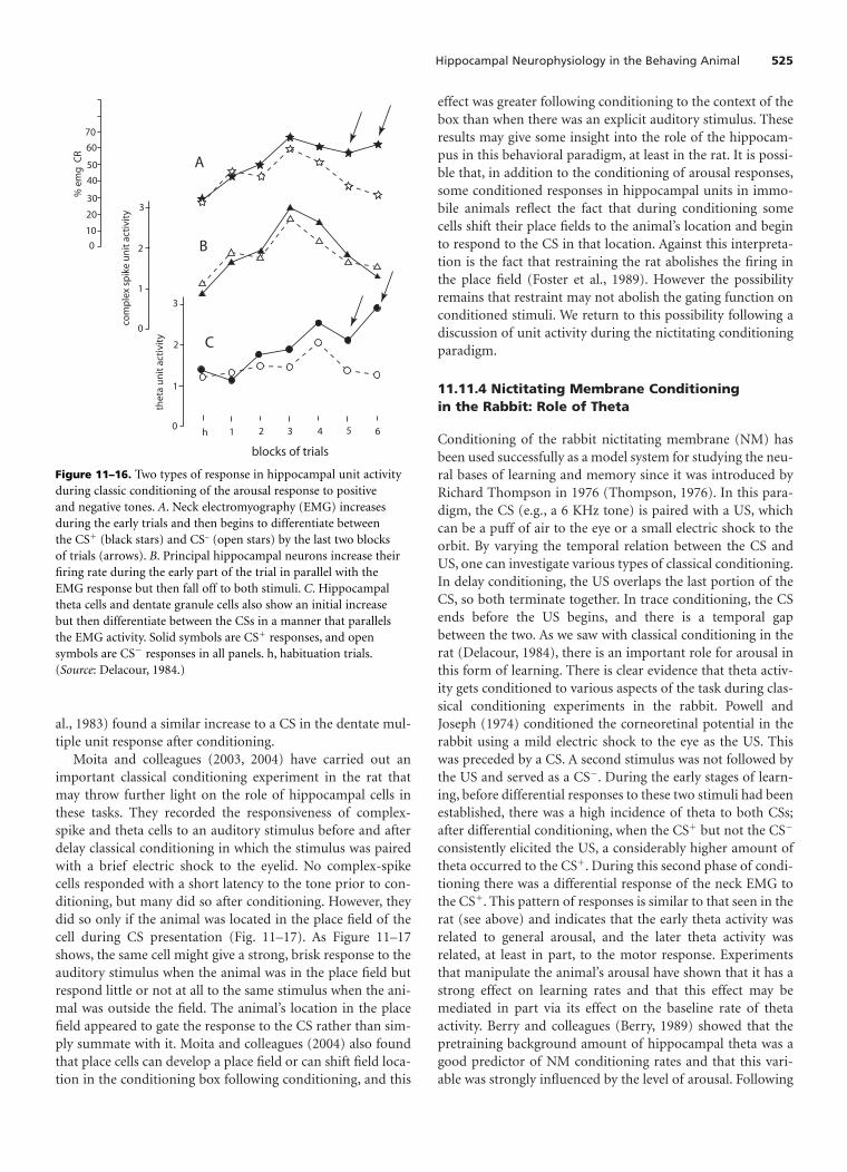
al., 1983) found a similar increase to a CS in the dentate mul-tiple unit response after conditioning.
Moita and colleagues (2003, 2004) have carried out animportant classical conditioning experiment in the rat thatmay throw further light on the role of hippocampal cells inthese tasks. They recorded the responsiveness of complex-spike and theta cells to an auditory stimulus before and afterdelay classical conditioning in which the stimulus was pairedwith a brief electric shock to the eyelid. No complex-spikecells responded with a short latency to the tone prior to con-ditioning, but many did so after conditioning. However, theydid so only if the animal was located in the place field of thecell during CS presentation (Fig. 11–17). As Figure 11–17shows, the same cell might give a strong, brisk response to theauditory stimulus when the animal was in the place field butrespond little or not at all to the same stimulus when the ani-mal was outside the field. The animal’s location in the placefield appeared to gate the response to the CS rather than sim-ply summate with it. Moita and colleagues (2004) also foundthat place cells can develop a place field or can shift field loca-tion in the conditioning box following conditioning, and this
effect was greater following conditioning to the context of thebox than when there was an explicit auditory stimulus. Theseresults may give some insight into the role of the hippocam-pus in this behavioral paradigm, at least in the rat. It is possi-ble that, in addition to the conditioning of arousal responses,some conditioned responses in hippocampal units in immo-bile animals reflect the fact that during conditioning somecells shift their place fields to the animal’s location and beginto respond to the CS in that location. Against this interpreta-tion is the fact that restraining the rat abolishes the firing inthe place field (Foster et al., 1989). However the possibilityremains that restraint may not abolish the gating function onconditioned stimuli. We return to this possibility following adiscussion of unit activity during the nictitating conditioningparadigm.
11.11.4 Nictitating Membrane Conditioningin the Rabbit: Role of Theta
Conditioning of the rabbit nictitating membrane (NM) hasbeen used successfully as a model system for studying the neu-ral bases of learning and memory since it was introduced byRichard Thompson in 1976 (Thompson, 1976). In this para-digm, the CS (e.g., a 6 KHz tone) is paired with a US, whichcan be a puff of air to the eye or a small electric shock to theorbit. By varying the temporal relation between the CS andUS, one can investigate various types of classical conditioning.In delay conditioning, the US overlaps the last portion of theCS, so both terminate together. In trace conditioning, the CSends before the US begins, and there is a temporal gapbetween the two. As we saw with classical conditioning in therat (Delacour, 1984), there is an important role for arousal inthis form of learning. There is clear evidence that theta activ-ity gets conditioned to various aspects of the task during clas-sical conditioning experiments in the rabbit. Powell andJoseph (1974) conditioned the corneoretinal potential in therabbit using a mild electric shock to the eye as the US. Thiswas preceded by a CS. A second stimulus was not followed bythe US and served as a CS. During the early stages of learn-ing, before differential responses to these two stimuli had beenestablished, there was a high incidence of theta to both CSs;after differential conditioning, when the CS� but not the CS
consistently elicited the US, a considerably higher amount oftheta occurred to the CS�. During this second phase of condi-tioning there was a differential response of the neck EMG tothe CS�. This pattern of responses is similar to that seen in therat (see above) and indicates that the early theta activity wasrelated to general arousal, and the later theta activity wasrelated, at least in part, to the motor response. Experimentsthat manipulate the animal’s arousal have shown that it has astrong effect on learning rates and that this effect may bemediated in part via its effect on the baseline rate of thetaactivity. Berry and colleagues (Berry, 1989) showed that thepretraining background amount of hippocampal theta was agood predictor of NM conditioning rates and that this vari-able was strongly influenced by the level of arousal. Following
Hippocampal Neurophysiology in the Behaving Animal 525
0
10
20
40
30
50
60
70
% e
mg
CR
0
1
2
3
thet
a u
nit
act
ivit
y
0
1
2
3
com
ple
x sp
ike
un
it a
ctiv
ity
h 1 2 3 4 5 6
A
B
C
blocks of trials
Figure 11–16. Two types of response in hippocampal unit activityduring classic conditioning of the arousal response to positiveand negative tones. A. Neck electromyography (EMG) increasesduring the early trials and then begins to differentiate betweenthe CS� (black stars) and CS– (open stars) by the last two blocksof trials (arrows). B. Principal hippocampal neurons increase theirfiring rate during the early part of the trial in parallel with theEMG response but then fall off to both stimuli. C. Hippocampaltheta cells and dentate granule cells also show an initial increasebut then differentiate between the CSs in a manner that parallelsthe EMG activity. Solid symbols are CS� responses, and opensymbols are CS responses in all panels. h, habituation trials.(Source: Delacour, 1984.)

mild water deprivation, there was a faster rate of learning. Asimilar pattern was found with trace conditioning (Kim et al.,1995).
11.11.5 Single-unit Recordingin the Hippocampus During NictitatingMembrane Conditioning of Rabbits
Several laboratories have recorded the activity of single unitsand multiple units from the hippocampus of rabbits duringclassical conditioning of the NM response. Berger and col-leagues (1983) recorded single units in the rabbit hippocam-pus during simple delay NM conditioning. Pyramidal cellswere identified by their antidromic activation from fornixstimulation. They comprised the largest proportion of cellsrecorded; and following conditioning, many increased theirfiring rates during the CS period (Fig. 11–18A–C). They typi-cally emitted one or more bursts of spikes during each trial,some showing a pattern of activity that closely modeled theNM response (Fig. 11–18A) whereas others were more selec-tive, firing during different time epochs of the trial (Fig.11–18B,C). One type of theta cell increased its overall level ofactivity during the trial (Fig. 11–18D,E), whereas another typeshowed an overall decrease in activity (Fig. 11–18F,G). Theta
cells were typically activated by the CS to fire a series of theta-like bursts, which often continued throughout the trial andin some cases continued beyond the termination of thetrial. Berger and colleagues also reported a third category ofcells, “silent” cells, which constituted 11% of the neuronsrecorded; they had exceptionally low spontaneous firing rates(� 0.2/second), were not activated by fornix stimulation, anddid not participate in the conditioned response. As we shallsee below, this estimate of the percentage of cells in this cate-gory may be low.
Weiss et al. (1996) recorded single units from rabbit hip-pocampus during trace conditioning of the NM, a version ofthe conditioning task that is sensitive to hippocampal lesions:Either the CR is abolished, or its timing is altered (see Chapter12). They also reported the existence of the same three classesof cells reported by Berger and colleagues in their delay con-ditioning experiments; but in other respects the results dif-fered markedly. Weiss et al. found a much larger percentage ofcells that did not participate in the conditioning (40% in con-trast to Berger’s 11%) and relatively few units that were signif-icantly excited (in contrast to inhibited) during the CS or traceperiod in the conditioned animals in comparison withunpaired controls. Thus, in CA1, 14% of pyramidal cells wereexcited during the CS period in contrast to 9% of the unpaired
526 The Hippocampus Book
Figure 11–17. Response of hippocampal complex spike cells toauditory CS following classic eyeblink conditioning. The cellsrespond only when the animal is in the place field. A. Responses intwo individual cells. On the left of each panel, place fields are shownwith the higher firing rates in the darker colors; on the right of eachpanel are histograms of the unit responses while the animal is insidethe place field (black color) and outside the field (light color). Themiddle panels show the portion of the environment included aspart of the field, the portion considered to be outside the field, and
an immediate zone not considered as part of either. Both cellsshowed a strong response to the stimulus inside the field and no(cell 1) or few (cell 2) spikes outside the field. B. Populationresponse histograms to stimulus presented inside (dark shading)and outside (light shading) the field of each cell. Across the popula-tion there is a significant difference between the in-the-field andout-of-field responses (histogram, right). The x-axes in the unitresponse histograms are 250 ms in total, which was the period ofCS presentation. (Source: Moita et al., 2003.)
cell 1 cell 2
field fieldout of fieldin fieldexcluded
out of fieldin fieldexcluded
A
pip
resp
onse
(Hz)
40
812162024
0pip
resp
onse
(Hz)
48
12162024
(Z-s
core
)no
rmal
ized
resp
onse
0.5
1.0
1.5
2.0
0
-0.5 0
0.2
0.4
0.6
0.8
1.0
(Z-s
core
)no
rmal
ized
resp
onse
Z in Zout
B

controls; and 11% were excited during the trace period incontrast to 9% of the controls. In contrast to the findings ofBerger and colleagues, the increase in inhibitory responses rel-ative to the controls was twice as large as the increase in exci-tatory responses. McEchron and Disterhoft (1997) obtainedsimilar results in animals conditioned in a trace paradigmwhen recordings were taken after asymptotic performancehad been reached. In addition, they recorded from some ani-mals during the earlier stages of learning. They found that themaximal activation in complex spike units occurred on thetrials just prior to the onset of learning; as behavioral condi-tioning proceeded and the conditioned responses appearedmore frequently, these unit responses actually diminished.Finally, when looked at on a trial-by-trial basis, there was nocorrelation between the unit activity and the occurrence of theconditioned response. This pattern suggests that the hip-pocampus may not be involved directly in the generation ortiming of the motor response. Rather, it may be involved onlyindirectly, perhaps playing a role in the creation of a tempo-rary behavioral state that precedes the motor learning but isa necessary condition for it to occur. Lesions of the hip-pocampus do not generally affect delay conditioning but doaffect trace conditioning, changing the timing of the condi-tioned response in the trace version of the task (for a reviewsee O’Keefe, 1999). Thus, if we compare the effects of hip-pocampal lesions on the NM conditioning with the resultsof single-unit recording experiments, we are left with a para-dox. Delay conditioning, in which there are a sizable numberof pyramidal cells whose temporal activation profile precedesand models the CR, does not require the hippocampus,
whereas trace conditioning, during which few such unitresponses are found, does require an intact hippocampus.
How might the hippocampus be involved in trace butnot delay conditioning? There are three distinct but relatedpossibilities. The first suggests that the hippocampus pro-vides the information that the animal is in a frightening place,the second that the animal is in a place where frighteningevents happen, and the third that it provides informationabout the timing of the unpleasant event. The first two makeclear links to the spatial functions of the hippocampus,whereas the third does not necessarily do so. We deal withthese in turn.
Our first two mechanisms relate to ones suggested byNadel and colleagues (1985). According to them, the spatialfunctions of the hippocampus might lead to its being involvedin conditioning in two ways: as the substrate for a direct asso-ciation between the background cues and the US or lessdirectly as the basis for an association between the CS–USevent and the overall context. Evidence for a hippocampal rolein conditioning to the background cues (the fearful contexthypothesis) comes from experiments showing that animalswith hippocampal lesions do not condition to the context(Phillips and LeDoux, 1994; Kim et al., 1995). This couldcome about either because there is an association of fear withthe entire testing box or with specific locations in it. Recallthat the ventral hippocampus contains place cells with largefields that may extend to an entire testing environment, andthey could provide the basis for context conditioning. Analternative is that the more localized place fields in the dorsalhippocampus could shift following context conditioning so
Hippocampal Neurophysiology in the Behaving Animal 527
A
B
cs ucs
C
D
E
F
G
Pyramidal cells Theta cells
nm
unit
Figure 11–18. Firing patterns of hippocampal unitsfollowing nictitating membrane conditioning in therabbit. A–C. Pyramidal cells. D, E. Excited theta cells.F, G. Inhibited theta cells. Top trace in each panel isthe nictitating membrane record; bottom trace is thehistogram of the average unit response. The firstarrow indicates CS onset and the second arrow UCSonset, an interval of 250 ms. Note the theta burstingat approximately 8 Hz following the CS onset in thetheta cells. (Source: Berger et al., 1983.)

the overall pattern of place cell firing was different, perhapssignifying a dangerous environment (Moita et al., 2004).
The second possibility is that the cells showing conditionedresponses are place cells whose fields coincide with the loca-tion of the testing box, and the responses to the CS are signal-ing the occurrence of an event in that place. As we have seenabove in the experiments of Moita and colleagues (2003),complex-spike cells in the rat, which are normally not respon-sive to auditory stimuli, begin to respond to the CS followingdelay conditioning but only if the animal occupies the firingfield of that cell. If the same changes are occurring in the rab-bit, this might explain the increased responsiveness of cellswith fields in the conditioning location; moreover, the fields ofsome cells might shift to that location following conditioning.With this interpretation, the increased responsiveness mightreflect the fact that the hippocampus is now signaling that theanimal is in a dangerous location and, furthermore, that theauditory cue predicts the onset of danger in that place. Moreevidence for a role for the hippocampus in contextual gatingof CS–US events comes from a context shift experiment.Although lesions of the hippocampu have no obvious effecton simple delay conditioning, they do affect the role the back-ground cues (i.e., the room in which conditioning takes place)play in that conditioning. Penick and Solomon (1991) showedthat simple delay conditioning was disrupted in normal rab-bits when the animal was moved into a new room after condi-tioning was completed but hippocampal-lesioned animalswere not affected. This result fits nicely with the idea that thehippocampus provides spatial contextual information thatgates the conditioning of fear to the CS. This informationmight not be necessary for simple delay conditioning, but itmight be essential for trace conditioning.
The third possibility is that the hippocampus providesinformation about the timing of the US. The interpretation ofthe altered timing response in trace conditioning followingdorsal hippocampal lesions is unclear. Why, for that matter,does the conditioned unit response occur just prior to theunconditioned stimulus in normals? The original rationale ofthe NM learning paradigm was to rule out any instrumentalcontribution to the learning and in particular the possibilitythat the conditioned response would protect the eye from theUS or otherwise attenuate its impact (Thompson, 1976). If, onthe other hand, one accepts that the conditioned response inthese conditioning paradigms is a reflection of the predictionof the US, the timing of the CS is important; and the lesionresults suggest that the hippocampus is involved in setting upthe conditions under which the short-term prediction ofstimuli can occur. Both Rawlins (1985) and Wallenstein andcolleagues (1998) have suggested that the hippocampus isneeded to bridge a temporal gap between two stimuli to beassociated. This role would be particularly important in para-digms such as trace conditioning, where there is a CS–USinterval and normal animals generate a CR just prior to theUS. What might the underlying mechanism be? One possibil-ity is that hippocampal a-theta is being used as a timingmechanism to allow a short-term signal to bridge the CS–US
gap. Theta oscillations, with their relatively constant period,could act as a clock over short intervals. During conditioningof the rabbit NM response, this clock signal can be reset by theCS and could therefore be used to predict the occurrence ofthe US. Even here there might be a secondary role for the spa-tial functions of the hippocampus. Recall that a-theta in therabbit can be driven by arousing stimuli (e.g., the CS) muchmore easily than in the rat (see Section 11.3). That requiredlevel of arousal might be based on conditioning to the appa-ratus and other nonspatial cues, but it might also be driven byhippocampus-mediated fear conditioning to the backgroundcontext as well. Under circumstances where the arousal level isappropriate, pyramidal cells may be able to count theta cyclesand use this clock signal to predict the timing of the US. Thistiming signal would then be available to brain stem regions tocontrol the occurrence of the CR. In the absence of this signal(e.g., following hippocampal damage), conditioning wouldstill occur but the timing of the CR would be controlled byother factors. Some support for this view comes from anexperiment on trace conditioning of the heart rate in rabbits(McEchron et al., 2003). Pairing a CS with an aversive USresults in slowing of the heart rate in anticipation of the US;this can be conditioned with trace intervals as long as 20 sec-onds. McEchron and colleagues used trace intervals of 10 and20 seconds in two groups of animals and found that one-fourth of complex-spike cells showed a burst of activity timedto coincide with the end of the trace interval. The effectwas weak on any given trial; but when summed over trialsthere was a discernible response. The problem for the theta-counting hypothesis is that 10 seconds is a long time to counttheta cycles. For further discussion of the contributions thatcontexts make to learning, see Chapter 13.
11.11.6 Hippocampal Unit RecordingDuring Operant Tasks
During operant conditioning tasks, the animal must emit aresponse to gain a reward or avoid punishment. Often theavailability of reinforcement is signaled by a sensory stimu-lus such as a tone. Christian and Deadwyler (1986) recordedfrom complex-spike and theta cells during an appetitive oper-ant conditioning task. They trained thirsty rats to poke theirnoses into a small antechamber in the wall of a box to receivea water reward. For some animals, the availability of rewardwas signaled by a tone, and no sensory discrimination wasrequired; for others, a differential CS�/CS procedure wasused. Following successful conditioning to the single tonestimulus (Fig. 11–19A), theta cells showed a consistent in-crease in firing rate during the 200 ms following tone onset(Fig. 11–19A, theta). In contrast, no change from the back-ground rate was seen in the complex spike cells (Fig. 11–19A,complex spike). During two-tone differential conditioning,the theta cells showed an increase to both stimuli with agreater increase to the CS� (Fig. 11–19B, theta). In contrast,the pyramidal cells registered a marginal but nonsignificantchange to the CS� (Fig. 11–19B, complex-spike). Recordings
528 The Hippocampus Book

taken from animals while they acquired the task showed thatthe changes in theta cell firing occurred in parallel to acquisi-tion of the conditioned EMG response and disappeared withsubsequent extinction. Again no changes were seen in com-plex-spike cells during the course of acquisition.
In a subsequent experiment from the same laboratory,Foster and colleagues (1987) did find a small but significantincrease in firing in their population of complex-spike cells toboth conditioned stimuli but still no differential activity to theCS�. A more detailed look at the differential response that didoccur in the theta cell group revealed that the difference inresponse to the two stimuli was due to an initial increase infiring to both stimuli, which peaked at about 80 to 100 msafter tone onset and was then maintained throughout the1-second tone period to the CS� but fell back to baseline inresponse to the CS.
The simplest explanation for the pattern of resultsobserved in these studies is that there are two independentfactors operating during conditioning: arousal and prepara-tion for the motor response. Both contribute to the firing oftheta cells, but only one of them, arousal, influences the
pyramidal cells. The initial short-latency response of hip-pocampal interneurons and granule cells to either a CS� ora CS is presumably activation reflecting an arousal inputfrom the brain stem; at about the same time, a small percent-age of pyramidal cells, in some studies, also show an arousalresponse. Depending on the stage and type of training, thiscan either be an inhibitory or a weak excitatory response. Thelater phase (� 200 ms after CS onset) of the unit activity isrelated to the behavioral response. For the theta and granulecells there is prolonged activation continuing throughout theCS� period but no such response to the CS. The pyramidalcells do not participate in this second longer-latency phase.
Eichenbaum and his colleagues have studied the behav-ioral correlates of hippocampal cells in various olfactoryrecognition and discrimination tasks. They recorded twomajor behavioral correlates: Some cells fired when the ani-mal sniffed at the odor cues, whereas others changed their fir-ing rates during various stages of the approach to the cuesor to the goal. In a successive go/no-go discrimination task(Eichenbaum et al., 1987), the rat was presented with oneodor of a pair and had to poke its nose into the single-odor
Hippocampal Neurophysiology in the Behaving Animal 529
** ** ** **
5040 30 20 10
0
10
0
50
40
30
20
3210
3
2
1
0
-200 200400 600 800 ms
-200 200 600 800 ms-200 200 400 600 800 ms
-200 200
0
10
20
30
40
50
-200 200 400 600 800 ms
tone onset
400 600 800 ms
tone onset
theta cell
complex spike cell
theta cell
complex spike cell
-200 200 400 600 800 ms
-200 200 400 600 800 ms
tone onset
tone onset
10
20
30
40
50
0
1
2
3
00
1
2
3
mea
n fir
ing
rate
(spi
kes
s-1)
mea
n fir
ing
rate
(spi
kes
s-1)
****** **
****
** **
*
*
*
CS+ CS-
60
A B
Figure 11–19. Theta and complex spike cell responses followingnose-poke conditioning to tones. Theta cells (top) show increasedfiring above the pre-tone background rate in response to the tone inboth simple (A) and compound (B) conditioning paradigms. In thelatter, the unit response to the CS� is more marked than to the CS.Complex spike cells (bottom) do not respond in either the simpleconditioning paradigm (A) or the complex compound conditioning
paradigm (B). Single asterisks indicate a significant difference frompre-tone firing rates at the 0.05 level of significance and doubleasterisks at the 0.01 level of significance. The apparent increase infiring rate in the complex spike cells 800 ms following the tone inboth A and B is an artifact from the reward dispenser. (Source:Christian and Deadwyler, 1986.)

port in response to the CS� but not to the CS. Water rewardwas available on the other side of the testing box, requiring theanimal to shuttle continuously between opposite sides of thebox. Three behavioral correlates of unit response were identi-fied: cells that fired when the animal was sniffing at the odor(“cue sampling” 15%); cells that fired when the animalapproached the sniff port or ran from the sniff port to thewater cup at the other end of the box (“reward/port approach”60%) (Fig. 11–20A); and theta cells (10%). Many of the cuesampling units had firing patterns that were maximally syn-chronized to the onset of cue sniffing. No evidence was foundfor cells that preferred one odor over others. Almost all cuesampling cells fired more to the positive stimulus that signaledthe availability of water reward in another part of the envi-ronment. In addition, there was evidence that in the trials thatfollowed a CS trial the cells gave a larger response than inthose that followed a CS�. A follow-up study (Otto andEichenbaum, 1992) looked at complex-spike cell firing duringa continuous recognition olfactory memory task in which anyone of 32 odors could be presented, and the availability of
water nearby was signaled by a mismatch between the currentand previous stimulus. Again, cells that had peak firing ratesat different points in the task were found, including 12% thatpeaked during the cue sampling period. Somewhat disap-pointingly however, only eight cells (3%) had firing patternsthat could be unambiguously classified as signaling a mis-match between successive stimuli. As we shall see below inthe section on primate studies of delayed nonmatch tosample, there are also very few cells in these studies thatresponded selectively to the familiarity or unfamiliarity of astimulus.
In a forced-choice discrimination trial, Wiener and col-leagues (1989) used simultaneous rather than successive odordiscrimination. Both the CS� and CS were presented at thesame time from two adjacent odor ports. In total, 22% ofcomplex-spike cells had increased activity during cue sam-pling (Fig. 11–20A, center panel). However, closer examina-tion of these data showed that only 13% of cue-sampling cellsdiscriminated between odors irrespective of location, and44% took the stimulus location into account. Figure 11–20B
530 The Hippocampus Book
port approach
cup approach
cue sampling
1 sec
odor 1/odor 2
odor 2 /odor 1
odor 3/odor 4
odor 4/odor 3
A B
Figure 11–20. Hippocampal cell firing correlates with variousaspects of an odor discrimination task. A. Firing rate histograms ofcells that fired best when the animal approached the odor samplingport (top trace), sniffed at the odors (middle trace), and approachedthe reward cup (bottom trace). Arrows in the top two traces indi-cate initiation of the behavior; arrow in the bottom trace indicates anose-poke into the reward cup. B. Firing pattern of an odor/spatial
cell that responded maximally when the animal sniffed at a particu-lar odor (odor 1) on the left side of the odor sampling port and asecond (different) odor (odor 2) on the right side. Top trace: bestresponse to odor 1 on the left and odor 2 on the right; second trace:same odors from the opposite ports gives a lower response; thirdand fourth traces: different odors are also less effective. Dotted linemarks initiation of the trials. (Source: Wiener et al., 1989.)

shows an example of a cell that had a strong spatial compo-nent. It fired best to a particular odor pair when one was pre-sented on the left and the other on the right. Somewhat moresurprising, 44% of cup-approach cells signaling approach tothe reward cup on the opposite side of the box also hadresponses that depended on the position of the prior nosepoke or the odor/position interaction. It is difficult to inter-pret these data, but one possibility is that the rats may havebeen turning in different directions away from the odor portas they headed toward the reward. If this is true, the cupapproach response in this ostensibly nonspatial task maydepend on whether the animal turns toward the left or righton its exit from the sniff port and thus passes through a placefield on one side of the sniff port as it turns in one directionbut not in the other. Alternatively, this may be an example ofthe dependence of some place cells firing on the prior turntaken by the animal before entry to the place field (see Section11.7.3). One can conclude that a small number of complex-spike cells respond to the CS� in a differential go/no-go dis-crimination but that, in general, few hippocampal cells codefor the specific odor quality, and that many cells take the loca-tion of the odor into account.
On the basis of these and other results (see Chapter 13),Eichenbaum and his colleagues proposed that the hip-pocampus stores information about a wide range of rela-tions, both nonspatial and spatial. Unit responses to odorsand approaches to the odor port or reward dispenser mightbe taken as evidence of nonspatial representations andunit responses signaling the identity between two successivestimuli as evidence of one type of nonspatial relationship: theidentity relation between two stimuli experienced at differenttimes.
As described earlier in the chapter (see Section 11.7.1),many place cells fire more when the animal sniffs at a locationwhen the stimulus in that location has been altered in someway, and these results from odor discrimination paradigmsmay provide additional information about the conditionsunder which this mismatch response occurs. To identify cellresponses as being due to the odor cue independently of itslocation, it is necessary to present the same odor in two ormore locations. The demonstration that place fields can berather large, encompassing a large part of the testing box (seeSection 11.7.2), means that it may be necessary to test thesame odors in two different laboratories as well as in two test-ing boxes in the same laboratory before concluding that hip-pocampal cells do not have a spatial correlate. Similarly, thecells that fire selectively during port approach and cupapproach have much in common with place cells recorded onlinear tracks. Recall that under conditions that constrain theanimal to move along narrow pathways, the place cells areunidirectional. To distinguish a goal-oriented response from amotivationally neutral place response, it is necessary to havetwo identical goals in the environment or two different goalsthat can be interchanged. O’Keefe (1976) reported that inter-changing the water and food at the end of a three-arm mazedid not change the location of complex-spike firing fields on
the maze. O’Keefe and Recce (1993) used a linear track withidentical food reward at both ends and found that some cellsfire in one direction and other cells fired in the opposite direc-tion. Even if one were tempted to describe these cells as goalapproach cells, it is not the approach to the food or the foodcontainer per se that is being signaled but their locations atdifferent ends of the track. It does not seem warranted todescribe the cells reported in the experiments of Eichenbaumand colleagues as cue-sampling or goal-approach cells in theabsence of similiar manipulations.
Two experiments have provided evidence about the effi-cacy of cues and goals in isolation from their location (Wiebeand Staubli, 1999; Wood et al., 1999). Wood et al. recordedfrom hippocampal cells during a variant of a continuousolfactory recognition task in which they tried to dissociatelocation, odor, and the match/mismatch aspects of the task.Rats were trained on an open platform to approach a smallcup containing sand scented with one of nine odors. In eachtrial the cup was placed in one of nine locations. If a cup hadan odor different from that of the previous one, it containedfood for which the animal could dig (nonmatch); if the odorwas the same, there was no food (match), and digging wentunrewarded. Cells were recorded during the 1 second prior toarrival at each cup. An analysis of variance showed that of thetotal 127 cells, 8% responded to odor in the absence of anyother correlate, 11% solely to location, and 10% solely to thematch/mismatch aspect of the task. The remainder of theresponsive cells took interactions between these variables intoaccount. In all, 20% of cells had nonspatial correlates, and32% took location into account. (Another 20% changed theirfiring rate as the animal approached any of the cups. The lat-ter may simply be movement- or speed-related—because thiswas not measured, it is not discussed further.) These resultsare notable as a higher percentage of cells with a match/mis-match correlate irrespective of location was found than in theprevious reports from the same laboratory (see above).Furthermore, there are fewer purely spatially coded cells thanare usually found. This may be because the experimentaldesign required the animal to approach each cup from a dif-ferent angle during different trials and therefore via differentlocations; this might lead to a significant underestimation ofthe number of place cells. Approaching a cup from the northwould not force the animal to cross the same region of spaceas approaching the same cups from the south. It is also notclear how much of the difference between cellular match andnonmatch responses can be attributed to the different behav-iors of digging and turning away from the cup that were usedas the response measures, rather than the relational judgmentitself. Nevertheless, taken at face value, these results provideevidence for the more general relational theory. Significantly,this task has been shown by the authors of the Wood et al.study themselves (Dudchenko et al., 2000) not to be disruptedby selective hippocampal damage.
Wiebe and Straubli (1999) used a Y maze task that forcedthe animal to traverse the same locations on the approaches tothe goals and came to a conclusion different from that of
Hippocampal Neurophysiology in the Behaving Animal 531

Wood and colleagues. Animals were trained on a delayed non-match-to-sample odor task with each trial comprised of threediscrete periods: a sampling period during which the animalsniffed one of two odors in the start arm, a delay period dur-ing which it was confined to the start arm and had to remem-ber the recently smelled odor, and a test period during whichit was required to choose one of the other two arms contain-ing the odor it had not recently experienced. During the firsttwo periods, everything happened within the same arm, andcellular activity could be correlated only with the odor pre-sented on that trial and with subsequent correct or incorrectperformance on the choice part of the trial. During the third(test) period the animal could enter one of two goal arms, andtherefore a correlation with location as well as odor and per-formance could be sought. Overall, most of the 1101 cellsrecorded from the dentate gyrus, CA3, and CA1 had signifi-cant correlations with one or more aspects of the task. Someof the results are shown in Table 11–1. Several conclusions canbe drawn from these results. First, during the sample phase,which always took place in the same arm and therefore did notallow the contribution of location to be examined, a smallpercentage of cells had significant changes in firing rate to thespecific odor presented (5%), to whether the subsequentchoice would be correct or incorrect (2%), or to the interac-tion between two (4%). None of these firing rate changes,however, carried over into the delay phase of the trial (Table11–1, delay) suggesting that the hippocampus does not main-tain an active trace of the correct odor choice, as it does oflocation in a comparable spatial memory task (O’Keefe andSpeakman, 1987). Second, in marked contrast to what wasseen in the sample phase, during the test phase only a tiny per-centage of cells fired to the odor, to the correctness of thechoice, or to the interaction between them (all � 1%). Thisrepresents a large discrepancy with the odor and performancecorrelates found in the sample arm. Third, in contrast to the
weak representation of odor or performance in the test arms,there was a strong representation of the animal’s location. Ofthe 571 cells with behavioral correlates in the test arms eitheralone or as an interaction with another correlate, 70% had aspatial correlate, whereas only 27% had an odor correlate and21% a performance correlate. More significantly, 37% of thecells with spatial correlates were pure place cells with no othercorrelates, whereas most of the cells with olfactory and per-formance correlates also had spatial correlates (92% and 93%,respectively). It is difficult to escape the conclusion that whenthe testing situation allows them to be identified spatialresponses predominate either as pure place responses or asplace combined with some other aspect of the task, evenin tasks where the spatial component is irrelevant to thesolution.
A potential criticism of the Wiebe and Straubli experimentis that they only used two odors, but there might be more thanone location in each of the test arms. This points up an impor-tant methodological problem in these studies. To equate twotasks on the number of odors and locations, it is important tohave an accurate measure of these items. It might be thought,for example, that the number of place fields in each arm of themaze is greater than the number of odors, but the odors usedin the tasks are typically compounds and may be distin-guished on the basis of many different elemental aspects of thecompound. In the absence of more information about theelemental components of locations and smells in any giventask, the best one can do is try to vary each variable systemat-ically to see what effect it has on the results.
11.11.7 Comparison of HippocampalCells During Operant Conditioningand Place Tasks in Rats
Are the neurons that show place responses the same as thosethat take part in nonspatial learning tasks conditioning, orare there two separate populations of cells? Several studieshave compared the response of the same hippocampal neu-rons during place tasks and conditioning tasks. Three studieshave asked whether complex-spike cells might be specificallyinvolved in the place task and the theta cells might be specifi-cally involved in the conditioning tasks. Christian andDeadwyler (1986) used antidromical stimulation of projec-tion pathways to identify complex-spike cells as pyramidalcells. They found a clear double dissociation of the cellsinvolved in the two tasks. In total, 81% of complex-spike cellshad place fields in the place task in comparison with none ofthe theta cells. Conversely, 81% of the theta cells participatedin the conditioning task whereas none of the complex-spikecells did. In two experiments, Eichenbaum and his colleagues(Eichenbaum et al., 1987; Wiener et al., 1989) asked whethercells that were related to events in their olfactory discrimina-tion task also had place fields in the same or different tasks. Inthe first study, they found that 43% of cells with correlates inthe odor discrimination task had place fields in the same
532 The Hippocampus Book
Table 11–1.Percentages of Total Number of Recorded Cellswith Significant Correlates to Various Aspects of anOlfactory Delayed Nonmatch-to-Sample Task
Sample Delay Test phasephase phase phase
Parameter (%) (%) (%)
Odor 4.8 0 0.9Performance 2.1 0 0.6Odor � performance 4.3 0 0.2Position – 26.8Position � odor – 8.4Position � performance – 5.6Position � odor � performance – 4.5Position exclusively – 13.5Odor exclusively 2.4 0.8Performance exclusively 2.1 0.4
After Wiebe & Staubli, 1999.

environment; in the second study, they recorded some com-plex-spike cells in a spatial task, some in the odor discrimina-tion task, and a third group in both tasks. They found 75% ofthe complex-spike cells had place fields in the spatial taskcompared to 58% with correlations in the odor discrimina-tion task. Of cells collected in both tasks, 85% had place fieldsin contrast to 54% that had correlates in the odor task.
We can conclude that in conditioning tasks involving ratsthat were not required to move around the environment toany great extent, most or all hippocampal cells taking part inthe conditioning response were theta cells. Complex-spikecells, by contrast, either did not take part or showed slightinhibition of their resting firing rate. In discrimination condi-tioning tasks in which the animal is required to move, thecomplex-spike cells also become engaged. The clearest exam-ple of cells that may not have a spatial correlate are the cue-sampling cells reported in the Eichenbaum studies. However,even some of these responses may be covert place responses,as clearly suggested by the Wiebe and Straubli experiments.
11.11.8 Hippocampal Units During NonspatialLearning in Nonhuman Primates
Tamura et al. (1991) reported that 10% of cells in the hip-pocampal formation responded differentially to the presenta-tion of three-dimensional objects, some of which had beenconditioned to rewarding (3%) or aversive (2%) stimuli. In afollow-up study (Tamura et al., 1992), they showed that 61%of object-responsive cells tested in the same apparatus variedtheir response as a function of the location of the stimulus inegocentric or allocentric space. The latter study indicates thatapparently perceptual responses in the hippocampus may bespatially modulated. Such responses may underlie object-in-place associativity, which like odor-place associativity, is oftenshown to be hippocampus-dependent, unlike odor-objectassociativity, which is hippocampus-independent (Gilbertand Kesner, 2002).
Hippocampal units have been recorded from primateswhile they performed in one of the paradigmatic relationaltasks: delayed match or non-match-to-sample. On these tasks,the animal must signal whether two successive stimuli sepa-rated by an interval are the same or different. It is now widelyaccepted that these tasks can be solved in two ways. The ani-mal can either assess the strength of the familiarity of the twostimuli presented during the test phase and choose the leastfamiliar, or it can remember which stimulus was previouslypresented in the present situation or context and choose theone it has not experienced there before. The second strategy isclearly both spatial and relational, but it could be argued thatthe first strategy is also relational although clearly nonspatial.To judge a stimulus as familiar or novel, it is necessary to com-pare it to the representation of a previous stimulus and todecide whether the relation between the two is one of equal-ity. Recognition tasks of this sort were originally thought todepend on the integrity of the hippocampus but are now
known to depend on the perirhinal/parahippocampal corticesand only minimally (Alvarez et al., 1995) or not at all (Murrayand Mishkin, 1998) on the hippocampus itself (see Chapter13) (Aggleton and Brown, 1999). In keeping with the lesionresults, it has been found that only a small number of cellsshow a differential response to the familiarity of the stimuli(0%–2.3% in various studies (Riches et al., 1991; Tamura etal., 1992; Rolls et al., 1993; Salzmann et al., 1993; Brown andXiang, 1998). In contrast, Wilson et al. (1990) found that 40%of their hippocampal cells in this task correlated with theresponse, signaling whether the animal was reaching to the leftor right position during their task. Salzmann and Creutzfeldt(Salzmann et al., 1993) reported that 28% of hippocampaland 33% of parahippocampal neurons responded to both pre-sentations of the visual stimuli in a delayed match-to-sampletask but that even more cells (38% in each area) fired whenthe animal was presented with a raisin. Vidyasagar andCreutzfeldt (Vidyasagar et al., 1991) reported similar resultsbut that arousing events such as the cage door being openedor closed or the experimenter entering or leaving the roomalso produced a response in these cells. On the basis of thesefindings, they attributed hippocampal single-unit activity tothe animal’s behavioral state rather than to any specific mem-ory. These studies provide no evidence for the representationof stimulus familiarity in the hippocampus.
In contrast and in keeping with the lesion results, there isevidence that cells in the rhinal cortex are involved in famil-iarity judgments. Brown and colleagues (Brown and Xiang,1998) reported that about one-fourth of cells in the rhinalcortex and area TE showed a decreased response to the secondpresentation of the stimulus in a delayed match-to-sampletask.
Rolls and his colleagues (Rolls et al., 1993) have testedmonkeys on object recognition tasks and compared unit res-ponses to object familiarity with those to object location. Onlya small percentage (2%) of cells responded to familiar objects(see above). In contrast, they reported that about 9% of cellsin the hippocampal region responded differentially to thelocation of the stimulus on a display screen. As part of a studyof the place properties of primate hippocampal neurons, Onoand his colleagues (1993) found that 17% of cells respondedto objects such as a moving experimenter or apple when pre-sented in a particular part of the visual field. Just over half ofthese were object-in-place cells, which responded differen-tially when the animal was shown the object in a particularlocation and not in other locations. They appear to have prop-erties similar to those of the misplace cells described in the rat(O’Keefe, 1976).
11.11.9 Hippocampal Units DuringNonspatial Learning in Humans
Single units have been recorded from medial temporal loberegions in patients with intractable epilepsy during bothrecognition and recall memory tasks (Heit et al., 1988; Fried et
Hippocampal Neurophysiology in the Behaving Animal 533

al., 1997, 2002; Cameron et al., 2001) as well as during the per-ceptual categorization tasks described in Section 11.11.1. Ingeneral, a sizable percentage of hippocampal units was foundto participate in both types of memory task. In a recognitionmemory task, Fried and colleagues (2002) presented a set ofmale and female faces portraying various emotions; and afteran interval of 1 to 12 hours they showed the same faces againtogether with an equal number of foils. A significant numberof hippocampal neurons responded to faces during encoding(25%) and a somewhat larger percentage during the recogni-tion phase (41%). Perhaps surprisingly, a large proportion ofthe hippocampal responses (62%) consisted of firing ratedecreases. Almost one-half the hippocampal neurons with anexcitatory response but only one-sixth of those with aninhibitory response showed some selectivity for gender oremotion. Similar findings were reported by Heit and col-leagues (1988), who used lists of words to be remembered andfound most hippocampal units selective for one of the wordson the list. When tested with a second list of different words,slightly less than half of these cells (13/30) were also selectivefor a word in that list. As the authors remarked, this suggeststhat rather than being preordained to respond to specificstimuli the cells were tuning into the current context andselecting one aspect of it (such as position along a list?). Theresult is reminiscent of the way in which a given pyramidal cellin the rat hippocampus may have unrelated place fields in twoor more environments. Hippocampal units have also beenrecorded during a paired-associate recall task (Cameron et al.,2001). A series of word pairs were presented, and after 1 to 2minutes the subject had to respond with the second of the pairwhen prompted with the first: 29% of hippocampal unitsresponded during stimulus encoding and 44% during recall.These percentages are similar to those found in the entorhinalcortex and amygdala in the same study and in the face recog-nition study of Fried et al. (2002) but significantly higher thanthe response to faces and other nonspatial visual stimuli in theperceptual classification study of Kreiman et al., 2000a), sug-gesting a specific role for learning. An interesting aspect of thestudy was that one-fifth of the hippocampal cells showed adifferential firing rate during encoding between word pairsthat were subsequently remembered correctly and those thatwere forgotten. Most intriguingly, most of these cells showeda smaller increase in firing rate to the word pairs that weresubsequently remembered than to the ones that were forgot-ten. Whatever the role being played by the hippocampus inthese tasks, it cannot simply be that increased firing in hip-pocampal neurons during encoding leads to a stronger mem-ory trace and therefore to better recall.
Although one must be cautious when comparing acrossstudies, even from the same laboratory, the results can besummarized as follows. (1) The percentage of cells respondingto faces and words is higher when they are part of a memorytask than when they are simply perceived; (2) these stimuli areequally well represented in both recognition and recall mem-ory tasks; and finally (3) the response of hippocampal units toa stimulus during the encoding phase of a recall test correlateswith subsequent recall of that stimulus but in a counterintu-
itive fashion. The cells increase firing to both remembered andforgotten stimuli but the increase is smaller to the subse-quently remembered stimuli than to those forgotten.
11.11.10 Conclusions
In all species tested, a small percentage of hippocampal cellshave been reported to fire to nonspatial stimuli. The data areclearest in humans, where about 12% of cells responded to thesight of famous faces, but this fell well below the proportion(29%) that responded to spatial stimuli such as pictures ofhouses or interiors. Hippocampal unit responses to stimuliused as discriminanda in learning experiments have beenreported in all species tested: rats, rabbits, monkeys, andhumans. There is some suggestion that the responses in the ratmay be due to a place-gated mechanism because the one studythat tested this carefully found that the conditioned stimulionly elicited a pyramidal cell response after conditioning ifit was presented while the animal was in the place field ofthe cell (Moita et al., 2003). Theta cells are much less sensitiveto location and may account for a large percentage of theresponses reported in the early studies, which did not distin-guish between cell types. In general, monkey experimentsreported few cells responding to objects per se but more tolocation or object-in-places. In keeping with the lesion results(see Chapter 13), units responding to objects and their famil-iarity were more plentiful in the rhinal cortex than in the hip-pocampus.
There are clearly cells in the human medial temporal lobethat respond to words as well as faces and other stimuli inboth recognition and recall paradigms. Furthermore, in onestudy of verbal paired associate learning, hippocampal unitresponses during encoding of a pair predicted the success ofsubsequent recall (Cameron et al., 2001). Somewhat surpris-ingly, there was only a hint of lateralization of these responsesto the left hemisphere, as might be expected from the lesiondata. It is clear that the human hippocampus is involved in thestorage of verbal as well as nonverbal material.
In general then, there is evidence that nonspatial informa-tion is represented in the hippocampus but that the numberof cells involved is considerably smaller than those involved inspatial information and often the nonspatial responses aresecondary to a primary spatial correlate. The larger percentageof responses to nonspatial stimuli reported in humans is con-sonant with the wider function attributed to the human hip-pocampus in narrative and episodic memory (O’Keefe andNadel, 1978; O’Keefe, 1996, 2001; Burgess et al., 2002).
Á11.12 Other Distinctive Cells in theHippocampal Formation and Related Areas
As we have seen, the hippocampal formation is part of a wide-spread system of anatomically related regions involved inmemory and navigation. In addition to the CA fields andthe dentate gyrus, the hippocampal formation contains two
534 The Hippocampus Book

anatomically distinct regions: the subicular complex andthe parahippocampal cortex. Response properties of cells insome of these other regions have been touched on at differentpoints in this chapter where appropriate. For example inSections 11.9 and 11.10, we summarized the properties ofhead-direction (HD) cells found in the presubiculum and, inaddition, regions of the Papez circuit, which forms the inputsto these cells. In this section, we briefly summarize the prop-erties of cells in some of these other related brain regions andtouch upon the question of whether these cells are afferent tothe hippocampal formation, providing spatial information, oralternately should be thought of as efferent output structures,receiving spatial and memory information from the hip-pocampal formation.
There are three ways this question might be approached.The first is to look at the effects of lesions elsewhere in the cir-cuit on hippocampal place cell firing (Brun et al., 2002; Caltonet al., 2003) and presubicular HD cell firing (Golob andTaube, 1997) as described in Sections 11.7.4 and 11.9/11.10,respectively. The second examines the relative latency of theneurons in each area to respond to the stimulus or behaviorunder study. This approach depends on the assumption thatneurons in a region that projects to a second region in a serialfashion, on average, fire earlier in response to the appropriatestimulus or during the relevant behavior than the neurons inthat second region. On the assumption that many cells in thehippocampal formation and related areas have spatial aspectsof behavior as their primary correlate, it is possible to com-pare the temporal correlation of cells in different hippocam-pal regions in response to manipulations of these spatialvariables to estimate the temporal relation between cells indifferent regions indirectly. For example, one might study thelatency to onset of firing of HD cells with the same preferredorientation in different parts of the Papez circuit. Ideally, thisought to be done with simultaneous recording from thetwo (or more) regions in question to control for variationsbetween rats in behavior and location of the tracking lightson the head. The third method is based on a comparison ofthe place correlates of the neurons in the two areas. If onehas a serial model of place field construction, it might bepossible to attribute some neuronal responses to earlier stagesin that process than others. For example, Barnes and col-leagues (1990) compared the field sizes of place cells in differ-ent regions of the hippocampal formation and subiculum.As we shall see, some generalizations arise from this thirdtype of analysis. The locational code in the CA fields ofthe hippocampus is more environment-specific than in otherregions. Hippocampal place cells often fire in distinct patternsin different environments (“remapping”); indeed, many hip-pocampal cells are or become silent in a given environment(see Section 11.7.4). By contrast, the locational region(s) sig-naled by nonhippocampal cells are much more likely to be insimilar places across dissimilar environments. Moreover, inother regions, such as the subiculum and superficial entorhi-nal cortex, principal cells are never silent.
Taking the results of all three methods into account, itis difficult but not impossible to make suggestions about the
functional circuit diagram of the interconnections betweenthese regions of the hippocampal formation that underliesspatial behavior and memory.
11.12.1 Subicular Region Has Fewer PlaceCells than the Hippocampus Proper,and Their Properties Differ
Place cells and theta cells have been recorded from severalsubdivisions of the subicular region. We consider the similar-ities and differences between cells in the next sections. Thesubiculum contains both place cells and theta cells (Sharpand Green, 1994; Sharp, 1997). The percentage of theta cellsis approximately the same as found in the hippocampusproper (10%), and most of the remaining cells have strongspatial signals. There are no complex-spike cells, but onegroup (29% of total) shows a bursting pattern with a peakinterspike interval of 2 to 4 ms. These cells do not seem to dif-fer in any other respect from the nonbursting types exceptthat they have a stronger theta modulation. The field sizes andaverage firing rates of the subicular place cells are much largerwhen compared to those in the hippocampus. A typical subic-ular cell fires over the entire environment with an average rateof 5 to 15 Hz (in contrast to 0.5–2.5 Hz in the hippocampus)and has one or more areas of increased firing. Like the placecells of the hippocampus, subicular place cells recorded in acylinder are under the influence of a cue card on the walland rotate in step with it. It has been claimed that the subicu-lar cells have a stronger directional component than thehippocampal cells, but this claim must be treated with cau-tion. The amount of variance in subicular firing rate that isexplained by direction is actually very small (Sharp andGreen, 1994).
One important difference from hippocampal place cells isthat subicular cells are far less likely to remap across environ-ments that differ in shape (Sharp and Green, 1994), shape andvisual markings (Sharp, 1997), or size (Sharp, 1999b). Forinstance, whereas many of the hippocampal cells displayedmarkedly different fields in a cyclinder and a rectangle, thesubicular fields were similar in the two environments. This isreminiscent of what is found in the spatially coded cells in thesuperficial layers of the entorhinal cortex (see below) and tohippocampal place cells during the animal’s initial experiencesof the two (see Section 11.8.3). A study of the timing of subic-ular cell firing relative to the animal’s location suggests thatsubicular cells fire slightly earlier on average than CA1 cells(70 vs. 40 ms), perhaps indicating that they come earlier in thecomputational chain (Sharp, 1999a). This result may seemparadoxical in light of the strong connections from CA1 to thesubiculum, but it is consistent with the data showing thatsubicular cells do not remap the environments that CA1 cellsdo.
In conclusion, various approaches to the functional rela-tion between the subiculum and the hippocampus propersuggest that the subicular place cells cannot receive their spa-tial properties solely by virtue of their inputs from the hip-pocampus proper. They may be more dependent on direct
Hippocampal Neurophysiology in the Behaving Animal 535

inputs from the entorhinal cortex and/or they themselves mayprovide a source of spatial inputs to the entorhinal cells.
11.12.2 Presubiculum Contains SeveralClasses of Spatial Cell
The dorsal presubiculum (or postsubiculum in some authors’terminology) contains other types of spatial cell in addition tothe HD cells described above (see Section 11.9). A surveystudy (Sharp, 1996) of presubicular cells in the standardcylinder also reported cells with correlates for angular veloc-ity, running speed (see also Lever et al., 2003), location anddirection, and location. The nature of these correlates suggestsan important role for the presubiculum in navigation and spa-tial memory. One class of these cells, called theta-modulatedplace-by-direction (TPD) cells, has been examined in detail(Cacucci et al., 2004) (Fig. 11–21B, see color insert). TPD cellsgenerally have a strong tendency to fire at or just before thetrough of the local theta oscillation. Their depth of thetamodulation is high and comparable to that of cells in themedial septum/diagonal band of Broca (King et al., 1998). Thequantitative analyses of Cacucci et al. indicate that both loca-
tional and directional signals are stable and robust in TPDcells, with the directional signal carrying more information.The directional and locational signals are dissociable in differ-ent environments. The preferred direction of a given TPDcell appears to be environment-invariant, being similar acrossdifferent locations in the testing room (like the HD cells) andacross different enclosures in the same location (cylinder ver-sus an open circular platform). In contrast, the locationalfields tend to differ more between environments; notably,TPD cells generally have different locational fields in the cylin-der and open circular platform. TPD cell firing representsan integration of location-related and direction-related infor-mation. It is possible that directional information may have tobe incorporated into and distributed in theta wave packets tobe used by the navigation system. Neither anterodorsal thala-mic nor presubicular HD cells show theta modulation.Cacucci et al. provided evidence that a robust orientationsignal operating in theta mode exists in the hippocampal for-mation.
There have been no published reports of cells recordedfrom the ventral portions of either the presubiculum or theparasubiculum. By analogy with the differences between dor-
536 The Hippocampus Book
Figure 11–21. Four spatial cell types in the hippocampal forma-tion. False color firing field maps (left) show the firing rate as afunction of the animal’s location in cylindrical environments irre-spective of heading direction; directional polar plots (right) showthe firing rate for the same cell as a function of the animal’s headingdirection irrespective of location. A. Place cell has single localizedfield and no directional selectivity. B. Place by directional cell hassingle localized field and strong directional selectivity. C. Head
direction cell does not have localized firing but has strong direc-tional selectivity. (Source: A–C. Courtesy of Francesca Cacucci.)D. Grid cell has multiple place fields and no directional selectivity.Directional grid cells also exist. Numbers associated with the firingrate plots represent the maximum firing rate (red regions in firingrate maps and peak x and y values in polar plots). Diameter ofcylinders in A– C is 79 cm; diameter in D is 2 m. (Source: Edvardand May-Britt Moser.)

sal and ventral hippocampus (Jung et al., 1994), these ventralregions might be expected to contain lower levels of spatialsignaling and/or larger spatial fields.
11.12.3 Parasubiculum
The parasubiculum contains location-specific neurons,although in much lower percentages (ca. 10%) than are foundin the hippocampus proper or the subiculum (Taube, 1995b).In contrast, it contains a much higher percentage of thetacells (41%). No complex-spike cells have been found there. Incomparison with the hippocampus, the firing fields of theparasubicular place cells are significantly larger and have lessspatial information content. In common with all place andHD cells recorded thus far, their place fields rotate in step withthe rotation of the cue card on the wall of the environment.Analysis of the temporal relations of the firing fields of thesecells to the animal’s location showed a variety of relations, buton average the cell firing preceded the animal’s location byabout 60 ms. This is less than the average of about 120 ms forhippocampal cells, suggesting that the parasubicular neuronsmay be later in the circuit than the hippocampal cells.
11.12.4 Spatial Cells in the Entorhinal Cortex
Quirk and colleagues (1992) recorded from cells in layers 2and 3 of the medial entorhinal cortex under conditions simi-lar to those in which hippocampal place cells have beenrecorded (Muller et al., 1987). They found both place-codedand theta cells. Two properties of the spatial cells were differ-ent from those displayed by the hippocampal place cells butsimilar to those found in the subiculum. The entorhinal cor-tex (EC) fields were larger and less spatially compact; andunlike the hippocampal cells but like the subicular cells, theyshowed less sensitivity to the shape of the enclosure. Whereasmany of the hipppocampal place cells displayed markedly dif-ferent fields in the cyclinder and the square, the EC cell fieldswere similar in the two environments. Unlike the hippocam-pus, where this similarity between fields in the two boxesoccurs only during the initial experience of the environments,the EC cells appear not to learn to discriminate. As we saw ear-lier (Mizumori et al., 1992; Jeffery et al., 1995), the theta cellsin the EC are dependent on the integrity of the medial septumin the same way as the hippocampal theta cells.
As discussed in Section 11.7, work by Fynn, Hafting, andcolleagues (Fyhn et al., 2004; Hafting et al., 2005) suggests thatat least one class of cells in the superficial layers of the medialentorhinal cortex lays a grid-like structure on every environ-ment the animal visits. Each grid cell fires in several locationsin each environment, with the locations forming a regularpattern as though they were nodes on a triangular grid (seeFigs 11–10 and 11–21D, see color insert). Different cellsrecorded at the same location have the same grid spacing andorientation relative to the environment but differ in the loca-tion of the nodes, such that the firing peaks of one cell areslightly shifted from those of its neighbor. The result is that
the overall set of fields of several such cells covers the entireenvironment. For each cell, the size of the grid appears to beindependent of the size or shape of the environment. As onegoes more ventral in the medial entorhinal cortex, the size ofthe grid gets larger. This grid system appears to be based onpath integration inputs generated by the animal’s own move-ments and may provide the hippocampal mapping systemwith distance and directional information on which to con-struct maps of individual environments.
Frank and colleagues (2000) recorded from neurons in thehippocampus proper and in the superficial and deep layers ofthe entorhinal cortex while rats ran along single or double U-shaped tracks (the latter termed W-shaped tracks). On thesingle U-shaped track, the animal shuttled back and forthbetween the two prongs of the U to receive rewards at eachend; on the W track it shuttled first between the left-hand andmiddle prongs and then between the middle and right-handones and vice-versa. Similar to the findings of Quirk et al. (seeabove), they found that: (1) EC cells fire at rates about fivetimes those of CA1 cells; (2) the locational fields of cellsin both the superficial and deep layers of the entorhinalcortex were larger (by about three times) and containedless locational information than those in CA1. Deep EC cellscontained more locational information than superficial ECcells. Some cells in all three regions were sensitive to the placefrom which the animals had recently come (retrospective cod-ing) or to which they were intending to go (prospective cod-ing) (cf. Section 11.7.3 for a discusssion of retrospective andprospective coding in hippocampal cells). However, the deepentorhinal cortex contained a significantly higher proportionof prospectively coding cells than the CA1 or the superficialentorhinal cortex. Cells in the deep entorhinal layers were alsomore likely to reflect environment-invariant aspects of thepaths taken along these tracks. For instance, one deep EC cellfired on both the U and W tracks as the animal ran away froma prong and turned left into the adjacent arm. These findingssuggest that the deeper layers of the entorhinal cortex, whichreceive strong inputs from the CA1 field and the subiculum,may be using spatial information provided by these regions toconstruct routes between known locations. Alternatively, theresults may be related to the regular field structure of grid cellsreported by Hafting and colleagues (see above).
11.12.5 Cells in the Perirhinal CortexCode for the Familiarity of Stimuli
Cells in the perirhinal cortex of the monkey and rat (Brown etal., 1987) are sensitive to the familiarity of the stimulus. Theamplitude of the cell’s response to the second and subsequentrepetitions of an object is smaller than that to the first presen-tation, and this decreased response recovers as a function ofthe time and number of intervening items since the previousexposure to that stimulus. This fits with the suggestion thatthe deficit in delayed non-match-to-sample in the monkey orhuman following large lesions in the mesial temporal lobes isdue primarily to the involvement of these rhinal structures
Hippocampal Neurophysiology in the Behaving Animal 537

(Brown and Aggleton, 2001) (see Section 11.11.8 and Chapter13 for further discussion).
11.12.6 Cells in the Medial SeptumAre Theta Cells
Cholinergic and GABAergic cells in the medial septum anddiagonal band of Broca (DBB) supply the driving inputs thatset the frequency of hippocampal theta. As we saw in Section11.3.3, lesions of this region abolish hippocampal theta.Ranck (1973) recorded from cells in the medial septum offreely moving rats and confirmed earlier reports by Petscheand Stumpf that cells there had a rhythmical bursting patternthat was phase-locked to hippocampal theta. About 50% ofthe septal cells showed this pattern. Attempts have been madeto classify septal cells into those likely to be cholinergic orGABAergic on the basis of their waveform (Matthews and Lee,1991; King et al., 1998). Cells have also been classified on thebasis of the strength of their correlation to the hippocampustheta. In one study (King et al., 1998) , 47% showed a strongphase relation to the hippocampal theta with strong peaks inthe auto-correlation at the theta period. Each cell had its ownindividual preferred phase, which was constant over days. Therest of the septal cells show weaker relations to theta. Thereis, however, only a slight preference for a particular phaseacross the whole population with all phases represented.Putative cholinergic cells (on the basis of wave shape) tendedto be concentrated in the less rhythmical class than putativeGABAergic cells. On the other hand, GABAergic medial septalneurons, which selectively target hippocampal inteneurons,appear to form two distinct populations tightly coupled eitherto the trough (178�) or the peak (330�) of hippocampal thetawaves (Borhegyi et al., 2004).
As we saw in Section 11.3.4 on the theta rhythms and theEEG, there is some evidence that the frequency of theta is cor-related with the speed with which an animal moves in an envi-ronment. A similar correlation was found in the rhythmicaltheta cells of the septum (King et al., 1998). Rhythmical cellsshowed a burst frequency that was correlated with the speedof movement on a linear track. The slope of the regression lin-ear curve was about 0.9 Hz/m/s with an intercept at 8.4 Hz. Asmall number of rhythmical septal cells had an interestingdirectional response. They fired with a strong rhythmicitywhen the animal ran in one direction but lost their rhythmic-ity when it ran in other directions.
11.12.7 Summary of ExtrahippocampalPlace Field Properties
The firing of extrahippocampal cells tends to be more envi-ronment-invariant than hippocampal place cell firing, gener-alizing across the various environments explored and routestaken. Thus, cells in the subiculum and entorhinal cortex maysignal broadly equivalent locations, or similar points along aroute, in different environments. Whereas extrahippocampalcells generalize, hippocampal place cells basically discriminate(especially after experience). This is revealed at the population
level: In any single environment, half or more of the cells inthe hippocampus proper are silent. Outside the hippocampusproper, principal cells are generally not silent and indeed tendto fire at higher rates than hippocampal place cells. Furtherstudy is required to disentangle the particular role played byeach of the regions in the hippocampal formation.
Relative to hippocampal place cells, cells in some neigh-boring extrahippocampal regions tend to, have larger fields,and are less sensitive to environmental changes such as alter-ations in the shape of the enclosure. It seems reasonable tosuggest that the presubicular HD cells are afferent to themedial EC grid cells, which in turn are afferent to the hip-pocampal place cells; and the anatomical connectionsbetween these regions are consonant with this functional rela-tion. Less clear is the relation between the subiculum and thehippocampus. The greater susceptibility of hippocampal placecells to environmental change and the latency data suggestthat the subiculum provides environmental inputs to the hip-pocampus perhaps in the form of the distance to one or moreboundaries; on the other hand, the anatomy suggests a strongprojection from the CA1 field to the subiculum that is notreciprocated. The perirhinal and lateral entorhinal cells mostlikely provide information about environmental landmarksand other objects to the hippocampus where it is integratedwith the place information to support behavior based onobject-in-place knowledge. The deeper layers and the lateralseptum are the major efferent targets of the CA1 and CA3 pyr-amids, respectively. Little is known about the behavioral cor-relates of deep layers of the entorhinal cortex and theprefrontal cortex, but they may be the first stages in the trans-formation of the hippocampal signal into environmentallyspecific route information or where it is used to program theapproach to goal locations. The hippocampal projection tothe EC may stabilize the grid cell location relative to a givenenvironment.
Á11.13 Overall Conclusions
This chapter has focused on the neural correlates of hip-pocampal EEG and single-unit activity. We found that theEEG could be categorized into several frequency bands: theta,beta, gamma, and the high-frequency ripples. Each of thesehas a different behavioral correlate and is reflected by differentfiring patterns in hippocampal interneurons. In rodents, thebeta and gamma oscillations relate primarily to olfactorystimuli, but theta and the ripples have much broader corre-lates. There are two types of theta, which are differentially sen-sitive to cholinergic drugs: a-Theta is activated during periodsof arousal or attention, and t-theta is related to movements(e.g., walking, swimming, jumping) that translate the animal’sposition relative to the environment. Hippocampal interneu-rons fire in synchrony with the concurrent theta; and varioustypes of interneuron, targeting different parts of the pyrami-dal cell, preferentially fire on different phases of theta. Onefunction of theta activity is to coordinate and perhaps bind
538 The Hippocampus Book

together neural activity in different parts of the nervous sys-tem. Theta-related firing patterns have been found in cells insuch disparate regions of the nervous system as the prefrontalcortex, amygdala, and inferior colliculus. Theta also organizesthe temporal relation between the firing of cells in the hip-pocampus such that inputs to a cell at one phase of theta pro-duce larger synaptic modifications (LTP) than at other phases.Finally, each theta cycle acts as a timing cycle against which thephase of hippocampal pyramidal cell firing can be measured.This phase coding complements the locational signal carriedin the gross firing rate and allows rate modulations above thebaseline to code for variables such as the speed of running inaddition to location (see below). Dual phase and firing ratecoding may be a general strategy for binding together the rep-resentations of different aspects of the world in the train ofaction potentials of a single cell. The high-frequency ripplesoccur in conjunction with the large irregular activity (LIA)state of the hippocampal EEG, which occurs during nontrans-lational activities such as sleeping, quiet resting, grooming,drinking, and eating. Theta and LIA/ripples appear to bemutually exclusive states of the hippocampus, never occurringat the same time. It has been suggested that the synchronizedbursts of activity that occur in hippocampal neurons duringthe ripples may reflect the transfer of information from thehippocampus to the neocortex as part of a memory consoli-dation process.
Three classes of cells with spatial correlates have beenreported in the hippocampal formation: the place cells in thehippocampus itself (Fig. 11–21A, see color insert); the headdirection (HD) cells in the dorsal presubiculum (Fig. 11–21C,see color insert); and the grid cells in the medial entorhinalcortex (Fig. 11–21D, see color insert). In addition, there arecells that combine two types of information, such as the “placeby direction” cells found in the presubiculum (Fig. 11–21B,see color insert). Place cells signal the animal’s location, HDcells signal the animal’s direction of heading, and grid cellsprovide information about distances moved in particulardirections. It seems reasonable to assume that the place cellfiring patterns are constructed from combinations of grid cellinputs, which in turn get their directional orientation fromthe head direction input. Together these three cell types pro-vide the information required to construct a mapping systemthat identifies the animal’s location in an environment andrelates these locations to each other on the basis of the dis-tance and direction from one to the other. It should also allowthe animal to move from one location in that environment toanother along any available path, supporting flexible naviga-tion. As we mentioned above, hippocampal pyramidal cellsalso code for events or stimuli that occur in particular places,laying the basis for the more general episodic memory systemseen in the human hippocampus. A true episodic memorysystem would incorporate the time of occurrence of events aswell as their location.
Encoding of events as well as locations in hippocampalpyramidal cells is made possible by a dual coding strategy:Location is conveyed by a combination of increased firing rateabove a low-level baseline and the phase of firing relative to
the ongoing hippocampal EEG theta activity; events and stim-uli occurring in that location are signaled by variations in thefiring rate above the baseline. The existence of temporal cod-ing strategies in the nervous system means that one must becautious when interpreting the results of recording techniquesthat reflect only relative rates of neural activity in contrast tothe timing of action potentials. This is especially true whenthe representation is distributed across a population of cellswith some cells increasing and others decreasing their firingrates in addition to shifting phases.
Several types of learning have been demonstrated inhippocampal pyramidal cells: short-term changes in the ori-entation of place fields relative to the environment lastingfor the duration of the trial; longer changes in the shape ofthe place field lasting for the duration of the testing session;and long-term discrimination between similar environmentsthat appears to be permanent. One type of environmental dis-crimination appears to involve the collective behavior ofpyramidal cells, perhaps operating as a discrete attractor inwhich the behavior of neighboring pyramidal cells, in addi-tion to environmental inputs, is taken into account in the finalrepresentation.
The location of place fields depends on factors in additionto the animal’s physical location in an environment. Undercertain circumstances, fields may depend on the animal’s pre-vious behavior, such as the turn that it has just made at achoice point or one that it intends to make. Training on aver-sive tasks such as eyeblink conditioning in the rat has alsobeen shown to shift the location of fields. Whether this meansthat the animal conceives of these environments as different isnot clear at present.
It has also been reported that pyramidal cells respond tononspatial stimuli such as the odors used in a running recog-nition task or the auditory cues in a nictitating membraneconditioning task. The existence of such responses togetherwith deficits in some types of conditioning tasks (e.g., traceconditioning following hippocampal lesions) has suggested abroader function for the hippocampus than purely spatial. Onthe other hand, several studies have suggested that when thelocations in which these stimuli occur are varied, these “non-spatial” responses are gated by the animal’s location. In a rateyeblink conditioning experiment, the auditory conditionedstimulus only elicited short latency response in hippocampalcells when the animal was in the place field of that cell. Inan olfactory delayed match-to-sample task, cells appearedto respond to the olfactory cues alone when position wasnot varied in the start arm of a Y maze; in contrast, only olfac-tory responses gated by position were seen in the two goalarms where position was available as a factor. To rule outposition as a contribution to hippocampal unit responses,it is important to provide the stimulus in more than onelocation.
Studies of single-unit activity in monkey hippocampussuggest that although place responses also dominate there isevidence of nonspatial inputs as well. As primate researchmoves in the direction of the use of freely moving monkeys,and especially when the animal can explore the walls as well as
Hippocampal Neurophysiology in the Behaving Animal 539
the floor of the environment, we will begin to get a better pic-ture of the relation between these nonspatial and spatialinputs. There are also a small number of studies of hip-pocampal unit activity in humans. Units responding to pic-tures of faces and household objects have been reported, butthe percentage of cells responding to pictures of houses andlocations in the same studies has been higher. Responses towords have been reported frequently, especially when they arepresented in a learning paradigm. This may be an indicationthat the function of the human hippocampus is broader thanpurely spatial. This is in line with the original suggestion ofthe cognitive map theory that the incorporation of verbal andtemporal inputs into its original spatial functions would resultin employment of the hippocampus as an episodic and narra-tive memory system as well a spatial one.
Á ACKNOWLEDGMENTS
I am grateful to my co-editors for extensive and helpful comments onearlier drafts of the chapter. I also thank Dr. Colin Lever (LeedsUniversity) who made extensive comments and suggestions on anearlier version. Support for the research from my own laboratorycited in this chapter came from the Medical Research Council UK,Wellcome Trust, and the BBSRC.
Á REFERENCES
Acsady L, Kamondi A, Sik A, Freund T, Buzsaki G (1998) GABAergiccells are the major postsynaptic targets of mossy fibers in the rathippocampus. J Neurosci 18:3386–3403.
Adey WR, Dunlop CW, Hendrix CE (1960) Hippocampal slow waves:distribution and phase relations in the course of approachlearning. Arch Neurol 3:74–90.
Aggleton JP, Brown MW (1999) Episodic memory, amnesia, andthe hippocampal-anterior thalamic axis. Behav Brain Sci 22:425–490.
Alonso A, Garcia-Austt E (1987a) Neuronal sources of theta rhythmin the entorhinal cortex of the rat. I. Laminar distribution oftheta field potentials. Exp Brain Res 67:493–501.
Alonso A, Garcia-Austt E (1987b) Neuronal sources of theta rhythmin the entorhinal cortex of the rat. II. Phase relations betweenunit discharges and theta field potentials. Exp Brain Res 67:502–509.
Alvarez P, Zola-Morgan S, Squire LR (1995) Damage limited to thehippocampal region produces long-lasting memory impair-ment in monkeys. J Neurosci 15:3796–3807.
Arnolds DE, Lopes da Silva FH, Aitink JW, Kamp A, Boeijinga P(1980) The spectral properties of hippocampal EEG relatedto behavior in man. Electroencephalogr Clin Neurophysiol 50:324–328.
Barnes CA, McNaughton BL, O’Keefe J (1983) Loss of place speci-ficity in hippocampal complex spike cells of senescent rat.Neurobiol Aging 4:113–119.
Barnes CA, McNaughton BL, Mizumori SJ, Leonard BW, Lin LH(1990) Comparison of spatial and temporal characteristics ofneuronal activity in sequential stages of hippocampal process-ing. Prog Brain Res 83:287–300.
Barnes CA, Suster MS, Shen J, McNaughton BL (1997) Multistabilityof cognitive maps in the hippocampus of old rats. Nature388:272–275.
Bassett JP, Taube JS (2001) Neural correlates for angular head veloc-ity in the rat dorsal tegmental nucleus. J Neurosci 21:5740–5751.
Battaglia FP, Sutherland GR, McNaughton BL (2004) Local sensorycues and place cell directionality: additional evidence of pros-pective coding in the hippocampus. J Neurosci 24: 4541–4550.
Berger TW, Thompson RF (1978) Identification of pyramidal cells asthe critical elements in hippocampal neuronal plasticity duringlearning. Proc Natl Acad Sci USA 75:1572–1576.
Berger TW, Rinaldi PC, Weisz DJ, Thompson RF (1983) Single-unitanalysis of different hippocampal cell types during classicalconditioning of rabbit nictitating membrane response.J Neurophysiol 50:1197–1219.
Berry SD, Swain RA (1989) Water deprivation optimizes hippocam-pal activity and facilitates nictitating membrane conditioning.Behav Neurosci 103:71–76.
Black A (1975) Hippocampal electrical activity and behavior. In: Thehippocampus: a comprehensive treatise ( Isaacson RL, PribramKH, eds), pp 129–167. New York: Plenum Press.
Blair HT, Sharp PE (1996) Visual and vestibular influences on head-direction cells in the anterior thalamus of the rat. BehavNeurosci 110:643–660.
Blair HT, Lipscomb BW, Sharp PE (1997) Anticipatory time intervalsof head-direction cells in the anterior thalamus of the rat: impli-cations for path integration in the head-direction circuit.J Neurophysiol 78:145–159.
Blair HT, Cho J, Sharp PE (1999) The anterior thalamic head-direc-tion signal is abolished by bilateral but not unilateral lesions ofthe lateral mammillary nucleus. J Neurosci 19:6673–6683.
Bland BH, Oddie SD (2001) Theta band oscillation and synchrony inthe hippocampal formation and associated structures: the casefor its role in sensorimotor integration. Behav Brain Res127:119–136.
Bland BH, Vanderwolf CH (1972) Diencephalic and hippocampalmechanisms of motor activity in the rat: effects of posteriorhypothalamic stimulation on behavior and hippocampal slowwave activity. Brain Res 43:67–88.
Bland BH, Konopacki J, Kirk IJ, Oddie SD, Dickson CT (1995)Discharge patterns of hippocampal theta-related cells inthe caudal diencephalon of the urethane-anesthetized rat.J Neurophysiol 74:322–333.
Bland BH, Trepel C, Oddie SD, Kirk IJ (1996) Intraseptal microinfu-sion of muscimol: effects on hippocampal formation theta fieldactivity and phasic theta-on cell discharges. Exp Neurol138:286–297.
Bloch V, Laroche S (1981) Conditioning of hippocampal cells: itsacceleration and long-term facilitation by post-trial reticularstimulation. Behav Brain Res 3:23–42.
Borhegyi Z, Varga V, Szilagyi N, Fabo D, Freund TF (2004) Phase seg-regation of medial septal GABAergic neurons during hip-pocampal theta activity. J Neurosci 24:8470–8479.
Bostock E, Muller RU, Kubie JL (1991) Experience-dependent modi-fications of hippocampal place cell firing. Hippocampus1:193–205.
Bragin A, Engel J Jr, Wilson CL, Fried I, Buzsaki, G (1999) High-frequency oscillation in human brain. Hippocampus 9: 137–142.
Bragin A, Jando G, Nadasdy Z, Hetke J, Wise K, Buzsaki G (1995)Gamma (40–100 Hz) oscillation in the hippocampus of thebehaving rat. J Neurosci 15:47–60.
540 The Hippocampus Book

Brankack J, Stewart M, Fox SE (1993) Current source density analysisof the hippocampal theta rhythm: associated sustained poten-tials and candidate synaptic generators. Brain Res 615:310–327.
Breese CR, Hampson RE, Deadwyler SA (1989) Hippocampal placecells: stereotypy and plasticity. J Neurosci 9:1097–1111.
Brown MW, Aggleton JP (2001) Recognition memory: what are theroles of the perirhinal cortex and hippocampus? Nat RevNeurosci 2:51–61.
Brown MW, Xiang JZ (1998) Recognition memory: neuronal sub-strates of the judgement of prior occurrence. Prog Neurobiol55:149–189.
Brown MW, Wilson FA, Riches IP (1987) Neuronal evidence thatinferomedial temporal cortex is more important than hip-pocampus in certain processes underlying recognition memory.Brain Res 409:158–162.
Brun VH, Otnass MK, Molden S, Steffenach HA, Witter MP, MoserMB, Moser EI (2002) Place cells and place recognition main-tained by direct entorhinal-hippocampal circuitry. Science296:2243–2246.
Bullock TH, Buzsaki G, McClune MC (1990) Coherence of com-pound field potentials reveals discontinuities in the CA1-subiculum of the hippocampus in freely-moving rats.Neuroscience 38:609–619.
Bures J, Fenton AA, Kaminsky Y, Rossier J, Sacchetti B, Zinyuk L(1997) Dissociation of exteroceptive and idiothetic orientationcues: effect on hippocampal place cells and place navigation.Philos Trans R Soc Lond B Biol Sci 352:1515–1524.
Burgess N, O’Keefe J (1996) Neuronal computations underlying thefiring of place cells and their role in navigation. Hippocampus6:749–762.
Burgess N, Recce M, O’Keefe J (1994) A model of hippocampal func-tion. Neural Networks 7:1065–1081.
Burgess N, Jeffery KJ, O’Keefe J (1999) Intergrating hippocampal andparietal functions: a spatial point of view. In: The hippocampaland parietal foundations of spatial cognition (Burgess N, JefferyKJ, O’Keefe J, eds), pp 3–29. Oxford, UK: Oxford UniversityPress.
Burgess N, Maguire EA, O’Keefe J (2002) The human hippocampusand spatial and episodic memory. Neuron 35:625–641.
Burgess N, Cacucci F, Lever C, O’Keefe J (2005) Characterizing mul-tiple independent behavioral correlates of cell firing in freelymoving animals. Hippocampus 15:149–153.
Buzsaki G (1989) Two-stage model of memory trace formation: arole for “noisy” brain states. Neuroscience 31:551–570.
Buzsaki G (2002) Theta oscillations in the hippocampus. Neuron33:325–340.
Buzsaki G (2005) Theta rhythm of navigation: link between pathintegration and landmark navigation, episodic and semanticmemory. Hippocampus 15:827–840.
Buzsaki G, Draguhn A (2004) Neuronal oscillations in cortical net-works. Science 304: 1926–1929.
Buzsaki G, Leung LW, Vanderwolf CH (1983) Cellular bases of hip-pocampal EEG in the behaving rat. Brain Res 287:139–171.
Buzsaki G, Horvath Z, Urioste R, Hetke J, Wise K (1992) High-frequency network oscillation in the hippocampus. Science256:1025–1027.
Cacucci F, Lever C, Wills TJ, Burgess N, O’Keefe J (2004) Theta-modulated place-by-direction cells in the hippocampal forma-tion in the rat. J Neurosci 24:8265–8277.
Calton JL, Stackman RW, Goodridge JP, Archey WB, DudchenkoPA, Taube JS (2003) Hippocampal place cell instability after
lesions of the head direction cell network. J Neurosci23:9719–9731.
Cameron KA, Yashar S, Wilson CL, Fried I (2001) Human hip-pocampal neurons predict how well word pairs will be remem-bered. Neuron 30:289–298.
Caplan JB, Madsen JR, Schulze-Bonhage A, Aschenbrenner-ScheibeR, Newman EL, Kahana MJ (2003) Human theta oscillationsrelated to sensorimotor integration and spatial learning.J Neurosci 23:4726–4736.
Chakraborty S, Anderson MI, Chaudhry AM, Mumford JC, JefferyKL (2004) Context-independent directional cue learning byhippocampal place cells. Eur J Neurosci 20: 281–292.
Chen LL, Lin LH, Green EJ, Barnes CA, McNaughton BL (1994)Head-direction cells in the rat posterior cortex. I. Anatomicaldistribution and behavioral modulation. Exp Brain Res101:8–23.
Cho J, Sharp PE (2001) Head direction, place, and movement corre-lates for cells in the rat retrosplenial cortex. Behav Neurosci115:3–25.
Cho YH, Giese KP, Tanila H, Silva AJ, Eichenbaum H (1998) Abnor-mal hippocampal spatial representations in alphaCaMKIIT286Aand CREBalphaDelta mice. Science 279:867–869.
Christian EP, Deadwyler SA (1986) Behavioral functions and hip-pocampal cell types: evidence for two nonoverlapping popula-tions in the rat. J Neurophysiol 55:331–348.
Chrobak JJ, Buzsaki G (1996) High-frequency oscillations in the out-put networks of the hippocampal- entorhinal axis of the freelybehaving rat. J Neurosci 16:3056–3066.
Clark RE, Broadbent, Squire LR (2005) Imparied remote spatialmemory after hippocampal lesions despite extensive trainingbeginning early in life. Hippocampus 15:340–346.
Cohen NJ, Eichenbaum H (1993) Memory, amnesisa and the hip-pocampal system. Cambridge, MA: MIT Press.
Colom LV, Bland BH (1987) State-dependent spike train dynamics ofhippocampal formation neurons: evidence for theta-on andtheta-off cells. Brain Res 422:277–286.
Colombo M, Fernandez T, Nakamura K, Gross CG (1998) Functionaldifferentiation along the anterior-posterior axis of the hip-pocampus in monkeys. J Neurophysiol 80:1002–1005.
Cooper BG, Mizumori SJ (2001) Temporary inactivation of the ret-rosplenial cortex causes a transient reorganization of spatialcoding in the hippocampus. J Neurosci 21:3986–4001.
Cressant A, Muller RU, Poucet B (1997) Failure of centrally placedobjects to control the firing fields of hippocampal place cells.J Neurosci 17:2531–2542.
Csicsvari J, Hirase H, Czurko A, Buzsaki G (1998) Reliability andstate dependence of pyramidal cell-interneuron synapses in thehippocampus: an ensemble approach in the behaving rat.Neuron 21:179–189.
Csicsvari J, Hirase H, Mamiya A, Buzsaki G (2000) Ensemble patternsof hippocampal CA3-CA1 neurons during sharp wave-associated population events. Neuron 28:585–594.
Csicsvari J, Jamieson B, Wise KD, Buzsaki G (2003) Mechanisms ofgamma oscillations in the hippocampus of the behaving rat.Neuron 37:311–322.
Czurko A, Hirase H, Csicsvari J, Buzsaki G (1999) Sustained activa-tion of hippocampal pyramidal cells by ‘space clamping’ in arunning wheel. Eur J Neurosci 11:344–352.
Delacour J (1984) Two neuronal systems are involved in a classicalconditioning in the rat. Neuroscience 13:705–15.
Draguhn A, Traub RD, Bibbig A, Schmitz (2000) Ripple (approxi-
Hippocampal Neurophysiology in the Behaving Animal 541
mately 200-Hz) oscillations in temporal structures. J ClinNeurophysiol 17:361–376.
Dudchenko PA, Wood ER, Eichenbaum H (2000) Neurotoxic hip-pocampal lesions have no effect on odor span and little effect onodor recognition memory but produce significant impairmentson spatial span, recognition, and alternation. J Neurosci20:2964–2977.
Eichenbaum H, Cohen N (2001) From conditioning to consciousrecollection. Oxford, UK: Oxford University Press.
Eichenbaum H, Kuperstein M, Fagan A, Nagode J (1987) Cuesampl-ing and goal-approach correlates of hippocampal unit activityin rats performing an odor-discrimination task. J Neurosci7:716–732.
Eichenbaum H, Wiener SI, Shapiro ML, Cohen NJ (1989) The organ-ization of spatial coding in the hippocampus: a study of neuralensemble activity. J Neurosci 9:2764–2775.
Ekstrom AD, Meltzer J, McNaughton BL, Barnes CA (2001) NMDAreceptor antagonism blocks experience-dependent expansion ofhippocampal “place fields.” Neuron 31:631–638.
Ekstrom AD, Kahana MJ, Caplan JB, Fields TA, Isham EA, NewmanEL, Fried I (2003) Cellular networks underlying human spatialnavigation. Nature 425:184–188.
Fenton AA, Muller RU (1998) Place cell discharge is extremely vari-able during individual passes of the rat through the firing field.Proc Natl Acad Sci USA 95:3182–3187.
Fenton AA, Csizmadia G, Muller RU (2000a) Conjoint control ofhippocampal place cell firing by two visual stimuli. I. The effectsof moving the stimuli on firing field positions. J Gen Physiol116:191–209.
Fenton AA, Csizmadia G, Muller RU (2000b) Conjoint control ofhippocampal place cell firing by two visual stimuli. I. A vector-field theory that predicts modifications of the representation ofthe environment. J Gen Physiol 116:211–221.
Ferbinteanu J, Shapiro ML (2003) Prospective and retrospectivememory coding in the hippocampus. Neuron 40:1227–1239.
Foster TC, Christian EP, Hampson RE, Campbell KA, Deadwyler SA(1987) Sequential dependencies regulate sensory evokedresponses of single units in the rat hippocampus. Brain Res408:86–96.
Foster TC, Castro CA, McNaughton BL (1989) Spatial selectivity ofrat hippocampal neurons: dependence on preparedness formovement. Science 244:1580–1582.
Fox SE, Ranck JB Jr (1975) Localization and anatomical identifica-tion of theta and complex spike cells in dorsal hippocampal for-mation of rats. Exp Neurol 49:299–313.
Fox SE, Ranck JB Jr (1981) Electrophysiological characteristics ofhippocampal complex-spike cells and theta cells. Exp Brain Res41:399–410.
Fox SE, Wolfson S, Ranck JB Jr (1986) Hippocampal theta rhythmand the firing of neurons in walking and urethane anesthetizedrats. Exp Brain Res 62:495–508.
Frank LM, Brown EN, Wilson M (2000) Trajectory encoding in thehippocampus and entorhinal cortex. Neuron 27:169–178.
Frank LM, Stanley GB, Brown EN (2004) Hippocampal plasticityacross multiple days of exposure to novel environments.J Neurosci 24:7681–7689.
Freeman WJ (1975) Mass action in the nervous system. San Diego:Academic Press.
Fried I, MacDonald KA, Wilson CL (1997) Single neuron activity inhuman hippocampus and amygdala during recognition of facesand objects. Neuron 18:753–765.
Fried I, Cameron KA, Yashar S, Fong R, Morrow JW (2002)Inhibitory and excitatory responses of single neurons in thehuman medial temporal lobe during recognition of faces andobjects. Cereb Cortex 12:575–584.
Fyhn M, Molden S, Witter MP, Moser EI, Moser MB (2004) Spatialrepresentation in the entorhinal cortex. Science 305:1258–1264.
Gevins A, Smith ME, McEvoy L, Yu D (1997) High-resolution EEGmapping of cortical activation related to working memory:effects of task difficulty, type of processing, and practice. CerebCortex 7:374–385.
Gilbert PE, Kesner RP (2002) Role of the rodent hippocampus inpaired-associate learning involving associations between a stim-ulus and a spatial location. Behav Neurosci 116:63–71.
Golob EJ, Taube JS (1997) Head direction cells and episodic spatialinformation in rats without a hippocampus. Proc Natl Acad SciUSA 94:7645–7650.
Golob EJ, Taube JS (1999) Head direction cells in rats withhippocampal or overlying neocortical lesions: evidence forimpaired angular path integration. J Neurosci 19:7198–7211.
Goodridge JP, Taube JS (1997) Interaction between the postsubicu-lum and anterior thalamus in the generation of head directioncell activity. J Neurosci 17:9315–9330.
Goodridge JP, Dudchenko PA, Worboys KA, Golob EJ, Taube JS(1998) Cue control and head direction cells. Behav Neurosci112:749–761.
Gothard KM, Skaggs WE, McNaughton BL (1996a) Dynamics ofmismatch correction in the hippocampal ensemble code forspace: interaction between path integration and environmentalcues. J Neurosci 16:8027–8040.
Gothard KM, Skaggs WE, Moore KM, McNaughton BL (1996b)Binding of hippocampal CA1 neural activity to multiple refer-ence frames in a landmark-based navigation task. J Neurosci16:823–835.
Grastyan, E., Lissak, K., Madarasz, I, Donhoffer, H. (1959) Hip-pocampal electrical activity during the development ofconditioned reflexes. Electroencephalogr Clin Neurophysiol11:409–430.
Gray JA (1971) Medial septal lesions, hippocampal theta rhythm andthe control of vibrissal movement in the freelymoving rat.Electroencephalogr Clin Neurophysiol 30:189–197.
Green JD, Arduini AA (1954) Hippocampal electrical activity inarousal. J Neurophysiol 17:533–557.
Guzowski JF, McNaughton BL, Barnes CA, Worley PF (1999)Environment-specific expression of the immediate-early geneArc in hippocampal neuronal ensembles. Nat Neurosci2:1120–1124.
Hafting T, Fyhn M, Molden S, Moser MB, Moser EI (2005)Microstructure of a spatial map in the entorhinal cortex. Nature436:801–806.
Halgren E, Babb TL, Crandall PH (1978) Human hippocampal for-mation EEG desynchronizes during attentiveness and move-ment. Electroencephalogr Clin Neurophysiol 44:778–781.
Hampson RE, Simeral JD, Deadwyler SA (1999) Distribution of spa-tial and nonspatial information in dorsal hippocampus. Nature402:610–614.
Harris KD, Henze DA, Hirase H, Leinekugel X, Dragoi G, Czurko A,Buzsaki G (2002) Spike train dynamics predicts theta-relatedphase precession in hippocampal pyramidal cells. Nature417:738–741.
Hasselmo ME (2005) What is the function of hippocampal thetarhythm? Linking behavioral data to phasic properties of
542 The Hippocampus Book

field potential and unit recording data. Hippocampus15:936–949.
Heit G, Smith ME, Halgren E (1988) Neural encoding of individualwords and faces by the human hippocampus and amygdala.Nature 333:773–775.
Hetherington PA, Shapiro ML (1997) Hippocampal place fields arealtered by the removal of single visual cues in a distance-dependent manner. Behav Neurosci 111:20–34.
Hill AJ (1978) First occurrence of hippocampal spatial firing in a newenvironment. Exp Neurol 62:282–297.
Hill AJ, Best PJ (1981) Effects of deafness and blindness on the spa-tial correlates of hippocampal unit activity in the rat. Exp Neurol74:204–217.
Hirase H, Czurko HH, Csicsvari J, Buzsaki G (1999) Firing rate andtheta-phase coding by hippocampal pyramidal neurons during‘space clamping.’ Eur J Neurosci 11:4373–4380.
Hok V, Save E, Lenck-Santini PP, Poucet B (2005) Coding for spatialgoals in the prelimbic/infralimbic area of the rat frontal cortex.Proc Natl Acad Sci USA 102:4602–4607.
Hollup SA, Molden S, Donnett JG, Moser MB, Moser EI (2001)Accumulation of hippocampal place fields at the goal locationin an annular watermaze task. J Neurosci 21:1635–1644.
Holscher C, Anwyl R, Rowan MJ (1997) Stimulation on the positivephase of hippocampal theta rhythm induces long-term potenti-ation that can be depotentiated by stimulation on the negativephase in area CA1 in vivo. J Neurosci 17:6470–6477.
Honzik CH (1936) The sensory basis of maze learning in rats. CompPsychol Monogr 13:1–113.
Hori E, Nishio Y, Kazui K, Umeno K, Tabuchi E, Sasaki K, Endo S,Ono T, Nishijo H (2005) Place-related neural responses in themonkey hippocampal formation in a virtual space.Hippocampus 15:991–996.
Huerta PT, Lisman JE (1995) Bidirectional synaptic plasticityinduced by a single burst during cholinergic theta oscillation inCA1 in vitro. Neuron 15:1053–1063.
Huxter J, Burgess N, O’Keefe J (2003) Independent rate and tempo-ral coding in hippocampal pyramidal cells. Nature 425:828–832.
Hyman JM, Wyble BP, Goyal V, Rossi CA, Hasselmo ME (2003)Stimulation in hippocampal region CA1 in behaving rats yieldslong-term potentiation when delivered to the peak of theta andlong-term depression when delivered to the trough. J Neurosci23:11725–11731.
Hyman JM, Zilli EA, Paley AM, Hasselmo ME (2005) Medial pre-frontal cortex cells show dynamic modulation with the hip-pocampal theta rhythm dependent on behavior. Hippocampus15:739–749.
Jarosiewicz B, Skaggs WE (2004a) Hippocampal place cells are notcontrolled by visual input during the small irregular activitystate in the rat. J Neurosci 24:5070–5077.
Jarosiewicz B, Skaggs WE (2004b) Level of arousal during the smallirregular activity state in the rat hippocampal EEG.J Neurophysiol 91:2649–2657.
Jarosiewicz B, McNaughton BL, Skaggs WE (2002) Hippocampalpopulation activity during the small-amplitude irregular activ-ity state in the rat. J Neurosci 22:1373–1384.
Jeffery KJ (1998) Learning of landmark stability and instability byhippocampal place cells. Neuropharmacology 37:677–687.
Jeffery KJ, Donnett JG, O’Keefe J (1995) Medial septal control oftheta-correlated unit firing in the entorhinal cortex of awakerats. Neuroreport 6:2166–2170.
Jeffery KJ, Donnett JG, Burgess N, O’Keefe JM (1997) Direc-
tional control of hippocampal place fields. Exp Brain Res117:131–142.
Jeffery KJ, Gilbert A, Burton S, Strudwick A (2003) Preserved per-formance in a hippocampal-dependent spatial task despitecomplete place cell remapping. Hippocampus 13:175–189.
Jensen O, Lisman JE (2000) Position reconstruction from an ensem-ble of hippocampal place cells: contribution of theta phase cod-ing. J Neurophysiol 83:2602–2609.
Jones MW, Wilson MA (2005) Theta rhythms coordinate hippocam-pal-prefrontal interactions in a spatial memory task. PLoS Biol3:e402.
Jung MW, Wiener SI, McNaughton BL (1994) Comparison of spatialfiring characteristics of units in dorsal and ventral hippocampusof the rat. J Neurosci 14:7347–7356.
Jung R, Kornmuller AE (1938) Eine methodik der ableitunglokalisierter Potentialschwankungen aus subcorticalen Hirnge-bieten. Arch Psychiatry 109:1–30.
Kahana MJ, Sekuler R, Caplan JB, Kirschen M, Madsen JR (1999)Human theta oscillations exhibit task dependence during vir-tual maze navigation. Nature 399:781–784.
Kali S, Dayan P (2000) The involvement of recurrent connections inarea CA3 in establishing the properties of place fields: a model.J Neurosci 20:7463–7477.
Kamondi A, Acsady L, Wang XJ, Buzsaki G (1998) Theta oscillationsin somata and dendrites of hippocampal pyramidal cells in vivo:activity-dependent phase-precession of action potentials.Hippocampus 8:244–261.
Kentros C, Hargreaves E, Hawkins RD, Kandel ER, Shapiro M, MullerRV (1998) Abolition of long-term stability of new hippocampalplace cell maps by NMDA receptor blockade. Science280:2121–2126.
Kim JJ, Clark RE, Thompson RF (1995) Hippocampectomy impairsthe memory of recently, but not remotely, acquired trace eye-blink conditioned responses. Behav Neurosci 109:195–203.
King C, Recce M, O’Keefe J (1998) The rhythmicity of cells of themedial septum/diagonal band of Broca in the awake freely mov-ing rat: relationships with behavior and hippocampal theta. EurJ Neurosci 10:464–477.
Kirk IJ, McNaughton N (1991) Supramammillary cell firing and hip-pocampal rhythmical slow activity. Neuroreport 2:723–725.
Klausberger T, Magill PJ, Marton LF, Roberts JD, Cobden PM,Buzsaki G, Somogyi P (2003) Brain-state- and cell-type-specificfiring of hippocampal interneurons in vivo. Nature421:844–848.
Klimesch W (1999) EEG alpha and theta oscillations reflect cognitiveand memory performance: a review and analysis. Brain ResBrain Res Rev 29:169–195.
Knierim JJ, Kudrimoti HS, McNaughton BL (1995) Place cells, headdirection cells, and the learning of landmark stability. J Neurosci15:1648–1659.
Kobayashi T, Nishijo H, Fukuda M, Bures J, Ono T (1997) Task-dependent representations in rat hippocampal place neurons.J Neurophysiol 78:597–613.
Kocsis B, Vertes RP (1997) Phase relations of rhythmic neuronal fir-ing in the supramammillary nucleus and mammillary body tothe hippocampal theta activity in urethane anesthetized rats.Hippocampus 7:204–214.
Kramis R, Vanderwolf CH, Bland BH (1975) Two types of hip-pocampal rhythmical slow activity in both the rabbit and therat: relations to behavior and effects of atropine, diethyl ether,urethane, and pentobarbital. Exp Neurol 49:58–85.
Hippocampal Neurophysiology in the Behaving Animal 543
Kreiman G, Koch C, Fried I (2000a) Category-specific visualresponses of single neurons in the human medial temporal lobe.Nat Neurosci 3:946–953.
Kubie JL, Ranck JB Jr (1983) Sensory-behavioral correlates of indi-vidual hippocampal neurons in three situations: space and con-text. In: Neurobiology of the hippocampus (Siefert W, ed), pp433–447, New York: Academic Press.
Kudrimoti HS, Barnes CA, McNaughton BL (1999) Reactivation ofhippocampal cell assemblies: effects of behavioral state, experi-ence, and EEG dynamics. J Neurosci 19:4090–4101.
Kyd RJ, Bilkey DK (2005) Hippocampal place cells show increasedsensitivity to changes in the local environment following pre-frontal cortex lesions. Cereb Cortex 15:720–731.
Laroche S, Falcou R, Bloch V (1983) Post-trial reticular facilitation ofassociative changes in multiunit activity; comparison betweendentate gyrus and entorhinal cortex. Behav Brain Res 9:381–387.
Lawson VH, Bland BH (1993) The role of the septohippocampalpathway in the regulation of hippocampal field activity andbehavior: analysis by the intraseptal microinfusion of carbachol,atropine, and procaine. Exp Neurol 120:132–144.
Leblanc MO, Bland BH (1979) Developmental aspects of hippocam-pal electrical activity and motor behavior in the rat Exp Neurol66:220–237.
Lee AK, Wilson MA (2002) Memory of sequential experience in thehippocampus during slow wave sleep. Neuron 36:1183–1194.
Lee I, Rao G, Knierim JJ (2004) A double dissociation between hip-pocampal subfields: differential time course of CA3 and CA1place cells for processing changed environments. Neuron42:803–815.
Lenck-Santini PP, Save E, Poucet B (2001a) Evidence for a relation-ship between place-cell spatial firing and spatial memory per-formance. Hippocampus 11:377–390.
Lenck-Santini PP, Save E, Poucet B (2001b) Place-cell firing does notdepend on the direction of turn in a Y-maze alternation task.Eur J Neurosci 13:1055–1058.
Lenck-Santini PP, Muller RU, Save E, Poucet B (2002) Relationshipsbetween place cell firing fields and navigational decisions byrats. J Neurosci 22:9035–9047.
Lengyel M, Szatmary Z, Erdi P. (2003) Dynamically detuned oscilla-tors account for the coupled rate and temporal code of place cellfiring. Hippocampus 13:700–714.
Leonard BJ, McNaughton BL, Barnes CA (1987) Suppression of hip-pocampal synaptic plasticity during slow-wave sleep. Brain Res425:174–177.
Leung LW (1985) Spectral analysis of hippocampal EEG in the freelymoving rat: effects of centrally active drugs and relations toevoked potentials. Electroencephalogr Clin Neurophysiol60:65–77.
Leung LS (1992) Fast (beta) rhythms in the hippocampus: a review.Hippocampus 2:93–98.
Leung LW, Borst JG (1987) Electrical activity of the cingulate cortex.I. Generating mechanisms and relations to behavior. Brain Res407:68–80.
Leutgeb S, Mizumori SJ (1999) Excitotoxic septal lesions result inspatial memory deficits and altered flexibility of hippocampalsingle-unit representations. J Neurosci 19:6661–6672.
Lever C, Cacucci F, Wills TJ, Burton S, McClelland, Burgess N,O’Keefe J (2003) Spatial coding in the hippocampal formation:input, information type, plasticity, and behavior. In: The neuro-biology of spatial behavior (Jeffrey KJ, ed), pp 199–225. Oxford,UK: Oxford University Press.
Lever C, Wills T, Cacucci F, Burgess N, O’Keefe J (2002) Long-termplasticity in hippocampal place-cell representation of environ-mental geometry. Nature 416:90–94.
Louie K, Wilson MA (2001) Temporally structured replay of awakehippocampal ensemble activity during rapid eye movementsleep. Neuron 29:145–156.
Ludvig N, Tang HM, Gohil BC, Botero JM (2004) Detecting location-specific neuronal firing rate increases in the hippocampus offreely-moving monkeys. Brain Res 1014:97–109.
Maquet P (2001) The role of sleep in learning and memory. Science294:1048–1052.
Markus EJ, Barnes CA, McNaughton BL, Gladden VL, Skaggs WE(1994) Spatial information content and reliability of hippocam-pal CA1 neurons: effects of visual input. Hippocampus4:410–421.
Markus EJ, Qin YL, Leonard B, Skaggs WE, McNaughton BL, BarnesCA (1995) Interactions between location and task affect the spa-tial and directional firing of hippocampal neurons. J Neurosci15:7079–7094.
Martin PD, Berthoz A (2002) Development of spatial firing in thehippocampus of young rats. Hippocampus 12:465–480.
Matthews RT, Lee WL (1991) A comparison of extracellular andintracellular recordings from medial septum/diagonal bandneurons in vitro. Neuroscience 42:451–462.
Maurer AP, Vanrhoads SR, Sutherland GR, Lipa P, McNaughtonBL (2005) Self-motion and the origin of differential spatialscaling along the septo-temporal axis of the hippocampus.Hippocampus 15:841–852.
McEchron MD, Disterhoft JF (1997) Sequence of single neuronchanges in CA1 hippocampus of rabbits during acquisi-tion of trace eyeblink conditioned responses. J Neurophysiol78:1030–1044.
McEchron MD, Tseng W, Disterhoft JF (2003) Single neurons in CA1hippocampus encode trace interval duration during trace heartrate (fear) conditioning in rabbit. J Neurosci 23:1535–1547.
McHugh TJ, Blum KI, Tsien JZ, Tonegawa S, Wilson MA (1996)Impaired hippocampal representation of space in CA1-specificNMDAR1 knockout mice. Cell 87:1339–1349.
McNaughton BL, Barnes CA, O’Keefe J (1983a) The contributionsof position, direction, and velocity to single unit activity inthe hippocampus of freely-moving rats. Exp Brain Res 52:41–49.
McNaughton BL, O’Keefe J, Barnes CA (1983b) The stereotrode:a new technique for simultaneous isolation of several singleunits in the central nervous system from multiple unit records.J Neurosci Methods 8:391–397.
McNaughton BL, Barnes CA, Gerrard JL, Gothard K, Jung MW,Knierim JJ, Kudrimoti H, Qin Y, Skaggs WE, Suster M, WeaverKL (1996) Deciphering the hippocampal polyglot: the hip-pocampus as a path integration system. J Exp Biol 199(Pt1):173–185.
Mehta MR, Barnes CA, McNaughton BL (1997) Experience-dependent, asymmetric expansion of hippocampal place fields.Proc Natl Acad Sci USA 94:8918–8921.
Mehta MR, Quirk MC, Wilson MA (2000) Experience-dependentasymmetric shape of hippocampal receptive fields. Neuron25:707–715.
Mehta MR, Lee AK, Wilson MA (2002) Role of experience and oscil-lations in transforming a rate code into a temporal code. Nature417:741–746.
Miller VM, Best PJ (1980) Spatial correlates of hippocampal unit
544 The Hippocampus Book
activity are altered by lesions of the fornix and endorhinal cor-tex. Brain Res 194:311–323.
Mitchell SJ, Ranck JB Jr (1980) Generation of theta rhythm inmedial entorhinal cortex of freely moving rats. Brain Res189:49–66.
Mizumori SJ, Williams JD (1993) Directionally selective mnemonicproperties of neurons in the lateral dorsal nucleus of the thala-mus of rats. J Neurosci 13:4015–4028.
Mizumori SJ, Barnes CA, McNaughton BL (1989) Reversible inacti-vation of the medial septum: selective effects on the sponta-neous unit activity of different hippocampal cell types. BrainRes 500:99–106.
Mizumori SJ, Ward KE, Lavoie AM (1992) Medial septal modulationof entorhinal single unit activity in anesthetized and freely mov-ing rats. Brain Res 570:188–197.
Mizumori SJ, Lavoie AM, Kalyani A (1996) Redistribution of spatialrepresentation in the hippocampus of aged rats performing aspatial memory task. Behav Neurosci 110:1006–1016.
Moita MA, Rosis S, Zhou Y, LeDoux JE, Blair HT (2003) Hippocam-pal place cells acquire location-specific responses to the condi-tioned stimulus during auditory fear conditioning. Neuron37:485–497.
Moita MA, Rosis S, Zhou Y, LeDoux JE, Blair HT (2004) Putting fearin its place: remapping of hippocampal place cells during fearconditioning. J Neurosci 24:7015–7023.
Muir GM, Bilkey DK (1998) Synchronous modulation of perirhinalcortex neuronal activity during cholinergically mediated (typeII) hippocampal theta. Hippocampus 8:526–532.
Muir GM, Bilkey DK (2001) Instability in the place field location ofhippocampal place cells after lesions centered on the perirhinalcortex. J Neurosci 21:4016–4025.
Muller RU, Kubie JL (1987) The effects of changes in the environ-ment on the spatial firing of hippocampal complex-spike cells.J Neurosci 7:1951–1968.
Muller RU, Kubie JL, Ranck JB Jr (1987) Spatial firing patterns of hip-pocampal complex-spike cells in a fixed environment. J Neurosci7:1935–1950.
Muller RU, Bostock E, Taube JS, Kubie JL (1994) On the directionalfiring properties of hippocampal place cells. J Neurosci14:7235–7251.
Murray EA, Mishkin M (1998) Object recognition and locationmemory in monkeys with excitotoxic lesions of the amygdalaand hippocampus. J Neurosci 18:6568–6582.
Nadel L, Willner J, Kurz E (1985) Cognitive maps and environmentalcontex. In: Context and learning (Balsam P, Tomie A, eds), pp385–406. Hillside, NJ: Erlbaum.
Nakazawa K, Quirk MC, Chitwood RA, Watanabe M, Yeckel MF, SunLD, Kato A, Carr CA, Johnston D, Wilson MA, Tonegawa S(2002) Requirement for hippocampal CA3 NMDA receptors inassociative memory recall. Science 297:211–218.
Nakazawa K, Sun LD, Quirk MC, Rondi-Reig L, Wilson MA,Tonegawa S (2003) Hippocampal CA3 NMDA receptors arecrucial for memory acquisition of one-time experience. Neuron38:305–315.
Nakazawa K, McHugh TJ, Wilson MA, Tonegawa S (2004) NMDAreceptors, place cells and hippocampal spatial memory. Nat RevNeurosci 5:361–372.
Natsume K, Hallworth NE, Szgatti TL, Bland BH (1999) Hippocam-pal theta-related cellular activity in the superior colliculus of theurethane-anesthetized rat. Hippocampus 9:500–509.
Nimmrich V, Maier N, Schmitz D, Draguhn A (2005) Induced sharp
wave-ripple complexes in the absence of synaptic inhibition.J Physiol 563(Pt 3):663–670.
Nishijo H, Ono T, Eifuku S, Tamura R (1997) The relationshipbetween monkey hippocampus place-related neural activity andaction in space. Neurosci Lett 226:57–60.
Nitz D, McNaughton B (2004) Differential modulation of CA1 anddentate gyrus interneurons during exploration of novel envi-ronments. J Neurophysiol 91:863–872.
Nowicka A, Ringo JL (2000) Eye position-sensitive units in hip-pocampal formation and in inferotemporal cortex of themacaque monkey. Eur J Neurosci 12:751–759.
Nunez A, de Andres, I, Garcia-Austt E (1991) Relationships ofnucleus reticularis pontis oralis neuronal discharge with sensoryand carbachol evoked hippocampal theta rhythm. Exp Brain Res87:303–308.
O’Keefe J (1976) Place units in the hippocampus of the freely mov-ing rat. Exp Neurol 51:78–109.
O’Keefe J (1991a) The hippocampal cognitive map and navigationalstrategies. In: Brain and space (Paillard J, ed), pp 273–295.Oxford, UK: Oxford University Press.
O’Keefe J (1991b) An allocentric spatial model for the hippocampalcognitive map. Hippocampus 1:230–235.
O’Keefe J (1996) The spatial prepositions in English, vector grammarand the cognitive map theory. In: Language and space (Bloom P,Peterson M, Nadel L, Garrett M, eds), pp 277–316. Cambridge,MA: MIT Press.
O’Keefe J (1999) Do hippocampal pyramidal cells signal non-spatialas well as spatial information? Hippocampus 9:352–364.
O’Keefe J (2001) Vector grammar, places, and the functional role ofthe spatial prepositions in English. In: Axes and vectors in lan-guage and space (van der Zee E, Slack J, eds). Oxford, UK:Oxford University Press.
O’Keefe J, Burgess N (1996) Geometric determinants of the placefields of hippocampal neurons. Nature 381:425–428.
O’Keefe J, Conway DH (1978) Hippocampal place units in the freelymoving rat: why they fire where they fire. Exp Brain Res31:573–590.
O’Keefe J, Dostrovsky J (1971) The hippocampus as a spatial map:preliminary evidence from unit activity in the freely-movingrat. Brain Res 34:171–175.
O’Keefe J, Nadel L (1978) The hippocampus as a cognitive map.Oxford, UK: Oxford University Press.
O’Keefe J, Nadel L (1979) Precis of O’Keefe and Nadel’s TheHippocampus as a Cognitive Map. Behav Brain Sci 2:487–533.
O’Keefe J, Recce ML (1993) Phase relationship between hippocam-pal place units and the EEG theta rhythm. Hippocampus3:317–330.
O’Keefe J, Speakman A (1987) Single unit activity in the rat hip-pocampus during a spatial memory task. Exp Brain Res 68:1–27.
O’Keefe J, Burgess N, Donnett JG, Jeffery KJ, Maguire EA (1998)Place cells, navigational accuracy, and the human hippocampus.Philos Trans R Soc Lond B Biol Sci 353:1333–1340.
O’Mara SM, Rolls ET, Berthoz A, Kesner RP (1994) Neuronsresponding to whole-body motion in the primate hippocam-pus. J Neurosci 14:6511–6523.
Olton DS, Branch M, Best PJ (1978) Spatial correlates of hippocam-pal unit activity. Exp Neurol 58:387–409.
Olypher AV, Lansky P, Fenton AA (2002) Properties of the extra-positional signal in hippocampal place cell discharge derivedfrom the overdispersion in location-specific firing. Neuroscience111:553–566.
Hippocampal Neurophysiology in the Behaving Animal 545

Olypher AV, Klement D, Fenton AA (2006) Cognitive disorganizationin hippocampus: a physiological model of the disorganizationin psychosis. J Neurosci 26:158–168.
Ono T, Nakamura K, Nishijo H, Eifuku S (1993) Monkey hippocam-pal neurons related to spatial and nonspatial functions.J Neurophysiol 70:1516–1529.
Otto T, Eichenbaum H (1992) Neuronal activity in the hippocampusduring delayed non-match to sample performance in rats: evi-dence for hippocampal processing in recognition memory.Hippocampus 2:323–334.
Pang K, Rose GM (1987) Differential effects of norepinephrine onhippocampal complex-spike and theta-neurons. Brain Res425:146–158.
Pang K, Rose GM (1989a) Differential effects of methionine5-enkephalin on hippocampal pyramidal cells and interneurons.Neuropharmacology 28:1175–1181.
Paré D, Gaudreau H (1996) Projection cells and interneurons of thelateral and basolateral amygdala: distinct firing patterns and dif-ferential relation to theta and delta rhythms in conscious cats.J Neurosci 16:3334–3350.
Pavlides C, Winson J (1989) Influences of hippocampal place cell fir-ing in the awake state on the activity of these cells during subse-quent sleep episodes. J Neurosci 9:2907–2918.
Pavlides C, Greenstein YJ, Grudman M, Winson J (1988) Long-termpotentiation in the dentate gyrus is induced preferentially onthe positive phase of theta-rhythm. Brain Res 439:383–387.
Paz-Villagran V, Lenck-Santini PP, Save E, Poucet B (2002) Propertiesof place cell firing after damage to the visual cortex. EurJ Neurosci 16:771–776.
Pedemonte M, Pena JL, Velluti RA (1996) Firing of inferior colliculusauditory neurons is phase-locked to the hippocampus thetarhythm during paradoxical sleep and waking. Exp Brain Res112:41–46.
Penick S, Solomon PR (1991) Hippocampus, context, and condition-ing. Behav Neurosci 105:611–617.
Phillips RG, LeDoux JE (1994) Lesions of the dorsal hippocampalformation interfere with background but not foreground con-textual fear conditioning. Learn Mem 1:34–44.
Pickenhain, L., and Klingberg, F. (1967) Hippocampal slow waveactivity as a correlate of basic behavioural mechanisms in therat. In Progress in brain research, Vol. 27 (Adley WR, Tokizane T,eds), pp 218–27. Elsevier, Amsterdam.
Poe GR, Nitz DA, McNaughton BL, Barnes CA (2000) Experience-dependent phase-reversal of hippocampal neuron firing duringREM sleep. Brain Res 855:176–180.
Powell DA, Joseph JA (1974) Autonomic-somatic interaction andhippocampal theta activity. J Comp Physiol Psychol 87:978–986.
Qin YL, McNaughton BL, Skaggs WE, Barnes CA (1997) Memoryreprocessing in corticocortical and hippocampocortical neu-ronal ensembles. Philos Trans R Soc Lond B Biol Sci 352:1525–1533.
Quirk GJ, Muller RU, Kubie JL (1990) The firing of hippocampalplace cells in the dark depends on the rat’s recent experience.J Neurosci 10:2008–2017.
Quirk GJ, Muller RU, Kubie JL, Ranck JB Jr (1992) The positional fir-ing properties of medial entorhinal neurons: description andcomparison with hippocampal place cells. J Neurosci 12:1945–1963.
Ranck JB (1973) Studies on single neurons in dorsal hippocampalformation and septum in unrestrained rats. I. Behavioral corre-lates and firing repertoires. Exp Neurol 41:461–531.
Ranck JB (1984) Head-direction cells in the deep cell layers of thedorsal presubiculum in freely moving rats. Soc Neurosci Abstr10:599.
Rawlins JNP (1985) Associations across time: the hippocampus as atemporary memory store. Brain Behav Sci 8:479–496.
Redish AD, Touretzky DS (1996) Modeling interactions of the rat’splace and head direction systems. In: Neural information pro-cessing systems 8 (Touretzky DS, Mozer MC, Hasselmo ME, eds),pp 61–67. Cambridge, MA: MIT Press.
Redish AD, Battaglia FP, Chawla MK, Ekstrom AD, Gerrard JL, LipaP, Rosenzweig ES, Worley PF, Guzowski JF, McNaughton BL,Barnes CA (2001) Independence of firing correlates of anatom-ically proximate hippocampal pyramidal cells. J Neurosci21:RC134.
Riches IP, Wilson FA, Brown MW (1991) The effects of visual stimu-lation and memory on neurons of the hippocampal formationand the neighboring parahippocampal gyrus and inferior tem-poral cortex of the primate. J Neurosci 11:1763–1779.
Ringo JL, Sobotka S, Diltz MD, Bunce CM (1994) Eye movementsmodulate activity in hippocampal, parahippocampal, andinferotemporal neurons. J Neurophysiol 71:1285–1288.
Rivas J, Gaztelu JM, Garcia-Austt E (1996) Changes in hippocampalcell discharge patterns and theta rhythm spectral properties as afunction of walking velocity in the guinea pig. Exp Brain Res108:113–118.
Rolls ET, Cahusac PM, Feigenbaum JD, Miyashita Y (1993) Responsesof single neurons in the hippocampus of the macaque related torecognition memory. Exp Brain Res 93:299–306.
Rolls ET, Robertson RG, Georges-Francois P (1997) Spatial view cellsin the primate hippocampus. Eur J Neurosci 9:1789–1794.
Rose GM, Pang KC (1989) Differential effect of norepinephrine upongranule cells and interneurons in the dentate gyrus. Brain Res488:353–356.
Rosenzweig ES, Barnes CA (2003) Impact of aging on hippocampalfunction: plasticity, network dynamics, and cognition. ProgNeurobiol 69:143–179.
Rosenzweig ES, Redish AD, McNaughton BL, Barnes CA (2003)Hippocampal map realignment and spatial learning. NatNeurosci 6:609–615.
Rossier J, Kaminsky Y, Schenk F, Bures J (2000) The place preferencetask: a new tool for studying the relation between behavior andplace cell activity in rats. Behav Neurosci 114:273–284.
Rotenberg A, Mayford M, Hawkins RD, Kandel ER, Muller RU (1996)Mice expressing activated CaMKII lack low frequency LTP anddo not form stable place cells in the CA1 region of the hip-pocampus. Cell 87:1351–1361.
Rotenberg A, Abel T, Hawkins RD, Kandel ER, Muller RU (2000)Parallel instabilities of long-term potentiation, place cells,and learning caused by decreased protein kinase A activity.J Neurosci 20:8096–8102.
Russell NA, Horii A, Smith PF, Darlington CL, Bilkey DK (2003)Long-term effects of permanent vestibular lesions on hip-pocampal spatial firing. J Neurosci 23:6490–6498.
Sainsbury RS, Harris JL, Rowland GL (1987a) Sensitization and hip-pocampal type 2 theta in the rat. Physiol Behav 41:489–493.
Sainsbury RS, Heynen A, Montoya CP (1987b) Behavioral correlatesof hippocampal type 2 theta in the rat. Physiol Behav39:513–519.
Salzmann E, Vidyasagar TR, Creutzfeldt OD (1993) Functional com-parison of neuronal properties in the primate posteriorhippocampus and parahippocampus (area TF/TH) during dif-
546 The Hippocampus Book

ferent behavioral paradigms involving memory and selectiveattention. Behav Brain Res 53:133–149.
Save E, Cressant A, Thinus-Blanc C, Poucet B (1998) Spatial firingof hippocampal place cells in blind rats. J Neurosci 18:1818–1826.
Save E, Nerad L, Poucet B (2000) Contribution of multiple sensoryinformation to place field stability in hippocampal place cells.Hippocampus 10:64–76.
Save E, Paz-Villagran V, Alexinsky T, Poucet B (2005) Functionalinteraction between the associative parietal cortex and hip-pocampal place cell firing in the rat. Eur J Neurosci 21:522–530.
Schenk F (1985) Development of place navigation in rats from wean-ing to puberty. Behav Neural Biol 43:69–85.
Seidenbecher T, Laxmi TR, Stork O, Pape HC (2003) Amygdalar andhippocampal theta rhythm synchronization during fear mem-ory retrieval. Science 301:846–850.
Shapiro ML, Tanila H, Eichenbaum H (1997) Cues that hippocampalplace cells encode: dynamic and hierarchical representation oflocal and distal stimuli. Hippocampus 7:624–642.
Sharp PE (1996) Multiple spatial/behavioral correlates for cells in therat postsubiculum: multiple regression analysis and comparisonto other hippocampal areas. Cereb Cortex 6:238–259.
Sharp PE (1997) Subicular cells generate similar spatial firing pat-terns in two geometrically and visually distinctive environ-ments: comparison with hippocampal place cells. Behav BrainRes 85:71–92.
Sharp PE (1999a) Comparison of the timing of hippocampal andsubicular spatial signals: implications for path integration.Hippocampus 9:158–172.
Sharp PE (1999b) Subicular place cells expand or contract their spa-tial firing pattern to fit the size of the environment in an openfield but not in the presence of barriers: comparison with hip-pocampal place cells. Behav Neurosci 113:643–662.
Sharp PE, Green C (1994) Spatial correlates of firing patterns of sin-gle cells in the subiculum of the freely moving rat. J Neurosci14:2339–2356.
Sharp PE, Blair HT, Etkin D, Tzanetos DB (1995) Influences ofvestibular and visual motion information on the spatial firingpatterns of hippocampal place cells. J Neurosci 15:173–189.
Sharp PE, Tinkelman A, Cho J (2001) Angular velocity and headdirection signals recorded from the dorsal tegmental nucleus ofgudden in the rat: implications for path integration in the headdirection cell circuit. Behav Neurosci 115:571–588.
Siapas AG, Wilson MA (1998) Coordinated interactions between hip-pocampal ripples and cortical spindles during slow-wave sleep.Neuron 21:1123–1128.
Siapas AG, Lubenov EV, Wilson MA (2005) Prefrontal phase lockingto hippocampal theta oscillations. Neuron 46:141–151.
Sinclair BR, Seto MG, Bland BH (1982) Theta-cells in CA1 and den-tate layers of hippocampal formation: relations to slow-wave activity and motor behavior in the freely moving rabbit.J Neurophysiol 48:1214–1225.
Singer W, Gray CM (1995) Visual feature integration and the tempo-ral correlation hypothesis. Annu Rev Neurosci 18:555–586.
Sinnamon HM (2000) Priming pattern determines the correlationbetween hippocampal theta activity and locomotor steppingelicited by stimulation in anesthetized rats. Neuroscience98:459–470.
Sinnamon HM, Jassen AK, Ilch C (2000) Hippocampal theta activityand facilitated locomotor stepping produced by GABA injec-tions in the midbrain raphe region. Behav Brain Res 107:93–103.
Skaggs WE, McNaughton BL (1996) Replay of neuronal firingsequences in rat hippocampus during sleep following spatialexperience. Science 271:1870–1873.
Skaggs WE, McNaughton BL, Wilson MA, Barnes CA (1996) Thetaphase precession in hippocampal neuronal populations andthe compression of temporal sequences. Hippocampus 6:149–172.
Slawinska U, Kasicki S (1995) Theta-like rhythm in depth EEG activ-ity of hypothalamic areas during spontaneous or electricallyinduced locomotion in the rat. Brain Res 678:117–126.
Slawinska U, Kasicki S (1998) The frequency of rat’s hippocampaltheta rhythm is related to the speed of locomotion. Brain Res796:327–331.
Sobotka S, Nowicka A, Ringo JL (1997) Activity linked to externallycued saccades in single units recorded from hippocampal,parahippocampal, and inferotemporal areas of macaques.J Neurophysiol 78:2156–2163.
Speakman A, O’Keefe J (1990) Hippocampal complex spike cells donot change their place fields if the goal is moved within a cuecontrolled environment. Eur J Neurosci 7:544–555.
Stackman RW, Taube JS (1997) Firing properties of head directioncells in the rat anterior thalamic nucleus: dependence onvestibular input. J Neurosci 17:4349–4358.
Stackman RW, Taube JS (1998) Firing properties of rat lateral mam-millary single units: head direction, head pitch, and angularhead velocity. J Neurosci 18:9020–9037.
Stackman RW, Clark AS, Taube JS (2002) Hippocampal spatial repre-sentations require vestibular input. Hippocampus 12: 291–303.
Stewart M, Fox SE (1991) Hippocampal theta activity in monkeys.Brain Res 538:59–63.
Stewart M, Luo Y, Fox SE (1992) Effects of atropine on hippocampaltheta cells and complex-spike cells. Brain Res 591:122–128.
Stickgold R, Hobson JA, Fosse R, Fosse M (2001) Sleep, learning,and dreams: off-line memory reprocessing. Science 294:1052–1057.
Tamura R, Ono T, Fukuda M, Nishijo H (1991) Role of monkey hip-pocampus in recognition of food and nonfood. Brain Res Bull27:457–461.
Tamura R, Ono T, Fukuda M, Nakamura K (1992) Spatial respon-siveness of monkey hippocampal neurons to various visual andauditory stimuli. Hippocampus 2:307–322.
Tanila H, Shapiro M, Gallagher M, Eichenbaum H (1997a) Brainaging: changes in the nature of information coding by the hip-pocampus. J Neurosci 17:5155–5166.
Tanila H, Shapiro ML, Eichenbaum H (1997b) Discordance of spatialrepresentation in ensembles of hippocampal place cells.Hippocampus 7:613–623.
Taube JS (1995a) Head direction cells recorded in the anterior thala-mic nuclei of freely moving rats. J Neurosci 15:70–86.
Taube JS (1995b) Place cells recorded in the parasubiculum of freelymoving rats. Hippocampus 5:569–583.
Taube JS (1998) Head direction cells and the neuropsychologicalbasis for a sense of direction. Prog Neurobiol 55:225–256.
Taube JS, Burton HL (1995) Head direction cell activity monitoredin a novel environment and during a cue conflict situation.J Neurophysiol 74:1953–1971.
Taube JS, Muller RU (1998) Comparisons of head direction cell activ-ity in the postsubiculum and anterior thalamus of freely mov-ing rats. Hippocampus 8:87–108.
Taube JS, Muller RU, Ranck JB Jr (1990a) Head-directioncells recorded from the postsubiculum in freely moving rats.
Hippocampal Neurophysiology in the Behaving Animal 547

I. Description and quantitative analysis. J Neurosci 10:420–435.
Taube JS, Muller RU, Ranck JB Jr (1990b) Head-direction cellsrecorded from the postsubiculum in freely moving rats. II.Effects of environmental manipulations. J Neurosci 10: 436–447.
Tesche CD, Karhu J (2000) Theta oscillations index human hip-pocampal activation during a working memory task. Proc NatlAcad Sci USA 97:919–924.
Thompson LT, Best PJ (1989) Place cells and silent cells in the hip-pocampus of freely-behaving rats. J Neurosci 9:2382–2390.
Thompson RF (1976) The search for the engram. Am Psychol31:209–227.
Tsien JZ, Huerta PT, Tonegawa S (1996) The essential role of hip-pocampal CA1 NMDA receptor-dependent synaptic plasticityin spatial memory. Cell 87:1327–1338.
Vanderwolf CH (1969) Hippocampal electrical activity and volun-tary movement in the rat. Electroencephalogr Clin Neurophysiol26:407–418.
Vanderwolf CH (2001) The hippocampus as an olfacto-motor mech-anism: were the classical anatomists right after all? Behav BrainRes 127:25–47.
Vidyasagar TR, Salzmann E, Creutzfeldt OD (1991) Unit activity inthe hippocampus and the parahippocampal temporobasal asso-ciation cortex related to memory and complex behavior in theawake monkey. Brain Res 544:269–278.
Vinogradova OS, Brazhnik ES (1977) Neuronal aspects of septo-hippocampal relations Ciba Found Symp 145–177.
Wallenstein GV, Eichenbaum H, Hasselmo ME (1998) The hip-pocampus as an associator of discontiguous events. TrendsNeurosci 21:317–323.
Watanabe T, Niki H (1985) Hippocampal unit activity and delayedresponse in the monkey. Brain Res 325:241–254.
Weiss C, Kronforst-Collins MA, Disterhoft JF (1996) Activity of hip-pocampal pyramidal neurons during trace eyeblink condition-ing. Hippocampus 6:192–209.
Wesierska M, Dockery C, Fenton AA (2005) Beyond memory, navi-gation, and inhibition: behavioral evidence for hippocampus-dependent cognitive coordination in the rat. J Neurosci25:2413–2419.
Whishaw IQ, Vanderwolf CH (1971) Hippocampal EEG and behav-ior: effects of variation in body temperature and relation of EEGto vibrissae movement, swimming and shivering. Physiol Behav6:391–397.
Whishaw IQ, Vanderwolf CH (1973) Hippocampal EEG and behav-ior: changes in amplitude and frequency of RSA (theta rhythm)associated with spontaneous and learned movement patterns inrats and cats. Behav Biol 8:461–484.
Wiebe SP, Staubli UV (1999) Dynamic filtering of recognition mem-ory codes in the hippocampus. J Neurosci 19:10562–10574.
Wiener SI (1993) Spatial and behavioral correlates of striatal neuronsin rats performing a self-initiated navigation task. J Neurosci13:3802–3817.
Wiener SI, Paul CA, Eichenbaum H (1989) Spatial and behavioralcorrelates of hippocampal neuronal activity. J Neurosci9:2737–2763.
Wiener SI, Korshunov VA, Garcia R, Berthoz A (1995) Inertial, sub-stratal and landmark cue control of hippocampal CA1 place cellactivity. Eur J Neurosci 7:2206–2219.
Wills TJ, Lever C, Cacucci F, Burgess N, O’Keefe J (2005) Attractordynamics in the hippocampal representation of the local envi-ronment. Science 308:873–876.
Wilson FA, Riches IP, Brown MW (1990) Hippocampus and medialtemporal cortex: neuronal activity related to behavioralresponses during the performance of memory tasks by pri-mates. Behav Brain Res 40:7–28.
Wilson MA, McNaughton BL (1993) Dynamics of the hippocampalensemble code for space. Science 261:1055–1058.
Wilson MA, McNaughton BL (1994) Reactivation of hippocampalensemble memories during sleep. Science 265:676–679.
Wirth S, Yanike M, Frank LM, Smith AC, Brown EN, Suzuki WA(2003) Single neurons in the monkey hippocampus and learn-ing of new associations. Science 300:1578–1581.
Wood ER, Dudchenko PA, Eichenbaum H (1999) The global recordof memory in hippocampal neuronal activity. Nature397:613–616.
Wood ER, Dudchenko PA, Robitsek RJ, Eichenbaum H (2000)Hippocampal neurons encode information about differenttypes of memory episodes occurring in the same location.Neuron 27:623–633.
Yamaguchi Y, Aota Y, McNaughton BL, Lipa P (2002) Bimodality oftheta phase precession in hippocampal place cells in freely run-ning rats. J Neurophysiol 87:2629–2642.
Ylinen A, Bragin A, Nadasdy Z, Jando G, Szabo I, Sik A, Buzsaki G(1995) Sharp wave-associated high-frequency oscillation (200Hz) in the intact hippocampus: network and intracellularmechanisms. J Neurosci 15:30–46.
Zhang K (1996) Representation of spatial orientation by the intrinsicdynamics of the head-direction cell ensemble: a theory.J Neurosci 16:2112–2126.
Zhang K, Ginzburg I, McNaughton BL, Sejnowski TJ (1998)Interpreting neuronal population activity by reconstruction:unified framework with application to hippocampal place cells.J Neurophysiol 79:1017–1044.
Zinyuk L, Kubik S, Kaminsky Y, Fenton AA, Bures J (2000) Under-standing hippocampal activity by using purposeful behavior:place navigation induces place cell discharge in both task-rele-vant and task- irrelevant spatial reference frames. Proc Natl AcadSci USA 97:3771–3776.
Zugaro MB, Tabuchi E, Wiener SI (2000) Influence of conflictingvisual, inertial and substratal cues on head direction cell activ-ity. Exp Brain Res 133:198–208.
Zugaro MB, Berthoz A, Wiener SI (2001) Background, but not fore-ground, spatial cues are taken as references for head directionresponses by rat anterodorsal thalamus neurons. J Neurosci21:RC154.
Zugaro MB, Arleo A, Berthoz A, Wiener SI (2003) Rapid spatial reori-entation and head direction cells. J Neurosci 23:3478–3482.
Zugaro MB, Monconduit L, Buzsaki G (2005) Spike phase precessionpersists after transient intrahippocampal perturbation. NatNeurosci 8:67–71.
548 The Hippocampus Book