) in reef-building corals and its integral role in coral health · 2015-10-26 · 5 ! Reef-building...
162
This file is part of the following reference: Raina, Jean-Baptiste (2013) Production and fate of dimethylsulfoniopropionate (DMSP) in reef-building corals and its integral role in coral health. PhD thesis, James Cook University. Access to this file is available from: http://researchonline.jcu.edu.au/41076/ The author has certified to JCU that they have made a reasonable effort to gain permission and acknowledge the owner of any third party copyright material included in this document. If you believe that this is not the case, please contact [email protected] and quote http://researchonline.jcu.edu.au/41076/ ResearchOnline@JCU
Transcript of ) in reef-building corals and its integral role in coral health · 2015-10-26 · 5 ! Reef-building...

This file is part of the following reference:
Raina, Jean-Baptiste (2013) Production and fate of
dimethylsulfoniopropionate (DMSP) in reef-building
corals and its integral role in coral health. PhD thesis,
James Cook University.
Access to this file is available from:
http://researchonline.jcu.edu.au/41076/
The author has certified to JCU that they have made a reasonable effort to gain
permission and acknowledge the owner of any third party copyright material
included in this document. If you believe that this is not the case, please contact
[email protected] and quote
http://researchonline.jcu.edu.au/41076/
ResearchOnline@JCU

1
Production and fate of dimethylsulfoniopropionate (DMSP) in reef-building corals and its integral role in coral
health
PhD Thesis submitted by
Jean-Baptiste RAINA
In March 2013
For the degree of Doctor of Philosophy School of Marine and Tropical Biology
James Cook University

2
Statement of contribution of others
Research fundings: • James Cook University (JCU) • AIMS@JCU • Australian Institute of Marine Science (AIMS) • Australian Microscopy & Microanalysis Research Facility (AMMRF) • Australian National Network in Marine Science (ANNIMS)
Supervision:
• Professor Bette Willis, JCU • Dr. David Bourne, AIMS • Dr. Dianne Tapiolas, AIMS
Experimental set-up and instrumentation:
• Adrian Lutz (AIMS@JCU) (Chapter 2 and 3) • Dr. Peta Clode (Chapter 3 and 5) • Dr. Jeremy Shaw, Dr. Matt Killburn and Dr. Anthony Reeder (UWA) (Chapter 5) • Dr. Cherie Motti (AIMS) (Chapter 2, 3 and 6) • Dr. Victor Beltran (AIMS) (Chapter 5) • Dr. Sylvain Forêt (ANU) (Chapter 3) • Dr. David Abrego (AIMS) (Chapter 3)
Editorial assistance:
• Prof. Bette Willis and Dr. David Bourne (whole thesis) • Dr. Emmanuelle Botté (whole thesis) • Dr. Dianne Tapiolas and Dr. Cherie Motti (Chapter 2, 3, 4, 5 and 6) • Dr. Peta Clode (Chapter 3 and 5) • Dr. Melissa Garren (Chapter 3) • Prof. David Yellowlees (Chapter 3) • Dr. Walt Dunlap (Chapter 3) • Dr. Janja Ceh (Chapter 3)

3
Acknowledgments I would like to start by expressing my gratitude to two persons that enabled me to start my Australian adventure: Kim Lema for giving me the courage to leave Marseille and apply at JCU, I do not know where I would be now without her... certainly not in Townsville; and to Noel Leonetti, my English teacher, who permitted me to pass the TOEFL exam, the entry gate to JCU and the first step leading me to where I am today.
I am grateful to have had three wonderful and complementary supervisors; Bette Willis, Dianne Tapiolas and David Bourne.
I truly thank Bette for agreeing to take me on as a student at a time when I was barely able to speak English. Her love of coral reefs and astounding knowledge of their biology and ecology have been truly inspiring and her brilliant suggestions have greatly improved my work and enabled to move my scientific writing to the next level. Not only does she provide scientific supervision for the plethora of students she supervises, but she also truly cares about their well-being and I feel lucky that I have been one of them.
I am grateful to Dave for taking me on board when I knew nothing about bacteria. I grew, by his side, from a student that was dumping sharps into the wrong bin to a polyvalent microbial ecologist and I will never thank him enough for that. It was reassuring to know that he was guiding the boat and pointing the way forward even in the heart of the multiple storms we went through.
I am also thankful to Dianne, for not only almost succeeding turning me into a chemist, but also for being the kind and caring person she is. I would have not been able to tackle my PhD topic from a chemistry angle without her and I truly thank her involvement and constant support with all the chemistry techniques I used.
Many people have also been instrumental to the success of my project: Cherie Motti, for being always there and ready to help after Dianne’s departure; Peta Clode, for welcoming me in Perth and being constantly supportive and positive despite the many unsuccessful attempts on my NanoSIMS journey; Adrian Lutz, for sharing the sleepless nights associated with two extensive experiments; Janja Ceh, for giving me a roof during my numerous trips to Perth but more importantly for all the good moments we spent together and for being the annoying person she is; other people have helped me at various stages of my PhD and I would also like to thank them for their precious support: Victor Beltran, Peter Thomas-Hall, Matt Killburn, Jeremy Shaw, Mike House, Anthony Reeder, Elizabeth Dinsdale, David Abrego, Murray Logan and Sylvain Forêt.
I would like to thank my friends in Townsville, that have been sharing the ups and downs of my life here by my side: Alex Anderson, Brooke Bateman, Ben Plumb, Patricia Warner, Allison Paley, Joe Pollock, Adrian Lutz, David Abrego, Enéour Puill-Stephan, François Seneca, Gergely Torda, Aurélie Moya, Vivian Cumbo, Yui Sato, Emily Howells, Ana Caño-Gomez, Peter Thomas-Hall, Kim Lema, Janja Ceh, Hugo Harrison, Clement Fay, Morena Mills, Véronique Mocellin, Jorge Alvarez-Romero, Elisha Wood-Charlson, Anita Giraldo Ospina, Miwa Takahashi, Sam Noonan, Zouzou, and Andrea Monge.
I also would like to thank my friends from France and beyond for encouraging me to start this Australian adventure: Angela Randazzo, Valentine Cuillier, Flora Cordoleani, Anthony Malkassian, Celia Pastorelli, Sam Rommelaere, Annette Scheffer, Andrea Luna, Soledad Tolosa, Anna Lagaria, Medhi Boutrif, Adrien Cheminée, Marie La Rivière, Nina Hoareau, Yann LeHuerou, Mathieu Barthozzi, Benoit Minvielle and Maryline Verkein.
I am more than grateful to Manue, who have coped with all the craziness of my PhD and coated me with all her love and support throughout.
Lastly, my family deserve a special thank you, for permitting me to pursue my childhood dreams. This thesis is dedicated to them.

4
Abstract
Bacteria play crucial roles in most biogeochemical cycles in the oceans because of their high
abundance and metabolic capabilities. Each square centimetre of coral surface harbours between 106
and 108 bacterial cells, and significantly, bacterial assemblages tend to be highly specific to their coral
host. Although the phylogenetic diversity and dynamics of coral-associated bacterial communities
have been studied for more than a decade, their ecological and functional roles in the coral holobiont
are still poorly understood. The taxonomic composition of these bacterial communities is likely to be
greatly influenced by chemicals produced by coral hosts, as well as by their endosymbiotic algae
Symbiodinium. Dimethylsulfoniopropionate (DMSP) is a ubiquitous compound found within reef-
building corals and is a central molecule in the marine sulfur cycle, particularly as a precursor to the
climate-regulating gas dimethylsulfide (DMS). Marine bacteria are the primary organisms that
degrade DMSP into DMS, and consequently play a critical role in linking the marine environment and
the atmosphere in the global sulfur cycle. To date, the role of these organic sulfur compounds in the
metabolism of coral-associated bacteria has not been investigated. Consequently, this thesis aims to
provide new insights into the roles of DMSP in corals, and more specifically in coral-bacterial
associations, with a particular focus on the production and metabolism of this sulfur molecule.
To investigate the roles of DMSP in corals, I developed a new direct approach to accurately
and rapidly quantify DMSP and one of its breakdown products, acrylate, based on quantitative nuclear
magnetic resonance (qNMR) spectroscopy (Chapter 2). This method overcomes inaccuracies
associated with indirect methods that convert DMSP to DMS and measure this volatile molecule. The
method was tested on a range of coral genera, and enabled simultaneous and direct quantification of
multiple molecules from the same extract, as well as rapid processing with high reproducibility. Thus
large numbers of samples can be processed in short time periods. The method was successfully
applied to environmental samples and provides the first baseline information on diel variation of
DMSP and acrylate concentrations in the coral Acropora millepora. The lack of diel variation found
raises questions about the role of endosymbiotic dinoflagellates in DMSP biosynthesis in corals.

5
Reef-building corals are among the most prolific DMSP producers in the ocean, but their
DMSP production has been attributed entirely to the activities of their algal symbiont, Symbiodinium.
Combining chemical, genomic and molecular approaches, I show that coral juveniles from the genus
Acropora produce DMSP in the absence of associated microalgae (Chapter 3). DMSP levels increased
through time (by up to 54% over 6 days) in coral juveniles raised without access to photosynthetic
symbionts. Increased DMSP levels in juvenile and adult corals exposed to experimentally elevated
temperature treatments suggest a role for DMSP in thermal stress responses. Discovery of coral
orthologs of two algal genes recently identified in DMSP biosynthesis suggests that corals possess the
enzymatic machinery necessary for DMSP production. My findings overturn the current paradigm
that photosynthetic organisms are the sole biological source of DMSP, and highlight a direct role for
corals in climate regulation.
In order to investigate the influence of DMSP and DMS on coral-associated bacteria, the
bacterial communities of two coral species, Acropora millepora and Montipora aequituberculata,
were characterized by both culture-dependent and molecular techniques (Chapter 4). Three genera,
Roseobacter, Spongiobacter, and Alteromonas, which were isolated on media with either DMSP or
DMS as the sole carbon source, comprised the majority of bacterial communities in these two corals
based on both clone library and pyrosequencing approaches. Bacteria capable of degrading DMSP
represented 37% of the communities in Montipora and between 67 and 92% in Acropora. These
results demonstrate that DMSP and potentially DMS act as nutrient sources for coral-associated
bacteria, and that these sulfur compounds are likely to play a role in structuring bacterial communities
in corals. Exploration of the publically available metagenome databases revealed that genes
implicated in DMSP metabolism are abundant in the viral component of coral-reef-derived
metagenomes, indicating that viruses can act as a reservoir for such genes (Chapter 4).
The metabolic potential of bacteria in pure culture does not necessarily reflect their metabolic
activities within the coral holobiont, therefore I used state-of-the-art imaging techniques (NanoSIMS),
coupled with analytical chemistry approaches, to determine linkages between DMSP-synthesising

6
Symbiodinium and DMSP-degrading bacteria (Chapter 5). DMSP-degrading bacteria were co-
incubated with Symbiodinium cells previously grown in a medium with isotopically labelled sulfate as
sole sulfur source. This experiment confirmed that the sulfur used for DMSP biosynthesis comes from
sulfate assimilation in Symbiodinium and enabled visualization of sulfur isotope hotspots adjacent to
Symbiodinium cells that correlated with the location of bacteria observed with electron microscopy.
These results confirm the role of coral-associated bacteria in the sulfur cycle and constitute the first
empirical evidence of the bacterial assimilation of Symbiodinium secondary metabolites in vivo.
Bacterial communities associated with healthy corals have been suspected to produce
antimicrobial compounds that inhibit the colonization and growth of invasive microbes and potential
pathogens; however, antimicrobial molecules derived from coral-associated bacteria have not been
identified. In chapter 6, I describe the isolation of an antimicrobial compound produced by
Pseudovibrio sp., a bacterium commonly associated with reef-building corals and able to degrade
dimethylsulfoniopropionate (DMSP). Bioassay-guided fractionation and spectroscopic techniques,
including NMR and mass spectrometry (MS), identified the antimicrobial as tropodithietic acid
(TDA), a sulfur-containing compound likely derived from DMSP metabolism. TDA was produced in
large quantities by Pseudovibrio spp. and prevented the growth of two known coral pathogens, V.
coralliilyticus and V. owensii, at very low concentrations (0.5 µg/mL) in agar diffusion assays. Its
production was significantly reduced at temperatures causing thermal stress in corals, indicating a role
for DMSP-metabolizing bacterial communities in coral disease prevention under ambient
temperatures and the potential disruption of this protection during thermal stress events.
In summary, this thesis presents novel information on the production and fate of DMSP in
reef-building corals. It identifies the coral animal as a DMSP producer, provides corroborative
evidence of the important role of DMSP for numerous coral-associated bacteria using both in vitro
and in vivo approaches, and isolates an antimicrobial compound likely derived from DMSP
metabolism. Together, these results constitute the first comprehensive study of DMSP in reef-building
corals and underscore the remarkable contribution of this molecule to coral health.

7
Table of contents
Chapter 1: General introduction ............................................................................................................ 12
The importance of sulfur and its biogeochemical cycle in coral reefs .................................................. 12
1.1. The marine sulfur cycle ......................................................................................................... 13
1.2. A sulfurous smell over coral reefs ......................................................................................... 14
1.3. Production of methyl-sulfur compounds on coral reefs ........................................................ 17
1.4. The role of microorganisms in the methyl-sulfur cycle ........................................................ 18
1.4.1. Microbes involved in DMSP degradation ..................................................................... 18
1.4.2. Bacteria involved in the degradation of DMS ............................................................... 19
1.4.3. Coral-associated bacteria ............................................................................................... 20
1.5. Putative roles of DMSP and DMS in the coral host .............................................................. 21
1.5.1. Reported roles of DMSP and DMS ............................................................................... 21
1.5.2. Relevance of these roles for corals ................................................................................ 22
1.5.1.1. Antioxidant function .............................................................................................. 22
1.5.1.2. Antimicrobial properties ........................................................................................ 23
1.6. Study aims and objectives ..................................................................................................... 23
1.7. Thesis structure ...................................................................................................................... 25
Chapter 2: Direct measurement of dimethylsulfoniopropionate (DMSP) in reef-building corals ........ 26
2.1. Introduction ........................................................................................................................... 27
2.2. Methods ................................................................................................................................. 29
2.2.1. Sample collection and analysis for method development ............................................. 29
2.2.2. Stability and recovery of the compound ........................................................................ 29
2.2.3. Sample collection for comparative survey of coral genera ........................................... 30
2.2.4. Sample collection and experimental design for diel circadian rhythm study ................ 30
2.2.5. qNMR analysis .............................................................................................................. 30
2.2.6. Surface area calculation ................................................................................................. 31
2.3. Results and Discussion .......................................................................................................... 32
2.3.1. Compound identification using NMR spectroscopy ..................................................... 32
2.3.2. Quantification method development ............................................................................. 34
2.3.3. DMSP and acrylate detection and quantification across coral genera .......................... 34
2.3.4. DMSP concentrations in A. millepora throughout the day measured by qNMR .......... 37
2.3.5. Conclusion ..................................................................................................................... 39
Chapter 3: DMSP biosynthesis by an animal: implication for reef-building corals in a warming world ............................................................................................................................................................... 40
3.1. Introduction ........................................................................................................................... 41

8
3.2. Methods ................................................................................................................................. 42
3.2.1. Adult corals ................................................................................................................... 42
3.2.1.1. Thermal stress experiment ..................................................................................... 42
3.2.1.2. Quantitative NMR analysis ................................................................................... 43
3.2.1.3. Symbiodinium densities ......................................................................................... 43
3.2.1.4. Symbiodinium genotype ........................................................................................ 44
3.2.1.5. Surface area calculation ......................................................................................... 44
3.2.1.6. Transmission electron microscopy (TEM) ............................................................ 44
3.2.1.7. PAM fluorometry measurements .......................................................................... 44
3.2.1.8. Data analyses ......................................................................................................... 45
3.2.2. Coral juveniles ............................................................................................................... 45
3.2.2.1. Sample collections ................................................................................................. 45
3.2.2.2. qNMR analysis ...................................................................................................... 46
3.2.2.3. DNA extractions and PCR amplification .............................................................. 46
3.2.2.4. Identification of candidate genes ........................................................................... 47
3.2.2.5. Data analyses ......................................................................................................... 47
3.3. Results ................................................................................................................................... 47
3.3.1. DMSP production in adult corals .................................................................................. 47
3.3.2. DMSP production in juvenile corals without photosynthetic symbionts ...................... 51
3.3.3. Identification of genes involved in DMSP synthesis in corals ...................................... 53
3.4. Discussion ............................................................................................................................. 55
Chapter 4: Coral-associated bacteria and their role in the biogeochemical cycling of sulfur ............... 60
4.1. Introduction ........................................................................................................................... 61
4.2. Methods ................................................................................................................................. 62
4.2.1. Sample collections ......................................................................................................... 62
4.2.2. Isolation of bacterial strains using DMSP, DMS, and acrylate as carbon sources ........ 62
4.2.3. DNA extraction and purification ................................................................................... 64
4.2.4. PCR amplification of bacterial 16S rRNA genes .......................................................... 64
4.2.5. Clone library construction and sequencing ................................................................... 65
4.2.6. GC analysis .................................................................................................................... 65
4.2.7. NMR analysis ................................................................................................................ 65
4.2.8. Data analysis .................................................................................................................. 65
4.2.9. Nucleotide sequence accession numbers ....................................................................... 66
4.2.10. Pyrosequencing ............................................................................................................. 66
4.2.11. Occurrence of DMSP-degrading genes in environmental metagenomes ...................... 66

9
4.2.12. Phylogenetic analysis .................................................................................................... 67
4.3. Results ................................................................................................................................... 67
4.3.1. Isolation of coral-associated bacteria degrading DMSP ............................................... 67
4.3.2. Isolation of coral-associated bacteria involved in DMS metabolism ............................ 69
4.3.3. Bacterial 16S rRNA gene clone libraries ...................................................................... 69
4.3.4. Comparison between isolates and retrieved clone library sequences ............................ 71
4.3.5. Pyrosequencing data ...................................................................................................... 71
4.3.6. Occurence of DMSP-degrading genes in environmental metagenomes ....................... 72
4.3.7. Isolation of coral-associated bacteria involved in acrylate metabolism ........................ 75
4.4. Discussion ............................................................................................................................. 76
4.4.1. Bacterial strains metabolizing DMSP ........................................................................... 76
4.4.2. Bacterial strains metabolizing DMS .............................................................................. 77
4.4.3. Coral-associated microbial communities ...................................................................... 77
4.4.4. Genes for DMSP degradation in the environment ........................................................ 78
4.4.5. Acrylate, the forgotten story .......................................................................................... 79
4.4.6. Conclusion ..................................................................................................................... 80
Chapter 5: In vivo imaging of Symbiodinium-bacterial interactions using nanoscale secondary ion mass spectrometry ................................................................................................................................. 81
5.1. Introduction ........................................................................................................................... 82
5.2. Methods ................................................................................................................................. 85
5.2.1. Isolation of Symbiodinium and bacteria ........................................................................ 85
5.2.2. Synthesis of labelled magnesium sulfate (Mg34SO4) .................................................... 86
5.2.3. Symbiodinium growth and experimental conditions ..................................................... 86
5.2.4. Cryo-preservation .......................................................................................................... 88
5.2.5. NanoSIMS sample preparation ..................................................................................... 89
5.2.6. Transmission electron microscopy (TEM) .................................................................... 90
5.2.7. High pressure liquid chromatography-mass spectrometry (HPLC-MS) ....................... 90
5.2.8. Quantitative nuclear magnetic resonance (qNMR) ....................................................... 91
5.3. Results ................................................................................................................................... 91
5.3.1. Labelled MgSO4 synthesis ............................................................................................ 91
5.3.2. Symbiodinium growth .................................................................................................... 92
5.3.3. Incorporation ................................................................................................................. 93
5.3.4. TEM observation ........................................................................................................... 95
5.3.5. NanoSIMS imaging of Symbiodinium cells .................................................................. 95
5.4. Discussion ............................................................................................................................. 98
Chapter 6: Identification of a sulfur-based antimicrobial produced by coral-associated bacteria ...... 101

10
6.1. Introduction ......................................................................................................................... 102
6.2. Materials and Methods ........................................................................................................ 104
6.2.1. Bacterial isolation ........................................................................................................ 104
6.2.2. Well diffusion assay with bacterial isolates ................................................................ 104
6.2.3. DNA extraction, sequencing and analysis ................................................................... 105
6.2.4. DMSP metabolic capabilities of the isolate P12 ......................................................... 105
6.2.5. Preparation of crude extract for antagonist assays ...................................................... 106
6.2.6. Purification and characterization of active compound ................................................ 106
6.2.7. NMR and FTMS analysis ............................................................................................ 107
6.2.8. Temperature dependant activity .................................................................................. 107
6.2.9. Genomic analyses ........................................................................................................ 108
6.2.10. Preparation of coral extracts ........................................................................................ 108
6.3. Results ................................................................................................................................. 109
6.3.1. Isolate P12: antimicrobial production, taxonomy and metabolic capabilities ............. 109
6.3.2. Identification of antimicrobial compounds produced by P12 ..................................... 112
6.3.3. Investigating the presence of TDA in coral samples ................................................... 113
6.4. Discussion ........................................................................................................................... 113
Chapter 7: General discussion: Production and fate of DMSP in reef-building corals and its integral role in coral health ............................................................................................................................... 119
7.1. From sulfate assimilation to DMSP production in reef-building corals ............................. 120
7.2. The importance of DMSP for coral-associated bacteria ..................................................... 123
7.3. The fate of DMSP in coral reef systems .............................................................................. 125
7.5. Concluding remarks ............................................................................................................ 128
References...........................................................................................................................................128
Appendix A.........................................................................................................................................143
Appendix B.........................................................................................................................................147
Appendix C.........................................................................................................................................148
Appendix D.........................................................................................................................................153
Appendix E.........................................................................................................................................160

11

12
Chapter 1: General introduction
The importance of sulfur and its biogeochemical cycle in coral
reefs A portion of this chapter is published as: Raina, J.B., Dinsdale, E.A., Willis, B.L., and D.G. Bourne (2010) Do the organic sulfur
compounds DMSP and DMS drive coral microbial associations? Trends in Microbiology 18:101-108.

13
1.1. The marine sulfur cycle
Biogeochemical cycles involve the transport and transformations of chemical elements
between living (biotic) and nonliving (abiotic) compartments, and are intrinsically linked to the
abundance and distribution of organisms. Six elements undergoing cycling are essential for living
organisms, constituting more than 95% of their biomass: carbon, nitrogen, hydrogen, oxygen,
phosphorus, and sulfur (Johnson and Risser 1975, Fagerbakke et al. 1996). In addition,
biogeochemical cycles are central to global climate regulation (Post et al. 1990, Galloway 1998).
Although the carbon cycle has been studied extensively for its influence on climate, an ocean-
atmosphere linkage occurs in the sulfur cycle, and as far-reaching consequences for local climate
regulation (Charlson et al. 1987, Andreae 1990, Ayers and Gras 1991). Although geochemical aspects
of the sulfur cycle are well known, biogenic processes involved in sulfur cycling are comparatively
less well studied. Understanding the sources and sinks of major chemical elements, like sulfur, as
well as the biological, chemical and physical processes regulating them are fundamental to
understanding interactions within and between ecosystems.
The ocean represents one of the largest reservoirs of sulfur on Earth and the marine sulfur
cycle has a significant influence on atmospheric chemistry and climatic processes (Andreae 1990,
Sievert et al. 2007). In the photic zone, the largest quantities of sulfur are present as dissolved sulfate,
which constitutes the main source of exogenous sulfur for phytoplankton and marine algae (Stefels
2000). Most of this sulfur is assimilated by these organisms into the sulfur-based amino acids cysteine
and methionine, and ultimately recycled as dimethylsulfoniopropionate (DMSP) (Stefels 2000).
DMSP represents up to 10% of the carbon fixed by marine primary producers in the photic zone
(Archer et al. 2001, Simo et al. 2002) and is also the precursor of the volatile dimethylsulfide (DMS).
DMS is a gas responsible for the largest natural flux of sulfur into the atmosphere, thus exerting
considerable influence on atmospheric chemistry (Sievert et al. 2007). In the atmosphere, DMS is
oxidized into aerosol particles that induce the formation of clouds and increase their reflectivity,
thereby playing an important role in reducing light levels and water temperatures over marine

14
ecosystems (Ayers and Gras 1991, Andreae and Crutzen 1997). DMSP production by marine
photosynthetic organisms is well documented, and phytoplankton species belonging to
prymnesiophyte, chrysophyte and dinoflagellate taxa (Stefels et al. 2007) are believed to produce
more than half of the biogenic sulfur emitted to the atmosphere each year (Andreae 1990). Recently,
significant concentrations of DMSP and DMS were recorded in invertebrates harbouring symbiotic
dinoflagellates, such as scleractinian corals and giant clams (Broadbent et al. 2002, Van Alstyne et al.
2006), suggesting that coral reefs might play a substantial role in sulfur cycling.
1.2. A sulfurous smell over coral reefs
The importance of coral reefs in the carbon and nutrient biogeochemical cycles of shallow,
tropical waters is well established, but their contribution to biogenic sulfur cycling has not been
investigated (Broadbent et al. 2002). Coral reefs are typically confined to oligotrophic waters with
extremely low phytoplankton densities; consequently, their productivity is largely dependent on the
cycling of nutrients and trace elements by reef-associated bacterial communities. In this type of
ecosystem, benthic organisms, particularly those containing symbiotic dinoflagellates (Symbiodinium
spp.), dominate primary production (Broadbent et al. 2002). The densities of symbiotic dinoflagellates
present in coral tissues are equivalent to those recorded in phytoplankton blooms (Van Alstyne et al.
2006). As dinoflagellates are among the biggest producers of DMSP, and high intracellular
concentrations of this compound have been found in cultured Symbiodinium (Keller et al. 1989), it is
reasonable to predict that symbiotic dinoflagellates might play an integral role in sulfur cycling in reef
waters, but this has not been empirically demonstrated to date.

15
Figure 1.1: Production of DMSP in marine dinoflagellates and its possible functions (in orange) for these organisms.
In the oceanic system, DMSP produced by planktonic dinoflagellates (Figure 1.1) is released
into the surrounding seawater through exudation, grazing or viral infection (Christaki et al. 1996,
Laroche et al. 1999, Evans et al. 2007). An important fraction of the released DMSP is converted to
DMS through a process mainly mediated by bacteria (Figure 1.2) (Moran et al. 2004). However, most
of the DMS produced is consumed by bacteria before reaching the atmosphere (Kiene and Bates
1990). Bacteria are highly abundant in corals (Rohwer et al. 2001), although their role in the
physiology and health of the coral holobiont (a complex symbiosis between the coral animal,
endosymbiotic algae and an array of microorganisms) is still poorly understood. The species-specific
nature of coral-microbial associations suggests that microbial communities provide benefits to their
hosts, such as fixation and passage of nitrogen and carbon to other members of the coral holobiont
(Williams et al. 1987, Shashar et al. 1994, Rohwer et al. 2002, Lesser et al. 2004, Lema et al. 2012),
SO42-
Cysteine
Methionine
DMSP
COOHH2N
SH
S
NH2
COOH
COO -‐S+
Antioxidant(breakdown products)
Osmoprotection
Signal molecule
Antimicrobials(breakdown products)

16
along with production of secondary metabolites (e.g. antibiotics) that inhibit growth of potentially
pathogenic microbes (Castillo et al. 2001, Ritchie 2006). As corals produce high levels of DMSP and
DMS, and metabolism of these compounds typically involves bacteria, it is reasonable to predict that
coral-associated bacteria might be involved in the cycling of these compounds. However, this
possibility was unexplored before I commenced this PhD study.
Figure 1.2: Following its production by Symbiodinium, dimethylsulphoniopropionate (DMSP) is subjected to bacterial degradation following two general routes: i) the demethylation pathway, leads to the production of methylmercaptopropionate (MMPA) and ultimately methanethiol (MeSH); ii) whereas the cleavage pathway produces dimethyl sulphide (DMS) and either acrylate or 3-hydroxypropionate. DMS can then be further transformed by DMS-degrading bacteria or released into the atmosphere, where it can be converted to sulphate aerosols, enhancing the formation of cloud condensation nuclei (CCN). MeSH, methanethiol; DMSO, dimethyl sulfoxide; X, tetrahydrofolate.
SO4-2
Coral-dinoflagellates DMSP
Bacterial cleavage pathways
genes: dddD, dddLdddP, dddWdddY, dddQ
Bacterialdemethylation pathway gene: dmdA
DMS MMPA X-CH3+
MeSHAcetate +
Bacterial amino-acids
DMSO, MeSHand tetrathionate
DMS(g)
SO2
CH3SO3H
Sulfateaerosols CCN
Light
Cloud albedoLight
dmdB/C/D

17
1.3. Production of methyl-sulfur compounds on coral reefs
Despite variability between species, DMSP concentrations measured in corals are typically
one to three orders of magnitude greater than values obtained for benthic algae (Broadbent et al. 2002,
Van Alstyne and Puglisi 2007). Furthermore, DMSP concentrations measured in coral ‘mucus ropes’
(i.e. patches of mucus present at the sea surface during low tides) (Broadbent and Jones 2004) are one
order of magnitude greater than the highest levels measured from highly productive polar waters of
Antarctica (Fogelqvist 1991, Trevena et al. 2000, 2003). Similarly, concentrations of the breakdown
product DMS in mucus ropes are the highest on record (Broadbent and Jones 2004). These extremely
high concentrations of DMSP and DMS measured in coral mucus suggest that coral reefs play a
disproportionately larger role in global sulfur cycling than would be predicted from their
geographically relatively modest distribution.
Studies conducted in the Northern Great Barrier Reef (GBR) revealed that atmospheric DMS
concentrations increase during daytime and are positively correlated with tidal height (Jones and
Trevena 2005, Broadbent and Jones 2006). A seasonal and diurnal study carried out on One Tree
Island reef (Southern GBR), showed an increase in both atmospheric and dissolved DMS
concentrations during summer (Broadbent and Jones 2006). This increase in DMS was directly linked
to higher levels of dissolved DMSP in reef waters, probably as a result of a seasonal increase in the
photosynthetic activity of corals and benthic algae (Broadbent and Jones 2006). Moreover, dissolved
and atmospheric DMS concentrations were three times lower after a coral bleaching event (i.e. loss of
the symbiotic dinoflagellate partner, Symbiodinium, from coral tissues) (Jones et al. 2007). The same
pattern of diminished DMS concentrations following coral bleaching was observed in closed chamber
temperature experiments involving the coral Acropora formosa and Acropora intermedia (Jones et al.
2007, Fischer and Jones 2012), providing evidence that corals are likely driving seasonal variations in
DMS production observed in reef waters (Broadbent and Jones 2006).

18
1.4. The role of microorganisms in the methyl-sulfur cycle
1.4.1. Microbes involved in DMSP degradation
The chemical stability of DMSP in seawater (pH 8.3) is eight years (Dacey and Blough 1987),
however the majority of DMSP released into seawater appears to be degraded and taken up by
bacteria (Howard et al. 2006, Howard et al. 2008, Tripp et al. 2008). This molecule acts as a sulfur
and carbon source for a wide range of microorganisms, contributing 50-100% of the total sulfur
requirements for heterotrophic bacteria in the surface oceans (Kiene et al. 2000). DMSP-consuming
bacteria use at least seven degradation routes (Howard et al. 2006, Todd et al. 2007, Curson et al.
2008, Todd et al. 2009, Todd et al. 2010, Curson et al. 2011, Todd et al. 2011): the demethylation
pathway, which converts approximately 70% of dissolved DMSP into methyl-mercaptopropionate and
ultimately to methanethiol and acetaldehyde (Howard et al. 2006, Reisch et al. 2011), and six different
cleavage pathways, each mediated by different enzymes, but all resulting in the formation of DMS
(Figure 1.2).
The first gene involved in DMSP degradation to be characterized, dmdA, was identified in
2006 and encodes the enzyme for the first step in the demethylation pathway (Howard et al. 2006),
whereas the other genes dddD, dddL, dddP, dddW, dddQ and dddY are involved in the degradation of
DMSP into DMS (Todd et al. 2007, Curson et al. 2008, Todd et al. 2009, Todd et al. 2010, Curson et
al. 2011, Todd et al. 2011). Orthologs of these seven genes have been found in all subdivisions of the
Proteobacteria and in the Cytophaga–Flavobacterium cluster, confirming that the capacity to degrade
DMSP is common to all major phylogenetic groups of marine bacteria (Howard et al. 2008, Todd et
al. 2009). The capacity of marine bacteria to degrade DMSP is further supported by the isolation of a
range of phylogenetically diverse bacteria from DMSP-enrichment cultures (Appendix A; Tables S1.1
and S1.2). The widespread capacity of marine microbes to metabolize DMSP and their preference for
DMSP over other abundant sources of sulfur is remarkable, considering, for example, that seawater
contains 10 million times more sulfate than DMSP (Kiene et al. 1999, Kiene et al. 2000). Some
DMSP-degrading bacteria are also extremely abundant. For example, members of SAR11, the most

19
numerous and ubiquitous clade of marine bacteria, have an incomplete set of genes for assimilatory
sulfate reduction and require reduced sulfur (such as DMSP) for growth (Tripp et al. 2008). Similarly,
Roseobacter spp. (Alphaproteobacteria, Rhodobacterales) a taxon accounting for up to 30% of the
bacterioplankton and dominating communities during phytoplankton blooms (Gonzalez and Moran
1997, Moran et al. 2007) display high chemotactic attraction towards DMSP (Miller et al. 2004). This
could explain why Roseobacter spp. are early colonisers of coral larvae from Pocillopora species, a
group of corals that vertically transmit DMSP-producing Symbiodinium to their larvae during
reproduction (Apprill et al. 2009). Therefore, DMSP could act as a primary chemical cue enabling the
establishment of bacterial communities associated with corals.
1.4.2. Bacteria involved in the degradation of DMS
The importance of DMS was revealed in 1972, when it was identified as the missing gaseous
compound that enables the steady-state flow of sulfur between marine and terrestrial environments,
making DMS emission a key step in the global sulfur cycle (Lovelock et al. 1972). Concentrations of
this volatile compound are typically two orders of magnitude lower in the atmosphere than in ocean
surface waters, resulting in a net flux of sulfur (estimated to be between 15 and 40 million tons per
year) from the oceans to the atmosphere (Andreae and Crutzen 1997, Kettle and Andreae 2000,
Bentley and Chasteen 2004). DMS flux to the atmosphere was thought to be the major removal
pathway of this compound from the ocean; however, more recent studies have revealed that between
50% and 80% of the DMS produced is directly consumed by bacteria (Kiene and Bates 1990).
Degradation of DMS has been described for a range of aerobic and anaerobic prokaryotes
(Visscher and Taylor 1993), and several very abundant DMSP-degrading bacteria can also degrade
DMS (Appendix A; Table S1.2). However, to date, all known DMS-degrading organisms belong to
the Proteobacteria class. Two degradation pathways have been identified in these organisms: (i) the
aerobic pathway, in which DMS is degraded by an NADH-dependant monooxygenase to form
methanethiol and formaldehyde (De Bont et al. 1981, Suylen et al. 1986); and (ii) the

20
methyltransferase pathway, which does not use oxygen as a substrate and therefore allows growth on
DMS with nitrate or nitrite as the electron acceptors (Visscher and Taylor 1993). However, the genes
encoding DMS monooxygenase, DMS methyltransferases and other key enzymes involved in DMS
degradation (Schafer 2007) have not been identified. Owing to the lack of genetic markers targeting
the genes encoding these enzymes, culture-based techniques have been used to identify the DMS
degraders. As only a small percentage of marine bacteria can grow on artificial culture media, it is
probable that only a similarly small percentage of the DMS-degrading bacteria have been isolated.
1.4.3. Coral-associated bacteria
In nutrient-poor coral reef environments, bacteria are extremely dependent on organic
compounds produced by photoautotrophic organisms, such as the coral endosymbiont Symbiodinium
(Ritchie and Smith 2004). The photosynthetic products that are released into coral tissues lead to
mucus production and potentially govern the microbial communities present in corals (Ritchie and
Smith 2004). A detailed understanding of the bacterial communities closely associated with corals is
only beginning to emerge (Rohwer et al. 2002, Wegley et al. 2007, Bourne et al. 2009), and the nature
of their interactions with the coral host remains an important research question.

21
Figure 1.3: Comparison of marine bacterial taxa involved in degradation of DMSP/DMS with those associated with corals. The bacterial genera implicated in the degradation of DMSP (left oval) and DMS (right oval) in the water column overlap extensively with the bacterial taxa found in corals (orange oval). Further details, including references, can be found in Appendix A; Tables S1.1 and S1.2.
To explore the links between sulfur metabolism and coral-associated microbial communities,
I completed an extensive survey comparing bacterial species associated with corals to those
implicated in the degradation of DMSP and DMS. I found that more than 65% of the bacterial genera
known to be involved in DMSP/DMS metabolism have also been reported to be associated with corals
(Figure 1.3, Appendix A; Table S1 and S2 (Raina et al. 2010)). This survey indicates that corals
harbour a large number of bacterial strains potentially involved in the metabolism of methylated
sulfur compounds, suggesting that these compounds might be important in structuring coral-
associated microbial communities.
1.5. Putative roles of DMSP and DMS in the coral host
1.5.1. Reported roles of DMSP and DMS
DMSP is involved in several protective physiological functions within phytoplankton and
macroalgal cells, including osmotic regulation and cryoprotection, which may explain its high
concentrations in bacterial communities in polar waters (Trevena et al. 2000). It is also an important

22
signal molecule in the marine environment, acting as a foraging cue for herbivorous fishes (DeBose et
al. 2008) and attracting a diverse array of bacteria (Seymour et al. 2010). DMSP reacts rapidly with
reactive oxygen species such as hydroxyl radicals (·∙OH), and therefore can be considered an effective
cellular scavenger of damaging free radicals (Sunda et al. 2002). However, DMS and acrylate, the
breakdown products of DMSP, are even more effective ·∙OH scavengers, being 20–60 times more
reactive than DMSP in laboratory-based conditions (Sunda et al. 2002). Therefore, these three
compounds, (DMSP, DMS and acrylate) acting together, might constitute an extremely effective
antioxidant system (Sunda et al. 2002). Enzymatic cleavage of DMSP could substantially increase
antioxidant protection of cells, highlighting an important metabolic function of bacterial degradation
(Sunda et al. 2002). Extremes of ultraviolet radiation, CO2 and iron limitation, plus high H2O2 and
·∙OH levels, have been correlated with a significant increase in concentrations of cellular DMSP and
its breakdown products in marine algae and anemones (Lesser and Shick 1989, Butow et al. 1998,
Okamoto et al. 2000, Sunda et al. 2002). Furthermore, antimicrobial activities are frequently
associated with the presence of DMS and acrylate in both bacteria and phytoplankton (Sieburth 1960,
1961). Laboratory experiments revealed that millimolar concentrations of both DMS and acrylate
reduced bacterial growth and they were most effective when applied in combination (Slezak et al.
1994, Evans et al. 2006).
1.5.2. Relevance of these roles for corals
1.5.1.1. Antioxidant function
It remains unknown whether an antioxidant mechanism involving DMSP and its breakdown
products is present in corals; however, the high light intensity of reef environments and the presence
of coral-associated bacteria able to cleave DMSP suggest that such a mechanism is possible and
potentially important for corals. Loss of Symbiodinium during coral bleaching events could result in a
decrease in associated DMSP production, possibly amplifying physiological stress to the coral host
and potentially increasing rates of coral mortality.

23
1.5.1.2. Antimicrobial properties
To maximize the uptake of prey and nutrients, most coral species have a high surface area to
volume ratio. However, this condition might also maximize exposure to potentially pathogenic
bacteria. Although lacking a sophisticated immune system, corals have previously been reported to
inhibit the growth of some invasive bacterial species (Geffen and Rosenberg 2005, Ritchie 2006,
Geffen et al. 2009). The lower pH (around 7.7) of coral mucus compared with the surrounding
seawater (~8.2) (Wild et al. 2005) indicates the presence of acidic compounds in mucus, and acrylate
seems a likely candidate to explain this phenomenon. Coral mucus and tissues contain high
concentrations of DMS and acrylate and could therefore play a significant role in the prevention of
pathogenic bacterial colonization.
1.6. Study aims and objectives
My over-arching goal in this study was to investigate the roles of the methyl-sulfur
compounds DMSP and DMS in the coral holobiont, and more specifically in coral-bacterial
associations, with a particular focus on the production and metabolism of these two sulfur molecules.
To accomplish this goal, I addressed the following five specific objectives:
1. To accurately quantify DMSP production in corals by developing a direct technique that
enables rapid and precise measurement of DMSP. Current methods measure DMSP
indirectly via its transformation into DMS. Indirect measurement techniques are not suitable
for coral samples since they naturally contain high concentrations of DMS. Development of a
direct technique using nuclear magnetic resonance (NMR) spectroscopy would significantly
advance the field and was critical to enabling DMSP quantification in corals in my
subsequent experiments.
2. To determine the impact of thermal stress on the production of DMSP in coral juveniles
and adults. Understanding the impact of thermal stress on DMSP production by corals

24
provides the first insights into whether DMSP is likely to play a significant role in coral stress
responses. Comparison of DMSP production under thermal stress between adults with
established photosymbionts and newly settled juveniles lacking photosymbionts untangles the
respective contribution of the coral host and its photosynthetic symbionts to the DMSP pool
produced by coral reefs.
3. To examine the potential roles that the sulfur compounds DMSP and DMS play in the
maintenance of coral-associated bacterial communities and the health of the coral
holobiont. By using culture-dependant techniques as well as molecular tools ranging from
clone libraries to metagenomic analyses, this study will determine if these two compounds
provide important nutrient sources for coral-associated bacteria, significantly advancing
current understanding of how coral-associated bacterial communities are structured and the
role they play in coral health.
4. To visualise the translocation and metabolism of DMSP between the coral endosymbiont
Symbiodinium and coral-associated bacteria. Using nano-scale secondary Ion Mass
Spectrometer (NanoSIMS) to detect, visualise and localise compounds at the single cell level,
I will follow labelled DMSP molecules synthesised in vivo by Symbiodinium and their uptake
by coral-associated bacteria. This study will provide corroborative evidence of the central role
of DMSP for coral-associated bacteria and the first visualisation of interactions between
Symbiodinium and bacteria.
5. To explore the function of DMSP in coral defence against pathogens. Isolation of the first
antimicrobial molecule produced by coral-associated bacteria, growing on DMSP as sole the
carbon source, highlights the significant functional role that DMSP-degrading bacteria play in
the health of the coral host.

25
1.7. Thesis structure
The five objectives listed above are addressed in Chapters 2 to 6. In Chapter 2, I introduce a
new technique allowing quick and direct quantification of DMSP in coral samples. I test the
applicability of the method to a range of coral species and investigate potential variations in DMSP
concentration throughout a 24 hour period. In Chapter 3, I characterise the production of DMSP by
the common reef-building coral Acropora millepora at different life-stages in controlled aquarium
experiments. This chapter clarifies the respective roles of the coral animal and its photosynthetic
symbionts in DMSP production and investigates the effect of thermal stress on the production of this
molecule. In Chapters 4, 5 and 6, I investigate the fate of DMSP in the coral holobiont and its usage
by coral-associated bacteria. More specifically, in Chapter 4, I examine the role of this molecule as a
nutrient source, isolating coral bacteria capable of using DMSP to sustain their growth and estimating
the abundance of DMSP-degrading consortia using molecular profiling. Chapter 5 underpins findings
in Chapter 4, using state-of-the-art imaging techniques to visualise in vivo the bacterial uptake of
DMSP produced by Symbiodinium associated with corals. This chapter provides additional support for
the critical role that this molecule plays in structuring bacterial assemblages associated with corals. In
Chapter 6, I investigate the functional role of DMSP-degrading bacteria and more specifically their
antimicrobial properties. The isolation of a sulfur-based antimicrobial molecule provides new insights
into the fate of DMSP in corals and the role that this molecule plays in coral health. Lastly, in Chapter
7, I discuss results obtained in the above chapters in an ecological context and synthesise discoveries
from this complete body of work into a conceptual framework following the fate of sulfur in the coral
holobiont.

26
Chapter 2: Direct measurement of dimethylsulfoniopropionate
(DMSP) in reef-building corals
This chapter is published as:
Raina, J.B., Tapiolas, D.M, Lutz, A., Willis, B.L. and C.A. Motti (2013) Direct measurement of dimethylsulfoniopropionate (DMSP) in reef-building corals using quantitative Nuclear Magnetic Resonance (qNMR) spectroscopy. JEMBE 443: 85-89

27
2.1. Introduction
The compound dimethylsulfoniopropionate (DMSP) and its breakdown products
dimethylsulfide (DMS) and acrylate have numerous functions in both marine and terrestrial
ecosystems. In the marine environment, DMSP is an important signaling molecule, attracting a
diverse array of bacteria (Seymour et al. 2010) and acting as a foraging cue for herbivorous fishes
(DeBose et al. 2008) and marine birds (Cunningham et al. 2008, Nevitt 2008). In addition, DMSP has
been involved in osmoregulation in marine algae (Kirst 1996) and cryoprotection in ice algae (Kirst et
al. 1991). Acrylate has significant antimicrobial properties (Sieburth 1961) and, in combination with
DMS and DMSP, this group of compounds constitutes an effective antioxidant system in marine algae
(Sunda et al. 2002). Moreover, DMS has been the focus of considerable attention because of its
possible role in climate regulation. DMS diffuses from seawater into the atmosphere where it is
subsequently oxidized to form non-sea-salt sulfate aerosol particles, such as sulfur dioxide (SO2)
(Charlson et al. 1987, Andreae and Crutzen 1997). These aerosols enable the condensation of water
molecules and can significantly affect solar radiation and sea surface temperature over coral reefs
(Deschaseaux et al. 2012, Fischer and Jones 2012).
Although DMSP concentrations in sea water are usually in the nM range (Kettle et al. 1999),
concentrations reported from reef-building corals are several orders of magnitude higher, with values
in coral mucus as high as 54 µM (Broadbent and Jones 2004). These high concentrations of DMSP in
corals are assumed to be due to their endosymbiotic microalgae (Symbiodinium spp.) (Broadbent et al.
2002, Broadbent and Jones 2004, Van Alstyne et al. 2006). Overall, DMSP concentrations appear to
be highly variable between coral species (Van Alstyne and Puglisi 2007), potentially reflecting
differences between their associated Symbiodinium clades, which are also known to vary in DMSP
concentration (Steinke et al. 2011). The reasons for the production and accumulation of DMSP in
coral-Symbiodinium symbioses have not been elucidated, nor has the effect of anthropogenic stressors
on the concentration of this compound in situ been fully addressed. The capacity to readily quantify
DMSP is a critical prerequisite for addressing these fundamental physiological questions.

28
Traditionally, DMSP quantification in corals has been achieved with gas chromatography
(GC). This method has the advantage of being highly sensitive, with a detection limit of 1 nmol (Yost
and Mitchelmore 2010); however, DMSP has to be measured indirectly, via alkaline hydrolysis to
DMS. DMS is volatile and unstable when stored improperly (Sulyok et al. 2001), and is itself
naturally present in high concentrations in coral samples (Broadbent et al. 2002). Thus, the in vitro
conversion of DMSP into DMS for quantification purposes potentially introduces a bias, mixing
natural DMS content with DMS produced in the alkaline hydrolysis step. These issues, inherent to this
indirect method of measurement can be solved using a direct measurement technique, such as that
developed by Spielmeyer et al. (Spielmeyer and Pohnert 2010). In this direct measurement technique,
derivatization of DMSP with 1-pyrenyldiazomethane followed by reverse phase ultra high
performance liquid chromatography coupled with mass spectrometry (UPLC-MS) enabled direct
determination of DMSP levels in marine algae (Spielmeyer and Pohnert 2010). Another unexplored
technique for directly detecting DMSP is nuclear magnetic resonance (NMR) spectroscopy (Chudek
et al. 1987).
NMR spectroscopy has been used extensively for both the identification and quantification of
chemicals from complex mixtures (Taggi et al. 2004, Pierens et al. 2005, Motti et al. 2009). Since the
area under a 1H NMR signal (the integral) is directly proportional to the number of protons giving rise
to that signal, it allows very precise quantification of the compound of interest (Pauli 2001). The error
of quantitative NMR (qNMR) has been reported to be less than 2% when acquisition parameters are
optimized (Malz and Jancke 2005). NMR has a number of unique advantages over UPLC-MS and
GC, as it is a non-destructive technique, allowing samples to be recovered for further analysis or
purification if needed. It also allows simultaneous quantification of multiple compounds from the
same extract without the addition of internal standards. Furthermore, it is highly reproducible and the
analysis time required per sample is relatively short (between 1 and 15 minutes) (Silvestre et al.
2001), enabling extensive replicate numbers and complex experimental designs.

29
Here, I apply qNMR to samples of reef-building corals from the Great Barrier Reef (GBR) to:
(i) determine the optimal parameters needed to ensure precise and accurate quantification of DMSP
and acrylate, (ii) assess the applicability of the proposed method in a variety of reef-building coral
genera, and (iii) determine the influence of diel circadian rhythms on DMSP and acrylate
concentrations in the common reef-building coral Acropora millepora, demonstrating the suitability
of the method for analyzing large numbers of samples.
2.2. Methods
2.2.1. Sample collection and analysis for method development
Colonies of A. millepora (n=3) were collected from Davies Reef, Great Barrier Reef,
Australia (18°05’ S/147°39’ E) and single fragments (nubbins) were extracted into 2 mL of deuterated
methanol (CD3OD) for 2 minutes. CD3OD was chosen for its ability to solubilize DMSP (Chudek et
al. 1987) which allowed immediate analysis of the samples. The extracts were spun down, transferred
directly into 5 mm Norell 509-UP NMR tubes and analyzed immediately by 1H NMR. 1H NMR
spectra were recorded on a Bruker Avance 600 MHz NMR spectrometer (Bruker, Germany) with a
TXI cryoprobe, referenced using CD3OD (δH 3.31). Spectra were acquired spinning at 298 K, using
the standard Bruker zg pulse program.
2.2.2. Stability and recovery of the compound
To monitor the stability of the measured compounds, three CD3OD extracts in NMR tubes
were stored at -20°C for 24 hours, and then left at room temperature (25°C) on a laboratory bench for
an additional 24 hours and 1H NMR spectra were sequentially re-acquired at four different time
points. The technique’s ability to recover a known amount of DMSP was also assessed. Three extracts
were spiked with 14 µL of 50 mM DMSP in 700 µL extracts (leading to a 1 mM increase in DMSP
concentrations). 1H NMR spectra were acquired before and after spiking to estimate the percent of
recovery.

30
2.2.3. Sample collection for comparative survey of coral genera
Samples of 17 different genera of corals, including 18 different species (n=1 sample per
species) were collected from Davies Reef (central GBR) using a small bone-cutters. Collected
samples were handed over to a boat attendant and immediately preserved in liquid nitrogen.
2.2.4. Sample collection and experimental design for diel circadian rhythm study
Colonies of A. millepora (n=9) were collected from Trunk Reef, Great Barrier Reef, Australia
(18°17’ S/146°53’ E), transferred to the outdoor aquarium facility at the Australian Institute of Marine
Science (Townsville, Queensland, Australia) and acclimated for 2 weeks prior to starting the
experiment. The colonies were arranged in three different aquaria (n=3 colonies per aquarium) and
were exposed to natural sunlight. The amount of solar irradiance reaching the colonies was adjusted
using 50% light reduction shade cloth to mimic the high light (midday irradiance 1200 µmol photons
m-2 s-1) and clear water environment of the collection site (3m depth, mid-shelf reef flat).
Samples were collected every two hours over a 24 hour period starting at midnight. The time
points preceding sunrise (06.30 am) and following sunset (05.30 pm) were monitored every hour. At
every sampling time, one coral fragment (nubbin) was collected per coral colony (n=9 colonies) and
immediately snap-frozen and stored in liquid nitrogen until analysis.
2.2.5. qNMR analysis
Based on the results from the method development, samples from the comparative genera and
the circadian rhythm studies were extracted using the following protocol: coral fragments were
extracted in 5 mL of HPLC-grade methanol (CH3OH) for 3 hours with sonication at room
temperature, followed by a second extraction with an additional 1 mL of CH3OH (for 10 min). The
two extracts were pooled and dried using a vacuum centrifuge (Savant). The dried extracts were
resuspended in a mixture of deuterated methanol (CD3OD, 750 µL) and deuterium oxide (D2O, 250
µL), vortexed to solubilize the compounds and then centrifuged to pelletize the debris. A 700 µL

31
aliquot of the particulate-free extract was transferred into a 5 mm Norell 509-UP NMR tube and
analyzed immediately by 1H NMR.
1H NMR spectra were recorded on a Bruker Avance 600 MHz NMR spectrometer (Bruker,
Germany) with a TXI cryoprobe, referenced using CD3OD (δH 3.31). Spectra were acquired spinning
at 298 K, using the standard Bruker zg pulse program, with a sweep width of 12ppm (7184 Hz), a 90°
pulse to maximize sensitivity, a relaxation delay of 35 sec, receiver gain of 16, 2 dummy scans, 16
acquisition scans and 64 k data points corresponding to an acquisition time of 4.6 s. Quantification
was performed using the ERETIC method (Electronic REference To access In vivo Concentrations)
(Akoka and Trierweiler 2002). This technique electronically generates an external reference signal
during the data acquisition, which is calibrated using stock solutions of 4 mM acrylate and DMSP.
The data were Fourier transformed with exponential filtering (em), line broadening of 0.7 Hz and no
zero filling (SI = 32 k). The spectra were phased manually and the baseline automatically corrected
with a fifth-order polynomial. After calibration, the concentrations of acrylate and DMSP in the NMR
sample were determined by comparing the signal intensities of well resolved non-exchangeable
protons (CH2=CHCO2- centered at 6.15 ppm for acrylate and (CH3)2SCH2CH2CO2
- centered at 2.95
ppm for DMSP) in a 0.20 ppm window against the intensity of the reference signal (through signal
integration) (Akoka and Trierweiler 2002).
2.2.6. Surface area calculation
Coral skeletons remaining after extraction were dried overnight under vacuum (Dynavac FD12) and
their surface area calculated using a wax dipping technique (Veal et al. 2010). The surface area of
each individual fragment was used to normalize the qNMR data.

32
2.3. Results and Discussion
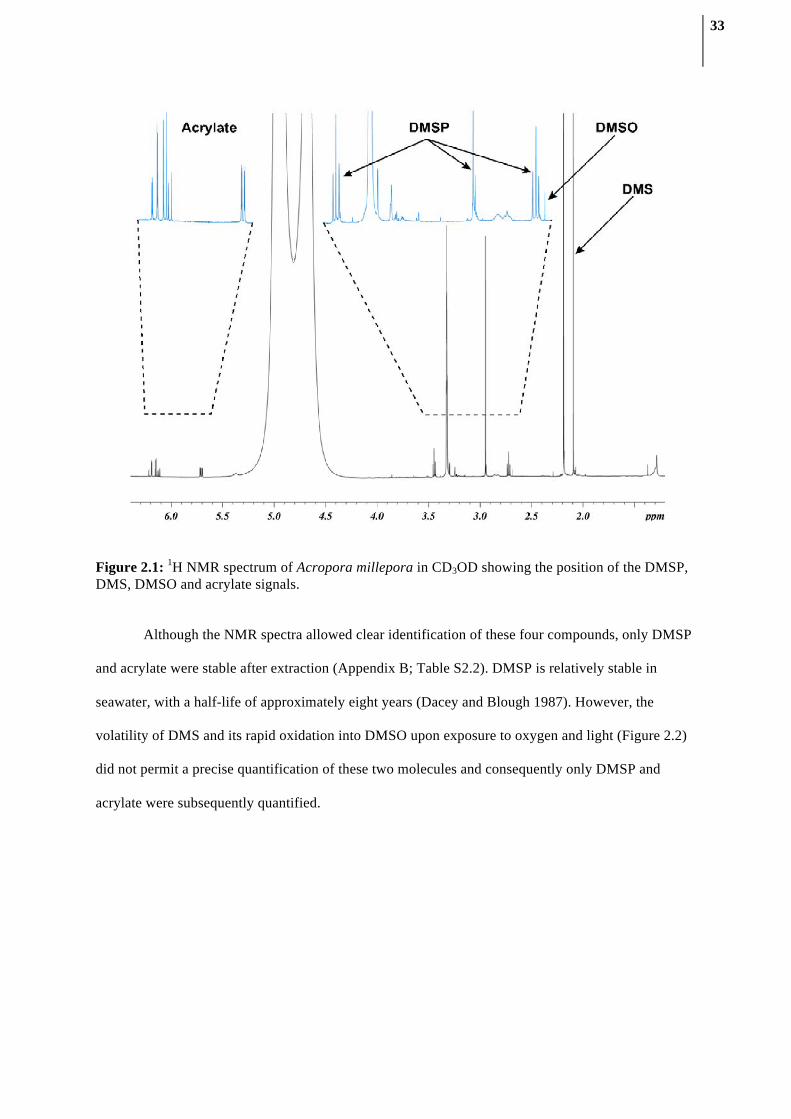
2.3.1. Compound identification using NMR spectroscopy
1H NMR spectra of the direct CD3OD extract of A. millepora contained several well-resolved
signals (Figure 2.1). Three multiplet signals (δH 5.71, 6.13 and 6.20 ppm, CH2=CH-) diagnostic of
acrylate protons were observed as described previously (Tapiolas et al. 2010). Two triplet signals at
δH 3.45 and 2.72 ppm (2×CH2), and a singlet signal at δH 2.95 ppm (2×CH3) were indicative of
DMSP. Furthermore, a singlet signal at δH 2.09 ppm (2×CH3) corresponded to DMS; while a singlet at
δH 2.69 ppm established the presence of DMSO (2×CH3), the chemical oxidation product of DMS.
The presence of DMSP, DMS, acrylate and DMSO was confirmed by comparison with 1D and 2D
spectra of the commercially available compounds and by spiking experiments. Spiking was also used
to estimate the extent of matrix effects on the position of the diagnostic signals and to determine the
method’s ability to recover a known amount of DMSP added to coral extracts (Appendix B; Table
S2.1). The DMSP recovery was 97.1% (±1.3), which is in line with previous error estimations using
qNMR, reported to be approximately 2% when acquisition parameters are optimized (Malz and
Jancke 2005).
33
Figure 2.1: 1H NMR spectrum of Acropora millepora in CD3OD showing the position of the DMSP, DMS, DMSO and acrylate signals.
Although the NMR spectra allowed clear identification of these four compounds, only DMSP
and acrylate were stable after extraction (Appendix B; Table S2.2). DMSP is relatively stable in
seawater, with a half-life of approximately eight years (Dacey and Blough 1987). However, the
volatility of DMS and its rapid oxidation into DMSO upon exposure to oxygen and light (Figure 2.2)
did not permit a precise quantification of these two molecules and consequently only DMSP and
acrylate were subsequently quantified.

34
Figure 2.2: 1H NMR spectra of the same Acropora millepora extract in CD3OD through time. The sample was kept at -20°C for 24 hours and subsequently at room temperature (25°C). Note conversion of DMS into DMSO starting after the sample was left at 25°C (from 24 hours onward).
2.3.2. Quantification method development
Based on the above optimization, a mixture of CD3OD and D2O was used in the acquisition of
1H NMR spectra to disperse and enhance the resolution of the diagnostic signals. The 90° pulse length
and the T1 relaxation times for both acrylate and DMSP were determined prior to the quantification to
give the best signal-to-noise ratio. Regions containing the two downfield signals from acrylate (6.00 -
6.20 ppm), and the singlet signal arising from DMSP (2.94 - 2.97 ppm) were selected for integration
(Figure 2.1).
2.3.3. DMSP and acrylate detection and quantification across coral genera
DMSP was unambiguously detected in 15 coral species, with the highest concentration
measured in Acropora millepora and the lowest in Merulina ampliata. In these corals, at least two of
the three DMSP proton signals were clearly visible and well resolved (Table 2.1). However, in four

35
species (Goniastrea aspera, Porites cylindrica, Diploastrea heliopora, Hydnophora exesa), the
resolution of the signals was poor, due to the presence of overlapping signals from other compounds
(Figure 2.3) which did not enable DMSP quantification. The DMSP concentrations measured were
comparable to those reported using classic GC methods (Broadbent et al. 2002, Wilson et al. 2002,
Van Alstyne et al. 2006), ranging from 0.03 to 2.47 nmol/mm2. In comparison, acrylate was observed
in 16 of the 18 GBR coral species. The lack of other signals from co-extracted compounds in the
region between 6.20 ppm and 5.50 ppm of the 1H NMR spectra facilitated the identification and
quantification of this molecule. Interestingly, acrylate concentrations in Acropora and few other
branching corals, such as Echinopora spp. and Porites cylindrica were consistently one order of
magnitude greater than those of DMSP. This result correlates well with previous measurements on A.
millepora (Tapiolas et al. 2010) and further suggests that either the turn-over of acrylate is slower than
DMSP or that it is stored in these corals for an unknown purpose. These results show that quantitative
1H NMR spectroscopy is a suitable technique to investigate both DMSP and acrylate concentrations in
a large array of reef-building corals, including the commonly studied genera Acropora, Pocillopora,
Seriatopora, Stylophora and Montipora.

36
Figure 2.3: 1H NMR spectra of three different reef-building coral species, Acropora millepora, Hydnophora exesa and Porites cylindrica. Note the large number of overlapping signals in Porites cylindrica and Hydnophora exesa, making DMSP quantification difficult.

37
Table 2.1: Measurements of DMSP and acrylate in 18 species of hard corals from the Great Barrier Reef. ND: not detectable.
Family Species DMSP (nmol/mm2)
Acrylate (nmol/mm2)
Faviidae Diploastrea heliopora ND ND Faviidae Platygyra sinensis 0.355 0.936 Faviidae Goniastrea aspera ND 0.253 Faviidae Echinopora spp. 0.467 8.235 Acroporidae Acropora millepora 2.473 15.223 Acroporidae Montipora spp. 0.092 0.387 Pocilloporidae Seriatopora hystrix 0.362 0.083 Pocilloporidae Pocillopora damicornis 0.333 0.035 Pocilloporidae Stylophora pistillata 0.774 0.130 Poritidae Porites spp. 0.271 1.083 Poritidae Porites cylindrica ND 2.945 Merulinidae Merulina ampliata 0.042 2.588 Merulinidae Hydnophora exesa ND 0.759 Agariciidae Pachyseris spp. 0.080 0.803 Euphyllidae Physogyra lichtensteini 1.048 0.330 Fungiidae Fungia spp. 0.353 ND Mussidae Symphyllia recta 1.517 0.171 Oculinidae Galaxea fascicularis 0.156 1.457
2.3.4. DMSP concentrations in A. millepora throughout the day measured by qNMR
To explore potential daily fluctuations in DMSP levels in corals and establish a clear baseline
for variations in relation to time of sampling, DMSP concentrations in A. millepora were measured
over a 24 hour period. This experiment also allowed the suitability of the qNMR technique for the
analysis of large numbers of samples to be tested. Despite the large number of samples collected
(n=135), less than 48 hours of work were required to process and analyze all samples. Contrary to my
expectations, DMSP concentrations did not change in response to high light conditions that are known
to increase photosynthetic activity and oxidative stress in corals (Figure 2.4) (Sunda et al. 2002). the
data showed that DMSP concentrations were not influenced by light or potential diel metabolic
patterns, but remained constant throughout the 24 hour period, ranging between 2.3 and 3.6
nmol/mm2 (Figure 2.4). Similarly, acrylate did not display a diel light-related pattern, even though
greater variability in its concentration was observed. As with the comparison between different coral

38
genera, acrylate concentrations were one order of magnitude greater than DMSP over the course of
the experiment, ranging from 27.2 to 42.6 nmol/mm2. The absence of light influence on both DMSP
and acrylate concentrations could reflect adaptation of colonies to their high light environment, as
revealed by oxidative and photochemical stress measurements conducted on the same colonies (A.
Lutz, unpublished results). In summary, under “normal” conditions, light levels do not influence
DMSP concentration and temporal variability is minimal in A. millepora. This implies that samples
could be collected at any time of the day in future studies investigating DMSP concentrations in
Acropora.
Figure 2.4: Concentration of DMSP and acrylate in A. millepora throughout a day. Photosynthetically active radiation (PAR; 400-700 nm) are indicated on the right-hand axis. DMSP and acrylate concentrations remained consistent over the diurnal cycle with 2.3-3.6 nmol mm-2 and 27.2 – 42.6 nmol mm-2 respectively.
0 2 4 5 6 8 14 16 17 18 20 22 240
10
20
30
40
50
60AcrylateDMSPLight
10 12
Time (hours)
Con
cent
ratio
n of
DM
SP a
nd a
cryl
ate
(nm
ol m
m-2
)
200
400
1000
1200
800
600
PAR
(um
ol p
hoto
ns m
-2 s
-1)

39
2.3.5. Conclusion
Quantitative NMR allows for the direct and simultaneous quantification of DMSP and
acrylate in a wide range of reef-building corals. It has several advantages over measurement methods
using GC: it allows direct measurements of DMSP without chemical conversion, reducing the
potential risks of sample degradation or concentration artifact through DMS conversion. Furthermore,
it allows for the quantification of several compounds simultaneously in a relatively short amount of
time, without complex sample preparation. Therefore, qNMR is an efficient and rapid method for
quantifying DMSP and acrylate in corals and the method presented here could easily be optimized to
quantify these compounds in other DMSP producing organisms.

40
Chapter 3: DMSP biosynthesis by an animal: implication for reef-
building corals in a warming world

41
3.1. Introduction
Only a few classes of marine algae and some species of higher plants have been reported to
produce DMSP, the main producers belonging to phytoplanktonic prymnesiophyte and dinoflagellate
taxa (Keller et al. 1989, Scarratt et al. 2002). Marine invertebrates that harbour photosynthetic
symbiotic dinoflagellates (Symbiodinium spp.), such as reef-building corals, sea anemones and giant
clams, also produce DMSP (Broadbent et al. 2002, Van Alstyne et al. 2006). To date, the highest
concentrations of DMSP and its breakdown products DMS and acrylate have been recorded in reef-
building corals, making coral reefs one of the most important ecosystems in the world in terms of
DMSP production (Broadbent and Jones 2004, Tapiolas et al. 2010). However, since it is widely
accepted that the production of DMSP is restricted to photosynthetic organisms (Karsten et al. 1990,
Kiene et al. 1996, Malin and Kirst 1997, Stefels 2000, Otte et al. 2004), it has been assumed that
DMSP produced by corals and other marine invertebrates is derived from the high density of
Symbiodinium present in their tissues. However, evidence that the total amounts of DMSP recorded in
corals are consistently higher than levels present in Symbiodinium cells alone (Hill et al. 1995, Yost
and Mitchelmore 2010, Yost et al. 2012) raise the possibility of a cryptic source of DMSP in reef-
building corals.
Here, I investigate DMSP production in two life history stages of corals from the genus
Acropora: i) coral juveniles prior to the acquisition of photosynthetic symbionts, and ii) adult colonies
with established endosymbiotic populations of Symbiodinium, under both ambient and elevated
temperatures constituting thermal stress. Corals in the genus Acropora are the most abundant reef-
building organisms in the Indo-Pacific region (Veron 2000) and as broadcast-spawning species, they
acquire Symbiodinium from their surrounding environment following larval development (Harrison et
al. 1984). The Symbiodinium-free coral larvae of these species provide a unique opportunity to
investigate Symbiodinium-independent production of DMSP by the coral animal, enabling us to
elucidate the roles of both corals and their photosymbionts in DMSP production in marine
ecosystems.

42
3.2. Methods
3.2.1. Adult corals
3.2.1.1. Thermal stress experiment
Acropora millepora colonies (n=10) were collected from Pelorus Island, Great Barrier Reef,
Australia (18°33’ S/146°29’ E) and transferred to the Australian Institute of Marine Science. Coral
colonies were fragmented to give a total of 24 fragments, each comprising approximately 25 branches
(nubbins). Fragments were arranged in eight indoor tanks in a randomized bloc design, resulting in the
allocation of 12 coral fragments to each of the control and thermal stress temperature treatments
(27°C and 32°C respectively). All tanks were continuously supplied with fresh 1 µm filtered seawater
(FSW), which was maintained at 27°C (±0.1°C) via computer control using a flow-through system at
a rate of 1.5 liters min−1. UV-filtered lights were mounted above each tank and provided an average
underwater light intensity of 350 µE over a 12:12 h light/dark cycle (400 W metal halide lamps, BLV,
Germany), typical of light intensities recorded at the collection site. The fragments were acclimatized
for two weeks prior to starting the experiment. Seawater temperatures in four tanks were slowly and
continuously ramped to 32°C (±0.05°C) over a 7 day period, via computer control, whilst the
remaining four control tanks were maintained at 27°C for the entire duration of the experiment.
Coral nubbins were sampled four times during the experiment: before any temperature
changes when both treatments were at 27°C (t = -7); once the 32°C target temperature had been
reached in the thermal stress treatment (t = 0); after 5 days at 32°C, when the first physiological
effects of temperature stress were visible (t = 5); and after 10 days at 32°C when all colonies in the
32°C treatment were completely bleached (t = 10). At each time point, one coral nubbin
(approximately 50 mm in length) was collected from each coral fragment (n=24) and immediately
transferred to a tube containing 2 mL of HPLC-grade methanol for qNMR analysis. Another coral
nubbin was collected from each coral fragment (n=24) to evaluate Symbiodinium densities. In
addition, one coral nubbin was collected at each time point from 4 different coral fragments,
transferred directly into fixative (1.25% glutaraldehyde + 0.5% paraformaldehyde in 0.2 µm-FSW)

43
and stored at 4°C until processed for structural investigations by transmission electron microscopy
(TEM).
3.2.1.2. Quantitative NMR analysis
The coral nubbins were extracted in methanol for 2 h with sonication followed by a second
extraction with an additional 1 mL of HPLC-grade methanol for 10 min. The two extracts were
pooled and dried using a vacuum-centrifuge then resuspended in a mixture of deuterium oxide (D2O,
D 99.8 %, 250 µL) and deuterated methanol (CD3OD, D 99.8 %, 750 µL) (Cambridge Isotope
Laboratories, Andover, MA, USA). A 700 µL aliquot of the particulate-free extract was transferred
into a 5 mm Norell 509-UP-7 NMR tube (Norell Inc., Landisville, NJ, USA) and analyzed
immediately by 1H NMR.
1H NMR spectra were recorded on a Bruker Avance 600 MHz NMR spectrometer with TXI 5
mm probe and quantification performed using the ERETIC method (Akoka and Trierweiler 2002).
This technique generates an internal electronic reference signal, calibrated using commercial stock
solutions of 4 mM acrylate and DMSP. The concentrations of DMSP and acrylate were determined by
integration of their respective signals in a 0.10 ppm window.
3.2.1.3. Symbiodinium densities
Freshly collected coral nubbins were airbrushed (80 lb.in2) in individual plastic bags in 4 mL
of 0.2 µm-FSW. The slurry was homogenized to breakdown aggregates and centrifuged at 3000 rcf.
The supernatant was removed and the pellet resuspended in 1 mL of 10% formalin. Homogeneous
extracts were placed on a hemocytometer (depth 0.1 mm) and Symbiodinium cells were counted under
a light microscope (8 technical replicates were averaged per sample).

44
3.2.1.4. Symbiodinium genotype
In hospite Symbiodinium populations of each experimental coral fragment were characterized
based on sequence differences in the nuclear ribosomal DNA internal transcribed spacer 1 region
using single-strand conformation polymorphism (SSCP) analysis (van Oppen et al. 2001). Total DNA
was extracted using a modified protocol (Wilson et al. 2002). Symbiodinium ITS1 region was
amplified with fluorescently labelled Sym ITS1 PCR primers. Genotype was determined using SSCP
with known reference samples running alongside experimental samples and scored manually using gel
images.
3.2.1.5. Surface area calculation
Coral skeletons remaining after samples were extracted for qNMR analyses and
Symbiodinium densities were lyophilized overnight and their surface area determined using a wax
dipping technique (Veal et al. 2010). The surface area of each individual nubbin was used to
normalize the qNMR and Symbiodinium data.
3.2.1.6. Transmission electron microscopy (TEM)
Fixed coral nubbins were decalcified in a formic acid:fixative mixture (1:3), with the solution
changed every 12 h until complete dissolution of the skeleton. Three individual polyps per sample
were post-fixed in osmium and subsequently dehydrated with increasing concentrations of ethanol
followed by dry acetone. Dehydrated samples were infiltrated in increasing concentrations of Araldite
resin before being cured for 24 h at 60°C. Longitudinal sections 90 nm thick were collected on copper
grids and imaged at 120 kV in a JEOL 2100 TEM (Tokyo, Japan).
3.2.1.7. PAM fluorometry measurements
Photosystem II (PSII) photochemical efficiency was measured with a Diving-PAM (Walz
Gmbh, Germany) on three random nubbins per coral fragment. Minimum and maximum fluorescence

45
(FO and FM) were recorded daily, two hours before the start of the light cycle. PS II photochemical
efficiency was expressed as maximum quantum yields ((FM-FO)/FM) = (FV/FM).
3.2.1.8. Data analyses
All data were square root transformed and no significant tank effect was detected for the
DMSP, acrylate and PAM fluorometry data (Nested ANOVA, p>0.05) (Statistica 7, Statsoft, Tulsa,
USA). Repeated measures ANOVA were carried out on the time series data, (data met all assumptions
of the test). Simple main effect tests (Quinn and Keough 2002) were used to compare the results
between the two temperature treatments, and between temperatures at each time point (Table S1).
This statistical technique was used to minimize the number of multiple comparisons (focusing only on
comparisons of interest), decreasing the likelihood of type I error.
3.2.2. Coral juveniles
3.2.2.1. Sample collections
Colonies of Acropora millepora (n=10) and A. tenuis (n=3) were collected from Orpheus
Island, Great Barrier Reef, Australia (18°34’ S/146°30’ E) and transferred to the Australian Institute
of Marine Science outdoor aquarium facility 4 days prior to the predicted spawning event in
November 2011. One hour before spawning, the colonies were isolated in 70 L tanks with 1 µm-FSW.
Gametes were collected from the surface of these tanks and fertilized in separate 70 L tanks with
FSW. After fertilization, embryos were gently rinsed three times by transferring to new containers and
were subsequently transferred to 500 L tanks (containing 0.5 µm-FSW) where they were kept through
larval development. After 12 days, Symbiodinium-free coral larvae were collected using a 1 µm mesh
net and washed three times in 0.2 µm-FSW. Larvae were subsequently settled in sterile 6-well plates
(8 plates per species, 40 larvae per well; each well filled with 10 mL of 0.2 µm-FSW). Eight hours
after settlement, the plates were separated between two temperature regimes: 4 plates per species were
incubated at 27ºC (control temperature), and the other 4 plates were ramped to 32ºC over 5 hours
(thermal stress treatment); all plates were maintained in the dark (in order to prevent the growth of

46
potential photosynthetic organisms). Settled juveniles were incubated at their respective temperature
treatments and six random wells were sampled every 2 days. The size of the sampled juveniles was
measured using a motorized stereomicroscope with a high resolution camera (Leica MZ16A and
Leica DFC500, Leica Microsystems, Wetzlar, Germany); sizes did not vary significantly between day
2 and day 6 with an average size of 0.79 mm2 (±0.05) for A. millepora juveniles and of 0.82 mm2
(±0.07) for A. tenuis juveniles.
3.2.2.2. qNMR analysis
After the required incubation time, seawater was removed from each well with a pipette, and
a sterile cotton bud was used to soak up any residual seawater, taking care not to disturb the settled
juvenile corals. Juveniles in six wells were extracted by adding 300 µL of deuterated methanol
(CD3OD) to each well, followed by 30 s of gentle shaking; 200 µL of this extract was transferred into
a 3 mm Bruker MATCH NMR tube and analyzed immediately. In addition, negative control wells
without settled juveniles were extracted following the same procedure. The concentrations of DMSP
and acrylate were normalized initially to the number of settled coral juveniles in the respective well.
They were then normalized to the averaged surface area of the juveniles (note that juveniles were
approximated to perfect circles).
3.2.2.3. DNA extractions and PCR amplification
At each time point, the contents of two wells were scraped into a 2 mL tube using a scalpel
blade, snap frozen with liquid nitrogen, and used for total DNA extraction, according to methods in
Bourne and Munn (Bourne and Munn 2005). Multiple sets of primers were subsequently used to
target different taxonomic groups: Symbiodinium (van Oppen et al. 2001), coral (Suzuki et al. 2008),
and general primers for the algal 23S rDNA plastid (Sherwood et al. 2008) and chloroplast DNA
(Taberlet et al. 1991) (Table S3). The PCR consisted of 1 µL of DNA template (dilution series from 1
to 10-5 of the original concentrations), 10 µL of buffer containing dNTP and MgCl2 (Bioline, London,
UK), 1.5 µL of each primer (10 µM), and 0.5 µL of Taq polymerase (Bioline, London, UK), adjusted

47
to a final volume of 50µL with sterile MilliQ water. Amplified PCR products were visualized by
electrophoresis on 1% agarose gel stained with ethidium bromide.
3.2.2.4. Identification of candidate genes
Orthology between coral and diatom genes was inferred based on best reciprocal BLAST hits
(Moreno-Hagelsieb and Latimer 2008). Orthologs in other species were identified using release 5
(March 2011) of the OrthoMCL database (Feng et al. 2007).
3.2.2.5. Data analyses
Repeated measure ANOVA were performed on the normalized DMSP and acrylate
concentrations (data met all assumptions of the test, except the acrylate data from Acropora tenuis
that violated sphericity assumption; in that unique case, Greenhouse-Geisser correction was applied to
the degrees of freedom). Simple main effect tests (Quinn and Keough 2002) were then used to
compare the results from the temperature treatment within species (Table S2).
3.3. Results
3.3.1. DMSP production in adult corals
Adult colonies of A. millepora (n=12 for each treatment) were subjected to ambient (27°C) or
thermal stress (32°C) conditions during a 17 day experiment, during which their DMSP and acrylate
contents were measured using quantitative nuclear magnetic resonance (qNMR) (23). All colonies
contained only Symbiodinium type C2 (GenBank Accession AF380552 (25)) (Single strand
conformation polymorphism (SSCP) profiles from all samples were single bands identical to the
reference). After 5 days of thermal stress, cell counts revealed a 25% decrease in Symbiodinium cell
numbers within coral tissues (Figure 3.1A). Transmission electron microscopy (TEM) showed that all
Symbiodinium cells observed within coral tissues (n=1304) were structurally compromised (Figure
3.2). Unexpectedly, DMSP concentrations in thermally stressed coral tissues increased significantly in

48
comparison to controls (ANOVA, n=12, p<0.005; Figure 3.3 and Appendix C; Table S3.1), despite
the substantial degradation and loss of Symbiodinium cells.
Figure 3.1: Density and photosynthetic efficiency (mean±SE) of Symbiodinium cells within adult colonies of the coral Acropora millepora maintained under control (27°C) or thermal stress (32°C) conditions for 10 days. (A) Density of Symbiodinium cells in the same coral fragments through time. (B) Comparison of photosystem II photochemical efficiency (maximum quantum yields: FV/FM) through time (repeated measure ANOVA, *p < 0.001; post-hoc simple main effect test, *p < 0.01). See also Appendix C; Table S3.1.
-7 0 5 10Time (days)
F V/F
M (d
ark
adap
ted)
*
*
27oC32oC
-7 0 5 10Time (days)
0.3
0.4
0.5
0.6
0.7
0.8
Sym
biod
iniu
m (n
umbe
r/mm
2 )
0
2,000
4,000
6,000
8,000 BA

49
Figure 3.2: Structurally damaged Symbiodinium cells (%) within adult colonies of the coral Acropora millepora maintained under control (27°C) or thermally stressed (32°C) conditions. The numbers above the bars refers to the total number of Symbiodinium cells observed.
After 10 days of thermal stress, the number of Symbiodinium cells remaining in coral tissues
was reduced to 16% of their original density (Figure 3.1A and Figure 3.3A-C) and structural damage
increased. In particular, thylakoid membranes within chloroplasts were completely disrupted and
general cell structure was lost in all cells examined (n=272) (Figure 3.2 and 3.4). Consistent with
these observations, Pulse Amplitude Modulation (PAM) fluorometry measurements revealed
significant reductions in the photochemical efficiency (FV/FM) of photosystem II in thermally stressed
corals (ANOVA, n=12, p<0.005; Figure 3.1B). Despite the severe degradation and depletion of algal
symbionts from coral tissues, thermally stressed corals contained 68% more DMSP and concomitantly
36% less acrylate than control colonies (Figure 3.3), suggesting the existence of an alternative source
of DMSP production in coral tissues, which dramatically escalates DMSP production in response to
thermal stress.
1304 1456 1648
1502
1216
1304
1352
272
-7 0 5 10Time (days)
Dam
aged
Sym
biod
iniu
m (%
cel
ls)
27oC32oC
20
40
60
80
100
0

50
Figure 3.3: Effects of thermal stress on adult colonies of the coral Acropora millepora. Temperature was raised gradually over 7 days to 32°C, mimicking a realistic thermal stress scenario, therefore thermal stress commenced on day 0. The total duration of the experiment was 17 days. (A) Micrograph of representative nubbins showing visual differences in the density of Symbiodinium cells present in the tissues of A. millepora maintained under control (27°C, brown, right) or thermal stress (32°C, white, left) conditions for 10 days. (B-C) Higher magnification micrographs of coral polyps showing visual differences in tissue color as intracellular Symbiodinium densities decline between (B) 27°C and (C) 32°C (scale bars: 1 mm). (D) Changes in DMSP concentration (mean±SE) in adult corals (n=12) exposed to control (27°C) and thermal stress (32°C) treatments (ANOVA simple main effect test, F1,22=10.79, *p<0.005). (E) Corresponding changes in acrylate concentration (mean±SE) in adult corals (n=12) exposed to control (27°C) and thermal stress (32°C) treatments (ANOVA simple main effect test, F1,22=8.4, *p<0.01).
Acr
ylat
e co
ncen
trat
ion
(nm
ol/m
m2 )
-7 0 5 10 -7 0 5 10
Time (days) Time (days)
D E27oC
*
*
**
A B C
32oC
DM
SP c
once
ntra
tion
(nm
ol/m
m2 )
0
10
20
30
40
50
0
5
10
15
20
25

51
Figure 3.4: Representative transmission electron micrographs showing the effects of thermal stress on the internal structure of endosymbiotic Symbiodinium cells associated with the coral Acropora millepora. (A-B) Symbiodinium cells after 10 days at 27°C, showing intact cell structures and intact thylakoid membranes of chloroplasts (arrows), the photosynthetic centre of cells; (C-D) Symbiodinium cells after 10 days at 32°C, showing structurally degraded cells with highly disrupted thylakoid membranes (arrows). Scale bars: 1 µm. nu: nucleus, ch: chloroplast.
3.3.2. DMSP production in juvenile corals without photosynthetic symbionts
To further investigate the role of Symbiodinium-independent DMSP production in Acropora,
DMSP and acrylate concentrations were also measured in juveniles from two coral species, A.
millepora and A. tenuis, lacking photosynthetic symbionts. The absence of any photosynthetic
organisms in these coral juveniles was confirmed using five different DNA markers, ranging from
Symbiodinium-specific to universal 23S rRNA plastids primers, targeting all known lineages of
microalgae and cyanobacteria. A lack of any detectable amplification was observed for all
markers (Appendix C, Table S3.3). Despite the complete absence of photosynthetic microalgae, high
concentrations of DMSP were recorded in all coral juvenile samples (Figure 3.5).

52
Figure 3.5: Changes in DMSP and acrylate concentrations (mean±SE) in coral juveniles lacking photosynthetic symbionts (n=6) through a six-day period after coral settlement. Patterns are compared for two thermal regimes: ambient (27°C, blue) and thermal stress (32°C, red), and for the coral species (A) Acropora millepora and (B) Acropora tenuis. DMSP concentrations increase significantly through time in the juveniles exposed to ambient temperature (ANOVA Simple main effect test, F2,20=6.30, *p<0.01 for Acropora millepora and F2,20=7.51, *p<0.005 for A. tenuis). Furthermore, DMSP concentrations in coral juveniles exposed to elevated temperature (32°C) for 6 days were significantly higher than controls kept at ambient temperature (27°C) (Simple main effect test, F1,10=27.68, *p<0.0005 for A. millepora and F1,10=10.44, *p<0.01 for A. tenuis). Conversely, acrylate concentrations decreased significantly in juveniles of the corals (C) Acropora millepora and (D) Acropora tenuis when exposed to elevated temperature (Simple main effect test, F1,10=5.58, *p<0.05 for A. millepora and F1,10=61.68, *p<0.0005 for A. tenuis). Scale bar: 1mm.
2 4 60
1
2
3
4
5
6
7
DM
SP c
once
ntra
tion
(nm
ol/m
m2 )
Time (days)2 4 6
0
1
2
3
4
5
6
7
DM
SP c
once
ntra
tion
(nm
ol/m
m2 )
Time (days)
2 4 60
1
2
3
4
5
6
7
8
9
Acr
ylat
e co
ncen
trat
ion
(nm
ol/m
m2 )
Time (days)2 4 6
0
1
2
3
4
5
6
7
8
9
Acr
ylat
e co
ncen
trat
ion
(nm
ol/m
m2 )
Time (days)
27oC32oC
*
*
*
*
*
*
*
A B
C D
Acropora millepora Acropora tenuis

53
Repeated sampling over a six day period after larval settlement revealed that initially high
concentrations of DMSP in coral juveniles continued to increase significantly over time (ANOVA,
n=6, p<0.005; Figure 3.5 and Appendix C; Table S3.2). At ambient water temperatures (27°C), mean
DMSP concentration in A. millepora juveniles increased by 44% (i.e. by 1.1 nmol/mm2) in four days.
A similar trend was observed in A. tenuis, with mean DMSP concentration increasing by 54% (i.e. by
1.7 nmol/mm2). These DMSP concentrations measured in non-photosynthetic coral juveniles are
approximately half of those present in adult colonies.
As found in temperature experiments with adult corals, thermal stress resulted in significant
increases in DMSP levels in juvenile corals (ANOVA, n=6, p<0.005; Figure 3.3 and Appendix C;
Table S3.2), with A. tenuis and A. millepora showing 30% and 48% increases in DMSP
concentrations, respectively, after six days of exposure to elevated temperatures (see Figure 3.5A-B
and Appendix C; Table S3.2). Conversely, concentrations of the DMSP breakdown product acrylate
decreased in both species, with thermally stressed juveniles containing 33% (A. millepora) and 61%
(A. tenuis) less acrylate than juveniles at ambient temperature after six days (Figure 3.5C-D).
3.3.3. Identification of genes involved in DMSP synthesis in corals
A recent study of the diatom Fragilariopsis cylindrus identified candidate genes for each of
the four steps of the DMSP biosynthesis pathway (Lyon et al. 2011). In an attempt to identify the
possible molecular mechanisms underlying DMSP production in corals, we searched for potential
orthologs of these genes in the comprehensive molecular resources available for two corals, the A.
millepora transcriptome (Moya et al. 2012) and the A. digitifera genome (Shinzato et al. 2011). Two
genes previously identified in diatoms had orthologs in both of these coral species (Appendix C;
Tables S3.4). These genes encode a NADPH-reductase and an AdoMet-dependant methyltransferase,
which mediate the second and third steps of the biosynthesis process respectively (Figure 3.6). The
orthologous relationship between the coral and diatoms genes is supported by best reciprocal blast

54
hits, and their mapping to the same OrthoMCL clusters (Feng et al. 2007): OG5_131390 and
OG5_156314 for the reductase and methyltransferase, respectively.
Figure 3.6: (A) Pathway of DMSP biosynthesis present in marine algae (Gage et al. 1997). The first two steps are reversible. The proposed diatom genes encoding the second and third steps (in green) have orthologs in Acropora millepora and Acropora digitifera genomes (see Appendix C; Table S3.4). (B-C) Color coded gene expression percentiles of the two putative genes involved in DMSP biosynthesis in Acropora millepora though five coral life-stages, from embryos (Pregast: pregastrula; Postgast: postgastrula) to adults, based on transcriptomic data from Moya et al. (Moya et al. 2012).
The reductase enzyme mediates a reversible step in the DMSP biosynthesis process (Gage et
al. 1997, Stefels 2000) (Figure 4). The gene encoding this enzyme is highly expressed throughout all
life history stages of A. millepora (Moya et al. 2012) (Figure 3.6A) and the phylogenetic distribution

55
of its OrthoMCL cluster revealed 45 orthologs spread throughout all Kingdoms (Appendix C; Figure
S3.1), in line with the widespread presence of this enzyme among plants that do not produce DMSP
(Stefels 2000). In contrast, the methyltransferase has been shown to mediate a non reversible step
regulating intracellular DMSP levels and is believed to be specific to this pathway (Stefels 2000). The
methyltransferase gene has high expression levels in early coral life stages but its expression
decreases after settlement and remains relatively low in adult corals (Moya et al. 2012) (Figure 3.6C).
This expression pattern could reflect the establishment of symbiosis with DMSP-producing
Symbiodinium around the time of settlement. The OrthoMCL cluster corresponding to this
methyltransferase has an unusually sparse phyletic pattern with only nine orthologs, including seven
in photosynthetic organisms and two in other eukaryotes (Appendix C; Figure S3.1).
We also searched for potential homologues of the diatom genes putatively involved in DMSP
synthesis in a comprehensive transcriptome assembly available for Symbiodinium (Bayer et al. 2012).
Interestingly, we identified two Symbiodinium sequences belonging to the same OrthoMCL clusters as
the methyltransferase and reductase identified in diatoms and corals (Appendix C; Table S3.4 and
Figure S3.1). This suggests that the function of these enzymes in DMSP synthesis may be conserved
between diatoms, alveolates, green plants and corals.
3.4. Discussion
Contrary to the current paradigm, which assumes that DMSP biosynthesis is limited to
photosynthetic organisms, this study reveals that corals (Kingdom: Animalia) also produce this
compound and were up-until-now a cryptic source of DMSP in coral reefs. Coral juveniles that did
not harbour symbiotic photosynthetic organisms contained high DMSP concentrations, approximately
half the concentrations measured in symbiont-bearing adult corals. Moreover, DMSP concentrations
in juveniles increased significantly through time, demonstrating unambiguously that the DMSP levels
measured were not simply inherited from parent colonies but produced by coral juveniles growing in
the absence of photosynthetic symbionts.

56
This result sheds new light on the relevance and importance of corals as DMSP producers in
coral reef ecosystems, highlighting the need to consider the influence of these animals in the global
sulfur cycle. These data also explain previous discrepancies reported in the literature between the total
amount of DMSP measured in corals and levels measured in Symbiodinium alone, which are typically
two to three times lower (Hill et al. 1995, Yost and Mitchelmore 2010, Yost et al. 2012). Interestingly,
DMSP concentrations are well-correlated with symbiont densities in giant clams (Hill et al. 2000),
and undetectable in sea anemones lacking photosynthetic symbionts (Van Alstyne et al. 2009).
Therefore, corals are likely to be the exception, rather than the rule, in terms of DMSP production by
marine invertebrates harboring photosynthetic symbionts.
Thermal stress of juvenile corals triggered a significant increase in DMSP production and a
simultaneous decrease in acrylate concentrations. Similar patterns have been reported in marine algae
subjected to a variety of stressors, with significant increases in intracellular DMSP concentrations
correlated with exposure to ultraviolet radiation, CO2 and iron limitation, and high levels of reactive
oxygen species (Sunda et al. 2002). This response relates to the antioxidant properties of DMS and
acrylate, the breakdown products of DMSP. During thermal stress, the production of reactive oxygen
species by coral mitochondria (and Symbiodinium, when present) increases, damaging coral cells
(Weis 2008). DMS and acrylate are very efficient scavengers of hydroxyl radicals and other reactive
oxygen species (Sunda et al. 2002); thus the observed decrease in acrylate concentrations in coral
juveniles during thermal stress is a likely consequence of its reactivity with reactive oxygen species.
This suggests that DMSP and its breakdown products fulfill an important role in coral stress
responses, presumably reducing cellular damage from reactive oxygen species.
Similar experimental results with adult corals, the building blocks of coral reefs and therefore
the most ecologically relevant life history stage in terms of DMSP production, further corroborate the
conclusion that enhanced DMSP production in response to elevated temperatures may be an important
mechanism for ameliorating thermal stress. Adult colonies of A. millepora exposed for ten days to a

57
temperature 2°C above their upper thermal threshold, showed a two-thirds increase in DMSP
concentration and a simultaneous halving in acrylate concentration within their tissues. These trends
are in close accordance to those observed in coral juveniles lacking photosymbionts. Remarkably,
despite an 84% reduction in their Symbiodinium cell density, DMSP concentrations increased. The
Symbiodinium cells remaining were metabolically dysfunctional, as they had suffered extensive
structural damage (including disruption of the thylakoid membranes, where the first stage of
photosynthesis takes place) and their photosynthetic efficiency was significantly reduced. Previous
reports investigating the effect of thermal stress on Symbiodinium in corals concluded that these
symptoms are characteristic of an advanced stage of necrosis (Strychar et al. 2004). In further support
of our conclusion that Symbiodinium were not contributing significantly to DMSP production,
previous experiments on Symbiodinium cultures have shown that DMSP content per cell volume
decreases under thermal stress (McLenon & DiTullio 2012).
Taken together, experimental results for juvenile corals lacking photosymbionts and thermally
stressed adult corals with dysfunctional photosymbionts provide conclusive evidence that the
observed increases in DMSP concentrations in thermally stressed corals cannot be attributed to the
activity of Symbiodinium cells. These results identify the coral animal as responsible for increased
DMSP production and imply that this production is not restricted to juvenile life-stages, but also
occurs at high levels in adult reef-building colonies subjected to thermal stress.
Studies have shown that marine algae produce DMSP from methionine via a pathway that
involves the successive action of four different enzymes (Gage et al. 1997), but until recently, very
little was known about genes involved in the biosynthesis of DMSP. The recent identification of
candidate genes in a diatom is a significant step toward the functional characterization of this pathway
in marine algae (Lyon et al. 2011). Remarkably, two of the four genes identified in diatoms have clear
orthologs in Symbiodinium and corals. The homologous genes are potentially involved in the second
and third steps of DMSP biosynthesis and encode, respectively, a reductase and a sulfur-

58
methyltransferase enzyme. Significantly, the sulfur-methyltransferase mediates the committing step of
DMSP synthesis and has been shown to regulate intracellular levels of DMSP in algae (Ito et al.
2011). The presence of these genes in coral genomes further supports the conclusion that DMSP is
synthesized directly by coral species in the genus Acropora.
A survey of the distribution of aerosol particles over Australia identified the Great Barrier
Reef (GBR) as a considerable emission hotspot (Bigg and Turvey 1978), and sulfur aerosols were
identified as a major constituent of these particles (Modini et al. 2009). The GBR is the largest
biological structure on the planet and the release of these particles along its 2600 km length would
constitute a major source of cloud condensation nuclei. During summer, the northern regions of the
GBR (from 10 to 18°S) experience considerably lower solar radiation than the south based on
significant increases in cloud cover (Masiri et al. 2008). This result can be ascribed to the south-
easterly winds prevailing along the GBR that carry sulfur aerosols emitted by the reefs northwards
(Jones and Trevena 2005). Coral reef-derived sulfur aerosol emission might therefore play a central
role in cloud formation in areas of the world with large coral densities, such as the GBR and the Coral
Triangle.
In summary, the novel finding of this study - that two common coral species produce large
amounts of DMSP in the absence of photosynthetic partners - has major implications across
physiological, ecological and biogeochemical scales. Notably, DMSP production originating from
corals is important at the scale of: i) coral physiology, because concentrations of DMSP and its
antioxidant breakdown product acrylate are strongly affected by thermal stress, indicating a role for
these molecules in coral-stress responses; ii) coral reef ecology, because the large concentrations
present in both juveniles lacking photosymbionts and adults with non-functional photosymbionts
indicate that corals contribute extensively to the DMSP pool produced by this ecosystem; and iii) the
global sulfur cycle, because coral-derived DMSP production is significant and likely to be integral to
sulfur aerosol production from the marine environment in areas of the world where coral cover is still

59
high. Considering predicted increases in coral mortality worldwide caused by anthropogenic stressors,
the associated decline in sulfur aerosol production from coral reefs may further destabilize local
climate regulation and accelerate degradation of this globally important and diverse ecosystem.

60
Chapter 4: Coral-associated bacteria and their role in the
biogeochemical cycling of sulfur
A portion of this chapter is published as: Raina, J.B., Tapiolas D., Willis B.L., and D.G. Bourne. (2009) Coral-associated bacteria and
their role in the biogeochemical cycling of sulfur. Applied and Environmental Microbiology. 75(11): 3492-3501.

61
4.1. Introduction
DMSP plays a fundamental role as sulfur and carbon source for marine bacteria (Sievert et al.
2007, Reisch et al. 2011) and can be metabolized by these organisms via multiple pathways (Howard
et al. 2006, Todd et al. 2007, Curson et al. 2008, Todd et al. 2009, Todd et al. 2010, Curson et al.
2011, Todd et al. 2011). The most widespread pathway among bacteria converts DMSP into
methanethiol, and enables bacteria to assimilate the reduced sulfur (Howard et al. 2006, Reisch et al.
2011). The other pathways cleave DMSP into DMS (plus acrylate in some case) (Todd et al. 2007,
Curson et al. 2008, Todd et al. 2009, Todd et al. 2010, Curson et al. 2011, Todd et al. 2011), the DMS
produced by these enzymes is then released into the surrounding water (Kiene et al. 2000). Prior to the
1980s, diffusion of DMS from the oceans to the atmosphere was thought to be its major removal route
from the marine system (Lovelock et al. 1972, Yoch 2002). More recently, however, it has been
estimated that between 50 and 80% of the DMS produced by DMSP-degrading bacteria is degraded
directly by other types of bacteria (Simo et al. 2002, Simo 2004), although the identity of the bacteria
involved has not been extensively studied.
In oligotrophic environments, such as coral reefs, bacteria depend on organic compounds
produced by primary producers and reef-building corals (Ritchie and Smith 2004) and these
compounds may determine the composition of coral-associated bacterial communities (Ritchie and
Smith 2004, Rosenberg et al. 2007b). The presence of high levels of DMSP and DMS in reef-building
corals suggests that they are likely to harbor bacterial species capable of metabolising these
compounds. To investigate the potential of these methyl-sulfur compounds to drive coral-associated
microbial communities, we used them as sole carbon sources to isolate bacteria from two common
coral species (Montipora aequituberculata and Acropora millepora) and directly compared these
microbial isolates with coral-associated microbiota identified using culture-independent analyses. We
also investigated the distribution and abundance of the genes involved in DMSP degradation within
metagenomic datasets derived from a range of marine and terrestrial environments, to evaluate their
relative roles in these ecosystems and their potential relevance to coral reefs.

62
4.2. Methods
4.2.1. Sample collections
Three colonies of the corals A. millepora and M. aequituberculata were collected from Davies
Reef, Great Barrier Reef, Australia (18°05’S/147°39’E) and maintained in aquaria at the Australian
Institute of Marine Science (Townsville, Queensland, Australia). Five replicate samples of coral
mucus were taken from each colony using sterile 50-ml syringes. Samples (1 ml) were used
immediately for selective enrichment cultures, and the remaining volume was filtered through 0.22-
µm-pore-size Sterivex filter columns (Durapore; Millipore), which were filled with 1.6 ml of lysis
buffer (0.75 M of sucrose, 40 mM EDTA, 50 mM Tris-base; pH 8.3), and stored at -80°C for
subsequent molecular analyses. Seawater samples (1 liter each) were also taken from the aquaria and
processed in the same manner. Coral tissue slurry samples were obtained from five replicate coral
fragments (25 mm in length, 60 to 70 polyps) from each colony and washed in autoclaved, 0.22-µm-
pore-size-filtered artificial seawater (ASW) to remove loosely attached microbes. The samples were
airbrushed (80 lb/in2) to form a slurry with 5 ml of ASW to remove coral tissues and associated
microbes from the skeleton. This slurry was homogenized to break down aggregates and divided into
1-ml aliquots and either stored at -80°C for DNA extraction or used immediately for selective
enrichment cultures. All corals appeared healthy when the samples were collected.
4.2.2. Isolation of bacterial strains using DMSP, DMS, and acrylate as carbon sources
After air brushing, the remaining coral skeleton was crushed by using a sterilized pestle and
mortar. Dilution series in basal medium were performed for each of the five replicates of mucus,
tissue slurry, and crushed skeleton samples for each species. Sterile 125-ml crimp-top vials were used
for the dilution series, and the vials were inoculated with coral bacteria to a final volume of 10 ml. All
vials were sealed by using sterile Teflon coated butyl rubber septa. Two different basal media (lacking
a carbon source) were tested for their ability to support the growth of coral bacteria: a modified
marine ammonium mineral salt (MAMS) medium and a modified ASW media (MASW) (Table 4.1).
The carbon sources used for enrichments were DMSP (50 µM), DMS (50 µM), and acrylate (50 µM).

63
These carbon sources were added aseptically through the septa of crimp-top vials with a syringe and
needle. Control bottles containing only the basal medium and the carbon source were set up, along
with enrichment cultures, to account for the chemical breakdown of the carbon sources. Cultures were
incubated at 28°C for 7 days, and the presence of DMS in the headspace gas was monitored by gas
chromatography (GC) analysis. Enrichments were respiked daily with an additional dose (20 µM) of
the carbon source to avoid the potential deleterious effect of high dosage concentrations. The
degradation of acrylate was assessed by nuclear magnetic resonance (NMR) analysis.
Table 4.1: Basal media composition (in grams per liter) used for the isolation of DMSP-, DMS- and acrylate-degrading bacteria. The Marine Ammonium Mineral Salts medium was modified from Goodwin et al. (2001). All solutions were autoclaved separately before combining. SL10 trace metal solution from Widdel et al. (1983); Vitamin solution from Pfennig (1978).
Solution Components (g/liter) MAMS medium MASW medium
1 NaCl (25), (NH4)2SO4 (1), CaCl2 · 2H2O (0.2) NaCL (25), MgSO4 · 7H2O (2.44), KCl (0.6), NaNO3 (1), CaCl2 · 2H2O (0.3)
2 MgSO4 · 7H2O (1),FeSO4 · 7H2O (0.002), Na2MoO4 · 2H2O (0.002) KH2PO4 (0.5)
3 KH2PO4 (3.6) K2HPO4 (0.9) 4 K2HPO4 (4.6) NH4Cl (0.267) 5 SL10 trace metal solution (1 ml/l) SL10 trace metal solution (1 ml/l) 6 Pfennig's vitamin solution (1 ml/l) Pfennig's vitamin solution (1 ml/l)
A 50-µl aliquot of each enrichment culture was spread onto the appropriate basal medium
plate (MAMS or MASW) containing the appropriate carbon source (DMSP, DMS, or acrylate). DMS
enrichment plates were kept in gas-tight jars, and 200 µM DMS was added to each jar. The jars were
regularly vented and replenished with DMS (every 3 days). All enrichment plates were incubated at
28°C in the dark for 14 days. To isolate single strains capable of metabolizing the carbon source for
growth, individual colonies were picked from isolation plates and resuspended in 10 ml of MAMS or
MASW medium containing the appropriate carbon source (50 µM DMSP, 50 µM DMS, or 50 µM
acrylate). Degradation of DMSP and DMS by the isolates was monitored by GC analysis, while the
degradation of acrylate was assessed by NMR analysis. A 50-µl aliquot of each liquid culture was
then inoculated back onto appropriate plates as described above to confirm the cultures formed

64
colonies with a consistent morphology. Again, a single colony was picked and grown in liquid culture
as described above, and degradation of the carbon source was confirmed.
4.2.3. DNA extraction and purification
DNA was extracted from isolated single strain liquid cultures by using a DNA extraction kit
(Promega, Madison, WI), according to the manufacturer’s instructions and resuspended in 30 µl of
sterile Milli-Q water. DNA from the seawater and coral mucus samples was extracted according to the
procedure of Schauer et al. (2000), resuspended in 30 µl of Milli-Q water, and used directly for PCR
amplification. DNA from the coral tissue samples was extracted according to a modified urea
extraction protocol (Bourne and Munn 2005). Extracted DNA (30 µl) from coral tissues was purified
to remove coextracted PCR-inhibitory humic and phenolic materials by passage through a 0.7%
agarose gel, and DNA fragments larger than 2 kb were cut from the gel and purified by using a
QIAquick gel extraction kit (Qiagen, Hilden, Germany) according to the manufacturer’s instructions.
All DNA was resuspended in 50 µl of sterile Milli-Q water, quantified with a Nanodrop
spectrophotometer (Amersham Pharmacia Biotech, Uppsala, Sweden), and stored at -20°C until
required.
4.2.4. PCR amplification of bacterial 16S rRNA genes
Primers 63F and 1387R, specifically targeting a 1,300-bp section of the genomic bacterial
16S rRNA gene (Marchesi et al. 1998), were used for PCR amplifications. The PCRs included 2.5
µmol of each deoxyribonucleotide triphosphate and 5×PCR buffer containing MgCl2, 10 pmol of each
primer, and 0.25 µl of Taq DNA polymerase, adjusted to a final volume of 50 µl with sterile Milli-Q
water. The reaction conditions were as follows: 94°C for 3 min; followed by 30 cycles of 94°C for 1
min, 55°C for 1 min, and 72°C for 1.5 min; and then a final extension of 72°C for 7 min. Amplified
PCR products were visualized by electrophoresis on 1% agarose gel stained with ethidium bromide.

65
4.2.5. Clone library construction and sequencing
PCR products from seawater, coral mucus, and coral tissue samples were purified using a
QIAquick gel extraction kit (Qiagen, Hilden, Germany) according to the manufacturer’s instructions.
Purified DNA was ligated into a TOPO-TA cloning vector (Invitrogen, Carlsbad, CA) according to
the manufacturer’s instructions, and ligated vectors and inserts were sent to the Australian Genome
Research Facility for clone library construction and sequencing. A total of 96 clones were sequenced
from each library. Inserts of the wrong size and chimeric sequences were removed from subsequent
analysis.
4.2.6. GC analysis
Determination of DMS in the vials was performed by injecting 50 µl of headspace gas into a
GC-ECD gas chromatograph (HP GC-500; Hewlett-Packard, Palo Alto, CA) fitted with a 1-m-by-4
mm glass column containing Poropack-Q and using nitrogen as the carrier gas (flow rate, 30 ml/min)
at 200°C. A flame ionization detector was used to detect the compound. A DMS standard (Fluka,
Evry, France) was used to confirm retention times.
4.2.7. NMR analysis
High-pressure liquid chromatography-grade methanol (20 ml) was added to each vial
containing the acrylate enrichments and subsequently transferred to a glass round-bottom flask. The
mixtures were dried in vacuo using a Buchi rotary evaporator and qNMR were run as described in
Chapter 2.
4.2.8. Data analysis
All sequence data were edited by using the Sequencher program (Gene Codes Corp.) for
removal of the vector and primer sequences and assembled into a single file for BLAST search
comparisons (http://www.ncbi.nlm.nih.gov/) to determine the closest sequence affiliation to reference
organisms or previously sequenced 16S rRNA genes. Chimeric sequences (checked with the

66
CHECK_CHIMERA software of the Ribosomal Database Project (Maidak et al. 1996)) were removed
from the analysis. Coverage values were calculated by the equation: C = 1 – (n/N) × 100, where n is
the number of unique clones, and N the total number of clones examined in the libraries. Simpson’s
(Magurran 1988) and Shannon-Weaver (Shannon and Weaver 1963) diversity indices were generated
by using DOTUR software (Schloss and Handelsman 2005).
4.2.9. Nucleotide sequence accession numbers
The nucleotide sequences obtained in the present study have been deposited in GenBank
database (http://www.ncbi.nlm.nih.gov/) under accession numbers FJ463226 to FJ463252 (isolates),
and FJ809043 to FJ809713 (clone libraries).
4.2.10. Pyrosequencing
In order to test the robustness of the results obtained with clone libraries, 10 biological
replicates of A. millepora were collected from Pelorus Island, Great Barrier Reef, Australia (18°33’
S/146°29’ E) and their DNA was extracted and purified following the protocol described above.
Primers 63F and 533R, specifically targeting a 450-bp section of the genomic bacterial 16S rRNA
gene (Engelbrektson et al. 2010, Bourne et al. 2013), were used for PCR amplifications as previously
described. A total of 5 PCR reactions was performed per samples, replicate PCRs were purified using
a QIAquick PCR purification kit (Qiagen, Hilden, Germany) and pooled to generate more than 1 µg
of amplicon sequences. PCR products were sequenced on a GS-FLX Titanium (454 Life Sciences,
Branford, Connecticut, USA). Sequences were checked for chimerae using UCHIME version 3.0.617
(Edgar et al. 2011), denoised using Acacia (Bragg et al. 2012), normalized to 2,000 reads per samples
and then parsed using the QIIME pipeline with default settings (Caporaso et al. 2010).
4.2.11. Occurrence of DMSP-degrading genes in environmental metagenomes
Genes implicated in DMSP degradation (dmdA, dddD, dddL and dddP) were compared with
the content of 152 metagenomes derived from a variety of marine and terrestrial ecosystems (publicly

67
available from the San Diego State University: Center for Universal Microbial Sequencing, SCUMS
database; http://scums.sdsu.edu/). These metagenomes were constructed by size fractionation of
environmental samples, which provided microbial metagenomes (corresponding to bacteria and
archaea) and viral metagenomes (derived mostly from bacteriophages). Only sequence hits with an E-
value <10-4 were considered. The gene sequences used for the comparison were described in (Howard
et al. 2008, Todd et al. 2009) and consisted of 2097 dmdA, 59 dddL, 22 dddD and 120 dddP. Results
were normalized to take into account the size difference between the metagenomes.
4.2.12. Phylogenetic analysis
A maximum-likelihood tree was constructed in ARB (Ludwig et al. 2004), with the James-
Taylor-Thornton model for distance calculation, on the peptide sequences of DmdA used by Howard
et al. (2008) and additional sequences coming from virus metagenomes of the SCUMS database. A
column filter, focusing on 360 amino-acids shared between the different sequences was used to
compare bacteria and viruses-derived sequences.
4.3. Results
4.3.1. Isolation of coral-associated bacteria degrading DMSP
Six different bacterial genera capable of metabolising DMSP were isolated from coral mucus,
tissue, and skeleton of A. millepora and M. aequituberculata. Some bacteria were isolated more than
once and were retrieved in enrichments inoculated from different coral derived samples (e.g., mucus,
tissue, and/or skeleton). Of the two basal media formulations (MAMS and MASW), only enrichments
grown on MAMS demonstrated the ability to degrade DMSP. This was confirmed by DMS formation
detected by GC analysis in the culture headspace, typically 24 hours after culture inoculation. After 2
weeks, no DMS formation was observed in the headspace gas of the MASW enrichment cultures or in
controls, hence these samples were not analysed further. The majority of isolates capable to degrade
DMSP belonged to the Gammaproteobacteria class as determined by 16S rRNA gene sequence
similarity, and included Alteromonas, Arhodomonas, Idiomarina, Pseudomonas, and Spongiobacter-

68
related organisms (Table 4.2). Organisms related to Roseobacter (Alphaproteobacteria) were also
isolated.
Table 4.2: Bacteria isolated from DMSP, DMS and Acrylate enrichment cultures of mucus, tissue and skeletal samples of the corals, Acropora millepora and Montipora aequituberculata and PCR detection of genes homologous to dddL and dddD previously implicated in DMSP degradation. 1Enrichment on MAMS media, 2 Enrichment on MASW media (Table 1), 3Sequences were aligned to the closest relative using BLAST, the similarity was calculated without gaps taken into account.
Enrichment Closest taxonomic
related strain(s)3 Strains isolated from Taxonomic description
Alignment (bp)
Sequence identity
(%)
DMSP1
Arhodomonas sp. EL-201 Acropora mucus
γ-Proteobacteria
682/699 97
Spongiobacter nickelotolerans Acropora tissues 667/699 95
Pseudomonas sp. CJ11075 Montipora tissues 666/670 99
Idiomarina sp. PR53-12 Acropora tissues & skeleton 666/670 99
Alteromonas sp. S1613
Acropora mucus, skeleton & Montipora tissues
700/700 99-100
Roseobacter sp. SOEmb11
Acropora & Montipora tissues α-Proteobacteria 685/700 98
DMS2
Alteromonas sp. S1613 and CF6-3
Acropora mucus, skeleton & Montipora mucus
γ-Proteobacteria
698/699 99-100
Pseudoalteromonas ruthenica
Acropora tissues & skeleton 695/700 99
Vibrio tubiashi Acropora tissues 699/700 99
Vibrio sp. 6G8 and 1G4 Montipora tissues, Acropora tissues & skeleton
700/700 100
Photobacterium sp. 3F8 Montipora tissues 698/699 98
Idiomarina sp. JL110-118 Acropora skeleton 700/700 100
Acrylate2
Vibrio harveyi Acropora mucus Acropora tissues
γ-Proteobacteria
700/700 100
Vibrio fischeri Acropora tissues 700/700 100
Vibrio fortis Acropora mucus Acropora tissues 641/642 89-98
Photobacterium sp. 3F8 Acropora tissues 694/695 99 Halomonas sp. s2151 Acropora tissues 644/667 96
Shewanella piezotolerans Montipora tissues 676/698 96

69
4.3.2. Isolation of coral-associated bacteria involved in DMS metabolism
In contrast to enrichments grown on DMSP, only samples inoculated into MASW medium
were able to completely deplete the headspace gas of DMS, generally within 3 to 4 days after
inoculation. Samples inoculated into the other medium did not degrade DMS, even after 2 weeks, and
were not analysed further. Six different bacterial genera demonstrated the ability to degrade DMS. All
belonged to the Gammaproteobacteria class, and included Alteromonas, Idiomarina, Photobacterium,
Pseudoalteromonas, and Vibrio-related organisms (Table 4.2). These isolates have not previously
been implicated in the degradation of DMS; the present study therefore widens the identity of
taxonomic microbial species able to metabolize this organic sulfur compound.
4.3.3. Bacterial 16S rRNA gene clone libraries
Comparison between bacterial 16S rRNA clone libraries from mucus and tissue samples of A.
millepora and M. aequituberculata revealed species-specific differences. The bacterial assemblages
from two independent A. millepora tissue samples were highly similar and were dominated by the
Gammaproteobacteria class (86 and 88% of the total number of affiliated sequences) (Figure 4.1B).
Similarly, libraries constructed from M. aequituberculata tissues were highly consistent, but were
dominated by members of the Alphaproteobacteria (Figure 4.1A) (62 and 57% of affiliated
sequences). Clones derived from mucus samples of both A. millepora and M. aequituberculata were
markedly different from their tissue counterpart and exhibited a higher proportion of sequences that
belonged to unidentified bacterial groups (Figures 4.1C and D).

70
Figure 4.1: Composition of the clone libraries. (A) M. aequituberculata tissues (181 clones); (B) A. millepora tissues (172 clones); (C) M. aequituberculata mucus (91 clones); (D) A. millepora mucus (91 clones); (E) seawater libraries (91 clones). The large pie charts represent the contents of the libraries at the class level. The smaller pies represent the percentage proportions of the different isolates in red (at the genus level). Replicate tissue libraries were highly similar, and therefore clone sequence data were pooled for this figure.

71
4.3.4. Comparison between isolates and retrieved clone library sequences
All bacterial isolates that could metabolize DMSP and DMS were present in the clone
libraries. Spongiobacter and Roseobacter-related organisms represented a large fraction of the clone
sequences retrieved from the libraries. Spongiobacter-affiliated sequences accounted for 59% of the
A. millepora tissues libraries and Roseobacter accounted for 28% of the M. Aequituberculata tissues,
15% of the M. aequituberculata mucus, and 10% of the A. millepora mucus libraries. The other
bacterial isolates represented a smaller fraction of the clone library sequences (ranging between 1 and
10%) (Figure 4.1).
4.3.5. Pyrosequencing data
Since clone libraries are limited both in their coverage of the bacterial communities and the
number of replicates they allow, high-throughput sequencing was undertaken on 10 biological
replicates of A. millepora tissues. This approach enabled the retrieval of 2,000 reads per samples,
vastly improving the coverage of the bacterial assemblages. The 10 replicates exhibited very
consistent bacterial communities, dominated by the Gammaproteobacteria class (Figure 4.2),
similarly to the tissues clone libraries. These bacterial assemblages contained high proportions of
bacterial isolates metabolizing DMSP or DMS (between 77.9 and 92.1% of the reads per samples),
the most abundant being Spongiobacter (Endozoicomonas) and Roseobacter-related organisms (70.7
to 74.8% and 2.3 to 7.6% respectively).

72
Figure 4.2: Composition of the 16S rRNA gene sequences retrieved from 10 different biological replicates of Acropora millepora tissues using pyrosequencing. The proportion of the different DMSP/DMS isolates is represented in red.
4.3.6. Occurence of DMSP-degrading genes in environmental metagenomes
Sequences putatively encoding DMSP-degrading enzymes were identified in 26 of the 152
metagenomes (Figure 4.3 and Appendix D; Table S4.1). The dmdA gene, the most widespread gene
for DMSP degradation in the ocean (Howard et al. 2006, Howard et al. 2008), was also predominant
in the SCUMS metagenomes; being present in 16 microbial metagenomes including that of the coral
Porites astreoides (Wegley et al. 2007) and metagenomes from coral reef water (Dinsdale et al.
2008b). Not surprisingly, dmdA was also highly abundant in DMSP-enriched seawater samples (Mou
et al. 2008). Two other genes, dddP and dddD were also observed in coral reef water samples and
DMSP enriched seawater.

73
Figure 4.3: The presence of genes for DMSP degradation in marine metagenomes. The plain bars represent the microbial metagenomes, whereas the dashed bars represent the viral metagenomes (sequence notations from the SCUMS database can be found in Appendix D; Table S4.1); the abundance of genes is normalized to the size of the metagenomes.
Unexpectedly, these four DMSP-degrading genes were also well represented in viral
metagenomes. The dmdA gene in particular, was present in viral particles collected from coral reef
water of the Line Islands in the central Pacific (Kiritimati, Tabuaeran, Palmyra and Kingman)
(Dinsdale et al. 2008b). Indeed, the viral fraction of the Kiritimati reef sample contained twice as
many dmdA sequences as the microbial fraction in the DMSP-enriched water samples (Figure 4.3). To
determine if all dmdA sequences from bacteriophages (virus infecting bacteria) had a common origin,
a subset of these sequences was translated and incorporated into a phylogeny analysis of bacterial
DmdA protein sequences (Howard et al. 2008), revealing that the viral sequences were
phylogenetically diverse, with orthologs occurring in the SAR11- and Roseobacter-clusters as well as
the dmdA clade C (Figure 4.4). Because most of the bacteriophages known are specific to the bacterial

74
taxa they infect (Kutter and Sulakvelidze 2004), it is probable that these viral dmdA genes were
originally captured by the phages from their cognate host bacteria. The presence of phylogenetically
diverse dmdA sequences in viral metagenomes indicates that multiple gene transfer events might have
occurred between bacteria and their associated phages.
Figure 4.4: Maximum-Likelihood tree showing the diversity of DmdA orthologs and comparing the viral (red) and the bacterial derived protein sequences (black).
Alphaproteobacterium HTCC2255JCVI PEP 1105093805471
JCVI PEP 1105082855831JCVI PEP 1105101554007
JCVI PEP 1105130718533JCVI PEP 1105164074575
JCVI PEP 1105164216937JCVI PEP 1105122146401Pelagibacter ubique HTCC1002Pelagibacter ubique HTCC1062
JCVI PEP 1105106659955
JCVI PEP 1105081489897JVCI PEP 1105121688217
JCVI PEP 1105155569747
JCVI PEP 1105086228975JCVI PEP 1105087059453
JVCI PEP 1105079873787JVCI PEP 1105091037263
JCVI PEP 1105090816541Sargasso sea SLAVR50T F
JCVI PEP 1105085102631JCVI PEP 1105081406357
JCVI PEP 1105141522093JCVI PEP 1105147807789
JCVI PEP 1105122952415
JCVI PEP 1105100711351JCVI PEP 1105118907203
JCVI PEP 1105110676845JCVI PEP 1105123383461
JCVI PEP 1105121925203JCVI PEP 1105098111349JCVI PEP 1105156501545JCVI PEP 1105152162073
JCVI PEP 1105133358505JCVI PEP 1105104083509
JCVI PEP 1105136982284JCVI PEP 1105115335143
Rhodo bacterales bacterium HTCC
Roseobacter sp SK209Ruegeria po meroyi DSS 3
JCVI PEP 1105165113863JCVI PEP 1105145888155
JCVI PEP 1105079653637JCVI PEP 1105111136441JCVI PEP 1105125207349
JCVI PEP 1105102382585JCVI PEP 1105108660345
JCVI PEP 1105143016463Alphaproteobacterium BAL199
Silicibacter sp TM1040Roseobacter sp MED193
Roseovarius nubinhibens ISMRoseobacter denitrificans OCH1
Dinoroseobacter shibae DFL12Jannaschia sp CCS1JCVI PEP 1105109230597Roseobacter sp CCS2
JCVI PEP 1105107717195JCVI PEP 1105111641189JCVI PEP 1105096095309
Roseovarius sp TM1035Roseovarius sp 217Roseobacter sp AZWK 3B
Sargasso sea UAAO069T RSargasso sea UBAMW22T R
JCVI PEP 1105092316315JCVI PEP 1105094107881JCVI PEP 1105119952301
JCVI PEP 1105135901534JCVI PEP 1105096757125JCVI PEP 1105133796599
JCVI PEP 1105086639891JCVI PEP 1105102381777JCVI PEP 1105088905183
JCVI PEP 1105133309231JCVI PEP 1105098085993JCVI PEP 1105158053213
JCVI PEP 1105105269055JCVI PEP 1105147024869
JCVI PEP 1105132287961Psychroflexus torqui s ATCC7007JCVI PEP 1105108276297
JCVI PEP 1105087479345Sargasso sea UEAVR02T RJCVI PEP 1105110015761
JCVI PEP 1105135593589JCVI PEP 1105139813855
JCVI PEP 1105158632353Gammaproteobacteria HTCC2080
JCVI PEP 1105111458137JCVI PEP 1105135390556JCVI PEP 1105124123603
Aminomethyl transferase DSS3Aminomethyl transferase proteiprotein
Sargasso sea phage 4332849
Kiritimati reef phage 5629708
Kingman reef phage 5073094
Sargasso sea phage 4132730
Kiritimati reef phage 5437405
Kiritimati reef phage 5460242Kingman reef phage 5053150Kiritimati reef phage 5489452
Kiritimati reef phage 5607791
Kingman reef phage 5084374
Kiritimati reef phage 5390778
Gammaproteobacteria-like DmdA (Clade E)
DmdA Clade C
DmdA Clade B
Roseobacter-like DmdA (Clade A)
Sar11-likeDmdA (Clade D)
Unclassified DmdA
0.1

75
4.3.7. Isolation of coral-associated bacteria involved in acrylate metabolism
Enrichment cultures derived from A. millepora mucus and tissues metabolised acrylate twice
as fast as cultures derived from M. aequituberculata. A total of six isolates were recovered from coral
enrichment cultures, and belonging to the Gammaproteobacteria class. The isolates included
organisms related to Photobacterium, Halomonas, and Shewanella species, as well as different
vibrios, including V. fortis, V. harveyi, and V. fischeri (Table 4.2). All isolates showed rapid
metabolism of acrylate, with complete depletion of the compound 7 days after inoculation (Figure
4.5). All isolates showed phylogenetic affiliation to sequences recovered from the coral clone
libraries, with members of the Vibrio genus in particular being a component of the A. millepora (10%)
and M. aequituberculata mucus (4%) libraries (Figure 4.1).
Figure 4.5: 1H NMR spectra of acrylate enrichment (50 µM) in MASW medium with Vibrio fortis (A) or Vibrio harveyi (B), both isolated from A. millepora tissues, and control (C) 7 days after inoculation. The three peaks of the acrylate signal (position 1) are only visible in the control; the other peaks represent the water (position 2) and methanol (position 3).
6.5 6.0 5.5 5.0 4.5 4.0 3.5 3.0 2.5 2.0 1.5 1.0 0.5 ppm
6.5 6.0 5.5 5.0 4.5 4.0 3.5 3.0 2.5 2.0 1.5 1.0 0.5 ppm
6.5 6.0 5.5 5.0 4.5 4.0 3.5 3.0 2.5 2.0 1.5 1.0 0.5 ppm
A
B
C
Acrylic Acid
Control
Vibrio harveyi
Vibrio fortis
1 2 3

76
4.4. Discussion
This study constitutes the first report of the isolation of DMSP/DMS-degarding bacteria from
reef-building corals. The strains isolated represented a major component of the bacterial communities
present in the two coral species investigated and it suggests that these two compounds might drive
healthy coral-associated bacterial communities.
4.4.1. Bacterial strains metabolizing DMSP
The present study isolated six different bacterial genera capable of using DMSP as sole
carbon source. Although Alteromonas (Ansede et al. 2001), Pseudomonas (Ledyard et al. 1993), and
Roseobacter (Ledyard et al. 1993, Gonzalez et al. 1999) genera have previously been demonstrated to
degrade DMSP, the Spongiobacter, Arhodomonas, and Idiomarina strains isolated from coral have
never previously been directly implicated in the degradation of this compound. Since corals harbor
both diverse and unique microbial diversity (Rohwer et al. 2001, Rohwer et al. 2002, Knowlton and
Rohwer 2003, Rosenberg et al. 2007b), the discovery of new DMSP-degrading species is not
surprising. Previously, the genus Spongiobacter has been reported in association with sponges
(Mohamed et al. 2008), acidians (Martinez-Garcia et al. 2007), and corals (Bourne et al. 2008,
Hansson et al. 2009). This genus represented more than half of retrieved sequences from A. millepora
tissues and has previously been reported as a dominant species harboured by healthy reef-building
corals (Bourne et al. 2008, Bourne et al. 2013).
Roseobacter-related organisms were isolated from both coral species sampled in the present
study and represented a large component (28%) of sequences retrieved from the clone library derived
from M. aequituberculata tissues. The ability of this bacterial genus to degrade DMSP has been
extensively studied (Ledyard et al. 1993, Gonzalez and Moran 1997, Gonzalez et al. 1999, Wagner-
Dobler and Biebl 2006), and it is known to dominate bacterioplankton communities in environments
with high DMSP concentrations, such as phytoplankton blooms (González et al. 2000, Riemann et al.
2000) or polar waters (Wagner-Dobler and Biebl 2006). This genus has antibacterial activities against

77
a wide range of fish and invertebrate pathogens (Hjelm et al. 2004), is widely associated with corals
(Rohwer et al. 2002, Bourne and Munn 2005, Bourne et al. 2008) and is likely involved in symbiotic
relationships with cultured Symbiodinium (Ritchie 2011). Given its strong association with corals and
the potential to produce antimicrobial compounds through the assimilation of DMSP, the Roseobacter
genus is potentially central to coral health. Other isolated DMSP metabolizing bacteria, such as
Pseudomonas and Alteromonas are also frequently reported from coral diversity studies and associate
with a variety of different coral species (Bourne and Munn 2005, Ritchie 2006, Kooperman et al.
2007, Bourne et al. 2013).
4.4.2. Bacterial strains metabolizing DMS
Despite the importance of DMS degradation mediated by bacteria (Simo 2004), bacterial
populations and the pathways involved in the metabolism of this compound are still poorly
understood. The dominant population of DMS-degrading organisms in the enrichments affiliated with
members of the Alteromonas, Idiomarina, Photobacterium, Pseudoalteromonas, and Vibrio genera.
The ability of these bacteria to degrade DMS has not been reported previously, which emphasizes the
need for a greater understanding of bacteria capable of degrading this compound and reducing the
amount of DMS reaching the atmosphere.
4.4.3. Coral-associated microbial communities
Overlap between the bacterial diversity detected in coral clone libraries/pyrosequencing and
bacteria implicated in methyl-sulfur metabolism highlights the potential importance of DMSP and
DMS in structuring coral associated bacterial communities. Previous studies have shown that some
bacterial genera are associated with corals from geographically separated locations (Rohwer et al.
2001, Littman et al. 2009), and some of these genera can metabolize DMSP and DMS. For example,
Pseudomonas, Roseobacter and Spongiobacter (Endozoicomonas) genera are ubiquitous in corals
(Rohwer et al. 2001, Rohwer et al. 2002, Bourne and Munn 2005, Koren and Rosenberg 2006,
Kooperman et al. 2007, Bourne et al. 2008, Bourne et al. 2013), providing further support that some

78
bacterial populations implicated in DMSP/DMS degradation are conserved between coral species, and
the availability of these methyl-sulfur compounds may drive the specificity of these coral-bacterium
associations.
4.4.4. Genes for DMSP degradation in the environment
The recent availability of metagenomic datasets from a wide range of environments provides
an opportunity to assess the presence of genes of interest in specific ecosystems and, therefore, the
potential importance of metabolic processes linked to these genes in the associated bacterial
communities. The presence of DMSP-degrading genes in all coral derived metagenomes present in
the database suggests the utilisation of this molecule to support bacterial growth in this ecosystem and
further support our hypothesis that methyl-sulfur compounds play a significant role in driving coral
bacterial associations.
Surprisingly, DMSP-degrading genes were also identified in viral metagenomes, constituting
the first report of the presence of these genes in viruses. The dmdA genes was highly abundant in
viruses and in one instance more abundant that in the bacterial metagenomes. Bacteriophages have
been shown to incorporate and carry extra pieces of bacterial DNA in their genomes, called ‘morons’,
which might confer an evolutionary advantage (Hendrix et al. 1999, Mann et al. 2003). For example,
cyanophages carry cyanobacterial genes that prevent photoinhibition of photosynthesis, thereby
guaranteeing the photosynthetic activity of infected cells and ensuring the provision of energy
required for their own replication (Mann et al. 2003). Mechanisms that exploit the bacterial
metabolism have been reported for other bacteriophages (van Oppen et al. 2009), and it is likely that
the additional function provided by ‘morons’ increases host cell fitness and helps support phage
replications (Hendrix et al. 2000). Similarly, because DMSP acts as a carbon or sulfur source for a
wide range of bacteria, the presence of DMSP degradation genes in phages potentially supports their
replication. In particular, carrying genes capable of degrading these methyl-sulfur compounds into the

79
host might increase the fitness of the host through broader substrate utilisation potential, which, in
turn, would increase the fitness of the phage.
Recently, Dinsdale et al. (Dinsdale et al. 2008a) demonstrated that a large number of bacterial
metabolic capabilities were encoded within the associated viromes, suggesting that phage
metagenomes provide a good representation of the functional diversity of associated bacterial
metagenomes. The presence of genes for DMSP degradation in the genomes of viruses inhabiting
high DMSP-producing environments (e.g. reef water, polar water and corals) supports this hypothesis.
Phages might act as reservoirs of DMSP metabolism genes for bacterial communities, thereby
influencing a wide range of processes, including short-term adaptation and long-term evolution of
bacteria acting in the biogeochemical cycling of sulfur.
4.4.5. Acrylate, the forgotten story
High concentrations of acrylate, one of the DMSP breakdown products, have recently been
reported in A. millepora (Tapiolas et al. 2010). The isolates derived from our coral samples were
related to Photobacterium, Halomonas, Shewanella spp., and Vibrio spp. Acrylate degradation has
been recorded previously for Halomonas (Johnston et al. 2008), and positive chemotactic responses
toward this molecule were observed for Vibrio (Sjoblad and Mitchell 1979). Vibrio-related organisms
have been implicated in several coral diseases (Kushmaro et al. 1996, Sussman et al. 2008), however
they constituted a major component of the healthy coral libraries, representing up to 10% of the
retrieved sequences in the A. millepora mucus. Similar results have been observed for the coral
Pocillopora damicornis, with Vibrio species representing up to 38% of clone libraries (Bourne and
Munn 2005), indicating that this group may constitute a natural part of healthy coral-associated
microbial communities. Acrylate it is a powerful antimicrobial compound (Sieburth 1960, 1961) and
might act as a selective filter for bacterial communities associated with other benthic organism
(Noordkamp et al. 2000). However its role in coral health is unknown and requires further study.

80
4.4.6. Conclusion
Microbial communities associated with corals are highly diverse and have been reported to be
species-specific, yet some bacterial genera are commonly associated with multiple coral species from
geographically separated locations. The factors that drive these associations are poorly understood,
although the passage of nutrients between the holobiont’s symbiotic partners, including both
Symbiodinium and associated microbial communities, will be important. The present study is the first
to report the isolation of coral-associated bacteria that are capable of metabolizing DMSP and DMS.
Three bacterial strains, Spongiobacter, Pseudomonas, and Roseobacter spp., represented between
36.8 and 92.1% of the bacterial communities associated with the species investigated, and all were
able to metabolize these sulfur compounds. Additional analyses indicated a high abundance of some
genes for DMSP degradation in coral-derived bacterial and viral metagenomes, highlighting the
potential role of viruses in shuffling these genes to their bacterial host in coral reefs. These results
demonstrate that DMSP, DMS, and acrylate can act as nutrient sources for coral-associated bacteria
and that these compounds are likely to play a role in structuring the bacterial communities present in
healthy corals.

81
Chapter 5: In vivo imaging of Symbiodinium-bacterial interactions
using nanoscale secondary ion mass spectrometry

82
5.1. Introduction
The significant influence that the marine sulfur cycle exerts on atmospheric chemistry and
climatic processes fully depends on the metabolic activities of microorganisms such as phytoplankton
and marine bacteria (Andreae 1990, Sievert et al. 2007). Although interactions between DMSP-
producing phytoplankton and DMSP-degrading bacteria are critical to sulfur cycling in the marine
environment, there have been no studies that directly visualize sulfur exchanges between these two
groups. Dinoflagellates are among the largest producers of DMSP on the planet (Keller et al. 1989,
Scarratt et al. 2002), with this compound comprising more than 50% of their total cellular sulfur
content (Matrai and Keller 1994, Keller et al. 1999). A large proportion of this DMSP production is
exuded into the water column, where it can be used by marine bacteria to produce the climate-
regulating gas dimethylsulfide (DMS) (Ayers and Gras 1991, Andreae and Crutzen 1997, Laroche et
al. 1999, Todd et al. 2007) (Fig. 5.1). Reef-building corals that have both endosymbiotic
dinoflagellates producing DMSP and bacterial communities capable of metabolising it provide a
useful model system for exploring direct sulfur-cycling linkages between these two microbial groups.
In tropical oligotrophic waters, the provision of photosynthetically-derived secondary
metabolites to the coral host by endosymbiotic dinoflagellates in the genus Symbiodinium, enables
corals to flourish in these nutrient-poor environments (Muscatine 1990). The high densities of
Symbiodinium present in coral tissues, coupled with the postulated role of the coral host in DMSP
synthesis (see Chapter 3), jointly contribute to the extremely high DMSP concentrations recorded in
reef-building corals. In addition to the role that DMSP plays in coral stress responses (see Chapter 3),
DMSP is also highly likely to play an important role in coral-associated bacterial communities (see
Chapter 4). It has been suggested that the specific bacterial assemblages associated with reef-building
corals are involved in symbiotic relationships with the coral host (Rohwer et al. 2002, Bourne and
Munn 2005, Rosenberg et al. 2007b), and also potentially with endosymbiotic Symbiodinium (Lesser
et al. 2004, Olson et al. 2009, Ritchie 2011, Lema et al. 2012). However, direct visualisation of such
interactions among corals and microbial members of the coral holobiont is lacking.

83
A large fraction of the bacterial consortia harboured by reef-building corals possesses the
metabolic machinery to degrade DMSP (see Chapter 1 and 4). The presence of large concentrations of
DMSP in corals, along with abundant bacterial communities capable of metabolizing it, strongly
suggests that this molecule plays a central role in coral-bacteria interactions. Evidence that bacteria
belonging to the Alpha- and Gamma-Proteobacteria classes, the dominant groups present in coral-
associated bacterial communities, can metabolize DMSP in artificial culture media (see Chapter 4),
suggests that DMSP is highly likely to influence the phylogenetic composition of the bacterial
communities living in association with corals and might play a significant role in structuring coral-
bacteria associations. However, this hypothesis is based on in vitro metabolic capabilities, which do
not necessarily translate in vivo. It is therefore important to test this hypothesis in vivo in order to
clearly elucidate the role played by DMSP in coral-bacteria associations.
Recent developments in high-resolution secondary ion mass spectrometry (NanoSIMS;
Cameca, France), enable the incorporation of stable isotope substrate tracers in complex symbioses to
be both quantified and visualised, providing an important tool for testing hypotheses about metabolic
linkages between microbial partners in the coral holobiont. NanoSIMS has been applied to reef-
building corals to investigate skeletal composition (Meibom et al. 2004, Meibom et al. 2008), as well
as carbon (Clode et al. 2007) and nitrogen assimilation (Pernice et al. 2012) but has not yet been used
to image and quantify the distribution of sulfur isotopes. In the environment, the most abundant sulfur
isotope is 32S (95%); however another stable isotope 34S is far less abundant, representing only 4.2%
of the total amount of sulfur. Enrichment of 34S can therefore be used to trace sulfur exchange
between Symbiodinium and coral-associated bacteria, providing a novel method for tracking the fate
of sulfur in microbial interactions associated with corals (Figure 5.1).

84
Figure 5.1: Hypothesized DMSP biosynthetic pathway in Symbiodinium spp. Sulfate (SO42-) uptake
from seawater is converted to sulfite (SO32-), sulfur-based amino acids and finally DMSP. A portion
of DMSP is then exuded from the cells and can be metabolized by some marine bacteria (sulfur atoms (S) and bacterial cells that have taken up sulfur are in red). For more details see Stefels (Stefels 2000).
The high solubility of DMSP in water (Stefels 2000) presents a unique set of challenges for
studies of DMSP metabolism in corals. Usual NanoSIMS preservation techniques for biological
samples involve chemical fixation of the tissue in aqueous glutaraldehyde, followed by multiple
rinsing and dehydration steps (Wagner 2009). While glutaraldehyde stabilizes most proteins and
amino acids, it does not prevent the extraction of highly soluble compounds like DMSP. Instead, the
use of advanced preservation techniques, namely high-pressure freezing (Smart et al. 2010), followed
by a water-free embedding procedure is required in order to effectively prevent the loss of DMSP
from samples. Since coral skeletons must be decalcified in dilute acid, which would also extract
DMSP into solution, NanoSIMS studies cannot be carried out on coral fragments. As a consequence,
to successfully preserve DMSP in samples, the experimental system used in this study was simplified
to focus solely on Symbiodinium and coral-associated bacteria.

85
Here, DMSP synthesis and translocation in Symbiodinium was investigated by: i) cultivating
Symbiodinium cells in a medium containing 34SO42- as the sole sulfur source, ii) using mass-
spectrometry (MS) and nuclear magnetic resonance (NMR) to assess the biosynthesis of 34S labelled
DMSP by Symbiodinium cells, and iii) visualizing the spatial distribution of 34S within Symbiodinium
cells and its potential uptake by coral-associated bacteria.
5.2. Methods
5.2.1. Isolation of Symbiodinium and bacteria
Cells of Symbiodinium type C1 used in this study were isolated from air-brushed tissues of the
coral Acropora tenuis, which had been collected from Magnetic Island, Great Barrier Reef, Australia
(latitude 19°10’S; longitude 146°50’E). Cells were sequentially washed three times (5 min at 1600 g)
with 0.2 µm filtered seawater. Clean Symbiodinium cells were inoculated into sterile IMK medium
(Wako Chemicals, Richmond, VA, USA) with the antibiotics penicillin (100 µg/mL), neomycin (100
µg/mL), streptomycin (100 µg/mL), nystatin (100 µg/mL), amphotericin (2.5 µg/mL), and GeO2 (50
µM)) (Ishikura et al. 2004, De Santos et al. 2011). Cultures were genotyped by SSCP of the ITS1
region (van Oppen et al. 2001).
Two species of coral-associated bacteria were isolated from healthy coral colonies:
Pseudovibrio sp. P12 from Pocillopora damicornis and Pseudomonas sp. A01 derived from
Montipora aequituberculata. Both coral species were collected from Davies Reef, Great Barrier Reef,
Australia (latitude 18°51’S; longitude 147°41’E) and maintained in aquaria at the Australian Institute
of Marine Science (Townsville, Queensland, Australia) prior to strain isolation. Both strains are
capable of metabolizing DMSP as sole carbon source and their isolation is described in detail in
Chapter 4 for Pseudomonas sp. A01, and in Chapter 6 for Pseudovibrio sp. P12. Escherichia coli (E.
coli W (ATCC 9637)), which is not associated with corals and not capable of degrading DMSP was
also included in the study as a control strain.

86
5.2.2. Synthesis of labelled magnesium sulfate (Mg34SO4)
Magnesium sulfate (Mg34SO4) was synthesized from pure sulfur 34S (purity > 98%,
Cambridge Isotope, MA) following a two-step reaction:
(1) 6HNO3 + 34S → H234SO4 + 6NO2 + 2H2O
(2) H234SO4 + MgCO3 → Mg34SO4 + H2O + CO2
Elemental sulfur 34S (0.1069 g) was ground into a fine powder and transferred to a pear-shaped flask.
Nitric acid (65%, 4 mL) was added to the flask and heated at 80°C for 5 hours. The temperature was
subsequently raised to 130°C and maintained for an additional 24 hours in order to completely oxidise
the nitric acid. The resulting sulfuric acid (H234SO4) was then converted to Mg34SO4 by adding
magnesium carbonate (MgCO3) (0.2643 g), giving a yield of 0.3780 g. The solution was subsequently
heated at 100°C until all water had completely evaporated. Elemental analysis of the dried crystals
was carried out with an electron probe microanalyser (EPMA, Jeol JXA8200), equipped with an
energy dispersive spectrometer (EDS), to confirm the production of Mg34SO4.
5.2.3. Symbiodinium growth and experimental conditions
Symbiodinium C1 cells were inoculated into sterile IMK medium (Wako Chemicals,
Richmond, VA, USA) (starting density: 500 000 cells/mL) and incubated at 27°C for 18 days. LED
lights were mounted above the culture, providing an average light intensity of 50 µE over a 14:10-
hour light/dark cycle (AI Super Blue LED module 1003, IA, USA). Temperature and light intensities
were monitored every 2 minutes for the entire duration of the experiment (using a HOBO UA-002-64,
64K temperature/light data logger).
After 18 days, Symbiodinium cells were inoculated into 5 different falcon tubes (5 mL per
tube) in equal cell densities (1 500 000 cells/mL). The tubes contained sulfur-free artificial sea water
(ASW) (24.72 g of NaCl, 0.67 g of KCl, 1.36 g of CaCl2·2H2O, 4.66 g of MgCl2·6H2O, 0.18 g of
NaHCO3, and 3.8 mL of modified ASP-8A solution (Table 5.1) in 1 L of MilliQ water). Magnesium
sulfate (MgSO4·7H2O, 6.29 g/L) was used as the sole sulfur source, with the sulfur atom being either

87
34S (hereafter called 34S-ASW) or 32S (32S-ASW) (Fig. 5.2). This sulfate concentration was adopted
from recommended conditions for cultures of marine invertebrates (Karp and Solursh 1974). Tubes 1
to 4 were incubated in 34S-ASW, whereas the control, tube 5, contained only 32S-ASW. All treatments
were incubated for a further 18 days (with growth media replaced every 5 days). Symbiodinium cell
numbers were monitored every 3 days under a light microscope using a haemocytometer (depth 0.1
mm, 8 technical replicates were averaged per time point) and cell mortality assessed using a 0.05%
(w/v) Evans Blue solution (Morera and Villanueva 2009).
Table 5.1: ASP-8A supplement composition used for Symbiodinium cultures modified from (Blank 1987).
Compound
Mass (g) to make
stock
Stock volume
(ml)
Stock (mM)
Volume of stock/L of media (ml)
NaNO3 10.1988 40 3000 0.43333 Na3NTA 10.284 40 1000 0.28 H3BO3 1.2366 40 500 0.36 KH2PO4 2.7218 40 500 0.296 Na2EDTA 0.74448 40 50 1.8 NH4NO3 1.6008 40 500 0.03 Thymine 0.06305 40 12.5 0.256 FeCl3·∙6 H2O 0.5406 40 50 0.27 Pyridoxine (HCl) 0.08226 40 10 0.0097 Riboflavin 0.00301 40 0.2 0.05 Cyanocobalamin 0.00542 40 0.1 0.05
After 18 days, the medium in each tube was decanted and discarded. The remaining
Symbiodinium cultures were rinsed three times with 32S-ASW prior to the addition of bacteria. The
34S-ASW was replaced by 32S-ASW in tubes 1-4, in order to prevent direct bacterial uptake of 34SO42-
from the medium. Tubes 1 and 2 were subsequently inoculated with the DMSP-degrading bacteria
Pseudovibrio sp. P12 and Pseudomonas sp. A01, respectively; tube 3 with E. coli; tube 4 acted as a
control without bacteria added; and tube 5 was never in contact with 34S and acted as negative control

88
for sulfur isotope incorporation (Fig. 5.2). All bacteria strains were inoculated at a density of 106
cells/mL and samples collected six hours after bacterial inoculation.
Figure 5.2: Sampling design showing culture conditions for the five treatments. After 18 days of incubation in IMK medium, Symbiodinium cells were transferred into 5 different tubes and further incubated into artificial sea water containing either Mg34SO4 (red, 34S-ASW) or Mg32SO4 (green, 32S-ASW). After a further 18 days, all treatments were rinsed with 32S-ASW and inoculated with different bacterial strains for 6 hours.
5.2.4. Cryo-preservation
Because of the high solubility of DMSP in water, typical preservation techniques for
NanoSIMS involving aqueous glutharaldehyde/paraformaldehyde fixative could not be used. An
alternative cryopreparation method was therefore employed to preserve the DMSP in samples for
NanoSIMS analysis: Symbiodinium cultures pre-incubated with bacteria (20 µL) were placed in drops
on Thermanox strips (4 × 18 mm) in triplicate. After 10 minutes, the excess medium was carefully
removed with filter paper and the strips were rapidly plunged into liquid nitrogen slush (liquid
nitrogen under low vacuum). Samples required for structural imaging by electron microscopy (2 µL)
IMK
34S 34S 34S34S 32S
P12A01 E. coli P12
Treatments
18 days
18 days
Innoculation
1 2 3 4 5
IMK IMK IMK IMK

89
were also collected. These were deposited in a gold planchet and high-pressure frozen using a Leica
EMPACT2 high-pressure freezer. Both sample types were stored in liquid nitrogen until required.
For analysis of DMSP by NanoSIMS, frozen samples were freeze-substituted over a
molecular sieve in anhydrous 10% acrolein in diethyl ether, and warmed progressively to room
temperature over 3 weeks in a Leica EM AFS2 automatic freeze-substitution unit. The temperature
cycle was: 24 hours at -100°C, 168 hours at -90°C, 336 hours at -70°C, 24 hours at -20°C and a final
step holding the cells at 0°C until required. This slow temperature ramp using freeze substitution was
needed to avoid the formation of ice crystals within the cells as they thawed. These samples were
subsequently infiltrated and embedded in anhydrous Araldite 502 resin, after which the thermanox
strip was removed and the sample re-embedded and stored in a dessicator. For structural imaging,
samples were freeze-substituted in 1% OsO4 in acetone over 2 days and similarly infiltrated and
embedded.
5.2.5. NanoSIMS sample preparation
Resin sections (750 nm thick) with embedded Symbiodinium cells were cut dry using a
Diatome-Histo diamond knife on a Leica EM UC6 Ultramicrotome (Leica Microsystems, Wetzlar,
Germany), mounted on a silicon wafer and coated with 10 nm of gold. Sections were imaged using a
Cameca NanoSIMS 50 ion microprobe (Cameca, France) in order to quantify the distribution and
abundance of the sulfur species 34S and 32S in the samples. The presence of sulfur within and outside
Symbiodinium cells was assessed by simultaneously acquiring a mass image for 32S and another one
for 34S. The incorporation and enrichment of the stable isotope 34S was measured by determining the
increase of 34S/32S ratio compared to the natural abundance ratio (0.0429). In addition, the sample
structure was imaged in parallel to the sulfur measurements with the acquisition of the secondary ions
12C2 and 12C14N. The isotope ratio values are represented hereafter using a colour-coded transform
(hue saturation intensity (HSI)) showing natural 34S/32S abundance levels in blue, and grading to high
34S enrichment in pink.

90
Images were acquired at a 256 × 256 pixel resolution using a raster size of 20 µm. Detailed,
high resolution images were also acquired using a raster size of 10 µm. A beam current of 100 pA,
corresponding to a beam diameter of approximately 100 nm, was applied. Images were processed and
analysed using the ImageJ software with the Open-MIMS plug-in. Quantitative data were extracted
from the mass images through manually drawn regions of interest.
5.2.6. Transmission electron microscopy (TEM)
High pressure frozen samples for structural analysis were dehydrated with increasing
concentrations of ethanol followed by dry acetone. Dehydrated samples were infiltrated with
increasing concentrations of Araldite resin before being cured for 24 hours at 60°C. Longitudinal
sections 90 nm thick were cut on a diamond knife, collected on copper grids and imaged unstained at
120 kV in a JEOL 2100 TEM (Tokyo, Japan).
5.2.7. High pressure liquid chromatography-mass spectrometry (HPLC-MS)
Six hours after bacterial inoculation, all Symbiodinium cultures were centrifuged (3000 g),
medium was discarded and the pelletized cells were extracted with 5 mL of HPLC-grade methanol for
2 hours with sonication. Crude methanol extracts were then analyzed by reverse-phase (RP18) HPLC-
MS in triplicate along with pure DMSP standards.
A 10 µl aliquot of sample was chromatographed by RP18-HPLC -MS using a Waters
Alliance 2695 HPLC system comprising a quaternary pump, autosampler and photodiode array
detector (200-400 nm) coupled to a Waters Micromass LCT Premier orthogonal acceleration time-of-
flight (oa-TOF) mass spectrometer. Separation was achieved on a Waters XBridge Shield RP18-
HPLC column (3.5 µm; 2.1 × 100 mm) maintained at 20°C with a methanol (MeOH) and water linear
gradient (20% MeOH:H2O to 100% MeOH over 10 min at flow rate of 0.3 ml min-1).

91
TOF-MS accurate mass measurements (scan-range m/z 100–1000 at 4GHz, resolution =
9500) were acquired using a electrospray ionization (ESI) source in W positive mode with the
following operation parameters: capillary voltage: 3000 V; cone voltage: 80V; ion source
temperature: 80 °C; desolvation temperature: 350 °C; cone gas flow: 10 L/hr; desolvation gas flow:
750 L/hr; ion energy: 33 V; acceleration voltage: 100 V. MassLynx software (version 4.1, Waters)
was used for operating the HPLC-MS, as well as for data acquisition and processing.
5.2.8. Quantitative nuclear magnetic resonance (qNMR)
The methanol extract remaining after HPLC-MS analysis was dried using a vacuum-
centrifuge and dissolved in a mixture of deuterium oxide (D2O, D 99.8 %, 250 µL) and deuterated
methanol (CD3OD, D 99.8 %, 750 µL) (Cambridge Isotope Laboratories, Andover, MA, USA). A 700
µL aliquot of the particulate-free extract was transferred into a 5 mm Norell 509-UP-7 NMR tube
(Norell Inc., Landisville, NJ, USA) and analyzed immediately by 1H NMR.
1H NMR spectra were recorded on a Bruker Avance 600 MHz NMR spectrometer with TXI 5
mm probe and quantification performed using the ERETIC method (see Chapter 2). This technique
generates an internal electronic reference signal, calibrated using commercial stock solutions of 4 mM
acrylate and DMSP. The concentrations of DMSP and acrylate were determined by integration of
their respective signals in a 0.10 ppm window.
5.3. Results
5.3.1. Labelled MgSO4 synthesis
EDS spectra of the synthesized crystals revealed that Mg and S were the only elements
present in the synthesized crystals (the instrument used cannot detect oxygen) and their mass ratio
corresponded to the values expected for MgSO4. Magnesium and sulfur represented 39.8% and
59.4%, respectively, of the mass in the compound (expected value: Mg=41.3% and 34S=58.7%, when
oxygen is not considered). Furthermore, the elongated crystal morphology observed by SEM (Fig.

92
5.3) was in accordance with previous MgSO4 observations (Cullen and Baker 2002), confirming the
identity of the synthesized compound.
Figure 5.3: Scanning electron microscopy images showing crystal structure of the synthesized compound, MgSO4.
5.3.2. Symbiodinium growth
Symbiodinium cells grew and divided actively in the 34S-ASW. Their number nearly doubled
during the 18 day incubation period, reaching 280,000 (±24,000) cells/mL by the end of the
experiment (Fig. 5.4). The number of dead cells remained constant throughout the incubation, likely
due to frequent medium changes (every 5 days) that removed floating cells from the culture.

93
Figure 5.4: Growth kinetics of Symbiodinium cells (strain C1) incubated at 27°C in artificial sea water containing 34SO4
2- as sole sulfur source.
5.3.3. Incorporation
The production of 34S DMSP by Symbiodinium cultures was confirmed using HPLC-MS. The
natural abundance of 34S in the environment is 4.29%, which correlates well with the amount of 34S-
DMSP detected in controls (5.01% of total DMSP) (Fig. 5.5A, Table 5.2). At the end of the
experiment, six hours after all treatments were transferred into 32S-ASW, all cultures that were
initially incubated with 34S-ASW were highly enriched in 34S-DMSP (up to 62.64% of total DMSP)
(Fig. 5.5B, Table 5.2). However, none of the DMSP precursors (the amino acids 34S-methionine or
34S-cysteine) could be detected in the samples. In addition, 32S-DMSP was present in all 34S-ASW
incubated samples, at levels ranging from 37.36% to 55.19% of total DMSP (Table 5.2). Since the
growth medium contained only traces of 32S, these large amounts of 32S-DMSP are likely to have been
biosynthesized during the six hours preceding the sampling, when the Symbiodinium cells were
transferred into 32S-ASW prior to the addition of bacteria. Quantitative NMR revealed that the
concentrations of total DMSP were significantly smaller in treatments with coral-associated bacteria
present (T-Test, n=3, F1,3=1.14, *p<0.005) compared to those with no bacteria or bacteria incapable of
metabolising DMSP present (Table 5.2).
0
500000
1000000
1500000
2000000
2500000
3000000
0 3 6 9 12 15 18
Dead cellsLiving cells
Time (days)
Sym
biod
iniu
m d
ensi
ty (c
ells
/mL)

94
Figure 5.5: Representative HPLC-MS spectra showing the presence and relative abundance of 32S DMSP (green) and 34S DMSP (red) in methanol extracts derived from Symbiodinium cultures: (A) incubated with 32S (treatment 5); (B) incubated with 34S (treatment 3). Data were collected from m/z 133.5 to 139.0.
134 135 136 137 138 139m/z
137.0426
135.0467100
50
0
137.0428
135.0468
134 135 136 137 138 139m/z
100
50
0
A
B
Abu
ndan
ce (%
)A
bund
ance
(%)

95
Table 5.2: DMSP concentrations in the five different Symbiodinium culture treatments, as measured by qNMR and HPLC-MS.
Treatment DMSP (µmol)
32S-DMSP (% of total)
34S-DMSP (% of total)
32S-DMSP (µmol)
34S-DMSP (µmol)
34S + Pseudomonas sp. 2.29 45.61 54.39 1.04 1.24 34S + Pseudovibrio sp. 2.45 55.19 44.81 1.35 1.10 34S + Escherichia coli 3.90 43.12 56.88 1.68 2.22
34S + No bacteria 3.64 37.36 62.64 1.36 2.28 32S + Pseudovibrio sp. 2.06 94.99 5.01 1.96 0.10
5.3.4. TEM observation
Transmission electron microscopy (TEM) showed that the cryopreservation technique used
did not compromise the structural integrity of the Symbiodinium cells. All cells observed were well
preserved, with chloroplasts, pyrenoids, mitochondria and other organelles appearing intact. In
addition, bacteria were clearly visible around the cells (Fig. 5.6A-B) in all treatments except number 4
(Fig. 5.6C), where no bacteria were added.
Figure 5.6: Representative transmission electron micrographs showing Symbiodinium cells from different treatments: (A) Symbiodinium and Pseudomonas sp. A01; (B) Symbiodinium and Pseudovibrio sp. P12; (C) control without bacteria. Arrows point to bacterial cells. Scale bars: 1 µm.
5.3.5. NanoSIMS imaging of Symbiodinium cells
Analysis of Symbiodinium cells in the control cultures with no 34S enrichment (treatment 5)
confirmed that their 34S/32S ratio (±SE), 0.0428 (±0.0007; n=10 regions), was highly similar to the
natural abundance ratio (Fig. 5.7C and F). In contrast, Symbiodinium cells incubated for 18 days in

96
34S-ASW were 27 times more enriched, giving an average value of 1.1549 (±0.1015; n=12 regions).
In the two treatments where coral-associated bacteria were added (treatments 1 and 2), 34S hotspots
were recorded close to the Symbiodinium cell membranes (Fig. 5.7D and E). These hotspots measured
0.53 (±0.07) µm on average, which is consistent with the size of the bacterial cells observed with
TEM (Fig. 5.6A and B) and their 34S/32S ratio was 3.8482 (±0.2851; n=10), which is 3 times more
enriched than their neighbouring Symbiodinium cells and 90 times more than the cells in the control.
The enrichment of these hotspots was also well illustrated by 34S/32S transects (Fig. 5.7G and H),
which revealed the relative homogeneity of 34S enrichment within Symbiodinium cells compared to
the high values recorded on their periphery.

97
Figure 5.7: Representative NanoSIMS ion images of Symbiodinium cells exposed to 34S- or 32S-ASW for 18 days. (A-C): 12C14N/12C2 mass images showing cellular structures for comparison with the same cells in images directly below. (D-F): 34S/32S ratio image of the same regions. These mass images are shown as HSI images where the colour scale indicates the value of the 34S/32S ratio, with natural abundance in blue, changing to pink with increasing 34S levels. (A and D) contained the bacteria Pseudomonas sp. A01 (treatment 1), (B and E) contained the bacteria Pseudovibrio sp. P12 (treatment 2), (C and F) acted as negative control without prior enrichment with 34S (treatment 5). Bars = 1 µm. (G-I): 34S/32S ratio transects from the regions between the orange arrows for (D and E), and between the blue arrows for (F).
D F
A C
E
B
G H I3
2
1
14
10
6
2
10
12
2
4
6
8
34S/
32S
34S/
32S
34S/
32S
Microns22 4 6 8 2 4 4
MicronsMicrons
200 200 200

98
5.4. Discussion
This study demonstrates that metabolic linkages occur between microbial members of the
coral holobiont, specifically between the endosymbiont Symbiodinium and coral-associated, DMSP-
degrading bacteria. A combination of HPLC-MS and qNMR confirmed the biosynthesis of high
concentrations of 34S-labelled DMSP by Symbiodinium in cultures. High resolution mapping of the 34S
tracer by a NanoSIMS ion microprobe documented high abundance of the tracer in micrometer-size
particles closely associated with dinoflagellates that were consistent in size and position with bacteria.
This represents the first demonstration of tight cycling of sulfur through DMSP biosynthesis and
metabolism by microbial members of the coral holobiont.
Symbiodinium cells grew and divided normally in the medium containing 34SO42- as the sole
source of sulfur, suggesting that Symbiodinium must have incorporated the sulfur isotope.
Incorporation was verified at the end of the experiment, when high concentrations of 34S-DMSP were
found in Symbiodinium extracts using HPLC-MS and qNMR. These measurements confirm that the
sulfur atom used for DMSP biosynthesis in Symbiodinium originates from inorganic sulfate
incorporation (Stefels 2000). Symbiodinium cells in culture are known to exude proteins and sugars
into the surrounding medium (Markell et al. 1992, Markell and Trench 1993), however the methionine
and cysteine content of these exudates is extremely low (Markell and Trench 1993). DMSP is
therefore the largest component of the 34S exuded from Symbiodinium cells, and enriched 34S/32S
ratios in NanoSIMS images can be confidently attributed to exuded DMSP.
NanoSIMS analysis confirmed that all Symbiodinium cells incubated with 34S-sulfate were
highly enriched in 34S. Within these enriched Symbiodinium, the spatial distribution of the sulfur
tracer was relatively heterogeneous, with some organelles such as the nucleus showing lower levels
(but always above natural abundance); however, hotspots of much greater enrichment were detected
outside Symbiodinium cells in the treatments inoculated with DMSP-degrading bacteria. These
hotspots were at least 3 times more enriched in 34S than levels within Symbiodinium cells and were

99
similar in size to bacterial cells observed with TEM. It is therefore highly likely that these hotspots
represent coral-associated bacteria actively taking up DMSP excreted from Symbiodinium cells. This
hypothesis is further supported by markedly lower concentrations of DMSP in those treatments
inoculated with DMSP-degrading bacteria compared to treatments with either no bacteria or bacteria
unable to degrade DMSP, indicating that DMSP was being actively metabolized.
The techniques used here represent a novel approach to follow metabolic exchange in
symbiotic systems. By following the fate of 34S, from its uptake by Symbiodinium as inorganic sulfate
(34SO42-) to its biosynthesis and exudation as 34S-labelled DMSP, this study goes a long way towards
unravelling linkages in sulfur synthesis and metabolism between microbial members of the coral
holobiont. However, the complete picture of Symbiodinium-bacteria interactions in terms of sulfur
metabolism will only be obtained when the location of bacterial cells is unambiguously determined. In
theory, this could be achieved by using fluorescent or other in situ hybridization techniques (X-ISH)
to target bacteria of interest before using the same section for NanoSIMS, enabling bacterial
phylogenetic identity to be correlated with their metabolic capabilities (Orphan et al. 2001, Kuypers
and Jorgensen 2007, Behrens et al. 2008, Musat et al. 2008, Thompson et al. 2012). However, this
approach was not feasible in this study because these procedures include numerous steps in aqueous
buffers, which would extract soluble compounds such as DMSP. Similarly, correlation of structural
data from TEM micrographs directly with NanoSIMS analysis, as done for microorganisms associated
with plant roots (Clode et al. 2009), requires thin sections to be cut onto water, and could not be used
here. One option for future studies is to grow the bacteria of interest overnight in a medium containing
a 13C or 15N labelled sources prior to their inoculation with the Symbiodinium, allowing their co-
localization with NanoSIMS. Alternatively, swimming coral larvae with and without Symbiodinium
could be incubated in 34S-ASW prior to inoculation of 13C-labelled bacteria, providing a more realistic
model in an intact symbiosis.

100
The present study opens up new avenues for exploring the functional role of coral-associated
bacteria in vivo. The combination of NanoSIMS, HPLC-MS, qNMR, TEM and high-pressure freezing
techniques represents a powerful approach, which in this study enabled: i) confirmation of the
biosynthesis of high concentrations of 34S-DMSP resulting from Symbiodinium assimilation of 34S-
labelled sulfate, ii) effective preservation of soluble DMSP in samples, iii) visualization and
measurement of 34S enrichment in Symbiodinium cells, and iv) determination of 34S hotspots adjacent
to Symbiodinium cells that correlate well with the size and position of bacteria observed with TEM.
Taken together, these results constitute the first empirical evidence of the usage of Symbiodinium
secondary metabolites by coral-associated bacteria in vivo. Results also emphasize the importance of
DMSP for coral-associated bacteria and further support its role in structuring bacterial communities
associated with corals.

101
Chapter 6: Identification of a sulfur-based antimicrobial
produced by coral-associated bacteria

102
6.1. Introduction
Coral reefs are one of the most biologically diverse ecosystems on the planet (Pauley 1997,
Bellwood and Hughes 2001, Knowlton 2001); however, despite the myriad of iconic reef fishes and
coral groups, most of the biodiversity on coral reefs is invisible to the naked eye and of unknown
functional significance. Each square centimetre of a coral’s surface harbours several thousand
bacterial species (Sunagawa et al. 2010) and up to 107 bacterial cells (Koren and Rosenberg 2006,
Garren and Azam 2010), an abundance ten times greater, on average, than the surrounding sea water.
These bacterial assemblages are often highly specific to their coral host and include large numbers of
rare and sometimes even unique taxa (Sunagawa et al. 2010). Although the phylogenetic diversity and
dynamics of coral-associated bacterial communities have been studied for more than a decade
(Rohwer et al. 2001, Bourne and Munn 2005, Rosenberg et al. 2007a, Littman et al. 2009, Sunagawa
et al. 2010), their ecological and functional roles in the biology and health of corals are still poorly
understood.
Recent studies have started to unravel the roles that coral-associated bacteria and their
interactions with their coral hosts are likely to play within the coral holobiont. For example, some
members of the Cyanobacteria, Rhizobiales and Vibrionaceae taxa are likely to fix dissolved
nitrogen, a particularly important process in oligotrophic environments such as coral reefs (Lesser et
al. 2004, Olson et al. 2009, Lema et al. 2012). Others like Roseobacter, Pseudomonas and
Spongiobacter can metabolize dimethylsulfoniopropionate (DMSP), an organic sulfur compound
produced in large amounts by corals and suspected to play a role in structuring coral-associated
bacterial communities (Raina et al. 2009, Raina et al. 2010). It has also been hypothesized that
bacteria act as a line of defence against invasive pathogens, either by competing for space and
occupying coral niches (Ritchie and Smith 2004), or by directly producing antimicrobial compounds
that inhibit the growth of invasive microbes in coral mucus (Ritchie 2006).

103
In artificial culture conditions, approximately 25% of the cultivable coral-bacteria consortia
produce antimicrobial compounds that prevent the growth of pathogenic micro-organisms (Ritchie
2006, Shnit-Orland and Kushmaro 2008). Several of these antimicrobial-producing taxa, such as
Pseudoalteromonas, Pseudomonas, Spongiobacter and the Roseobacter clade are found in association
with numerous coral species (Radjasa et al. 2008, Nissimov et al. 2009, Shnit-Orland and Kushmaro
2009, Rypien et al. 2010). However, production of secondary metabolites, such as antimicrobial
compounds, in artificial culture medium does not necessary imply that they are also produced in
corals.
Although the presence of antimicrobial defences in reef-building corals has been reported in
numerous studies (Koh 1997, Geffen and Rosenberg 2005, Gochfeld and Aeby 2008, Geffen et al.
2009), attempts to isolate active compounds have been unsuccessful (Koh 1997, Kelman 2004). The
most important issue that has hindered the identification of biologically active compounds in reef-
building corals is their very low concentrations (Munro et al. 1999, Radjasa et al. 2008). The small
amounts of these compounds per coral, coupled with possible geographic and seasonal variations in
their production further complicate the isolation of these molecules. Therefore, it is currently
impossible to determine the source of these antimicrobial compounds and to ascertain the role of
associated bacteria in the defence of the coral host against pathogens.
My goal in this study was to apply new approaches for determining if bacteria produce
antimicrobial compounds in the coral host, to enhance understanding of the functional roles of coral-
associated bacteria in their natural environment. My specific objectives were to: i) identify coral-
associated bacteria with antimicrobial activity; ii) isolate an antimicrobial compound produced by a
pure culture of a common coral-associated bacterium; iii) evaluate the susceptibility of the coral
pathogens Vibrio coralliilyticus and Vibrio owensii to the pure isolated compound; iv) compare the
susceptibility of these pathogens when the antimicrobial-producing bacterium was grown under

104
ambient versus elevated temperatures; and v) investigate the natural abundance of this compound in
coral extracts.
6.2. Materials and Methods
6.2.1. Bacterial isolation
Healthy colonies of the corals Pocillopora damicornis, Acropora millepora and Montipora
aequituberculata (one colony per species) were collected from Davies Reef, Great Barrier Reef,
Australia (latitude, 18°51’S; longitude, 147°41’E) and maintained in aquaria at the Australian Institute
of Marine Science (Townsville, Queensland, Australia). Five replicate coral fragments (approximately
25 mm in length, containing 60 to 70 polyps) were collected from each colony and washed in sterile
artificial seawater (ASW) to remove loosely attached microbes. Tissue slurries were produced by
airbrushing (80 lb/in2) each coral fragment into 5 mL of ASW to remove coral tissues and associated
microbes. These tissue slurries were homogenized to break down tissue clumps, and a dilution series
was plated immediately on minimal marine agar (1% bacteriological agar; 0.3% casamino acids; 0.4%
glucose; in 1 L ASW [Instant Ocean]) (Hjelm et al. 2004). After 2 days of incubation at 28°C, single
colonies were transferred into Marine Broth (Difco) and grown overnight. Liquid cultures were re-
plated on minimal marine agar and the procedure was repeated until pure cultures were obtained.
6.2.2. Well diffusion assay with bacterial isolates
Fifty bacteria isolated from the coral tissue slurries of the 3 species combined were tested for
growth-inhibitory activity against the known coral pathogens Vibrio coralliilyticus P1 (LMG23696)
and V. owensii DY05 (LMG25443) in a well diffusion agar assay. In brief, the Vibrio strains were
seeded into two different batches of minimal marine agar (after the agar temperature cooled to 40°C).
Following solidification, wells (diameter 5 mm) were cut into the agar and loaded with 20 µL of dense
overnight cultures of the test isolates grown in Marine Broth (MB; Difco) (28°C, 170 rpm). Plates
were incubated at 28°C and observed every 24 h for a period of 72 h for inhibition zones.

105
Phaeobacter strain 27-4 was used as a positive antagonistic control on each plate because of its broad
spectrum inhibitory activity against Vibrio (Hjelm et al. 2004, Bruhn et al. 2007).
6.2.3. DNA extraction, sequencing and analysis
One isolate named P12, from Pocillopora damicornis, produced the strongest growth-
inhibitory activity against the two target Vibrio strains. Total DNA was extracted from a pure culture
of P12 grown in Marine Broth (Difco, BD, Franklin Lakes, NJ) media using a Wizard genomic DNA
purification kit (Promega, Madison, WI), as per manufacturer’s instructions. The near complete 16S
rRNA gene of the strain was PCR amplified with bacterial specific primers 63F and 1387R, as
outlined in Marchesi et al. (1998). Amplified PCR products were visualized by electrophoresis on 1%
agarose gel stained with ethidium bromide. The amplified DNA was dried in a vacuum centrifuge
(Savant DNA 120) and sequenced (Macrogen, Inc., Seoul, Korea). The 16S rRNA gene sequence of
isolate P12 was used for phylogenetic comparisons and Maximum Likelihood trees were constructed
using the ARB software.
6.2.4. DMSP metabolic capabilities of the isolate P12
Two different minimal media were used to examine the DMSP metabolic capabilities of P12:
a modified marine ammonium salt medium (MAMS) (Raina et al. 2009) lacking a carbon source, and
a modified basal salt medium lacking a sulfur source (Fuse et al. 2000) (f25 g of NaCl, 0.7 g of KCl,
0.05 g of KH2PO4, 1 g of NH4NO3, 0.2 g of MgCl2·∙H2O, 0.02 g of CaCl2·∙2H2O, 0.005 g of FeEDTA, 1
g of Tris, 5 g of sodium succinate, 1.35 g of glucose in 1 L of distilled water). DMSP was added to
both media (1 mM), acting either as the sole carbon or sulfur source. Five millilitres of each culture
media were inoculated in triplicate with single P12 colonies and incubated at 28°C for 6 days.
Negative controls containing only the basal media and DMSP were set up, along with the ones
inoculated with P12, to account for possible chemical breakdown of DMSP. Bacterial growth was
assessed via optical density measurement (NanoDrop, Thermo Fisher, Waltham, MA). DMSP
metabolism was assessed by 1H NMR, which involved adding MeOH (40 mL) to each culture tube

106
and subsequently dried the mixture in vacuo using a rotary evaporator (Buchi, Flawil, Switzerland).
The dried extracts were resuspended in deuterated methanol (CD3OD, 1 mL) and their 1H NMR
spectra recorded using quantitative NMR (see Chapter 2).
6.2.5. Preparation of crude extract for antagonist assays
An overnight culture of P12 (8 mL) was used to inoculate 4×250 mL of MB (total culture
volume = 1L). Cultures were incubated for two days at 28°C (120 rpm) to reach stationary phase; the
culture broth was then acidified to pH 2 with sulphuric acid before being extracted three times with
ethyl acetate. The extract was washed three times with MilliQ water and the organic soluble layer
dried in vacuo using a rotary evaporator (Buchi). It was then resuspended in MeOH (which was
chosen for its ability to solubilise a wide range of compounds and its volatility, plus it is innocuous
towards both V. coralliilyticus and V. owensii) and tested in well-diffusion assays to confirm the
extraction of the antimicrobial compound.
6.2.6. Purification and characterization of active compound
Purification of the crude extract was carried out via solid phase extraction on a silica gel C18
flash column. Eleven fractions were eluted sequentially with: 20% Aq. methanol (MeOH), 40% Aq.
MeOH, 60% Aq. MeOH, 80% Aq. MeOH, 90% Aq. MeOH, 100% Aq. MeOH, 20% dichloromethane
(DCM)/MeOH, 50% DCM /MeOH, 100% DCM, 40% hexane/DCM, and 100% hexane. The fractions
were dried and resuspended in MeOH (1 mg/mL of dry weight). Well diffusion assays were prepared
as described above. On each plate, test wells were inoculated with 20 µL of each chromatographic
fraction, whilst a control well was filled with 20 µL of MeOH and Vibrio growth monitored.
The active 80% MeOH fraction presented an intense yellow coloration. The fraction was concentrated
and orange-red crystals precipitated. These crystals were labelled compound 1 (2.1 mg, 1.7 % organic
extract).

107
6.2.7. NMR and FTMS analysis
Structure elucidation of compound 1 was achieved using Nuclear Magnetic Resonance
(NMR) spectroscopy and Fourier Transform Mass Spectrometry (FTMS). 1H and 13C NMR spectra of
compound 1 were acquired in a 5 mm 509-UP Norell NMR tube on a Bruker Avance 600 MHz NMR
spectrometer (Bruker, Germany) with a TXI cryoprobe using standard Bruker pulse sequences. NMR
spectra were referenced to residual 1H and 13C resonances in deuterated chloroform (CDCl3).
High resolution mass spectra of compound 1 were measured with a Bruker BioApex 47e
Fourier Transform Mass Spectrometer (FTMS) fitted with an Analytica of Branford ESI source; ions
were detected in negative mode within a mass range m/z 200-1,000. Direct infusion was carried out
using a Cole Palmer 74900 syringe pump at a flow rate of 120 µl h-1. The instrument was calibrated
with methanolic trifluoroacetic acid (0.1 mg mL-1). Proton and carbon NMR shifts measured, and the
accurate mass of compound 1 were in accordance with data previously obtained for tropodithietic acid
(TDA) (Kintaka et al. 1984, Brinkhoff et al. 2004).
Compound 1 was an optically inactive, orange-red microcrystalline solid. IR (film) νmax
3420, 1660, 1280 cm-1; UV (PDA, MeOH) λmax 512 nm; 1H NMR spectrum showed four signals (600
MHz, CD3Cl): δ 7.12, 7.44, 7.45 and 16.70 ppm; 13C NMR spectrum showed eight signals (150 MHz,
CD3Cl): δ 120.26, 131.99, 135.97, 138.67, 149.46, 168.71, 171.68, and 183.50 ppm; HRESIMS m/z
found 210.9534 for [M-H]- (calculated for C8H3O3S2- 210.9529, Δ 2 ppm).
6.2.8. Temperature dependant activity
The activity of P12 grown at 32°C (upper limit of coral thermo-tolerance) was compared to
the control incubated at 28°C. The two cultures were grown overnight in Marine Broth (Difco, BD,
Franklin Lakes, NJ) at the two different temperatures, and their densities were measured based on
their optical densities (NanoDrop, Thermo Fisher, Waltham, MA). Cell numbers were normalized
prior to inoculation into agar wells, and their activities against the two pathogens were compared

108
using well-diffusion assays as described above. The same procedure was repeated using the pure
active compound 1: two vials containing equal concentrations (2 µM in MeOH) were incubated
overnight at 32°C or 28°C and their antimicrobial activities were compared using well diffusion
assay.
6.2.9. Genomic analyses
The presence of genes involved in DMSP breakdown (dmdA, dddD, dddL, dddP, dddY, dddQ
and dddW) and TDA production (tdaA-tdaF) was investigated using the fully sequenced genome of
Pseudovibrio sp. FO-BEG1 (KEGG genome T01669; isolated from a Caribbean coral), a strain
sharing 100% sequence similarity with P12 based on its 16S rRNA gene sequence. The BLASTP
function of the Kyoto Encyclopedia of Genes and Genomes (KEGG) was used to determine the
presence of the genes of interest.
6.2.10. Preparation of coral extracts
The coral species Montipora aequituberculata, M. turtlensis, Pocillopora damicornis,
Acropora millepora, A. muricata and Porites cylindrica (one colony per species) were collected from
Orpheus Island, Great Barrier Reef, Australia (latitude, 18°35’S; longitude, 146°20’E). The corals
were airbrushed (80 lb/in2) into 1µm filtered seawater (FSW) (total volume = 500 mL), acidified to
pH 2 with sulphuric acid and the solution extracted three times with equal volumes of ethyl acetate.
The crude extracts were washed with MilliQ water, dried and tested in well-diffusion assays as
previously described for the bacterial isolate extracts. The active species were then fractionated as
described above for the crude extract from P12. Each fraction was tested in well-diffusion assays and
the active ones were screened for TDA presence using both 1H NMR and FTMS.

109
6.3. Results
6.3.1. Isolate P12: antimicrobial production, taxonomy and metabolic capabilities
A total of 50 coral-associated bacterial isolates were obtained from tissue slurry homogenates
of the three coral species. Twelve of the 50 strains that were tested against the two pathogenic Vibrio
strains (V. coralliilyticus and V. owensii) inhibited growth of the pathogens in well diffusion assays.
The bioactive isolate that exhibited the strongest in vitro activity against both pathogens, called P12,
originated from Pocillopora damicornis and produced growth inhibition zones of 5 mm (±0.07 mm,
n=20) against V. owensii and 2 mm (±0.09 mm, n=20) against V. coralliilyticus. The activity of P12
was temperature-dependent and was significantly reduced when grown at 32°C compared to 28°C (T-
Test, n=20, F2,38=1.25, *p<0.001 for V. owensii and F2,38=3.45, *p<0.001 for V.
coralliilyticus) (Figure 6.1). Based on its bioactivity, the isolate P12 was selected for bioassay-
guided fractionation.

110
Figure 6.1: Representative well diffusion assays of P12 grown at two different temperatures (28°C and 32°C) and then inoculated onto agar plates with embedded: (A) Vibrio owensii or (B) Vibrio coralliilyticus [(+) Positive control: Phaeobacter sp. 27-4]. (C) Comparison of the radius of inhibition zones between the two temperature treatments (T-Test, n=20, F2,38=1.25, *p<0.001 for V. owensii and F2,38=3.45, *p<0.001 for V. coralliilyticus).
According to its 16S rRNA gene sequence, isolate P12 is an Alphaproteobacterium belonging
to the Rhodobacteraceae family and the Pseudovibrio genus. Its closest fully described relative is
Pseudovibrio denitrificans (100% identity to the type strain) (Figure 6.2). Like other P. denitrificans
strains (Enticknap et al. 2006), P12 colonies formed brown mucoid colonies when grown on Marine
Agar. The brown coloration was absent when the strain was grown on minimal marine agar, with
colonies appearing white. This strain was able to grow in minimal media using DMSP as either a sole
carbon or sole sulfur source. The complete utilisation of DMSP from the liquid media after 2 to 3 days
of incubation, as well as the presence of its metabolic byproduct dimethylsulfide (DMS), were

111
confirmed by 1H NMR, however acrylate, the other byproduct of DMSP metabolism, was not
observed.
Figure 6.2: (A) Pseudovibrio sp. P12 colonies on Marine Agar (Difco, BD). (B) Maximum likelihood phylogenetic tree based on 16S rRNA gene sequences showing the isolate used in this study (P12 in red) and closely associated Pseudovibrio spp. Note: the strain FO-BEG1 has been fully sequenced. Phaeobacter sp. 27-4 [AJ536669] was used as outgroup. Maximum parsimony bootstrap values (10,000 replicates) are given when different from 100. The scale bar indicates the number of substitution per nucleotide position.
Among the seven different DMSP degradation pathways currently identified, two gene
orthologs, dddD and dmdA, involved in DMSP metabolism were identified in the genome of
Pseudovibrio denitrificans strain FO-BEG1, which is closely affiliated to strain P12 (Table 1 and
Figure 6.2). The dddD gene is involved in a pathway that converts DMSP into DMS without
formation of acrylate (Todd et al. 2007), whereas dmdA encodes the sulfur assimilation route (Howard
et al. 2006). The presence of these two genes supports the 1H NMR measurements with observed
production of DMS without acrylate formation following DMSP metabolism (dddD pathway); and the
ability to use DMSP as sole sulfur source (dmdA pathway) (Table 6.1).
P. denitrificansFO-BEG1 [FR716549]
P. denitrificansF71059 [HQ908691]
P. denitrificansDN34 [AY486423]
P. ascidiaceicola [AB175663]P. japonicus [AB246748]P. axinellae [JN167515]
Phaeobacter sp. 27-4
P. sp. P1267
99
0.01
Isolate NW001 [AF295099]
P. strain JE062 [DQ097238]
A B

112
Table 6.1: Pseudovibrio sp. FO-BEG1 gene orthologs compared to known gene sequences involved in DMSP degradation and TDA biosynthesis.
Gene Function Best hit ortholog E value Accession
number
dmdA DMSP demethylation Gammaproteobacterium HTCC2080 [MGP20802] 2e-25 PSE_2975
dddD DMSP cleavage Marinomonas sp. MWYL1 [CP000749] 0 PSE_2912 tdaA Transcriptional regulator Ruegeria strain TM1040 [EF139200] 9e-51 PSE_2264 tdaB TDA biosynthesis Ruegeria strain TM1040 [EF139201] 4e-58 PSE_2263 tdaC TDA biosynthesis Ruegeria strain TM1040 [EF139202] 1e-58 PSE_2261 tdaD TDA biosynthesis Ruegeria strain TM1040 [EF139203] 2e-61 PSE_2260 tdaE TDA biosynthesis Ruegeria strain TM1040 [EF139204] 1e-152 PSE_2259 tdaF TDA biosynthesis Ruegeria strain TM1040 [EF139205] 6e-72 PSE_2247 paaI Phenylacetate oxygenase Ruegeria strain TM1040 [CP000376] 1e-54 PSE_1788 paaJ Phenylacetate oxygenase Ruegeria strain TM1040 [CP000376] 1e-42 PSE_1789 paaK Phenylacetate oxydoreductase Ruegeria strain TM1040 [CP000376] 5e-66 PSE_1790 cysI Sulfite reductase Ruegeria strain TM1040 [CP000377] 1e-169 PSE_1234 malY Cystathionase Ruegeria strain TM1040 [CP000377] 1e-103 PSE_1673
6.3.2. Identification of antimicrobial compounds produced by P12
Well diffusion assays revealed that the crude extract from P12 retained the antimicrobial
properties of the strain against both Vibrio species. Purification and subsequent chemical analysis of
the active fraction using a combination of chromatographic and spectroscopic techniques revealed that
the compound responsible for the antimicrobial activity was tropodithietic acid (TDA) (Brinkhoff et
al. 2004) (Figure 6.3A). Approximately 2.1 mg (yield=1.7%) was recovered from 1 L of P12 culture
and the identity of the molecule was confirmed using 2D NMR and FTMS (m/z 210.9534 for [M-H]-).
Eleven of the twelve genes involved in TDA production had clear orthologs in the Pseudovibrio sp.
FO-BEG1 genome, confirming the presence of a genetic basis for the observed production (Table 6.1)
(Geng et al. 2008).
The biosynthesis of TDA correlated with production of the brown pigmentation in the culture
medium, as previously reported (Brinkhoff et al. 2004, Bruhn et al. 2005, Porsby 2010); however this
pigment had no antimicrobial activity. Both pathogens were highly sensitive to TDA, with the pure
compound inhibiting their growth at concentrations as low as 0.5 µg/mL (Figure 6.3B). In contrast to

113
the decrease in antimicrobial activity exhibited by Pseudovibrio sp. P12 after incubation at 32°C,
TDA activity was not reduced after incubation at this temperature (Appendix E; Table S6.1).
Figure 6.3: (A) Molecular structure of tropodithietic acid (TDA) as determined by 1H and 13C NMR spectroscopy. (B) Dilution series of pure TDA, showing zones of growth inhibition against Vibrio coralliilyticus: [(1): 500 µg/mL, (2): 50 µg/mL, (3): 5 µg/mL, (4): 500 ng/mL, (5): 50 ng/mL, (-) negative control with solvent only].
6.3.3. Investigating the presence of TDA in coral samples
Although extracts of all of the coral species investigated exhibited antimicrobial activity
against the two pathogens, the inhibition zones were usually very small (1 mm on average). Only the
non acroporid species, P. cylindrica, M. aequituberculata, M. turtlensis and P. damicornis, displayed
high antimicrobial activity (ranging from 3 to 5 mm in radius). Extracts of these four species were
fractionated and their chemical content was explored using both 1H NMR and FTMS, however, I was
not able to confirm the presence of TDA in any of the active fractions.
6.4. Discussion
This study represents the first successful isolation and identification of an antimicrobial
compound produced by a coral-associated bacterium. Tropodithietic acid (TDA) was isolated from a
pure culture of Pseudovibrio sp. P12 and inhibited the growth of two coral pathogens at ecologically
relevant concentrations. These results indicate that this molecule could potentially be synthesized in

114
vivo by coral-associated bacteria to provide protective antimicrobial activity to the coral host and
prevent colonisation by invasive bacterial species.
The strain P12 strongly inhibited the growth of two coral pathogens, Vibrio coralliilyticus and
V. owensii, known to cause white syndromes (a collective term describing rapidly progressing tissue
loss, exposing band-like areas of white skeleton) (Ben-Haim et al. 2003, Willis et al. 2004, Sussman
et al. 2008, Ushijima et al. 2012). Vibrio coralliilyticus exhibits antimicrobial resistance to a wide
range of commercial antibiotics and is also resistant to the activities of a large number of coral-
associated bacteria (Shnit-Orland and Kushmaro 2009, Rypien et al. 2010, Vizcaino et al. 2010). Its
antimicrobial resistance is considerably greater than other marine pathogens such as V.
parahaemolyticus or V. vulnificus, and may contribute to its competitive advantage within the coral
holobiont, as well as its ability to infect corals (Vizcaino et al. 2010). However, whilst V.
coralliilyticus is resistant to many coral-associated bacteria, its growth was strongly repressed by the
strain P12, underlining the important antimicrobial activity of this isolate.
The isolate P12 belongs to the bacterial genus Pseudovibrio, which has only recently been
described (Shieh et al. 2004), although it is ubiquitously found in association with healthy sponges
(Webster and Hill 2001, Hentschel et al. 2003, Thiel and Imhoff 2003, Enticknap et al. 2006) and
corals (Ritchie 2006, Radjasa et al. 2008, Nissimov et al. 2009, Rypien et al. 2010, Sulistiyani et al.
2010, Vizcaino et al. 2010). Pseudovibrio is thought to be involved in symbiotic relationships with
various organisms, as supported by evidence that it is vertically transmitted in large densities by adult
sponges to their larvae (Enticknap et al. 2006) and its presence is required for the growth of the sulfur-
oxidizing bacteria Beggiatoa in culture (Schwedt 2011). Furthermore, its genome is organized
similarly to that of Rhizobia, a well-characterized symbiotic bacterium (Enticknap et al. 2006,
Kennedy et al. 2009, Schwedt 2011). The full genome sequences of the Pseudovibrio strain closely
related to P12 reveal the presence of genes involved in host-cell adhesion, interactions with eukaryotic

115
cell machinery, and production of secondary metabolites (Schwedt 2011), further suggesting that this
bacterium is involved in symbiotic relationships with its hosts.
The Pseudovibrio genus is also known for its antimicrobial properties, especially against
human pathogens such as Mycobacterium tuberculosis, Bacillus cereus, Yersinia enterocolitica,
Listeria monocytogenes or methicillin-resistant Staphylococcus aureus (Sulistiyani et al. 2010,
O'Halloran et al. 2011). To date, two active compounds have been isolated from different
Pseudovibrio strains: heptylprodigiocin in P. denitrificans Z143-1 (Sertan-de Guzman et al. 2007) and
tropodithietic acid (TDA) from P. ascidiaceicola D323 (Penesyan et al. 2011). In the present study, I
isolated TDA from P12, a strain not closely related to P. ascidiaceicola D323, but sharing 100%
sequence similarity based on the 16S rRNA gene with P. denitrificans strain FO-BEG1. The full
genome sequence of this strain confirmed the presence of genes essential for TDA biosynthesis (Geng
et al. 2008) and corroborated the observed TDA production with its genetic basis. TDA inhibits the
growth of a wide range of marine pathogens (Bruhn et al. 2005, Bruhn et al. 2007) and is produced
exclusively by bacteria from the Roseobacter clade, especially the genera Phaeobacter, Silicibacter,
and Ruegeria, that are commonly associated with microalgae (Brinkhoff et al. 2004, Bruhn et al.
2005, Geng et al. 2008, Geng and Belas 2010, Porsby 2010).
Many members of the Roseobacter clade, including coral-associated isolates, have been
implicated in sulfur cycling (Moran et al. 2003, Miller et al. 2004, Raina et al. 2010). Interestingly,
TDA contains two sulfur atoms (C8H4O3S2) and Pseudovibrio sp. P12 was able to use DMSP either as
sole carbon or sole sulfur source, a common trait among Alphaproteobacteria and especially the
Roseobacter clade (Bruhn et al. 2005, Wagner-Dobler and Biebl 2006). Bacteria from this clade
preferentially metabolize DMSP rather than sulphate (SO42-), despite the latter being between 106 to
107-fold more abundant in seawater (Kiene et al. 1999, Geng and Belas 2010). Genomic and
spectrometric analyses revealed that DMSP metabolism in P12 (and related strains) occurs either via
one of the cleavage pathways (encoded by the gene dddD) that release the climate-regulating

116
molecule DMS, or via the demethylation pathway (encoded by dmdA), by which the bacterium can
retain the sulfur contained in DMSP molecules (Figure 6.4).
Figure 6.4: Potential pathways for DMSP degradation and TDA synthesis in Pseudovibrio sp. P12 aggregations present in corals (in close-up). The DMSP cleavage pathway encoded by dddD leads to DMS formation and subsequent loss of sulfur from the system, whereas the demethylation pathway encoded by dmdA leads to sulfur incorporation. The organic skeleton of TDA comes from the Shikimate pathway (red), whereas the sulfur atoms might come from DMSP metabolism via the demethylation pathway (blue).
The biosynthesis of TDA is not fully understood, but labelling studies have shown that its
aromatic skeleton is derived from phenylacetyl-CoA produced by the shikimate pathway (Cane et al.
1992, Thiel et al. 2010). However, the sulfur donor allowing the incorporation of the two sulfur atoms
into the TDA molecule is currently unidentified. It has been proposed that sulfur originating from
DMSP metabolism might be used to synthesize TDA (Bruhn et al. 2005, Wagner-Dobler and Biebl
2006, Bruhn et al. 2007, Geng and Belas 2010, Porsby 2010). For example, DMSP increases TDA

117
synthesis two-fold in comparison to other sulfur sources (Geng and Belas 2010), suggesting that
DMSP is a preferred source of sulfur for TDA biosynthesis. Even though it is likely that other sources
of sulfur might be used to synthesize TDA (since artificial media like Marine Broth used in this study,
do not contain DMSP, but rather the sulfur-based amino-acids cysteine and methionine) (Porsby
2010), it is highly likely that in DMSP-rich environments, such as reef-building corals, DMSP
metabolism provides the sulfur needed to produce TDA via the demethylation pathway.
The activity of P12 against V. coralliilyticus sharply decreased at elevated temperatures
(32°C), although the activity of TDA itself was not temperature-dependent. This reveals that the loss
of antimicrobial activity observed for P12 at 32°C is not due to thermal sensitivity of TDA but to a
decrease in its production. Decreased production of TDA during anomalously high sea temperatures
could facilitate pathogen outbreaks in corals following thermal stress, especially since the virulence of
some disease-causing bacteria (i.e. V. coralliilyticus) increase at 32°C (Sussman et al. 2008). Indeed,
clear links have been identified between warm thermal anomalies and outbreaks of white syndromes
(Bruno et al. 2007, Heron et al. 2010, Maynard et al. 2011), highlighting the likely contribution that
TDA production makes to the disease resistance of corals.
I was not able to confirm the presence of TDA in coral sample homogenates derived from 6
coral species (Montipora aequituberculata, M. turtlensis, Pocillopora damicornis, Porites cylindrica,
Acropora millepora and A. muricata) despite extensive efforts in terms of sample preparation,
fractionation and testing. This result highlights a few issues that should be taken into consideration in
future studies. First, TDA concentrations produced in the environment are predicted to be close to the
detection limits of our instruments. In addition, the quantities of coral extracted in this study were
minimal (due to the restricted availability of certain species) compared with classic examples from the
literature, in which material extracted weighed as much as half a ton for a single species (Petit et al.
1983). Finally, the metabolites present in reef-building coral extracts are numerous and still poorly
characterized. The resulting background noise adds significantly to the difficulty of detecting a

118
compound of interest present at trace level in the extracts. However, this negative result does not
imply that TDA is not synthesized in vivo. Roseobacters are among the first bacteria to colonise the
surface of marine microalgae and corals (Dang and Lovell 2000, Miller et al. 2004, Apprill et al.
2009), likely because of their chemotactic behaviour toward algal metabolites such as DMSP (Miller
et al. 2004). Since Roseobacter species are highly abundant in numerous coral species, TDA
biosynthesis in corals is therefore not unrealistic.
This study demonstrates that a common coral-associated bacterium, Pseudovibrio sp. P12,
produces TDA, a potent antimicrobial compound that inhibits the growth of marine and coral
pathogens, including V. coralliilyticus. The bacterium can use DMSP as a sole sulfur or carbon source
and potentially uses this molecule for the biosynthesis of TDA. The production of TDA is greatly
reduced at temperatures causing thermal stress in corals, providing a window of opportunity for the
growth of pathogens that could lead to outbreaks of coral disease. These results provide additional
evidence for the integral role of DMSP in structuring healthy, coral-associated bacterial communities
and strongly suggest that these DMSP-metabolizing communities contribute importantly to the
prevention of coral diseases.

119
Chapter 7: General discussion: Production and fate of DMSP in
reef-building corals and its integral role in coral health

120
Research presented in this thesis significantly improves current understanding of sulfur
cycling in coral reef ecosystems, in particular by determining the importance of sulfur compounds for
coral-associated bacteria and conclusively establishing the functional roles of DMSP-metabolising
bacteria, some of the most abundant bacterial taxa present in corals. In this concluding chapter, I
synthesize results presented in this thesis to develop a broader view of coral-microbe interactions, and
highlight the implications of these findings for both biologists and coral reef managers. I conclude by
identifying future research directions that would further advance our understanding of coral
microbiology and further emphasize the importance of bacterial symbioses in maintaining the health
and fitness of reef-building corals.
7.1. From sulfate assimilation to DMSP production in reef-building corals
Despite the importance of DMSP in the marine sulfur cycle, its biosynthesis is still poorly
understood (Stefels 2000). According to the current paradigm, DMSP is produced exclusively by
photosynthetic organisms, especially marine phytoplanktonic taxa (Keller et al. 1999, Scarratt et al.
2002), and theoretical evidence suggests it arises from the assimilation of sulfate from seawater
(Stefels 2000). However, much of our understanding of the processes involved in DMSP biosynthesis,
from the uptake of dissolved sulfate to the incorporation of sulfur into DMSP, is derived from studies
of higher plants (Stefels 2000). Results presented in Chapter 5, which documented uptake of
isotopically labelled sulfate by coral-associated dinoflagellates in the genus Symbiodinium and
subsequent integration of the sulfur tracer into DMSP molecules, confirm that the identity of the
sulfur source used for DMSP biosynthesis in marine algae is indeed sulfate.
DMSP production in marine algae is believed to be enhanced by light and photosynthetic
activity (Karsten et al. 1990, Simo et al. 2002), and in accordance with this belief, DMSP synthesis
and primary production in free-living dinoflagellate assemblages are strongly correlated and both
peak around noon (Simo et al. 2002). Although reef-building corals produce some of the highest
DMSP concentrations recorded in the environment (Broadbent et al. 2002, Broadbent and Jones

121
2004), baseline data describing factors affecting DMSP synthesis have not been available. Results
presented in Chapter 2 provide the first insights into the dynamics of DMSP synthesis in corals and
show that during a “typical” day (average temperature and light levels), DMSP concentrations in
corals are not influenced by light, remaining constantly high regardless the time of the day. This result
was unexpected and raised questions about the role of endosymbiotic dinoflagellates in DMSP
biosynthesis, which led me to investigate how DMSP concentrations are affected by temperature and
light levels high enough to induce the disruption of the coral-dinoflagellate endosymbiosis.
A thermal stress experiment conducted on adult colonies of the coral Acropora millepora
revealed an unexpected 68% increase in DMSP concentration in colonies subjected to increased
seawater temperatures (32°C) (Chapter 3). Significantly, increases in DMSP occurred despite thermal
stress causing major losses (i.e. bleaching) and degradation of DMSP-producing endosymbionts. To
investigate further these confounding results, I performed experiments on early life-stages of corals
devoid of photosynthetic endosymbionts. These additional studies unambiguously demonstrate that
high DMSP concentrations are present in juveniles of two coral species (A.millepora and A. tenuis),
and moreover, concentrations increased through time indicating active biosynthesis. This trend was
even more pronounced when coral juveniles were subjected to elevated temperatures, suggesting that
DMSP might contribute to mechanisms protecting coral tissues during thermal stress (Chapter 3).
These multiple lines of evidence strongly indicate that the coral animal direct produces DMSP. This
discovery is further supported by bioinformatic mining of two currently sequenced Acropora
genomes, which confirmed the presence of two DMSP synthesis gene orthologs related to diatom
genes recently implicated in DMSP biosynthesis (Lyon et al. 2011). These results overturn the current
paradigm for DMSP production and confirm for the first time that the coral animal is a major source
of DMSP in the marine environment. This poses further questions as to what other animals might also
be sources of this important sulfur-based compound, a promising area for future research.
The direct production of DMSP by the coral host itself indicates that this compound plays an
important ecological role in the coral holobiont. DMSP is a potent antioxidant (Sunda et al. 2002) and

122
could be an integral part of the coral tool-box to protect tissues from oxidative stress. This hypothesis
would explain the strong increase in DMSP concentrations when both adults and juveniles were
exposed to thermal stress (Chapter 3), as temperature stress is known to contribute to oxygen radical
formation in both Symbiodinium cells and coral mitochondria. DMSP is also an important signal
molecule in the marine environment (DeBose et al. 2008, Seymour et al. 2010) and many bacterial
taxa are very strongly attracted to DMSP at concentrations similar to those present in Acropora. The
production of DMSP by coral life stages still devoid of Symbiodinium might promote the early
recruitment of important bacterial taxa, such as Roseobacter sp., suspected to play a central role in
coral health (Miller et al. 2004, Apprill et al. 2009). Given the wide diversity of organisms using
DMSP as a cue (ranging from bacteria, to fishes and birds (Nevitt and Haberman 2003, Cunningham
et al. 2008, DeBose et al. 2008)), it also suggests that this molecule might be used to recruit other
important members of the coral holobiont, further underlining the ecological advantage of DMSP
biosynthesis by the coral animal. An important area for further research would be to investigate the
role of DMSP in attracting and establishing microbial communities associated with corals.
Studies presented in this thesis provide important and novel information about the origin of
the very high DMSP concentrations in corals, although many important questions about the
biosynthesis of DMSP remain to be addressed. Future research relating to DMSP production in corals
should determine if DMSP biosynthesis is common across reef-building corals or restricted to the
genus Acropora. Comparisons of DMSP concentration across coral genera revealed striking
heterogeneity (Chapter 2; (Van Alstyne et al. 2006, Van Alstyne and Puglisi 2007)), with Acropora
consistently exhibiting higher levels than other taxa. Low levels of DMSP in some coral genera might
indicate that they produce DMSP exclusively through their endosymbiotic algae Symbiodinium. A
clear understanding of the full diversity of DMSP producers is needed to accurately model and
estimate the contributions that corals make to the large DMSP-pool produced by coral reef
ecosystems. Furthermore, the pathway(s) by which the coral hosts acquire the sulfur needed for
DMSP biosynthesis remain to be elucidated; the assimilation of sulfate and its conversion to sulfide is

123
only carried out by microbes, fungi and photosynthetic organisms (Kredich 1996, Setya et al. 1996,
Droux 2004, Wirtz and Droux 2005). Animals have to acquire sulfur-containing amino acids (cysteine
and methionine) through their diet or potentially through endosymbioses with microbes or microalgae
(Kredich 1996, Droux 2004). Previous studies have shown that corals lack an essential enzyme for
cysteine biosynthesis (Shinzato et al. 2011). However they can produce methionine, the direct
precursor of DMSP, in the absence of Symbiodinium (Fitzgerald and Szmant 1997), but neither the
pathway involved nor the sulfur source have been elucidated. Since acroporid corals can produce both
methionine and DMSP in the total absence of photosynthetic organisms (Chapter 3), it is likely that
coral-associated microbes play a role in sulfate assimilation and its conversion into the amino acid
cysteine. Consequently, the full sulfur cycle, from the uptake of sulfate to DMSP production, should
receive more attention in corals, especially the role that coral-associated microbes might play in the
synthesis of the essential amino acids cysteine and methionine.
7.2. The importance of DMSP for coral-associated bacteria
Bacteria play crucial roles in most of the biogeochemical cycles in the oceans because of their
high abundance and metabolic capabilities (Azam and Malfatti 2007). Although they are 10 times
more abundant in corals than in the surrounding sea water (Koren and Rosenberg 2006, Garren and
Azam 2010), their functional roles in coral reefs remain largely unknown. Results from my thesis
unambiguously implicate coral-associated bacteria in the sulfur cycle and demonstrate that the sulfur
compounds DMSP and DMS support the growth of abundant bacterial taxa (Chapter 4 and 5). The
isolation of coral-associated bacteria growing on DMSP and DMS as sole carbon source (Chapter 4),
combined with confirmation of their capability to metabolise DMSP produced by Symbiodinium using
state-of-the-art imaging techniques (Chapter 5), suggest that bacteria are integral components of
DMSP metabolism in corals and that inter-kingdom linkages between microbial members of the coral
holobiont, specifically between Symbiodinium endosymbionts and bacterial assemblages, are
involved. This two-pronged approach, i.e. combining the isolation of methyl-sulfur degrading bacteria
in vitro with the confirmation of their metabolic potential in vivo, confirms the functional role of these

124
critical members of the coral holobiont and emphasizes the importance of DMSP and DMS in
structuring coral-associated bacterial communities.
Although DMSP/DMS degrading bacteria dominated microbial communities in Acropora
millepora (comprising 67 to 85% of the bacteria present), they were less abundant in Montipora
aequituberculata (37% of the bacterial assemblages) (Chapter 4). This observation might be due to
the low bacterial diversity present in Acropora, with DMSP-degrading bacteria from the genus
Spongiobacter (Endozoicomonas) typically dominate communities (Bourne et al. 2008, Littman et al.
2009). By contrast, the bacterial communities present in other coral species are often more diverse
(Rohwer et al. 2002, Bourne and Munn 2005, Wegley et al. 2007). The difference between Acropora
spp. and other coral genera at the bacterial level correlates well with marked divergences in the
metabolites they produce (see Chapter 2, Figure 2.3). Indeed, Acropora spp. contain only a few
compounds in large quantities (mainly DMSP, DMS, acrylate and lipids), whereas the metabolomes
of other species are much more complex, with high concentrations of DMSP and DMS but also a
plethora of undescribed molecules (Chapter 2). This could explain the large abundance of DMSP-
degrading bacteria in Acropora but also clearly indicates that other compounds influence the
taxonomic composition of the bacterial assemblages in non-acroporid corals. Future studies aimed at
identifying the metabolites produced by other major reef-building coral genera will provide valuable
information on the chemical landscapes that bacteria live within, enhancing our knowledge of the
functional roles that bacteria associated with corals might perform.
Comprehensive understanding of the functional roles of bacteria within the coral holobiont
depends on the capacity to isolate and cultivate coral-associated bacteria, but until recently, the vast
majority of microbes from the environment were thought to be unculturable (Kirchman 2000, Rappe
and Giovannoni 2003). In particular, artificial conditions intrinsic to culture media have been major
impediments, for example the absence of specific compounds required for growth or extremely high
substrate concentrations that enable fast-growing taxa to outcompete bacteria of interest (Zengler et

125
al. 2002). New high-throughput culturing techniques are now available, circumventing the flaws of
traditional methods and enabling isolation of bacterial taxa previously defined as “unculturable”
(Rappe et al. 2002, Zengler et al. 2002, Stingl et al. 2007). These new culturing techniques have not
yet been applied to corals, but constitute a very promising tool to elucidate the full metabolic potential
of the most abundant bacteria associated with corals, especially their potential role in vitamin and
essential amino-acid syntheses, nitrogen fixation, and their involvement in the coral host digestive and
immune systems.
7.3. The fate of DMSP in coral reef systems
Marine bacteria use at least two different routes to metabolise DMSP molecules. First, the
sulfur and carbon content of DMSP can be used to sustain bacterial growth (Howard et al. 2006).
Indeed, many marine bacteria appear to convert DMSP into methanethiol in order to assimilate the
reduced sulfur (Reisch et al. 2011). In this thesis, a sulfur-based antimicrobial compound called
tropodithietic acid (TDA) was isolated from a DMSP-degrading bacterium from the genus
Pseudovibrio (Chapter 6), suggesting that sulfur from the DMSP molecule can be used to produce
secondary metabolites capable of preventing the growth of coral pathogens. This result constitutes the
first identification of an antimicrobial molecule produced by coral-associated bacteria and suggests
that some DMSP-degrading bacteria play a critical role in the coral-host defence against pathogens.
The production of TDA is mainly restricted to Alphaproteobacteria from the Roseobacter clade
(Bruhn et al. 2005, Bruhn et al. 2007), which are well known for their DMSP-degrading capabilities
and for their ubiquitous association with reef-building corals (Rohwer et al. 2001, Rohwer et al. 2002,
Bourne and Munn 2005, Koren and Rosenberg 2006, Littman et al. 2009, Raina et al. 2009, Ceh et al.
2012, Bourne et al. 2013). Members of the Roseobacter clade represented nearly a third of the
bacterial communities harboured by Montipora aequituberculata (Chapter 4) and similar abundances
have been reported in other corals (Littman et al. 2009, Ceh et al. 2011). The widespread distribution
of Roseobacter among coral species coupled with their high abundance indicate that TDA is likely
produced in corals and might be used across multiple genera to prevent the growth of pathogens

126
(Chapter 5). Furthermore, the observed decrease of TDA production in Pseudovibrio at 32°C (Chapter
5) illustrates how pathogens can potentially overtake coral defences after thermal stress events (Miller
et al. 2009).
The second way marine bacteria can metabolise DMSP is through a cleavage pathway
ultimately releasing the gas DMS into the atmosphere (Todd et al. 2007, Curson et al. 2008, Todd et
al. 2009, Todd et al. 2010, Curson et al. 2011, Todd et al. 2011). This metabolic route can also be
found in coral-associated bacteria (Chapters 4 and 6) and explains the large quantities of DMS
released from coral reefs (Broadbent and Jones 2004, Deschaseaux et al. 2012). Therefore coral
associated-bacteria are involved in a crucial step linking the ocean and the atmosphere in the global
sulfur cycle, and ultimately, in climate regulation processes through cloud formation. However, under
predicted ocean warming scenarios (Harvell et al. 2002, Hansen et al. 2006), it is likely that the
integral role that coral reefs play in ameliorating local climate throughout the Indo-Pacific region (see
Chapter 3) will be dramatically decreased (Deschaseaux et al. 2012, Fischer and Jones 2012). DMS
emissions from coral reefs decline sharply during thermal stress (Fischer and Jones 2012), possibly
because of concomitant shifts in coral-associated bacteria and the loss of functionally important taxa
(Bourne et al. 2008). These biogeochemical processes will be greatly affected by the projected global
decline of corals over the coming century (Hoegh-Guldberg et al. 2007), highlighting another
important reason why coral reef managers are critical to efforts aimed at mitigating future climate
predictions.
7.4 A model for sulfur cyling in the coral holobiont
Results presented in Chapters 2-6 fill many gaps in current knowledge of sulfur cycling
within the coral holobiont, as synthesised in the model presented in Figure 7.1. A multidisciplinary
approach using a combination of metagenomic, metabolomic, and advanced imaging techniques,
coupled with traditional microbiology and analytical chemistry techniques tracked the journey of a
sulfur atom through the coral holobiont, from its uptake in the form of sulfate to the production of

127
DMSP and its subsequent release as antimicrobial or climate-regulating molecules (Figure 7.1).
Together, these results constitute the first comprehensive study of DMSP in reef-building corals and
underscore the remarkable contribution of this molecule in many aspects of coral health.
Figure 7.1: Schematic representation of interactions among the coral animal (tan rectangle), Symbiodinium cells (green circles) and associated bacteria (small multi-coloured rods) during sulfur cycling within the coral holobiont. The schematic synthesizes seven steps in sulfur cycling that summarise the main findings of my PhD research, i.e. 1) Sulfate is taken up by Symbiodinium cells, resulting ultimately in 2) the production of DMSP by these Symbiodinium cells. 3) Corals also produce DMSP, and the pool produced by the holobiont is then 4) metabolized by a large fraction of the associated bacterial communities. These bacteria can degrade DMSP to produce either 5) the climate regulating gas DMS, or 6) an antimicrobial compound called tropodithietic acid (TDA), which Alphaproteobacteria from the Roseobacter clade produce by incorporating sulfur from DMSP. Finally, 7) TDA produced by Alphaproteobacteria within the coral holobiont protects the coral host against potential pathogens.

128
7.5. Concluding remarks
Knowledge derived from my PhD study provides new insights into the sulfur cycle on coral
reefs and identifies key coral-associated bacteria integral to this cycle. This research ultimately
provides a stepping stone to gain a broader understanding of the role of microbes in coral reefs.
Investigating the functional roles of bacteria in corals is still in its infancy but this field has the
potential to progress rapidly and provide important scientific knowledge which will greatly enhance
our understanding of symbiotic relationships involving multi-partite microbial associations.
Understanding the complex microbial symbioses guaranteeing the health of reef-building corals and
the external factors disrupting them is a prerequisite to elaborate new management strategies and
enable the persistence of the most productive and biologically diverse marine ecosystem on the planet.

129
References
Akoka, S. and M. Trierweiler. 2002. Improvement of the ERETIC method by digital synthesis of the signal and addition of a broadband antenna inside the NMR probe. Instrum. Sci. Technol. 30:21-29.
Andreae, M. O. 1990. Ocean-atmosphere interactions in the global biogeochemical sulfur cycle. Mar. Chem. 30:1-29.
Andreae, M. O. and P. J. Crutzen. 1997. Atmospheric aerosols: biogeochemical sources and role in atmospheric chemistry. Science 276:1052-1058.
Ansede, J. H., R. Friedman, and D. C. Yoch. 2001. Phylogenetic analysis of culturable dimethyl sulfide-producing bacteria from a Spartina-dominated salt marsh and estuarine water. Appl. Environ. Microbiol. 67:1210-1217
Apprill, A., H. Q. Marlow, M. Q. Martindale, and M. S. Rappe. 2009. The onset of microbial associations in the coral Pocillopora meandrina. ISME J. 3:685-699.
Archer, S. D., C. E. Widdicombe, G. A. Tarran, A. P. Rees, and P. H. Burkill. 2001. Production and turnover of particulate dimethylsulfoniopropionate during a coccolithophore bloom in the northern North Sea. Aquat. Microb. Ecol. 24:225-241.
Ayers, G. P. and J. L. Gras. 1991. Seasonal relationship between cloud condensation nuclei and aerosol methanesulphonate in marine air. Nature 353:834-835.
Azam, F. and F. Malfatti. 2007. Microbial structuring of marine ecosystems. Nat. Rev. Microbiol. 5:782-791.
Bayer, T., M. Aranda, S. Sunagawa, L. K. Yum, M. K. DeSalvo, E. Lindquist, M. A. Coffroth, C. R. Voolstra, and M. Medina. 2012. Symbiodinium transcriptomes: genome insights into the dinoflagellate symbionts of reef-building corals. PLoS One 7:e35269.
Behrens, S., T. Loesekann, J. Pett-Ridge, P. K. Weber, N. Wing-On, B. S. Stevenson, I. D. Hutcheon, D. A. Relman, and A. M. Spormann. 2008. Linking microbial phylogeny to metabolic activity at the single-cell level by using enhanced element labeling-catalyzed reporter deposition fluorescence in situ hybridization (EL-FISH) and NanoSIMS. Appl. Environ. Microbiol. 74:3143-3150.
Beleneva, I. A., T. I. Dautova, and N. V. Zhukova. 2005. Characterization of communities of heterotrophic bacteria associated with healthy and diseased corals in Nha Trang Bay (Vietnam). Microbiol. 74:579-587.
Bellwood, D. R. and T. P. Hughes. 2001. Regional-scale assembly rules and biodiversity of coral reefs. Science 292:1532-1535.
Ben-Haim, Y., F. L. Thompson, C. C. Thompson, M. C. Cnockaert, B. Hoste, J. Swings, and E. Rosenberg. 2003. Vibrio coralliilyticus sp. nov., a temperature dependent pathogen of the coral Pocillopora damicornis. IJSEM 53:309-315.
Bentley, R. and T. G. Chasteen. 2004. Environmental VOSCs––formation and degradation of dimethyl sulfide, methanethiol and related materials. Chemosphere 55:291-317.
Bigg, E. K. and D. E. Turvey. 1978. Sources of atmospheric particles over Australia. Atmospheric Environ. 12:1643-1655.
Blank, R. J. 1987. Cell architecture of the dinoflagellate Symbiodinium sp. inhabiting the Hawaiian stony coral Montipora verrucosa. Marine Biology 94:143-155.
Bourne, D. and C. Munn. 2005. Diversity of bacteria associated with the coral Pocillopora damicornis from the Great Barrier Reef. Environ. Microbiol. 7:1162-1174.
Bourne, D. G., P. G. Dennis, S. Uthicke, R. M. Soo, G. W. Tyson, and N. S. Webster. 2013. Coral reef invertebrate microbiomes correlate with the presence of photosymbionts. ISME J. In press.
Bourne, D. G., M. Garren, T. M. Work, E. Rosenberg, G. W. Smith, and C. D. Harvell. 2009. Microbial disease and the coral holobiont. Trends in Microbiology 17:554-562.
Bourne, D. G., Y. Iida, S. Uthicke, and C. Smith-Keune. 2008. Changes in coral-associated microbial communities during a bleaching event. ISME J. 2:350-363.

130
Bragg, L., G. Stone, M. Imelfort, P. Hugenholtz, and G. W. Tyson. 2012. Fast, accurate error-correction of amplicon pyrosequencing using Acacia. Nat. Methods 9:425-426.
Brinkhoff, T., G. Bach, T. Heidorn, L. Liang, A. Schlingloff, and M. Simon. 2004. Antibiotic production by a Roseobacter clade-affiliated species from the German Wadden Sea and its antagonistic effects on the indigenous isolates. Appl. Environ. Microbiol. 70:2560-2565.
Broadbent, A. and G. Jones. 2006. Seasonal and diurnal cycles of dimethysulfide, dimethylsulfoniopropionate and dimethylsulfoxide at One Tree Reef lagoon. Environ. Chem. 3:260-267
Broadbent, A. D. and G. B. Jones. 2004. DMS and DMSP in mucus ropes, coral mucus, surface films and sediment pore waters from coral reefs in the Great Barrier Reef. Mar. Freshw. Res. 55:849-855.
Broadbent, A. D., G. B. Jones, and R. J. Jones. 2002. DMSP in corals and benthic algae from the Great Barrier Reef. Estuarine Coastal Shelf Sci. 55:547-555.
Bruhn, J. B., L. Gram, and R. Belas. 2007. Production of antibacterial compounds and biofilm formation by Roseobacter species are influenced by culture conditions. Appl. Environ. Microbiol. 73:442-450.
Bruhn, J. B., K. F. Nielsen, M. Hjelm, M. Hansen, J. Bresciani, S. Schulz, and L. Gram. 2005. Ecology, inhibitory activity, and morphogenesis of a marine antagonistic bacterium belonging to the Roseobacter clade. Appl. Environ. Microbiol. 71:7263-7270.
Bruno, J. F., E. R. Selig, K. S. Casey, C. A. Page, B. L. Willis, D. C. Harvell, H. Sweatman, and A. M. Melendy. 2007. Thermal stress and coral cover as driver of coral disease outbreaks. PLoS Biol. 5:e124.
Butow, B., D. Wynne, A. Sukenik, O. Hadas, and E. Tel-Or. 1998. Synergistic effect of carbon concentration and high temperature on lipid peroxidation in Peridinium gatunense. J. Plankt. Res. 20:355-369.
Cane, D. E., Z. Wu, and J. E. van Epp. 1992. Thiotropocin biosynthesis. Shikimate origin of a sulfur-containing tropolone derivative. J. Am. Chem. Soc. 114:8479-8483.
Caporaso, G. J., J. Kuczynski, J. Stombaugh, K. Bittinger, F. D. Bushman, E. K. Costello, N. Fierer, A. Gonzalez Pentildea, J. K. Goodrich, J. I. Gordon, G. A. Huttley, S. T. Kelley, D. Knights, J. E. Koenig, R. E. Ley, C. A. Lozupone, D. McDonald, B. D. Muegge, M. Pirrung, J. Reeder, J. R. Sevinsky, P. J. Turnbaugh, W. A. Walters, J. Widmann, T. Yatsunenko, Z. Zaneveld, and R. Knight. 2010. Qiime allows analysis of high-throughput community sequencing data. Nat. Methods 7:335-336.
Castillo, I., C. M. Lodeiros, and I. Campos. 2001. In vitro evaluation of antibacterial substances produced by bacteria isolated from different marine organisms. Rev. Biol. Trop. 49:1213-1222.
Ceh, J., J. B. Raina, R. M. Soo, M. van Keulen, and D. G. Bourne. 2012. Coral-bacterial communities before and after a coral mass spawning event on Ningaloo Reef. PLoS One 7:e36920.
Ceh, J., M. van Keulen, and D. G. Bourne. 2011. Coral-associated bacterial communities on Ningaloo reef, Western Australia. FEMS Microbiol. Ecol. 75:134-144.
Cervino, J. M., F. L. Thompson, B. Gomez-Gil, E. A. Lorence, T. J. Goreau, R. L. Hayes, K. B. Winiarski-Cervino, G. W. Smith, K. Hughen, and E. Bartels. 2008. The Vibrio core group induces yellow band disease in Caribbean and Indo-Pacific reef building corals. J. Appl. Microbiol. 105:1658-1671.
Charlson, R. J., J. E. Lovelock, M. O. Andreae, and S. G. Warren. 1987. Oceanic phytoplankton, atmospheric sulphur, cloud albedo and climate. Nature 326:655-661.
Christaki, U., S. Belviso, J. R. Dolan, and M. Corn. 1996. Assessment of the role of copepods and ciliates in the release to solution of particulate DMSP. Mar. Ecol. Prog. Ser. 141:119-127.
Chudek, J. A., R. Foster, D. J. Moore, and R. H. Reed. 1987. Identification and quantification of methylated osmolytes in algae using proton nuclear magnetic resonance spectroscopy. British Phycol. J. 22:169-173.
Clode, P. L., M. R. Kilburn, D. L. Jones, E. A. Stockdale, J. B. Cliff, A. M. Herrmann, and D. V. Murphy. 2009. In situ mapping of nutrient uptake in the rhizosphere using nanoscale secondary ion mass spectrometry. Plant Physiology 151:1751-1757.

131
Clode, P. L., R. A. Stern, and A. T. Marshall. 2007. Subcellular imaging of isotopically labeled carbon compounds in a biological sample by ion microprobe (NanoSIMS). Microscopy Research and Technique 70:220-229.
Cullen, D. and I. Baker. 2002. Observation of sulfate crystallites in Vostok accretion ice. Materials Characterization 48:263-269.
Cunningham, G. B., V. Strauss, and P. G. Ryan. 2008. African penguins (Spheniscus demersus) can detect dimethyl sulphide, a prey-related odour. J. Exp. Biol. 211:3123-3127.
Curson, A. R. J., R. Rogers, J. D. Todd, C. A. Brearley, and A. W. B. Johnston. 2008. Molecular genetic analysis of a dimethylsulfoniopropionate lyase that liberates the climate-changing gas dimethylsulfide in several marine α-proteobacteria and Rhodobacter sphaeroides. Environ. Microbiol. 10:757-767
Curson, A. R. J., M. J. Sullivan, J. D. Todd, and A. W. Johnston. 2011. DddY, a periplasmic dimethylsulfoniopropionate lyase found in taxonomically diverse species of Proteobacteria. ISME J. 5:1191-1200.
Dacey, J. W. H. and N. V. Blough. 1987. Hydroxide decomposition of dimethylsulfoniopropionate to form dimethylsulfide. Geophys. Res. Lett. 14:1246-1249.
Dang, H. Y. and C. R. Lovell. 2000. Bacterial primary colonization and early ssuccession on surfaces in marine waters as determined by amplified rRNA gene restriction analysis and sequences analysis of 16S rRNA genes. Appl. Environ. Microbiol. 66:467-475.
De Bont, J. A. M., J. P. Van Duken, and W. Harder. 1981. Dimethyl sulphoxide and dimethyl sulphide as a carbon, sulphur and energy source for growth of Hyphomicrobium sp. J. Gen. Microbiol. 127:315-323.
De Santos, E., N. Alvez Jr, G. M. Dias, A. M. Mazotto, A. Vermelho, G. J. Vora, B. Wilson, V. H. Beltran, D. G. Bourne, F. Le Roux, and F. L. Thompson. 2011. Genomic and proteomic analyses of the coral pathogen Vibrio coralliilyticus reveal a diverse virulence repertoire. ISME J. 5:1471-1483.
DeBose, J. L., S. C. Lema, and G. A. Nevitt. 2008. Dimethysulfoniopropionate as a foraging cue for reef fishes. Science 319:1356.
Derelle, R. and B. F. Lang. 2012. Rooting the eukariotic tree with mitochondrial and bacterial proteins. Mol. Biol. Evol. 29:1277-1289.
Deschaseaux, E., G. B. Jones, B. Miljevic, Z. Ristovski, H. Swan, and P. Vaattovaara. 2012. Can corals form aerosol particles through volatile sulphur compound Proceedings of the 12th International Coral Reef Symposium 4A Water motion, abiotic and biotic processes on coral reefs.
DeSouza, M. P. and D. C. Yoch. 1995. Purification and characterization of dimethylsulfoniopropionate lyase from an Alcaligenes-like dimethyl sulfide-producing marine isolate. Appl. Environ. Microbiol. 61:21-26.
Dinsdale, E. A., R. A. Edwards, D. Hall, F. Angly, M. Breitbart, J. M. Brulc, M. Furlan, C. Desnues, M. Haynes, L. Li, L. McDaniel, M. A. Moran, K. E. Nelson, C. Nilson, R. Olson, J. Paul, B. Rodrigez-Brito, Y. Ruan, B. K. Swan, R. Stevens, D. L. Valentine, R. Vega-Thurber, L. Wegley, B. A. White, and F. Rohwer. 2008a. Functional metagenomics profiling of nine biomes. Nature 452:629-633.
Dinsdale, E. A., O. Pantos, S. Smriga, R. A. Edwards, F. Angly, L. Wegley, M. Hatay, D. Hall, E. Brown, M. Haynes, L. Krause, E. Sala, S. A. Sandin, R. Vega-Thurber, B. L. Willis, F. Azam, N. Knowlton, and F. Rohwer. 2008b. Microbial ecology of four coral atolls in the Northern Line Islands. PLoS One 3:e1584.
Droux, M. 2004. Sulfur assimilation and the role of sulfur in plant metabolism: a survey. Photosynthesis Research 79:331-348.
Edgar, R. C., B. J. Haas, J. C. Clemente, C. Quince, and R. Knight. 2011. UCHIME improves sensitivity and speed of chimera detection. Bioinformatics 27:2194.
Engelbrektson, A., V. Kunin, K. C. Wrighton, N. Zvenigorodsky, F. Chen, H. Ochman, and P. Hugenholtz. 2010. Experimental factors affecting PCR-based estimates of microbial species richness and evenness. ISME J. 4:642-647.

132
Enticknap, J. J., M. Kelly, O. Peraud, and R. T. Hill. 2006. Characterization of a culturable Alphaproteobacterial symbiont common to many marine sponges and evidence for vertical transmission via sponge larvae. Appl. Environ. Microbiol.
Evans, C., S. V. Kadner, L. J. Darroch, W. H. Wilson, P. S. Liss, and G. Malin. 2007. The relative significance of viral lysis and microzooplankton grazing as pathways of dimethylsulfoniopropionate (DMSP) cleavage: an Emiliania huxleyi culture study. Limnol. Oceanogr. 52:1036-1045.
Evans, C., G. Malin, W. H. Wilson, and P. S. Liss. 2006. Infectious titers of Emiliania huxleyi virus 86 are reduced by exposure to millimolar dimethyl sulfide and acrylic acid. Limnol. Oceanogr. 51:2468:2471
Fagerbakke, K. M., M. Heldal, and S. Norlan. 1996. Content of carbon, nitrogen, oxygen, sulfur and phosphorus in native aquatic and cultured bacteria. Aquat. Microb. Ecol. 10:15-27.
Feng, C., A. J. Mackey, J. K. Vermunt, and D. S. Roos. 2007. Assessing performance of orthology detection strategies applied to eukariotic genomes. PLoS One 2:e383.
Fischer, E. and G. B. Jones. 2012. Atmospheric dimethylsulphide production from corals in the Great Barrier Reef and links to solar radiation, climate and coral bleaching. Biogeochemistry 110:31-46.
Fitzgerald, L. M. and A. M. Szmant. 1997. Biosynthesis of 'essential' amino acids by scleractinian corals. Biochem. J. 322:213-221.
Fogelqvist, E. 1991. Dimethylsulphide (DMS) in the Weddell Sea surface and bottom water. Marine Chemistry 35:169-177.
Frias-Lopez, J., A. L. Zerkle, G. T. Bonheyo, and B. W. Fouke. 2002. Partitioning of bacterial communities between seawater and healthy, black band diseased, and dead coral surfaces. Appl. Environ. Microbiol. 68:2214-2228.
Fuse, H., O. Takimura, K. Murakami, Y. Yamaoka, and T. Omori. 2000. Utilization of dimethyl sulfide as a sulfur source with the aid of light by Marinobacterium sp. strain DMS-S1. Appl. Environ. Microbiol. 66:5527-5532.
Gage, D. A., D. Rhodes, K. D. Nolte, W. A. Hicks, T. Leustek, A. J. L. Cooper, and A. D. Hanson. 1997. A new route for synthesis of dimethylsulfoniopropionate in marine algae. Nature 387:891-894.
Galloway, P. N. 1998. The global nitrogen cycle: changes and consequences. Environ. Pollut. 102:15-24.
Garren, M. and F. Azam. 2010. New method for counting bacteria associated with coral mucus. Appl. Environ. Microbiol. 76:6128-6133.
Geffen, Y., E. L. Ron, and E. Rosenberg. 2009. Regulation of release of antibacterials from stressed scleractinians corals. FEMS Microbiology Letters 295:103-109.
Geffen, Y. and E. Rosenberg. 2005. Stress-induced rapid release of antibacterials by scleractinian corals. Mar. Biol. 146:931-935.
Geng, H. and R. Belas. 2010. Expression of tropodithietic acid biosynthesis is controlled by a novel autoinducer. J. Bacteriol. 192:4377-4387.
Geng, H., J. B. Bruhn, K. F. Nielsen, L. Gram, and R. Belas. 2008. Genetic dissection of tropodithietic acid biosynthesis by marine roseobacters. Appl. Environ. Microbiol. 74:1535-1545.
Gochfeld, D. and G. S. Aeby. 2008. Antibacterial chemical defences in Hawaiian corals provide possible protection from disease. MEPS 362:119-128.
González, J. M., J. S. Covert, W. B. Whitman, J. R. Henriksen, F. Mayer, B. Scharf, R. Schmitt, A. Buchan, J. A. Fuhrman, R. P. Kiene, and M. A. Moran. 2003. Silicibacter pomeroyi sp. nov. and Roseovarius nubinhibens sp. nov., dimethylsulfoniopropionate-demethylating bacteria from marine environments. Int. J. Syst. Evol. Microbiol. 53:1261-1269.
González, J. M., R. P. Kiene, and M. A. Moran. 1999. Transformation of sulfur compounds by an abundant lineage of marine bacteria in the α-subclass of the Class Proteobacteria. Appl. Environ. Microbiol. 65:3810-3819.

133
González, J. M. and M. A. Moran. 1997. Numerical dominance of a group of marine bacteria in the a-subclass of the class Proteobacteria in coastal seawater. Appl. Environ. Microbiol. 63:4237-4242.
González, J. M., R. Simó, R. Massana, J. S. Covert, E. O. Casamayor, C. Pedrós-Alió, and M. A. Moran. 2000. Bacterial community structure associated with a dimethylsulfoniopropionate-producing North Atlantic algal bloom Appl. Environ. Microbiol. 66:4237-4246.
Goodwin, K. D., R. K. Varner, P. M. Crill, and R. S. Oremland. 2001. Consumption of tropospheric levels of methyl bromide by C-1 compound-utilizing bacteria and comparison to saturation kinetics. Appl. Environ. Microbiol. 67:5437-5443.
Hanlon, S. P., R. A. Holt, G. R. Moore, and A. G. McEwan. 1994. Isolation and characterization of Rhodobacter sulfidophilus: a bacterium which grows autotrophically with dimethylsulphide as electron donor. Microbiology 140:1953-1958.
Hansen, J., M. Sato, R. Ruedy, K. Lo, D. W. Lea, and M. Medina-Elizade. 2006. Global temperature change. Proc. Natl. Acad. Sci. USA 103:14288-14293.
Hansson, L., M. Agis, C. Maeir, and M. G. Weinbauer. 2009. Community composition of bacteria associated with cold-water coral Madrepora oculata: within and between colony variability. MEPS 397:89-102.
Harrison, P. L., R. C. Babcock, G. D. Bull, J. K. Oliver, C. C. Wallace, and B. L. Willis. 1984. Mass spawning in tropical reef corals. Science 223:1186-1188.
Harvell, D. C., C. E. Mitchell, J. R. Ward, S. Altizer, A. P. Dobson, R. S. Ostfeld, and M. D. Samuel. 2002. Climate warming and disease risks for marine biota. Science 296:2158-2162.
Hendrix, R. W., J. G. Lawrence, G. F. Hatfull, and S. Casjens. 2000. The origins and ongoing evolution of viruses. Trends in Microbiology 8:504-508.
Hendrix, R. W., M. C. M. Smith, R. N. Burns, M. E. Ford, and G. F. Hatfull. 1999. Evolutionary relationships among diverse bacteriophages and prophages: all the world's a phage. Proc. Natl. Acad. Sci. USA 96:2192-2197.
Hentschel, U., L. Fieseler, M. Wehrl, C. Gernert, M. Steinert, J. Hacker, and M. Horn. 2003. Microbial diversity of marine sponges. Pages 59-88 in W. E. G. Muller, editor. Sponges (Porifera). Springer, Berlin, Germany.
Heron, S. F., B. L. Willis, W. J. Skirving, M. Eakin, C. A. Page, and I. R. Miller. 2010. Summer hot snaps and winter conditions: modelling white syndrome outbreaks on Great Barrier Reef corals. PLoS One 5:e12210.
Hill, R. W., J. W. H. Dacey, and A. Edward. 2000. Dimethylsulfoniopropionate in giant clams (Tridacnidae). Biol. Bull. 199:108-115.
Hill, R. W., J. W. H. Dacey, and D. A. Krupp. 1995. Dimethylsulfoniopropionate in reef corals. Bulletin of Marine Science 57:489-494.
Hirano, H., T. Yoshida, H. Fuse, T. Endo, H. Habe, H. Nojiri, and T. Omori. 2003. Marinobacterium sp. strain DMS-S1 uses dimethylsulphide as sulphur source after light-dependent transformation by excreted flavins. Environ. Microbiol. 5:503-512.
Hjelm, M., O. Bergh, A. Riaza, J. Nielsen, J. Melchiorsen, S. Jensen, H. Duncan, P. Ahrens, H. Birkbeck, and L. Gram. 2004. Selection and identification of autochthonous potential probiotic bacteria from turbot larvae (Scophthalmus maximus) rearing units. Syst. Appl. Microbiol. 27:360-371.
Hoegh-Guldberg, O., P. J. Mumby, A. J. Hooten, R. S. Steneck, P. Greenfield, E. Gomez, D. C. Harvell, P. F. Sale, A. J. Edwards, K. Caldeira, N. Knowlton, C. M. Eakin, R. Iglesias-Prieto, N. Muthiga, R. H. Bradbury, A. Dubi, and M. E. Hatziolos. 2007. Coral reefs under rapid climate change and ocean acidification. Science 318:1737-1742.
Horinouchi, M., K. Kasuga, H. Nojiri, H. Yamane, and T. Omori. 1997. Cloning and characterization of genes encoding an enzyme which oxidizes dimethylsulfide in Acinetobacter sp. strain 20B. FEMS Microbiology Letters 155:99-105.
Horinouchi, M., T. Yoshida, H. Nojiri , H. Yamame, and T. Omori. 1999. Oxidation of dimethyl sulfide by various aromatic compound oxygenases from bacteria. . Biotechnology Letters 21:929-933.

134
Howard, E. C., J. R. Henriksen, A. Buchan, C. R. Reisch, H. Burgmann, R. Welsh, W. Ye, J. M. González, K. Mace, S. B. Joye, R. P. Kiene, W. B. Whitman, and M. A. Moran. 2006. Bacterial taxa that limit sulfur flux from the ocean. Science 314:649-652.
Howard, E. C., S. Sun, E. J. Biers, and M. A. Moran. 2008. Abundant and diverse bacteria involved in DMSP degradation in marine surface waters. Environ. Microbiol. 10:2397-2410.
Ishikura, M., K. Hagiwara, K. Takishita, M. Haga, K. Iwai, and T. Maruyama. 2004. Isolation of new Symbiodinium strains from Tridacnid giant clam (Tridacna crocea) and sea slug (Pteraeolidia ianthina) using culture medium containing giant clam tissue homogenate. Mar. Biotechnol. 6:378-385.
Ito, T., Y. Asano, Y. Tanaka, and T. Takabe. 2011. Regulation of biosynthesis of dimethylsulfoniopropionate and its uptake in sterile mutant of Ulva pertusa (Chlorophyta). J. Phycol. 47:517-523.
Jansen, M. and T. A. Hansen. 2001. Non-growth-associated demethylation of dimethylsulfoniopropionate by (homo)acetogenic bacteria Appl. Environ. Microbiol. 67:300-306.
Johnson, F. L. and P. G. Risser. 1975. Biomass, annual primary production, and dynamics of six mineral elements in a post oak-blackjack oak forest. Ecology 55:1246-1256.
Johnston, A. W. B., J. D. Todd, L. Sun, N. N. Nikolaidou-Katsaridou, A. R. J. Curson, and R. Rogers. 2008. Molecular diversity of bacterial production of the climate-changing gas, dimethyl sulphide, a molecule that impinges on local and global symbioses. J. Exp. Bot. 59:1059-1067.
Jones, G., M. Curran, A. Broadbent, S. King, E. Fisher, and R. J. Jones. 2007. Factors affecting the cycling of dimethylsulfate and dimethylsulfoniopropionate in coral reef waters of the Great Barrier Reef. Environ. Chem. 4:310-322.
Jones, G. B. and A. J. Trevena. 2005. The influence of coral reefs on atmospheric dimethylsulphide over the Great Barrier Reef, Coral Sea, Gulf of Papua and Solomon and Bismarck Seas. Marine and Freshwater Research 56:85-93.
Jonkers, H. M., S. de Bruin, and H. van Gemerden. 1998. Turnover of dimethylsulfoniopropionate (DMSP) by the purple sulfur bacterium Thiocapsa roseopersicina M11: ecological implications. FEMS Microbiology Letters 27:281-290.
Karp, G. C. and M. Solursh. 1974. Acid mucopolysaccharide metabolism, the cell surface, and primary mesenchyme cell activity in the sea urchin embryo. Developmental Biology 41:110-123.
Karsten, U., C. Wiencke, and G. O. Kirst. 1990. The beta-dimethylsuphoniopropionate (DMSP) content of macroalgae from Antarctica and southern Chile. Botanica Marina 33:143-146.
Keller, M. D., W. K. Bellows, and R. L. Guillard. 1989. Dimethylsulphide production in marine phytoplankton. Pages 167-182 in E. S. Saltzman and J. C. Cooper, editors. Biogenic Sulphur in the Environment. American Chemical Society, Washington D.C.
Keller, M. D., R. P. Kiene, P. A. Matrai, and W. K. Bellows. 1999. Production of glycine betaine and dimethylsulfoniopropionate in marine phytoplankton. I. Batch cultures. Mar. Biol. 135:237-248.
Kelman, D. 2004. Antimicrobial activity of sponges and corals. Pages 243-258 in E. Rosenberg and Y. Loya, editors. Coral health and disease. Springer, New York.
Kennedy, J., P. Baker, C. Piper, P. D. Cotter, M. Walsh, M. J. Mooij, M. B. Bourke, M. C. Rea, P. M. O'Connor, R. P. Ross, C. Hill, F. O'Gara, J. R. Marchesi, and A. D. W. Dobson. 2009. Isolation and analysis of bacteria with antimicrobial activities from the marine sponge Haliclona simulans collected from Irish waters. Mar. Biotechnol. 11:384-396.
Kettle, A. J. and M. O. Andreae. 2000. Flux of dimethylsulfide from the oceans: a comparison of updated data sets and flux models. J. Geophysical Research 105:26793-26808.
Kettle, A. J., M. O. Andreae, D. Amouroux, T. W. Andreae, T. S. Bates, H. Berresheim, H. Bingemer, R. Boniforti, M. A. Curran, G. R. DiTullio, G. Helas, G. B. Jones, M. D. Keller, R. P. Kiene, C. Leck, M. Levasseur, G. Malin, M. Maspero, P. Matrai, A. R. McTaggart, N. Mihalopoulos, B. C. Nguyen, A. Novo, J. P. Putaud, S. Rapsomanikis, G. Roberts, G. Schebeske, S. Sharma, R. Simo, R. Staubes, S. Turner, and G. Uher. 1999. A global database of sea surface

135
dimethylsulfide (DMS) measurements and a procedure to predict sea surface DMS as a function of latitude, longitude, and month. Global Biogeochem. Cycles 13:399-444.
Kiene, R. P. and T. S. Bates. 1990. Biological removal of dimethyl sulphide from sea water. Nature 345:702-705.
Kiene, R. P., L. J. Linn, J. Gonzalez, M. A. Moran, and J. A. Bruton. 1999. Dimethylsulfoniopropionate and methanethiol are important precursors of methionine and protein-sulfur in marine bacterioplankton. Appl. Environ. Microbiol. 65:4549-4558.
Kiene, R. P., L. J. Linna, and J. A. Bruton. 2000. New and important roles for DMSP in marine microbial communities. Journal of Sea Research 43:209-224.
Kiene, R. P., P. T. Visscher, M. D. Keller, and G. O. Kirst. 1996. Biological and environmental chemistry of DMSP and related sulphonium compounds. Plenum Press, New York.
Kintaka, K., H. Ono, S. Tsubotani, S. Harada, and H. Okazaki. 1984. Thiotropocin, a new sulfur-containing 7-membered-ring antibiotic produced by a Pseudomonas sp. J. Antibiotics 37:1294-1300.
Kirchman, D. L. 2000. Microbial ecology of the oceans. John Wiley & Sons, Hoboken, NJ, USA. Kirst, G. O. 1996. Osmotic adjustment in phytoplankton and macroalgae: the use of
dimethylsulfoniopropionate (DMSP). . Pages 121-129 in R. P. Kiene, P. T. Visscher, M. D. Keller, and G. O. Kirst, editors. Biological and environmental chemistry of DMSP and related sulfonium compounds. Plenum Press, New York.
Kirst, G. O., C. Thiel, H. Wolff, J. Nothnagel, M. Wanzek, and R. Ulmke. 1991. Dimethylsulfoniopropionate (DMSP) in ice-algae and its possible biological role. Mar. Chem. 35:381-388.
Klaus, J. S., I. Janse, J. Heikoop, R. A. Sanford, and B. W. Fouke. 2007. Coral microbial communities, zooxanthellae and mucus along gradients of seawater depth and coastal pollution. Environmental Microbiology 9:1291-1305.
Knowlton, N. 2001. Coral reef biodiversity - habitat size matters. Science 292:1493-1494. Knowlton, N. and F. Rohwer. 2003. Multispecies microbial mutualisms on coral reefs: the host as a
habitat. The American Naturalist 162:52-62. Koh, E. G. L. 1997. Do scleractinian corals engage in chemical warfare against microbes. Journal of
Chemical Ecology 23:379-398. Kooperman, N., E. Ben-Dov, E. Kramarsky-Winter, Z. Barak, and A. Kushmaro. 2007. Coral mucus-
associated bacerial communities from natural and aquarium environments. FEMS Microbiology Letters 276:106-113.
Koren, O. and E. Rosenberg. 2006. Bacteria associated with mucus and tissues of the coral Oculina patagonica in summer and winter. Appl. Environ. Microbiol. 75:254-259.
Kredich, N. M. 1996. Biosynthesis of cysteine. Pages 514-527 in F. C. Neidhardt, R. Curtiss, J. L. Ingraham, E. Lin, K. B. Low, B. Magasanik, W. S. Reznikoff, M. Riley, M. Schaechter, and H. E. Umbarger, editors. Escherishia coli and Salmonella typhimurium. Cellular and Molecular Biology. ASM Press, Washington DC.
Kushmaro, A., Y. Loya, M. Fine, and E. Rosenberg. 1996. Bacterial infection and coral bleaching. Nature 380:396.
Kutter, E. and A. Sulakvelidze, editors. 2004. Bacteriophages. CRC Press. Kuypers, M. M. M. and B. B. Jorgensen. 2007. The future of single-cell environmental microbiology.
Environ. Microbiol. 9:6-7. Lampert, Y., D. Kelman, Z. Dubinsky, Y. Nitzan, and R. T. Hill. 2006. Diversity of culturable
bacteria in the mucus of the Red Sea coral Fungia scutaria. FEMS Microbiology Letters 58:99-108.
Laroche, D., A. F. Vezina, M. Levasseur, M. Gosselin, J. Stefels, M. D. Keller, P. A. Matrai, and R. L. J. Kwint. 1999. DMSP synthesis and exudation in phytoplankton: a modeling approach. Mar. Ecol. Prog. Ser. 180:37-49.
Ledyard, K. M., E. F. DeLong, and J. W. H. Dacey. 1993. Characterization of a DMSP-degrading bacterial isolate from the Sargasso sea. Archives of Microbiology 160:312-318.
Lema, K. A., B. L. Willis, and D. G. Bourne. 2012. Corals form characteristic associations with symbiotic nitrogen-fixing bacteria. Appl. Environ. Microbiol. 78:3136-3144.

136
Lesser, M. P., C. H. Mazel, M. Y. Gorbunov, and P. G. Falkowski. 2004. Discovery of symbiotic nitrogen-fixing cyanobacteria in corals. Science 305:997-1000.
Lesser, M. P. and J. M. Shick. 1989. Effects of irradiance and ultraviolet radiation on photodaptation in the zooxanthellae of Aiptasia pallida: primary production, photoinhibition, and enzymic defenses against oxygen toxicity. Marine Biology 102:243-255.
Littman, R. A., B. L. Willis, C. Pfeffer, and D. G. Bourne. 2009. Diversity of coral-associated bacteria differ with location but not species for three Acroporids on the Great Barrier Reef. FEMS Microbiology Letters 68:152-163.
Lovelock, J. E., R. J. Maggs, and R. A. Rasmussen. 1972. Atmospheric dimethylsulfide and the natural sulfur cycle. Nature 237:425-453.
Ludwig, W., O. Strunk, R. Westram, L. Richter, H. Meier, A. Buchner, T. Lai, S. Steppi, G. Jobb, W. Forster, I. Brettske, S. Gerber, A. Ginhart, O. Gross, S. Grumann, S. Hermann, R. Jost, A. Konig, T. Liss, R. Lubmann, M. May, B. Nonhoff, B. Reichel, R. Strehlow, A. Stamatakis, N. Stuckmann, A. Vilbig, M. Lenke, T. Ludwig, A. Bode, and K. H. Schleifer. 2004. ARB: a software environment for sequence data. Nucleic Acids Res. 32:1363-1371.
Lyon, B. R., P. A. Lee, J. M. Bennett, G. R. DiTullio, and M. G. Janech. 2011. Proteomic analysis of a sea-ice diatom: salinity acclimation provides new insight into the dimethylsulfoniopropionate production pathway. Plant Physiology 157:1926-1941.
Magurran, A. E. 1988. Ecological diversity and its measurement. Princetown University Press, Princetown, N.J.
Maidak, B. L., G. L. Olsen, N. Larsen, M. J. McCaughey, and C. R. Woese. 1996. The ribosomal data project (RDP). Nucleic Acids Res. 24:82-85.
Malin, G. and G. O. Kirst. 1997. Algal production of dimethylsulphide and its atmospheric role. Journal of Phycology 33:889-896.
Malmstrom, R. R., R. P. Kiene, and D. L. Kirchman. 2004. Identification and enumeration of bacteria assimilating dimethylsulfoniopropionate (DMSP) in the North Atlantic and Gulf of Mexico. Limnol. Oceanogr. 49:597-606.
Malmstrom, R. R., R. P. Kiene, M. Vila, and D. L. Kirchman. 2005. Dimethylsufoniopropionate (DMSP) assimilation by Synechococcus in the gulf of Mexico and northwest Atlantic Ocean. Limnol. Oceano. 50:1924-1931.
Malz, F. and H. Jancke. 2005. Validation of quantitative NMR. J. Pharm. Biom. Anal. 38:813-823. Mann, N. H., A. Cook, A. Millard, S. Bailey, and M. Clokie. 2003. Bacterial photosynthesis genes in
a virus. Nature 424:741. Marchesi, J. R., T. Sato, A. J. Weightman, A. T. Martin, J. C. Fry, and W. G. Wade. 1998. Design and
evaluation of useful bacterium-specific primers that amplify genes coding for 16S rRNA. Appl. Environ. Microbiol. 64:795-799.
Markell, D. A. and R. K. Trench. 1993. Macromolecules exuded by symbiotic dinoflagellates in culture: amino acid and sugar composition. J. Phycol. 29:64-68.
Markell, D. A., R. K. Trench, and R. Iglesias-Prieto. 1992. Macromolecules associated with the cell walls of symbiotic dinoflagellates. Symbiosis 12:19-31.
Martinez-Garcia, M., M. Diaz-Valdes, G. Wanner, A. Ramos-Espla, and J. Anton. 2007. Microbial community associated with the colonial ascidian Cystodytes dellechiajei. Environ. Microbiol. 9:521-534.
Masiri, I., M. Nunez, and E. Weller. 2008. A ten year climatology of solar radiation for the Great Barrier Reef: implications for recent mass coral bleaching events. Int. J. Rem. Sens. 29:4443-4462.
Matrai, P. A. and M. D. Keller. 1994. Total organic sulfur and dimethylsulfoniopropionate in marine phytoplankton: intracellular variations. Mar. Biol. 119:61-68.
Maynard, J. A., K. R. N. Anthony, D. C. Harvell, M. A. Burgman, R. Beeden, H. Sweatman, S. F. Heron, J. B. Lamb, and B. L. Willis. 2011. Predicting outbreaks of a climate-driven coral disease in the Great Barrier Reef. Coral Reefs 30:485-495.
McLenon, A.L., and G.R. DiTullio. 2012. Effect of increased temperature on DMSP concentration and methionine synthase activity in Symbiodinium microadricaticus. Biogeochemistry 110: 17-29.

137
Meibom, A., J. P. Cuif, F. Hillion, B. R. Constantz, A. Juillet-Leclerc, Y. Dauphin, T. Watanabe, and R. B. Dunbar. 2004. Distribution of magnesium in coral skeleton. Geophys. Res. Lett. 31:L23306.
Meibom, A., J. P. Cuif, F. Houlbreque, S. Mostefaoui, Y. Dauphin, K. L. Meibom, and R. B. Dunbar. 2008. Compositional variation at ultra-structure length scales in coral skeleton. Geochim. Cosmochim. Ac. 72:1555-1569.
Miller, A., R. A. Scanlan, J. S. Lee, and L. M. Libbey. 1973. Volatile compounds produced in sterile fish muscle (Sebastes melanops) by Pseudomonas putrefaciens, Pseudominas fluorescens and an Achromobacter species. Applied Microbiology 26:18-21.
Miller, J., E. Muller, C. Rogers, R. Waara, A. Atkinson, K. R. T. Whelan, M. Patterson, and B. Witcher. 2009. Coral disease following massive bleaching in 2005 causes 60% decline in coral cover on reefs in the US Virgin Islands. Coral Reefs 28:925-937.
Miller, T. R., K. Hnilicka, A. Dziedzic, P. Desplats, and R. Belas. 2004. Chemotaxis of Silicibacter sp. strain TM1040 toward dinoflagellate products. Appl. Environ. Microbiol. 70:4692-4701.
Modini, R. L., Z. D. Ristovski, G. R. Johnson, C. He, N. Surawski, L. Morawska, T. Suni, and M. Kulmala. 2009. New particle formation and growth at a remote, sub-tropical coastal location. Atmosph. Chem. Physics 9:7607-7621.
Mohamed, N. M., E. L. Cicirelli, J. Kan, F. Chen, C. Fuqua, and R. T. Hill. 2008. Diversity and quorum-sensing signal production of Proteobacteria associated with marine sponges. Environ. Microbiol. 10:75-86.
Moran, M. A., R. Belas, M. A. Schell, J. M. González, L. Sun, S. Sun, B. J. Binder, J. Edmonds, W. Ye, B. Orcutt, E. C. Howard, C. Meile, W. Palefsky, A. Goesmann, Q. Ren, I. Paulsen, L. E. Ulrich, L. S. Thompson, E. Saunders, and A. Buchan. 2007. Ecological genomics of marine Roseobacters. Appl. Environ. Microbiol. 73:4559-4569.
Moran, M. A., A. Buchan, J. M. Gonzalez, J. F. Heidelberg, W. B. Whitman, R. P. Kiene, J. R. Henriksen, G. M. King, R. Belas, C. Fuqua, L. Brinkac, M. Lewis, S. Johri, B. Weaver, G. Pai, J. A. Eisen, E. Rahe, W. M. Sheldon, W. Ye, T. R. Miller, J. Carlton, D. A. Rasko, I. T. Paulsen, Q. Ren, S. C. Daugherty, R. T. Deboy, R. J. Dodson, A. S. Durkin, R. Madupu, W. C. Nelson, S. A. Sullivan, M. J. Rosovitz, D. H. Haft, J. Selengut, and N. Ward. 2004. Genome sequence of Silicibacter pomeroyi reveals adaptations to the marine environment. Nature 432:910-913.
Moran, M. A., J. M. González, and R. P. Kiene. 2003. Linking a bacterial taxon to sulfur cycling in the sea: studies of the marine Roseobacter group. Geomicrobiol. J. 20:375-388.
Moreno-Hagelsieb, G. and K. Latimer. 2008. Choosing BLAST options for better detection of orthologs as reciprocal best hits. Bioinformatics 24:319-324.
Morera, C. and M. A. Villanueva. 2009. Heat treatment and viability assessment by Evans blue in cultured Symbiodinium kawagutii cells. World J. Microbiol. Biotech. 25:1125-1128.
Motti, C. A., M. L. Freckelton, D. M. Tapiolas, and R. H. Willis. 2009. FTICR-MS and LC-UV/MS-SPE-NMR applications for the rapid dereplication of a crude extract from the sponge Ianthella flabelliformis. J. Nat. Prod. 72:290-294.
Mou, X., S. Sun, R. A. Edwards, R. E. Hodson, and M. A. Moran. 2008. Bacterial carbon processing by generalist species in the coastal ocean. Nature 451:708-711.
Moya, A., L. Huisman, E. E. Ball, D. C. Hayward, L. C. Grasso, C. M. Chua, H. N. Woo, J. P. Gattuso, S. Foret, and D. J. Miller. 2012. Whole transcriptome analysis of the coral Acropora millepora reveals complex responses to CO2-driven acidification during the initiation of calcification. Mol. Ecol. 21:2440-2454.
Munro, M. H. G., J. W. Blunt, E. J. Dumdei, S. J. H. Hickford, R. E. Lill, S. Li, C. N. Battershill, and A. R. Duckworth. 1999. The discovery and development of marine coumpounds with pharmaceutical potential. Biotechnol. 70:15-25.
Musat, N., H. Halm, B. Winterholler, P. Hoppe, S. Peduzzi, F. Hillion, F. Horreard, R. Amann, B. B. Jorgensen, and M. M. M. Kuypers. 2008. A single-cell view on the ecophysiology of anaerobic phototrophic bacteria. PNAS 105:17861-17866.
Muscatine, L. 1990. The role of symbiotic algae in carbon and energy flux in reef corals. Pages 75-87 in Z. Dubinsky, editor. Coral Reefs. Elsevier, Amsterdam.

138
Nevitt, G. A. 2008. Sensory ecology on the high seas: the odor world of the procellariiform seabirds. J. Exp. Biol. 211:1706-1713.
Nevitt, G. A. and K. Haberman. 2003. Behavioral attraction of Leach's storm-petrels (Oceanodroma leucorhoa) to dimethyl sulfide. J. Exp. Biol. 206:1497-1501.
Nissimov, J., E. Rosenberg, and C. B. Munn. 2009. Antimicrobial properties of resident coral mucus bacteria of Oculina patagonica. FEMS Microbiol. Lett. 292:210-215.
Noordkamp, D. J. B., W. W. C. Gieskes, J. C. Gottschal, L. J. Forney, and M. van Rijssel. 2000. Acrylate in Phaeocystis colonies does not affect the surrounding bacteria. J. Sea Research 43:287-296.
O'Halloran, J. A., T. M. Barbosa, J. P. Morrissey, J. Kennedy, F. O'Gara, and A. D. W. Dobson. 2011. Diversity and antimicrobial activity of Pseudovibrio spp. from Irish marine sponges. J. Appl. Microb. 110:1495-1508.
Okamoto, O. K., E. Pinto, L. R. Latorre, E. J. H. Bechara, and P. Colepicolo. 2000. Antioxidant modulation in response to metal-induced oxidative stress in algal chloroplasts. Environmental Contamination and Toxicology 40:18-24.
Olson, N. D., T. D. Ainsworth, R. D. Gates, and M. Takabayashi. 2009. Diazotrophic bacteria associated with Hawaiian Montipora corals: diversity and abundance in correlation with symbiotic dinoflagellates. J. Exp. Mar. Biol. and Ecol. 371:140-146.
Omori, T., S. Yuko, K. Kano, and K. Tohru. 1995. Desulfurization of Alkyl and Aromatic Sulfides and Sulfonates by Dibenzothiophene-desulfurizing Rhodococcus sp. Strain SY1. Bioscience, Biotechnology and Biochemistry 59:1195-1198.
Orphan, V. J., C. H. House, K. U. Hinrichs, K. D. McKeegan, and E. F. DeLong. 2001. Methane-consuming Archaea revealed by direct coupled isotopic and phylogenetic analysis. Science 293:484-487.
Otte, M. L., G. Wilson, J. T. Morris, and B. M. Moran. 2004. Dimethylsulphoniopropionate (DMSP) and related compounds in higher plants. J. Experimental Botany 55:1919-1925.
Parfrey, L. W., D. J. Lahr, A. H. Knoll, and L. A. Katz. 2011. Estimating the timing of early eukariotic diversification with multigene molecular clocks. Proc. Nat. Ac. Sci. USA 108:13624-13629.
Pauley, G. 1997. Diversity and distribution of reef organisms. Pages 298-345 in C. Birkeland, editor. Life and death of coral reefs. Chapman and Hall, New York.
Pauli, G. F. 2001. qNMR - A versatile concept for the validation of natural product reference compounds. Phytochem. Anal. 12:28-42.
Penesyan, A., J. Tebben, M. Lee, T. Thomas, S. Kjelleberg, T. Harder, and S. Egan. 2011. Identification of the antibacterial compound produced by the marine epiphytic bacterium Pseudovibrio sp. D323 and related sponge-associated bacteria. Mar. Drugs 9:1391-1402.
Pernice, M., A. Meibom, A. van Den Heuvel, C. Kopp, I. Domart-Coulon, O. Hoegh-Guldberg, and S. Dove. 2012. A single-cell view of ammonium assimilation in coral-dinoflagellate symbiosis. ISME J. 6:1314-1324.
Petit, G. R., C. L. Herald, Y. Kamano, D. Gust, and R. Aoyagi. 1983. The structure of Bryostatin 2 from the marine Bryozoan Bugula neritina. J. Nat. Prod. 46:528-531.
Pfennig, N. 1978. Rhodocyclus purpureus gen. nov. and sp. nov., a ring-shaped, vitamin B12-requiring member of the family Rhodospirillaceae. International Journal of Systematic and Evolutionary Microbiology 28:283-288.
Pierens, G. K., M. E. Palframan, C. J. Tranter, A. R. Carrol, and R. J. Quinn. 2005. A robust clustering approach for NMR spectra of natural product extracts. Magn. Reson. Chem. 43:359-365.
Porsby, C. H. 2010. Antagonism of Roseobacter clade bacteria against pathogenic bacteria. Technical University of Denmark. PhD Thesis.
Post, W. M., P. Tsunghung, W. R. Emanuel, A. W. King, V. H. Dale, and D. L. DeAngelis. 1990. The global carbon cycle. American Scientist 78:310-326.
Quinn, G. P. and M. J. Keough. 2002. Experimental design and data analysis for biologists. Cambridge University Press.

139
Radjasa, O. K., J. Wiese, A. Sabdono, and I. J.F. 2008. Coral as source of bacteria with antimicrobial activity. J. Coastal Development 11:121-130.
Raina, J. B., E. A. Dinsdale, B. L. Willis, and D. G. Bourne. 2010. Do the organic sulfur compounds DMSP and DMS drive coral microbial associations? Trends in Microbiology 18:101-108.
Raina, J. B., D. Tapiolas, B. L. Willis, and D. G. Bourne. 2009. Coral-associated bacteria and their role in the biogeochemical cycling of sulfur. Appl. Environ. Microbiol. 75:3492-3501.
Rappe, M. S., S. A. Connon, K. L. Vergin, and S. J. Giovannoni. 2002. Cultivation of the ubiquitous SAR11 marine bacterioplankton clade. Nature 418:630-633.
Rappe, M. S. and S. J. Giovannoni. 2003. The uncultured microbial majority. Annu. Rev. Microbiol. 57:369-394.
Reisch, C. R., M. J. Stoudemayer, V. A. Varaljay, I. J. Amster, M. A. Moran, and W. B. Whitman. 2011. Novel pathway for assimilation of dimethylsulfoniopropionate widespread in marine bacteria. Nature 473:208-211.
Riemann, L., G. F. Steward, and F. Azam. 2000. Dynamics of bacterial community composition and activity during a mesocosm diatom bloom. Appl. Environ. Microbiol. 66:578-587.
Ritchie, K. B. 2006. Regulation of microbial populations by coral surface mucus and mucus-associated bacteria. Marine Ecology Progress Series 322:1-14.
Ritchie, K. B. 2011. Bacterial symbionts of corals and Symbiodinium. Pages 139-150 in E. Rosenberg and U. Gophna, editors. Beneficial microorganisms in multicellular life forms. Springer.
Ritchie, K. B. and G. W. Smith. 2004. Microbial communities of coral surface mucopolysaccharide layer. Pages 259-263 in E. Rosenberg and Y. Loya, editors. Coral health and disease. Springer-Verlag, Berlin.
Rohwer, F., M. Breitbart, J. Jara, F. Azam, and N. Knowlton. 2001. Diversity of bacteria associated with the Caribbean coral Monastera franksi. Coral Reefs 20:85-91.
Rohwer, F., V. Segritan, F. Azam, and N. Knowlton. 2002. Diversity and distribution of coral-associated bacteria. Marine Ecology Progress Series 243:1-10.
Rosenberg, E., C. A. Kellogg, and F. Rohwer. 2007a. Coral microbiology. Oceanography 20:146-154. Rosenberg, E., O. Koren, L. Reshelf, R. Efrony, and I. Zilber-Rosenberg. 2007b. The role of
microorganisms in coral health, disease and evolution. Nature Reviews Microbiology 5:355-362.
Rypien, K. L., J. R. Ward, and F. Azam. 2010. Antagonistic interactions among coral-associated bacteria. Environmental Microbiology 12:28-39.
Scarratt, M. G., M. Levasseur, S. Michaud, G. Cantin, M. Gosselin, and S. J. de Mora. 2002. Influence of phytoplankton taxonomic profile on the distribution of dimethylsulfide and dimethylsulfoniopropionate in the northwest Atlantic. Mar. Ecol. Prog. Ser. 244:49-61.
Schafer, H. 2007. Isolation of Methylophaga spp. from marine dimethylsulfide degrading enrichment cultures and identification of polypeptides induced during growth on dimethylsulfide. Appl. Environ. Microbiol. 73:2580-2591.
Schauer, M., R. Massana, and C. Pedros-Alio. 2000. Spatial differences in bacterioplankton composition along the Catalan coast (NW Mediterranean) assessed by molecular fingerprinting. FEMS Microbiology Letters 33:51-59.
Schloss, P. D. and J. Handelsman. 2005. Introducing DOTUR, a computer program for defining operational taxonomic units and estimating species richness. Appl. Environ. Microbiol. 71:1501-1506.
Schwedt, A. 2011. Physiology of a marine Beggiatoa strain and the accompanying organism Pseudovibrio sp. - a facultative oligotrophic bacterium. University of Bremen. PhD Thesis.
Sertan-de Guzman, A. A., R. Z. Predicala, E. B. Bernardo, B. A. Neilan, S. P. Elardo, G. C. Mangalindan, D. Tasdemir, C. M. Ireland, W. L. Barraquio, and G. P. Concepcion. 2007. Pseudovibrio denitrificans strain Z143-1, a heptylprodigiosin-producing bacterium isolated from a Philippine tunicate. FEMS Microbiol. Lett. 277:188-196.
Setya, A., M. Murillo, and T. Leustek. 1996. Sulfate reduction in higher plants: molecular evidence for a novel 5'-adenylylsulfate reductase. Proc. Natl. Acad. Sci. USA 93:13383-13388.
Seymour, J. R., R. Simo, T. Ahmed, and R. Stocker. 2010. Chemoattraction to dimethylsulfoniopropionate throughout the marine food web. Science 329:342-345.

140
Shannon, C. E. and W. Weaver. 1963. The Mathematical Theory of Communication. University of Illinois Press, Urbana, IL.
Shashar, N., Y. Cohen, Y. Loya, and N. Sar. 1994. Nitrogen fixation (acetylene reduction) in stony corals: evidence for coral bacteria interactions. Mar. Ecol. Prog. Ser. 111:259-264.
Sherwood, A. R., Y. L. Chan, and G. G. Presting. 2008. Application of universally amplifying plastid primers to environmental sampling of a stream periphyton community. Mol. Ecol. Res. 8:1011-1014.
Shieh, W. Y., Y. Lin, and W. D. Jean. 2004. Pseudovibrio denitrificans gen. nov., sp. nov., a marine, facultative anaerobic, fermentative bacterium capable of denitrification. IJSEM 54:2307-2312.
Shinzato, C., E. Shoguchi, T. Kawashima, M. Hamada, K. Hisata, M. Tanaka, M. Fujie, M. Fujiwara, R. Koyanagi, T. Ikuta, A. Fujiyama, D. J. Miller, and N. Satoh. 2011. Using the Acropora digitifera genome to understand coral response to environmental change. Nature 476:320-323.
Shnit-Orland, M. and A. Kushmaro. 2008. Coral mucus bacteria as a source of antibacterial activity. Proceedings of the 11th International Coral Reef Symposium:257-259.
Shnit-Orland, M. and A. Kushmaro. 2009. Coral mucus-associated bacteria: a possible first line of defence. FEMS Microbiol. Lett. 67:371-380.
Sieburth, J. M. 1960. Acrylic acid, an "antibiotic" principle in phaeocystis blooms in antarctic waters. Science 132:676-677.
Sieburth, J. M. 1961. Antimicrobial properties of acrylic acid, a factor in the gastrointestinal antibiosis of polar marine animals. Science 142:527-530.
Sievert, S. M., R. P. Kiene, and H. N. Schulz-Vogt. 2007. The sulfur cycle. Oceanography 20:117-123.
Silvestre, V., S. Goupry, M. Trierweiler, R. Robins, and S. Akoka. 2001. Determination of substrate and product concentrations in lactic acid bacterial fermentations by proton NMR using the ERETIC method. Anal. Chem. 73:1862-1868.
Simo, R. 2004. From cells to globe: approaching the dynamics of DMS(P) in the ocean at multiple scales. Canadian Journal of Fisheries and Aquatic Sciences 61:673-684.
Simo, R., S. D. Archer, C. Pedros-Alio, L. Gilpin, and C. E. Stelfox-Widdicombe. 2002. Coupled dynamics of dymethylsulfoniopropionate and dimethylsulfide cycling and the microbial food web in surface waters of the North Atlantic. Limnol. Oceanogr. 47:53-61.
Sivela, S. 1980. Dimethylsulphide as a growth substrate for an obligately chemolithotrophic Thiobacillus. Commentationes Physico-Mathematicae Dissertationes 1:1-69.
Sjoblad, R. D. and R. Mitchell. 1979. Chemotactic responses of Vibrio alginolyticus to algal extracellular products. Canadian Journal of Microbiology 25:964-967.
Slezak, D. M., S. Puskaric, and G. J. Hemdl. 1994. Potential role of acrylic acid in bacterioplankton communities in the sea. Marine Ecology Progress Series 105:191-197.
Smart, K. E., J. A. C. Smith, M. R. Kilburn, B. G. H. Martin, C. Hawes, and C. R. M. Grovenor. 2010. High-resolution elemental localization in vacuolate plant cells by nanoscale secondary ion mass spectrometry. Plant J. 63:870-879.
Spielmeyer, A. and G. Pohnert. 2010. Direct quantification of dimethylsulfoniopropionate (DMSP) with hydrophilic interaction liquid chromatography/mass spectrometry. J. Chromatogr. B 878:3238-3242.
Stefels, J. 2000. Physiological aspects of the production and conversion of DMSP in marine algae and higher plants. J. Sea Research 43:183-197.
Stefels, J., M. Steinke, S. Turner, G. Malin, and S. Belviso. 2007. Environmental constrains on the production and removal of the climatically active gas dimethylsulphide (DMS) and implications for ecosystem modelling. Biogeochemistry 234:123-142.
Steinke, M., P. Brading, P. Kerrison, M. E. Warner, and D. J. Suggett. 2011. Concentration of dimethylsulfoniopropionate and dimethyl sulfide are strain-specific in symbiotic dinoflagellates (Symbiodinium sp., Dinophyceae). J. Phycol. 47:775-783.

141
Stingl, U., H. J. Tripp, and S. J. Giovannoni. 2007. Improvements of high-throughput culturing yielded novel SAR11 strains and other abundant marine bacteria from the Oregon coast and the Bermuda Atlantic Time Series study site. ISME J. 1:361-371.
Strychar, K. B., P. W. Sammarco, and T. J. Piva. 2004. Apoptotic and necrotic stages of Symbiodinium (Dinophyceae) cell deth activity: bleaching of soft and scleractinian corals. Phycologia 43:768-777.
Sulistiyani, S., A. Nugraheni, O. K. Radjasa, A. Sabdono, and M. M. Khoeri. 2010. Antibacterial activities of bacterial symbionts of soft coral Sinularia sp. against tuberculosis bacteria. J. Coastal Development 14:45-50.
Sulyok, M., C. Haberhauer-Troyer, E. Rosenberg, and M. Grasserbauer. 2001. Investigation of the storage stability of selected volatile sulfur compounds in different sampling containers. J. Chromatogr. A 917:367-374.
Sunagawa, S., C. M. Woodley, and M. Medina. 2010. Threatened corals provide underexplored microbial habitats. PLoS One 5:e9554.
Sunda, W., D. J. Keiber, R. P. Kiene, and S. Hunstsman. 2002. An antioxidant function for DMSP and DMS in marine algae. Nature 418:317-320.
Sussman, M., B. L. Willis, S. Victor, and D. G. Bourne. 2008. Coral pathogens identified for white syndrome epizootic in the indo-pacific. PLoS One 3:e2393.
Suylen, G. M. H., G. S. Stefess, and J. G. Kuenen. 1986. Chemolithotrophic potential of a Hyphomicrobium species, capable of growth on methylated sulphur compounds. Arch. Microbiol. 146:192-198.
Suzuki, G., T. Hayashibara, Y. Shirayama, and H. Fukami. 2008. Evidence of species-specific habitat selectivity of Acropora corals based on identification of new recruits by two molecular markers. Mar. Ecol. Prog. Ser. 355:149-159.
Taberlet, P., L. Gielly, G. Pautou, and J. Bouvet. 1991. Universal primers for amplification of three non-coding regions of chloroplast DNA. Plant Mol. Biol. 17:1105-1109.
Taggi, A. E., J. Meinwald, and F. C. Schroeder. 2004. A new approach to natural products discovery exemplified by the identification of sulfated nucleosides in spider venom. J. Am. Chem. Soc. 126:10364-10369.
Tapiolas, D. M., C. A. Motti, P. Holloway, and S. G. Boyle. 2010. High levels of acrylate in the Great Barrier Reef coral Acropora millepora. Coral Reefs 29:621-625.
Taylor, B. and D. C. Gilchrist. 1991. New route for aerobic biodegradation of dimethysulfoniopropionate. Appl. Environ. Microbiol. 57:3581-3584.
Thiel, V., T. Brinkhoff, J. S. Dickschat, S. Wickel, J. Grunenberg, I. Wagner-Dobler, M. Simon, and S. Schulz. 2010. Identification and biosynthesis of tropone derivatives and sulfur volatiles produced by bacteria of the marine Roseobacter clade. Org. Biomol. Chem. 2010:234-246.
Thiel, V. and J. F. Imhoff. 2003. Phylogenetic identification of bacteria with antimicrobial activities isolated from Mediterranean sponges. Biomolecular Engineering 20:421-423.
Thompson, A. W., R. A. Foster, A. Krupke, B. J. Carter, N. Musat, D. Vaulot, M. M. M. Kuypers, and J. P. Zehr. 2012. Unicellular Cyanobacterium symbiotic with single-celled Eukaryotic alga. Science 337:1546-1550.
Todd, J. D., A. R. J. Curson, M. Kirkwood, M. J. Sullivan, R. T. Green, and A. W. Johnston. 2010. DddQ, a novel, cupin-containing, dimethylsulfoniopropionate lyase in marine roseobacters and in uncultured marine bacteria. Environ. Microbiol. 13:427-438.
Todd, J. D., A. R. J. Curson, P. Nicholson, and A. W. B. Johnston. 2009. The dddP gene, encoding a novel enzyme that converts dimethylsulfoniopropionate into dimethyl sulfide, is widespread in ocean metagenomes and marine bacteria and also occurs in some Ascomycete fungi. Environ. Microbiol. 11:1376-1385.
Todd, J. D., M. Kirkwood, S. Newton-Payne, and A. W. B. Johnston. 2011. DddW, a third DMSP lyase in a model Roseobacter marine bacterium, Ruegeria pomeroyi DSS-3. ISME J. 6: 223-226.
Todd, J. D., R. Rogers, Y. G. Li, M. Wexler, P. L. Bond, L. Sun, A. R. J. Curson, G. Malin, M. Steinke, and A. W. B. Johnston. 2007. Structural and regulatory genes required to make the gas dimethylsulfide in bacteria. Science 315:666-669.

142
Trevena, A. J., G. B. Jones, S. W. Wright, and R. L. Van Den Enden. 2000. Profiles of DMSP, algal pigments, nutrients and salinity in pack ice from eastern Antarctica. J. Sea Research 43:265-273.
Trevena, A. J., G. B. Jones, S. W. Wright, and R. L. Van Den Enden. 2003. Profiles of dimethylsulphoniopropionate (DMSP), algal pigments, nutrients, and salinity in the fast ice of Prydz Bay, Antarctica. J. Geophysical Research 108:3145-3153.
Tripp, H. J., J. B. Kitner, M. S. Schwalback, J. W. H. Dacey, L. J. Wilhelm, and S. J. Giovannoni. 2008. SAR11 marine bacteria require exogenous reduced sulphur for growth. Nature 452:741-744.
Ushijima, B., A. Smith, G. S. Aeby, and S. M. Callahan. 2012. Vibrio owensii induces the tissue loss disease Montipora white syndrome in the Hawaiin reef coral Montipora capitada. PLoS One 7:10.
Van Alstyne, K. L., V. J. Dominique, and G. Muller-Parker. 2009. Is dimethylsulfoniopropionate (DMSP) produced by the symbionts or the host in an anemone-zooxanthellae symbiosis? Coral Reefs 28:167-176.
Van Alstyne, K. L. and P. Puglisi. 2007. DMSP in marine macroalgae and macroinnvertebrates: distribution, function and ecological impacts. Aquat. Sci. 69:394-402.
Van Alstyne, K. L., P. Schupp, and M. Stattery. 2006. The distribution of dimethylsulfoniopropionate in tropical Pacific coral reef invertebrates. Coral Reefs 25:321-327.
van der Maarel, M. J. E. C., P. Quist, L. Dijkhuizen, and T. A. Hansen. 1993. Anaerobic degradation of dimethylsulfoniopropionate to 3-S-methylmercaptopropionate by a marine Desulfobacteium strain. Arch. Microbiol. 160:411-412.
van der Maarel, M. J. E. C., S. van Bergeijk, A. F. van Werkhoven, A. M. Laverman, W. G. Meijer, W. T. Stam, and T. A. Hansen. 1996. Cleavage of dimethylsulfoniopropionate and reduction of acrylate by Desulfovibrio acrylicus sp. nov. Arch. Microbiol. 166:109-115.
van Oppen, M. J. H., J. A. Leong, and R. D. Gates. 2009. Coral-virus interactions: a double-edged sword? Symbiosis 47:1-8.
van Oppen, M. J. H., F. P. Palstra, A. M. T. Piquet, and D. J. Miller. 2001. Patterns of coral-dinoflagellate associations in Acropora: significance of local availability and physiology of Symbiodinium strains and host-symbiont selectivity. Proc. R. Soc. Lond. B 268:1759-1767.
Veal, C. J., G. Holmes, M. Nunez, O. Hoegh-Guldberg, and J. Osborn. 2010. A comparative study of methods for surface area and three dimensional shape measurement of coral skeletons. Limnol. Oceanogr. Meth. 8:241-253.
Veron, J. E. N. 2000. Corals of the World. Australian Institute of Marine Science, Townsville, Australia.
Visscher, P. T. and B. F. Taylor. 1993. A new mechanism for the aerobic catabolism of dimethyl sulfide. Appl. Environ. Microbiol. 59:3784-3789.
Visscher, P. T. and H. Van Gemerden. 1991. Photo-autotrophic growth of Thiocapsa roseopersicina on dimethyl sulfide. FEMS Microbiology Letters 81:247-250.
Vizcaino, M. I., W. R. Johnson, N. E. Kimes, K. Williams, M. Torralba, K. E. Nelson, G. W. Smith, E. Weil, P. D. R. Moeller, and P. J. Morris. 2010. Antimicrobial resistance of the coral pathogen Vibrio coralliilyticus and sister phylotypes isolated from a diseased Octocoral. Microb. Ecol. 59:646-657.
Vogt, C. and U. Fischer. 1998. Influence of reduced inorganic sulfur compounds and oxygen on DMS oxidation and DMS reduction by the marine purple "nonsulfur" bacterium Rhodovulum sulfidophilum strain W4. Microbiol. Res. 153:219-226.
Wagner-Dobler, I. and H. Biebl. 2006. Environmental biology of the marine Roseobacter lineage. Annual Reviews Of Microbiology 60:255-280.
Wagner, C. and E. R. Stadtman. 1962. Bacterial fermentation of dimethyl-beta-propiothetin. Arch. Biochem. Biophys. 98:331-336.
Wagner, M. 2009. Single-cell ecophysiology of microbes as revealed by Raman microspectrometry or secondary ion mass spectrometry imaging. Annu. Rev. Microbiol. 63:411-429.

143
Warren, B. R., R. L. Rouseff, K. R. Schneider, and M. E. Parish. 2007. Identification of volatile compounds produced by Shigella sonnei using gas chromatography-olfactometry. Food Control 18:179-182.
Webster, N. S. and R. T. Hill. 2001. The culturable microbial community of the Great Barrier Reef sponge Rhopaloeides odorabile is dominated by an Alphaproteobacterium. Marine Biology 138:843-851.
Wegley, L., M. Breitbart, R. A. Edwards, and F. Rohwer. 2007. Metagenomic analysis of the microbial community associated with the coral Porites astreoides. Environ. Microbiol. 9:2707-2719.
Weis, V. 2008. Cellular mechanisms of Cnidarian bleaching: stress causes the collapse of symbiosis. J. Exp. Biol. 211:3059-3066.
Widdel, F., G. W. Kohring, and F. Mayer. 1983. Studies on the dissimilatory sulfate that decomposes fatty acids. III. Characterization of the filamentous gliding Desulfonema limicola gen. nov., sp. nov., and Desulfonema magnum sp. nov. Microbiology 134:286-294.
Wild, C., H. Woyt, and M. Huettel. 2005. Influence of coral mucus on nutrient fluxes in carbonate sands. Marine Ecology Progress Series 287:87-98.
Williams, W. M., A. B. Viner, and W. J. Broughton. 1987. Nitrogen fixation (acetylene reduction) associated with the living coral Acropora variabilis. Mar. Biol. 94:531-535.
Willis, B. L., C. A. Page, and E. A. Dinsdale. 2004. Coral disease on the Great Barrier Reef. Pages 69-104 in E. Rosenberg and Y. Loya, editors. Coral health and disease. Springer.
Wilson, K., Y. Li, V. Whan, S. Lehnert, K. Byrne, S. Moore, S. Pongsomboom, A. Tassanakajon, G. Rosenberg, E. Ballment, Z. Fayazi, J. Swan, M. Kenway, and J. Benzie. 2002. Genetic mapping of the black tiger shrimp Penaeus monodon with amplified fragment length polymorphism. Aquaculture 204:297-309.
Wirtz, M. and M. Droux. 2005. Synthesis of the sulfur amino acids: cysteine and methionine. Photosynthetic Research 86:345-362.
Yoch, D. C. 2002. Dimethylsulfoniopropionate: its sources, role in the marine food web, and biological degradation to dimethylsulfite. Appl. Environ. Microbiol. 68:5804-5815.
Yost, D. M., R. Jones, C. L. Rowe, and C. L. Mitchelmore. 2012. Quantification of total and particulate dimethylsulfoniopropionate (DMSP) in five Bermudian coral species across a depth gradient. Coral Reefs 31:561-570.
Yost, D. M. and C. L. Mitchelmore. 2010. Determination of total and particulate dimethylsulfoniopropionate (DMSP) concentrations in four scleractinian coral species: a comparison of methods. J. Exp. Mar. Biol. Ecol. 395:72-79.
Zengler, K., G. Toledo, M. Rappe, J. Elkins, E. J. Mathur, J. M. Short, and M. Keller. 2002. Cultivating the uncultured. Proc. Natl. Acad. Sci. USA 99:15681-15686.
Zeyer, J., P. Eicher, S. G. Wakeham, and R. P. Schwarzenbach. 1987. Oxidation of dimethyl sulfide to dimethyl sulfoxide by phototrophic purple bacteria. Appl. Environ. Microbiol. 53:2026-2032.
Zhang, L., I. Kuniyoshi, M. Hirai, and M. Shoda. 1991. Oxidation of dimethyl sulfide by Pseudomonas acidovorans DMR-11 isolated from peat biofilter. Biotech. Letters 13:223-228.
Zubkov, M. V., B. M. Fuchs, S. D. Archer, R. P. Kiene, R. Amann, and P. H. Burkill. 2002. Rapid turnover of dissolved DMS and DMSP by defined bacterioplankton communities in the stratified euphotic zone of the North Sea. Deep-Sea Research 49:3017-3038.
Zwart, J. M. M., P. N. Nelisse, and J. G. Kuenen. 1996. Isolation and characterization of Methylophaga sulfidovorans sp. nov.: an obligately methylotrophic, aerobic, dimethylsulfide oxidizing bacterium from a microbial mat. FEMS Microbiology Ecology 20:261-270.

144
Appendix A
Supporting tables for Chapter 1
Table S1.1: Bacterial taxa previously implicated in the degradation of the sulfur compound dimethylsulfoniopropionate (DMSP), and overlap with the bacteria found in corals (in bold).
Genus Class DMSP degradation
References
For DMSP degradation
For presence in corals
Acetobacterium α-Proteobacteria Degradation shown (Jansen and Hansen 2001)
Achromobacter β-Proteobacteria DMS production (Miller et al. 1973)
(Bourne and Munn 2005, Bourne et al. 2008, Littman et al. 2009)
Alcaligene β-Proteobacteria Degradation shown (DeSouza and Yoch 1995)
(Bourne and Munn 2005, Kooperman et al. 2007)
Alteromonas γ-Proteobacteria Degradation shown (Raina et al. 2009)
(Frias-Lopez et al. 2002, Bourne and Munn 2005, Ritchie 2006, Kooperman et al. 2007, Raina et al. 2009)
Antarctobacter α-Proteobacteria Present in clone libraries from DMPS enrichments
(Zubkov et al. 2002) (Wegley et al. 2007, Raina et al. 2009)
Arhodomonas γ-Proteobacteria Degradation shown (Raina et al. 2009) (Raina et al. 2009)
Burkholderia β-Proteobacteria dddD (Todd et al. 2007) (Wegley et al. 2007)
Clostridium Firmicutes Degradation shown (Wagner and Stadtman 1962)
(Rohwer et al. 2001, Frias-Lopez et al. 2002, Rohwer et al. 2002)
Cytophaga Bacteroidetes FISH, present in clone libraries from DMPS enrichments
(Zubkov et al. 2002, Malmstrom et al. 2004)
(Frias-Lopez et al. 2002, Kooperman et al. 2007)
Desulfobacterium δ-Proteobacteria Degradation shown (van der Maarel et al. 1993) (Wegley et al. 2007)
Desulfovibrio δ-Proteobacteria Degradation shown (van der Maarel et al. 1996) (Klaus et al. 2007)
Dinoroseobacter α-Proteobacteria dddL, dmdA (Curson et al. 2008, Howard et al. 2008)
Eubacterium Firmicutes Degradation shown (Jansen and Hansen 2001) (Rohwer et al. 2001)
Flavobacterium Firmicutes FISH (Zubkov et al. 2002) (Kooperman et al. 2007, Littman et al. 2009)
Fulvimarina α-Proteobacteria dddL (Curson et al. 2008)

145
Hoeflea α-Proteobacteria dddD (Howard et al. 2008)
Halomonas γ-Proteobacteria Degradation shown (Johnston et al. 2008)
(Beleneva et al. 2005, Cervino et al. 2008, Raina et al. 2009)
Idiomarina γ-Proteobacteria Degradation shown (Raina et al. 2009) (Littman et al. 2009, Raina et al. 2009)
Jannaschia α-Proteobacteria dmdA, dddP (Howard et al. 2006)
(Kooperman et al. 2007)
Loktanella α-Proteobacteria dddL (Curson et al. 2008) (Littman et al. 2009)
Marinobacter γ-Proteobacteria dddD (Howard et al. 2008)
(Bourne et al. 2008, Littman et al. 2009)
Marinomonas γ-Proteobacteria Degradation shown, dddD
(Ansede et al. 2001, Todd et al. 2007)
Oceanibulbus α-Proteobacteria dmdA (Howard et al. 2008)
Oceanicola α-Proteobacteria dddL, dddP (Curson et al. 2008) (Littman et al. 2009)
Pelagibacter α-Proteobacteria dmdA (Howard et al. 2006, Tripp et al. 2008)
Phaeobacter α-Proteobacteria dddP (Todd et al. 2009)
Pseudomonas γ-Proteobacteria Degradation shown (Ledyard et al. 1993)
(Rohwer et al. 2001, Frias-Lopez et al. 2002, Bourne and Munn 2005, Koren and Rosenberg 2006, Kooperman et al. 2007, Raina et al. 2009)
Pseudoalteromonas γ-Proteobacteria Present in clone libraries from DMPS enrichments
(González et al. 2000)
(Rohwer et al. 2002, Koren and Rosenberg 2006, Kooperman et al. 2007, Raina et al. 2009)
Psychrobacter γ-Proteobacteria Degradation shown (Ansede et al. 2001) (Cervino et al. 2008)
Rhizobium β-Proteobacteria dddD (Todd et al. 2007) (Kooperman et al. 2007)
Rhodobacter α-Proteobacteria dddL (Curson et al. 2008) (Rohwer et al. 2001, Kooperman et al. 2007)
Roseobacter α-Proteobacteria Degradation shown, FISH, dddP
(Ledyard et al. 1993)
(Bourne and Munn 2005, Koren and Rosenberg 2006, Littman et al. 2009, Raina et al. 2009)
Roseovarius α-Proteobacteria Degradation shown, dmdA, dddP
(Gonzalez et al. 2003, Howard et al. 2006, Todd et al. 2009)
(Littman et al. 2009, Raina et al. 2009)
Ruegeria α-Proteobacteria Degradation shown, dmdA
(Gonzalez et al. 2003, Howard et al. 2006)
(Lampert et al. 2006)
Sagittula α-Proteobacteria dddD (Todd et al. 2007) (Kooperman et al. 2007)

146
Shewanella γ-Proteobacteria Degradation shown (Ledyard et al. 1993)
(Frias-Lopez et al. 2002, Bourne and Munn 2005, Kooperman et al. 2007)
Shigella γ-Proteobacteria Degradation shown (Warren et al. 2007) (Littman et al. 2009)
Sinorhizobium α-Proteobacteria dddD (Todd et al. 2007)
Spongiobacter γ-Proteobacteria Degradation shown (Raina et al. 2009) (Bourne et al. 2008, Littman et al. 2009, Raina et al. 2009)
Sporomusa Firmicutes Degradation shown (Jansen and Hansen 2001)
Stappia α-Proteobacteria dddL (Curson et al. 2008)
Sulfitobacter α-Proteobacteria Degradation shown, dddL (Curson et al. 2008) (Wegley et al. 2007)
Synechococcus Cyanobacteria Degradation shown (Malmstrom et al. 2005)
(Rohwer et al. 2002, Bourne and Munn 2005)
Trichodesmium Cyanobacteria Degradation shown (Taylor and Gilchrist 1991)
Thiocapsa γ-Proteobacteria Degradation shown (Jonkers et al. 1998)
Vibrio γ-Proteobacteria Degradation shown (Ansede et al. 2001)
(Bourne and Munn 2005, Bourne et al. 2008, Raina et al. 2009)
Table S1.2: Bacterial taxa previously implicated in the degradation of the sulfur compound dimethylsulfide (DMS), and overlap with the bacteria found in corals (in bold).
Genus Class DMS degradation References
For DMS degradation
For presence in corals
Acinetobacter γ-Proteobacteria Degradation shown, DMS oxidation
(Horinouchi et al. 1997)
(Rohwer et al. 2001, Rohwer et al. 2002, Bourne and Munn 2005, Wegley et al. 2007, Littman et al. 2009)
Alcaligenes β-Proteobacteria Degradation shown (Horinouchi et al. 1997)
(Rohwer et al. 2001, Bourne and Munn 2005, Kooperman et al. 2007)
Alteromonas γ-Proteobacteria Degradation shown (Raina et al. 2009)
(Bourne and Munn 2005, Lampert et al. 2006, Raina et al. 2009)

147
Comamonas β-Proteobacteria DMS-oxidizing potential
(Horinouchi et al. 1999)
(Rohwer et al. 2001, Littman et al. 2009)
Glaciecola γ-Proteobacteria Degradation shown (Schafer 2007)
Halomonas γ-Proteobacteria Degradation shown (Hirano et al. 2003) (Beleneva et al. 2005, Ritchie 2006, Cervino et al. 2008)
Hyphomicrobium γ-Proteobacteria Degradation shown (De Bont et al. 1981)
Idiomarina γ-Proteobacteria Degradation shown (Raina et al. 2009) (Littman et al. 2009, Raina et al. 2009)
Marinobacterium γ-Proteobacteria Degradation shown (Fuse et al. 2000)
Methylophaga γ-Proteobacteria Degradation shown (Zwart et al. 1996, Schafer 2007) (Rohwer et al. 2002)
Photobacterium γ-Proteobacteria Degradation shown (Raina et al. 2009) (Koren and Rosenberg 2006, Raina et al. 2009)
Pseudoalteromonas γ-Proteobacteria Degradation shown (Hirano et al. 2003) (Raina et al. 2009)
Pseudomonas γ-Proteobacteria Degradation shown (Zhang et al. 1991)
(Rohwer et al. 2001, Rohwer et al. 2002, Beleneva et al. 2005, Koren and Rosenberg 2006, Kooperman et al. 2007)
Rhodobacter α-Proteobacteria DMS as electron donor (Hanlon et al. 1994)
(Rohwer et al. 2001, Kooperman et al. 2007)
Rhodococcus Actinobacteria Degradation shown (Omori et al. 1995)
Rhodovulum α-Proteobacteria Degradation shown (Vogt and Fischer 1998)
Roseobacter α-Proteobacteria Degradation shown (Gonzalez et al. 1999)
(Rohwer et al. 2002, Bourne and Munn 2005, Koren and Rosenberg 2006, Raina et al. 2009)
Thiobacillus β-Proteobacteria Degradation shown (Sivela 1980)
Thiocapsa γ-Proteobacteria Degradation shown (Visscher and Van Gemerden 1991)
Thiocystis γ-Proteobacteria Degradation shown (Zeyer et al. 1987) (Wegley et al. 2007)
Vibrio γ-Proteobacteria Degradation shown (Raina et al. 2009)
(Bourne and Munn 2005, Bourne et al. 2008, Raina et al. 2009)

148
Appendix B
Supporting tables for Chapter 2
Table S2.1: Percent of recovery of a spike of DMSP in coral extracts (14 µL of a 50 mM DMSP solution into a 700 µL extract, resulting in a theoretic addition of 1 mM of DMSP). Integration of diagnostic 1H NMR signals for DMSP (2.98 – 2.96) in Acropora millepora CH3OD/D2O extracts. Spectra were all acquired using the following conditions described in the quantitative NMR method section.
Sample Original
concentrations (mM)
Spike (+1 mM) Recovery (%)
1 3.065 4.014 94.9 2 3.055 4.049 99.4 3 3.073 4.044 97.1
Table S2.2: Stability study. Integration of diagnostic 1H NMR signals for DMSP (3.50 – 3.40 and 2.95 – 2.92), DMS (2.10 – 2.07) and DMSO (2.683 – 2.674) in Acropora millepora CD3OD extract from T0 = 0.12 hrs to T4 = 48 hrs. Spectra were all acquired using the following conditions: ns = 16, d1 = 1, rg = 9, p1 = 8.40. All spectra were referenced to residual CD3OD (3.31 ppm), and baseline corrected using a polynomial degree = 5.
Time (hours) Treatment (sequential)
Integration of diagnostic signals
DMSP DMSP DMS DMSO CH2 (2H) CH3 (6H) CH3 (6H) CH3 (6H)
0 Initial extraction 2.51 7.41 4.73 0.38 24 Stored -20°C overnight 2.52 7.57 4.35 0.44 26 Exposed to light at 27°C 2.47 7.39 2.65 1.2 30 Exposed to light at 27°C 2.49 7.45 0.58 2.81 48 Exposed to light at 27°C 2.48 7.38 0.39 4.37

149
Appendix C
Supporting figures and tables for Chapter 3
Table S3.1: Sums of square (SS), mean squares (MS) and significance levels for ANOVAs of the thermal stress experiment on adult Acropora millepora corals: (A) DMSP concentrations, (B) acrylate concentrations, and (C) PAM data. Red lines show significant differences (p<0.05). (A)
Effect SS df MS F p Intercept
1308.777 1 1308.777 2314.789 0
Temperature 11.056 1 11.056 19.555 0.000215
Time 0 0.0105 1 0.0105 0.018584 0.893047
Time 7 1.9423 1 1.9423 3.437699 0.077184
Time 12 6.1001 1 6.1001 10.79664 0.003374
Time 17 7.2047 1 7.2047 12.75168 0.001707
Error 12.439 22 0.565 Time
8.787 3 2.929 11.272 0.000005
Time×Temperature 4.201 3 1.4 5.389 0.002226
27°C 1.5627 3 0.5209 2.003462 0.122017
32°C 11.4258 3 3.8086 14.64846 0
Error 17.151 66 0.26
(B)
Effect SS df MS F p Intercept
3531.668 1 3531.668 3456.576 0
Temperature 14.419 1 14.419 14.112 0.00109
Time 0 0.065 1 0.065 0.063601 0.803235
Time 7 3.0065 1 3.0065 2.941781 0.100373
Time 12 8.5848 1 8.5848 8.4 0.008338
Time 17 10.1534 1 10.1534 9.934834 0.004626
Error 22.478 22 1.022 Time
11.776 3 3.925 7.294 0.000269
Time×Temperature 7.391 3 2.464 4.578 0.005668
27°C 1.23 3 0.41 0.762082 0.519419
32°C 17.938 3 5.979 11.11338 0.000005
Error 35.521 66 0.538

150
(C)
Effect SS df MS F p Intercept
59.38719 1 59.38719 38632.32 0
Temperature 0.14436 1 0.14436 93.91 0
Time 0 0.0006 1 0.0006 0.38961 0.538929
Time 7 0.00204 1 0.00204 1.324675 0.262124
Time 12 0.02226 1 0.02226 14.45455 0.000977
Time 17 0.29276 1 0.29276 190.1039 0
Error 0.03382 22 0.00154 Time
0.16891 3 0.0563 103.23 0
Time×Temperature 0.17329 3 0.05776 105.91 0
27°C 0.00054 3 0.00018 0.327273 0.80568
32°C 0.34167 3 0.11389 207.0727 0
Error 0.036 66 0.00055
Table S3.2: Sums of squares (SS), mean squares (MS) and significance levels for ANOVAs of the Symbiodinium-free juvenile experiment: (A) DMSP concentrations in Acropora millepora, (B) DMSP concentrations in Acropora tenuis, (C) acrylate concentrations in Acropora millepora, and (D) acrylate concentrations in Acropora tenuis. Red lines show significant differences (p<0.05). (A)
Effect SS df MS F p Intercept
132.6612 1 132.6612 6959.5150 0.000000
Temperature 0.8075 1 0.8075 42.3610 0.000068
Time 2 0.0788 1 0.0788 4.1343 0.069420
Time 4 0.3015 1 0.3015 15.8185 0.002612
Time 6 0.5277 1 0.5277 27.6862 0.000367
Error 0.1906 10 0.0191 Time
1.2373 2 0.6186 24.7230 0.000004
Time×Temperature 0.1007 2 0.0504 2.0120 0.159840
27°C 0.3160 2 0.1580 6.3099 0.007500
32°C 1.0220 2 0.5110 20.4073 0.000015
Error 0.5005 20 0.0250

151
(B)
Effect SS df MS F p Intercept
162.4001 1 162.4001 6250.471 0
Temperature 0.5032 1 0.5032 19.368 0.001333
Time 2 0.08582 1 0.08582 3.30331 0.0991808
Time 4 0.17196 1 0.17196 6.618937 0.02776039
Time 6 0.27149 1 0.27149 10.44996 0.00898069
Error 0.2598 10 0.02598 Time
1.5042 2 0.7521 19.869 0.000018
Time×Temperature 0.0261 2 0.013 0.344 0.712926
27°C 0.56773 2 0.283865 7.511245 0.00368833
32°C 0.96251 2 0.481255 12.7343088 0.00027113
Error 0.7571 20 0.037792
(C)
Effect SS df MS F p Intercept
230.3004 1 230.3004 1830.6570 0.000000
Temperature 0.3525 1 0.3525 2.8020 0.125088
Time 2 0.0433 1 0.0433 0.3442 0.570427
Time 4 0.1583 1 0.1583 1.2583 0.288179
Time 6 0.7030 1 0.7030 5.5882 0.039685
Error 1.2580 10 0.1258 Time
0.4889 2 0.2444 5.4270 0.013110
Time×Temperature 0.5521 2 0.2760 6.1290 0.008394
27°C 0.0010 2 0.0005 0.0111 0.988964
32°C 1.0400 2 0.5200 11.5479 0.000463
Error 0.9008 20 0.0450
(D)
Effect SS df MS F p Intercept
903.6474 1 903.6474 589.2514 0
Temperature 94.6001 1 94.6001 61.6869 0.000014
Time 2 0.67816 1 0.67816 0.44221577 0.5210954
Time 4 1.82047 1 1.82047 1.1870953 0.30147489
Time 6 3.02883 1 3.02883 1.97504483 0.19021126
Error 15.3355 10 1.53355 Time
2.8944 2 1.4472 1.118 0.34653
Time×Temperature 5.6668 2 2.8334 2.1888 0.13816
27°C 0.0105 2 0.00525 0.00405567 0.99595336
32°C 0.67052 2 0.33526 0.25899103 0.77437888
Error 25.8897 20 1.294485

152
Table S3.3: Primer pairs used to target the genomic DNA of various microorganisms possibly responsible for DMSP production in order to assess their presence in coral juveniles. Mitochondrial DNA (mtDNA), chloroplast DNA (cpDNA), internal transcribed spacer (ITS), base pairs (bp).
Target organisms Target
region Primer name Amplicon size
Annealing temperature (°C)
Number of
cycles Reference Amplification
Coral mtDNA RNS2/GR 700 bp 54 30 Suzuki et al. (2008) Yes
Symbiodinium ITS1 ITSFP/ITSRP 350 bp 59 30 Van Oppen et al. (2004) No
Algae 23S rDNA plastid p23SrVf1/Vr1 410 bp Touchdown PCR
(from 66 to 58) 35 Sherwood et al. (2008) No
Photosynthetic organisms cpDNA a/b Variable 55 35 Taberlet et al.
(1991) No
Photosynthetic organisms cpDNA c/d Variable 55 35 Taberlet et al.
(1991) No
Photosynthetic organisms cpDNA e/f Variable 55 35 Taberlet et al.
(1991) No
Table S3.4: Description of the reductase and methyltransferase sequences in diatoms and corals. Database for A. digitifera genome: http://marinegenomics.oist.jp/genomes/download?project_id=3 annotation version 1.0; for A. millepora transcriptome: NCBI Transcriptome Shotgun Assembly (TSA) (Moya et al. 2012); for Symbiodinium transcriptome (Bayer et al. 2012).
Reductase Methyltransferase Fragilariopsis cylindrus jgi|Fracy1|173405|estExt
_Genewise1.C_220021 jgi|Fracy1|207357|estExt_Genewise1Plus.C_41074
Acropora digitifera adi_v1.10730 adi_v1.21031 Acropora millepora Cluster027405 Cluster022229 Symbiodinium (clade A, strain k8) kb8_c41244 kb8_rep_c2522 Pfam domain FMN_red (PF03358.8) Methyltransf_7 (PF03492.8)
OrthoMCL cluster OG5_131390 OG5_156314

153
Figure S3.1: Phylogenetic distribution of the reductase and methyltransferase orthologs (OrthoMCL groups OG5_131390 and OG5_156314 respectively). Note the unusually sparse distribution of OG5_156314. In red: co-occurrence of these two enzymes happens predominantly in DMSP producing organisms. The only species of bacteria in the OrthoMCL database where these two enzymes occur simultaneously is the marine cyanobacterium Synechococcus. (Phylogeny based on (Parfrey et al. 2011, Derelle and Lang 2012)).
Placozoa
Bacteria
Excavata
ChlorophytesRhodophytes
Ascomycota
Oomycetes
Nematodes
Alveolates
Lophotrochozoa
Embryophytes
Arthropods
Amoebozoa
Reducta
seMeth
yltran
sferas
e
Basidiomycota
Nematostella
Vertebrates
Acropora
Choanoflagelates
Diatomes
Urochordates
Ambulacria
Rhizaria
Archebacteria
CephalochordatesDeuterostomia
Protostomia
Metazoa
EcdysozoaBilateria
Unikonts
Stramenopiles
Prokaryotes
Cnidaria
Opisthokonts
Bikonts
Chordates
Fungi
Archeplastids
Eukaryotes
Microsporidia

154
Appendix D
Supporting table for Chapter 4
Table S4.1: Metagenomic sequences from the SCUMS database producing significant alignment with sequences of genes implicated in DMSP-degradation (dddD, dddL, dddP, dddR, dmdA), see Figure 4. Genes encoding enzymes for DMSP degradation Significant alignment in SCUMS database
Gene Source Metagenome Sequence name
dddD Sagittula stellata Bacteria Pond 11 3681139
Silicibacter Pomeroyi Bacteria Pond 11 3703408
Burkholderia phymatum Bacteria Pond 5 3875960
Rhizobium NGR234 Cow4 SID1015 12432207
Dinoroseobacter shibae Kirikimati Bacteria 5354770
Burkholderia phymatum Kirikimati phage 5471787
Burkholderia phymatum Saltern Pond 5 4734680
Burkholderia phymatum Saltern Pond LP1110 8344022
Burkholderia phymatum Saltern Pond MB1110 8505647
Rhizobium NGR233 Sample 3. Soudan Red Stuff 717221
Dinoroseobacter shibae Sample 3. Soudan Red Stuff 905095
dddL JCVI_READ_1093017122050 Arctic Phage 2314137
JCVI_READ_1093017122050 Arctic Phage 2410768
JCVI_READ_1093017122050 Arctic Phage 2500161
JCVI_READ_1093017122050 Arctic Phage 2532205
JCVI_READ_1095521066275 Bacteria Pond 11 3627313
JCVI_READ_1093017122050 Bacteria Pond 11 3675932
Oceanicola batsensis Bacteria Pond 11 3676188
JCVI_READ_1095521066275 Bacteria Pond 11 3736548
JCVI_READ_1093017122050 Bacteria Pond 11 3744232
JCVI_READ_1093017122050 Bacteria Pond 11 3745715
JCVI_READ_1095521066275 Bacteria Pond 11 3750056
JCVI_READ_1095521699328 Bacteria Pond 11 3784268
JCVI_READ_1093017122050 Bacteria Pond 11 3788727
JCVI_READ_1093017122050 Bacteria Pond 11 3807501
JCVI_READ_1095521066275 Bacteria Pond 11 3860393
JCVI_READ_1093017122050 Bacteria Pond 11 3865452
Dinoroseobacter sp. Bacteria pond 11 3661879
JCVI_READ_1093017122050 BBC Phage 2021964
JCVI_READ_1093017122050 Cow1 SID1012 11764215
JCVI_READ_1093017122050 GOM Phage 1422779

155
JCVI_READ_1093017122050 GOM Phage 1478032
JCVI_READ_1095522123195 Saltern Pond 5 4655064
JCVI_READ_1093017122050 Saltern Pond MB1110 8518791
dddP Roseovarius nubinhibens ISM Arctic Phage from world’s oceans 2296455
Roseovarius nubinhibens ISM Arctic Phage from world’s oceans 2362942
Silicibacter sp. TM1040 Arctic Phage from world’s oceans 2367586
JCVI_PEP_1105119658689 Arctic Phage from world’s oceans 2498429
Roseovarius nubinhibens ISM Arctic Phage from world’s oceans 2513792
JCVI_PEP_1105121008479 Arctic Phage from world’s oceans 2781136
Roseovarius nubinhibens ISM Arctic Phage from world’s oceans 2845736
Silicibacter sp. TM1040 Arctic Phage from world’s oceans 2938306
JCVI_PEP_1105121008479 Bacteria pond 10 3671148
JCVI_PEP_1105123494127 Bacteria pond 11 3816295
Roseovarius nubinhibens ISM Bacteria pond 11 3763744
Roseovarius nubinhibens ISM Bacteria pond 11 3847898
Roseovarius nubinhibens ISM Bacteria pond 11 3851959
JCVI_PEP_1105123494127 Bacteria pond 9 3607275
Roseovarius nubinhibens ISM GOM Phage from world’s oceans 1364115
JCVI_PEP_1105152730411 Kiritimati Reef phage 5390766
JCVI_PEP_1105152730411 Kiritimati Reef phage 5433454
JCVI_PEP_1105152730411 Kiritimati Reef phage 5495532
JCVI_PEP_1105152730411 Kiritimati Reef phage 5557429
JCVI_PEP_1105152730411 Kiritimati Reef phage 5558814
JCVI_PEP_1105129234453 Kiritimati Reef phage 5602053
JCVI_PEP_1105152730411 Kiritimati Reef phage 5613573
JCVI_PEP_1105133120683 Kiritimati Reef phage 5639520
JCVI_PEP_1105161276095 Mary Ann Moran pt 1 6606238
Silicibacter sp. TM1040 Mary Ann Moran pt 1 6616722
Roseovarius nubinhibens ISM Mary Ann Moran pt 2 6669848
JCVI_PEP_1105133120683 Mary Ann Moran pt 2 6651987
JCVI_PEP_1105152730411 Palmyra F8 Bacteria 6822704
JCVI_PEP_1105086865837 Palmyra F8 Bacteria 6832624
Rhodobacterales bacterium HTCC2150 Palmyra F8 Bacteria 6892790
Roseovarius nubinhibens ISM Saltern Pond MB1110 8531514
Roseovarius nubinhibens ISM Saltern Pond MB1110 8488116
P. gallaeciensis 2.10 Saltern Pond MB1110 8497696
Roseovarius nubinhibens ISM Saltern Pond MB1110 8517833
Roseovarius nubinhibens ISM Tilapia pond microbes 9905177
dddR Silicibacter pomeroyi Arctic Phages 2309079
Silicibacter pomeroyi Arctic Phages 2368386

156
Silicibacter pomeroyi Arctic Phages 2373281
Silicibacter pomeroyi Arctic Phages 2682457
Silicibacter pomeroyi Arctic Phages 2801317
Silicibacter pomeroyi Arctic Phages 2824869
Silicibacter pomeroyi Arctic Phages 2869537
Silicibacter pomeroyi BBC phages 2134795
dmdA JCVI_READ_1092256332829 Arctic Phage 2227575
JCVI_READ_1092256332829 Arctic Phage 2436745
Silicibacter Pomeroyi Arctic Phages 2311726
Silicibacter Pomeroyi Arctic Phages 2496421
Silicibacter Pomeroyi Arctic Phages 2566452
Silicibacter Pomeroyi Arctic Phages 2763116
JCVI_READ_1092257122408 Bacteria Pond 11 3679829
Roseobacter sp. Awz Bacteria Pond 11 3699676
Roseovarius nubinhibin Bacteria Pond 11 3714146
JCVI_READ_1091138106770 Bacteria Pond 11 3716611
Roseovarius nubinhibin Bacteria Pond 11 3717065
Roseovarius sp. 218 Bacteria Pond 11 3727882
Roseovarius sp. 217 Bacteria Pond 11 3773057
JCVI_READ_1091138106770 Bacteria Pond 11 3800591
Roseovarius sp. 220 Bacteria Pond 11 3808671
Roseovarius sp. 219 Bacteria Pond 11 3823849
Roseobacter denitrificans Bacteria pond 11 3710525
Silicibacter Pomeroyi Bacteria pond 11 3850696
Roseovarius nubinhibin Bacteria Pond 5 4014647
JCVI_READ_1092994700309 Cow4 SID1015 12521204
JCVI_READ_1092256235524 Healthy slime viruses 10333008
JCVI_READ_1095458149684 Highborne Cay stromatolite bacteria 9124647
JCVI_READ_1095458149684 Highborne Cay stromatolite bacteria 9170355
JCVI_READ_1095975059571 Kingman Reef Bacteria 4878982
JCVI_READ_1091140906836 Kingman Reef Bacteria 4960392
Sargasso Sea Kingman Reef Phage 5053150
JCVI_READ_1092959713993 Kingman Reef Phage 5084374
Sargasso Sea Kingman Reef Phage 5110397
JCVI_READ_1093006404075 Kingman Reef Phage 5113542
JCVI_READ_1095403702145 Kingman Reef Phage 5067334
JCVI_READ_1093012156421 Kingman Reef Phage 5073094
JCVI_READ_1092343732701 Kiritimati Reef Phage 5364958
Sargasso Sea Kiritimati Reef Phage 5370124 JCVI_READ_1092344085737 Kiritimati Reef Phage 5372837

157
JCVI_READ_1092343724689 Kiritimati Reef Phage 5380036
JCVI_READ_1092955209154 Kiritimati Reef Phage 5382467
Sargasso Sea Kiritimati Reef Phage 5386600
JCVI_READ_1095975136586 Kiritimati Reef Phage 5390778
JCVI_READ_1095460124329 Kiritimati Reef Phage 5394935
JCVI_READ_1092960003247 Kiritimati Reef Phage 5408546
JCVI_READ_1093011900577 Kiritimati Reef Phage 5410232
JCVI_READ_1093017480083 Kiritimati Reef Phage 5415960
JCVI_READ_1095975136586 Kiritimati Reef Phage 5419179
JCVI_READ_1093017385112 Kiritimati Reef Phage 5419235
JCVI_READ_1093011411295 Kiritimati Reef Phage 5426680
Sargasso Sea Kiritimati Reef Phage 5427866
Sargasso Sea Kiritimati Reef Phage 5437405
JCVI_READ_1095963235602 Kiritimati Reef Phage 5442722
Sargasso Sea Kiritimati Reef Phage 5444202
SAR Kiritimati Reef Phage 5444260
JCVI_READ_1095978306459 Kiritimati Reef Phage 5448412
JCVI_READ_1095978306459 Kiritimati Reef Phage 5448634
JCVI_READ_1092343724689 Kiritimati Reef Phage 5450875
JCVI_READ_1093011900577 Kiritimati Reef Phage 5454270
JCVI_READ_1095978306459 Kiritimati Reef Phage 5458989
SAR Kiritimati Reef Phage 5460242
JCVI_READ_1095462012136 Kiritimati Reef Phage 5464351
JCVI_READ_1093017559476 Kiritimati Reef Phage 5467818
JCVI_READ_1091143457749 Kiritimati Reef Phage 5467857
JCVI_READ_1095949462115 Kiritimati Reef Phage 5471009
JCVI_READ_1093015254418 Kiritimati Reef Phage 5479762
JCVI_READ_1093011411295 Kiritimati Reef Phage 5484366
JCVI_READ_1093011411295 Kiritimati Reef Phage 5486257
SAR Kiritimati Reef Phage 5487332
JCVI_READ_1095460051668 Kiritimati Reef Phage 5487638
JCVI_READ_1093011411295 Kiritimati Reef Phage 5488176
JCVI_READ_1093011411295 Kiritimati Reef Phage 5489282
JCVI_READ_1092955183754 Kiritimati Reef Phage 5489343
JCVI_READ_1092955183754 Kiritimati Reef Phage 5489452
SAR Kiritimati Reef Phage 5497292
JCVI_READ_1093011411295 Kiritimati Reef Phage 5497433
JCVI_READ_1093011411295 Kiritimati Reef Phage 5498837
SAR Kiritimati Reef Phage 5503684
JCVI_READ_1095978306459 Kiritimati Reef Phage 5503692

158
JCVI_READ_1093011411295 Kiritimati Reef Phage 5506337
JCVI_READ_1095462012136 Kiritimati Reef Phage 5507922
JCVI_READ_1091139216945 Kiritimati Reef Phage 5507953
JCVI_READ_1091143457749 Kiritimati Reef Phage 5508661
SAR Kiritimati Reef Phage 5524664
JCVI_READ_1093015382958 Kiritimati Reef Phage 5524855
JCVI_READ_1095901453524 Kiritimati Reef Phage 5530033
JCVI_READ_1092961140792 Kiritimati Reef Phage 5533732
JCVI_READ_1093017559476 Kiritimati Reef Phage 5535042
SAR Kiritimati Reef Phage 5537080
JCVI_READ_1095403702145 Kiritimati Reef Phage 5539880
SAR Kiritimati Reef Phage 5547247
SAR Kiritimati Reef Phage 5547538
JCVI_READ_1093015736344 Kiritimati Reef Phage 5548270
JCVI_READ_1091141777680 Kiritimati Reef Phage 5548826
JCVI_READ_1095403267546 Kiritimati Reef Phage 5550150
JCVI_READ_1095349032352 Kiritimati Reef Phage 5554976
JCVI_READ_1092256147551 Kiritimati Reef Phage 5555403
SAR Kiritimati Reef Phage 5555514
JCVI_READ_1093011411295 Kiritimati Reef Phage 5557969
JCVI_READ_1095467559483 Kiritimati Reef Phage 5558665
SAR Kiritimati Reef Phage 5563865
JCVI_READ_1093011411295 Kiritimati Reef Phage 5568243
JCVI_READ_1093017729705 Kiritimati Reef Phage 5568583
JCVI_READ_1093011411295 Kiritimati Reef Phage 5571604
SAR Kiritimati Reef Phage 5571909
JCVI_READ_1092322703750 Kiritimati Reef Phage 5580420
SAR Kiritimati Reef Phage 5585149
SAR Kiritimati Reef Phage 5590075
JCVI_READ_1095349032352 Kiritimati Reef Phage 5590889
SAR Kiritimati Reef Phage 5598291
SAR Kiritimati Reef Phage 5599434
JCVI_READ_1092961140792 Kiritimati Reef Phage 5606886
JCVI_READ_1093011411295 Kiritimati Reef Phage 5607202
JCVI_READ_1095462012136 Kiritimati Reef Phage 5607542
JCVI_READ_1095462012136 Kiritimati Reef Phage 5607791
JCVI_READ_1095467050886 Kiritimati Reef Phage 5610714
SAR Kiritimati Reef Phage 5615304
JCVI_READ_1095978306459 Kiritimati Reef Phage 5615570
JCVI_READ_1092963302515 Kiritimati Reef Phage 5616784

159
JCVI_READ_1095403267546 Kiritimati Reef Phage 5624422
JCVI_READ_1095978306459 Kiritimati Reef Phage 5628768
JCVI_READ_1092344081569 Kiritimati Reef Phage 5629708
JCVI_READ_1095349032352 Kiritimati Reef Phage 5630308
JCVI_READ_1095978306459 Kiritimati Reef Phage 5632582
JCVI_READ_1095898168770 Kiritimati Reef Phage 5634607
JCVI_READ_1091140902113 Kiritimati Reef Phage 5637704
JCVI_READ_1093011411295 Kiritimati Reef Phage 5643030
SAR Kiritimati Reef Phage 5646222
JCVI_READ_1095462316992 Kiritimati Reef phage 5384108
JCVI_READ_1095462316992 Kiritimati Reef phage 5414151
JCVI_READ_1091140902113 Kiritimati Reef phage 5435754
JCVI_READ_1092961104068 Kiritimati Reef Phage 5461958
JCVI_READ_1092343724689 Kiritimati Reef Phage 5462587
JCVI_READ_1092961104068 Kiritimati Reef phage 5537195
JCVI_READ_1092961104068 Kiritimati Reef phage 5580608
JCVI_READ_1092256577335 Kiritimati Reef phage 5583865
JCVI_READ_1095390079788 Kiritimati Reef phage 5645255
JCVI_READ_1092342060402 Lindas PA1 8015616
JCVI_READ_1092342060402 Lindas PA1 8130545
JCVI_READ_1092342060402 Lindas PA1 8178820
JCVI_READ_1092342060402 Lindas PA1 8258760
Roseobacter denitrificans MAM1 6582535
Silicibacter pomeroyi MAM1 6613469
Silicibacter pomeroyi MAM2 6637164
Silicibacter pomeroyi MAM2 6642178
Silicibacter pomeroyi MAM2 6648037
JCVI_READ_1093011411295 MAM2 6665189
JCVI_READ_1093011411295 MAM2 6667004
Roseobacter denitrificans MAM2 6672527
JCVI_READ_1092344085737 MAM2 6676947
Silicibacter pomeroyi MAM3 6686016
Silicibacter pomeroyi MAM4 6709772
JCVI_READ_1093017480083 MAM4 6714003
Silicibacter pomeroyi MAM4 6714177
JCVI_READ_1092970301894 Palmyra F8 Bacteria 6749262
JCVI_READ_1095460124329 Palmyra F8 Bacteria 6772206
JCVI_READ_1095901453524 Palmyra F8 Bacteria 6795397
JCVI_READ_1092959727843 Palmyra F8 Bacteria 6824585
JCVI_READ_1092961117404 Palmyra F8 Bacteria 6860162

160
JCVI_READ_1091140902113 Palmyra F8 Bacteria 6885935
JCVI_READ_1095456015748 Palmyra F8 Bacteria 6894768
JCVI_READ_1093011411295 Palmyra F8 Bacteria 6901218
JCVI_READ_1095403700801 Palmyra F8 Bacteria 6903119
JCVI_READ_1092977802640 Palmyra F8 Bacteria 6953061
JCVI_READ_1091140902113 Palmyra F8 Bacteria 6984981
JCVI_READ_1095403267546 Palmyra F8 Bacteria 6986596
JCVI_READ_1093017387794 Palmyra F8 Bacteria 6989949
JCVI_READ_1093016261191 Palmyra F8 Bacteria 7004884
JCVI_READ_1092351432286 Palmyra F8 Phage 7200751
JCVI_READ_1095898168770 Palmyra F8 Phage 7312012
JCVI_READ_1095390079788 Porites compressa pH treated viruses 11171008
JCVI_READ_1095390079788 Porites compressa pH treated viruses 11179557
JCVI_READ_1092344363543 Rios Mesquites Stromatolites bacteria
5676570
JCVI_READ_1092256332829 Saltern Pond 5 4704428
JCVI_READ_1092256332829 Saltern Pond 5 4750852
Dinoroseobacter shibae Saltern Pond LP1110 8399616
JCVI_READ_1095515491117 Sample 3. Soudan Red Stuff 615019
JCVI_READ_1095526022205 Sample 3. Soudan Red Stuff 673123
JCVI_READ_1095526022205 Sample 3. Soudan Red Stuff 914800
JCVI_READ_1095978306459 SAR Phage 4132730
JCVI_READ_1092214963960 SAR Phage 4259065
JCVI_READ_1095901449664 SAR Phage 4332849
JCVI_READ_1091140844121 SAR Phage 4338095
JCVI_READ_1095901434257 SAR Phage 4453793
JCVI_READ_1093017387794 SAR Phage 4508167
JCVI_READ_1093018642277 Streptococcus iniae 6247634
JCVI_READ_1091145353780 Tabuaren B1 Phage 7640336
JCVI_READ_1091140421823 Tabuaren B1 Phage 7648048
JCVI_READ_1095403700801 Tabuaren B1 Phage 7881218
JCVI_READ_1095403700801 Tabuaren B1 Phage 7931226

161
Appendix E
Supporting table for Chapter 6
Table S6.1: Comparison of the radius of inhibition zones exhibited by Vibrio coralliilyticus in contact to pure tropodithietic acid (TDA; 2µM in MeOH; pre-incubated overnight at two different temperatures) in well-diffusion assays.
Treatment Clear zone (mm)
27°C 6 27°C 6.5 27°C 6 27°C 6 27°C 6.5 27°C 6 27°C 6.5 27°C 6.5 27°C 6 27°C 6 32°C 6.5 32°C 6 32°C 6.5 32°C 6.5 32°C 6 32°C 6 32°C 6.5 32°C 6 32°C 6 32°C 6.5 32°C 6.5