`. Biomechanics of Tendon and Ligaments Akhtar RasulAkhtar Rasul.
42
`
-
Upload
marcia-williamson -
Category
Documents
-
view
248 -
download
0
Transcript of `. Biomechanics of Tendon and Ligaments Akhtar RasulAkhtar Rasul.

`
Biomechanics of Tendon and LigamentsAkhtar Rasul
Introduction
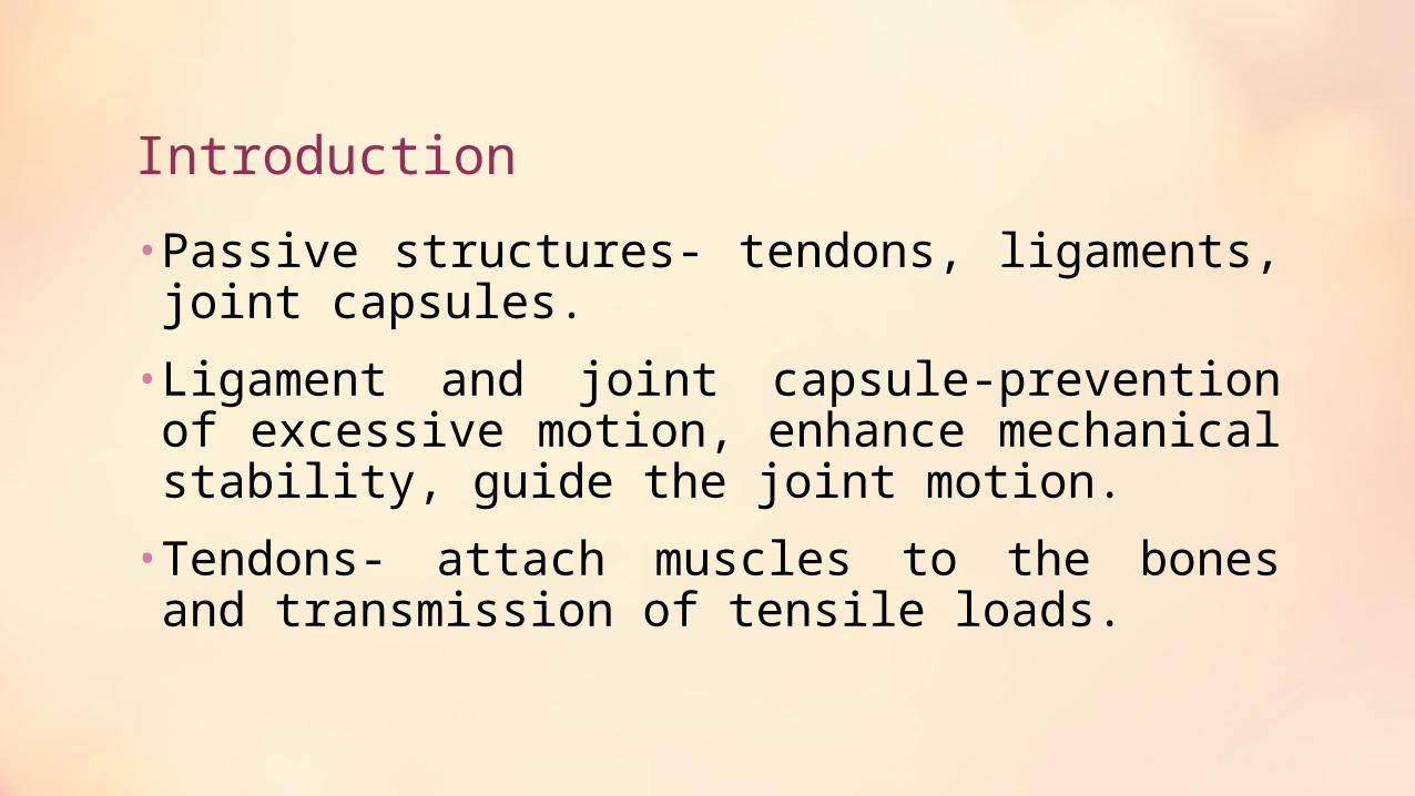
• Passive structures- tendons, ligaments, joint capsules.
• Ligament and joint capsule-prevention of excessive motion, enhance mechanical stability, guide the joint motion.
• Tendons- attach muscles to the bones and transmission of tensile loads.
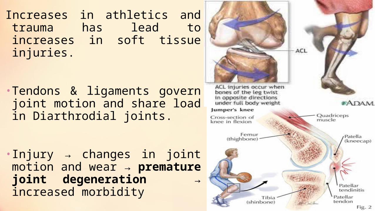
Increases in athletics and trauma has lead to increases in soft tissue injuries.
• Tendons & ligaments govern joint motion and share load in Diarthrodial joints.
• Injury → changes in joint motion and wear → premature joint degeneration → increased morbidity
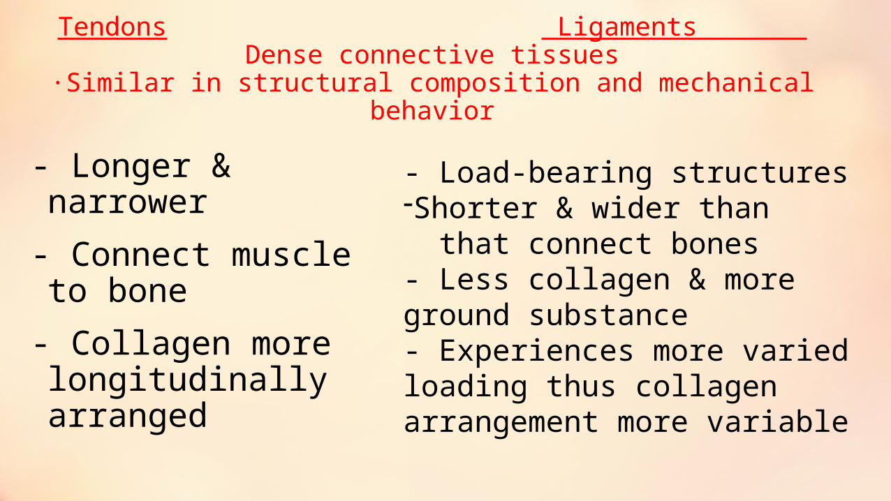
Tendons Ligaments Dense connective tissues
·Similar in structural composition and mechanical behavior
- Longer & narrower
- Connect muscle to bone
- Collagen more longitudinally arranged
- Load-bearing structures-Shorter & wider than that connect bones- Less collagen & more ground substance- Experiences more varied loading thus collagen arrangement more variable
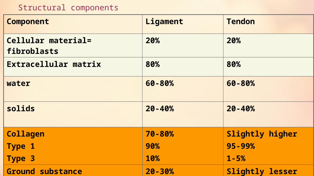
Structural components
Component Ligament Tendon
Cellular material= fibroblasts 20% 20%
Extracellular matrix 80% 80%
water 60-80% 60-80%
solids 20-40% 20-40%
Collagen Type 1Type 3
70-80%90%10%
Slightly higher95-99%1-5%
Ground substance 20-30% Slightly lesser
Anatomy
• Type I collagen is the predominant constituent of tendon and ligaments (86% fat-free dry weight)
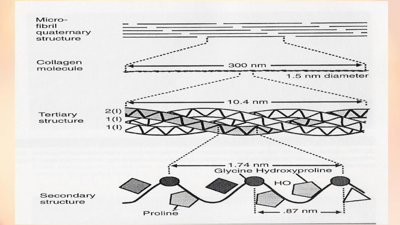
• structure: each collagen molecule consist of 3 polypeptide α chains combined making a collagen molecule, each coiled in helix with approximately 100 amino acids.Type I: two (peptide chain) α1….. and one α2 chains
slight different, form right-handed triple helix held together by hydrogen and covalent bonds.
Length=280nm diameter=1.5nm
Anatomy2/3 of collagen molecule consist of 3 amino acids, glycine,
33%, proline 15% & 15%hydroxyproline
• every third amino acid in each chain is glycine, and repetitive sequence essential for triple helix.
• Small size of glycine allows tight helical packing of collagen molecule, & enhances the stability of molecule by forming hydrogen bonds among three chains. Hydroxyproline and proline also form hydrogen bond.
• The intra- and interchain bonding or cross-linking. between specific groups on the chains is essential for the stability & strength of the molecule.
Factors effect the stability & strength• In newly developed collagen the crosslinkage are few..
• the collagen are soluble in natural salt and in acid solution,
• cross linkage easily damaged with high heat, as well aging reduce number of cross links decrease stability and strength.
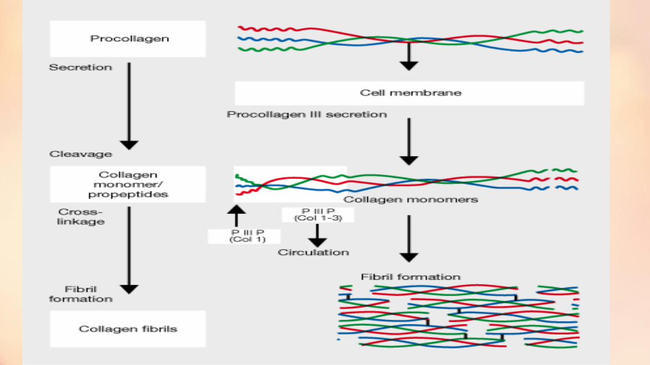
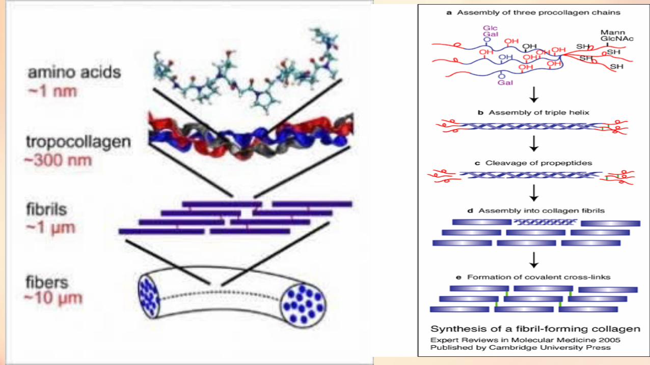
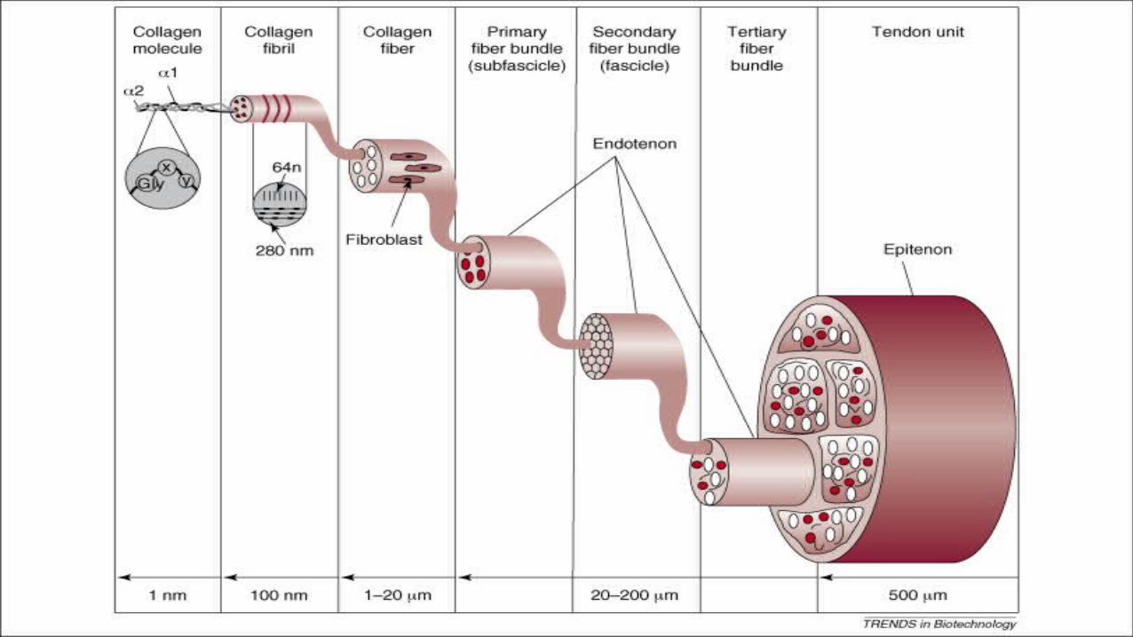
• overlapping and Organization of collagen molecules into a stable low-energy biologic unit →microfibril
• The arrangement of adjacent molecules aligns oppositely-charged acidic and basic Amino Acids makes a very stable construct that takes a lot of energy to separate its molecules
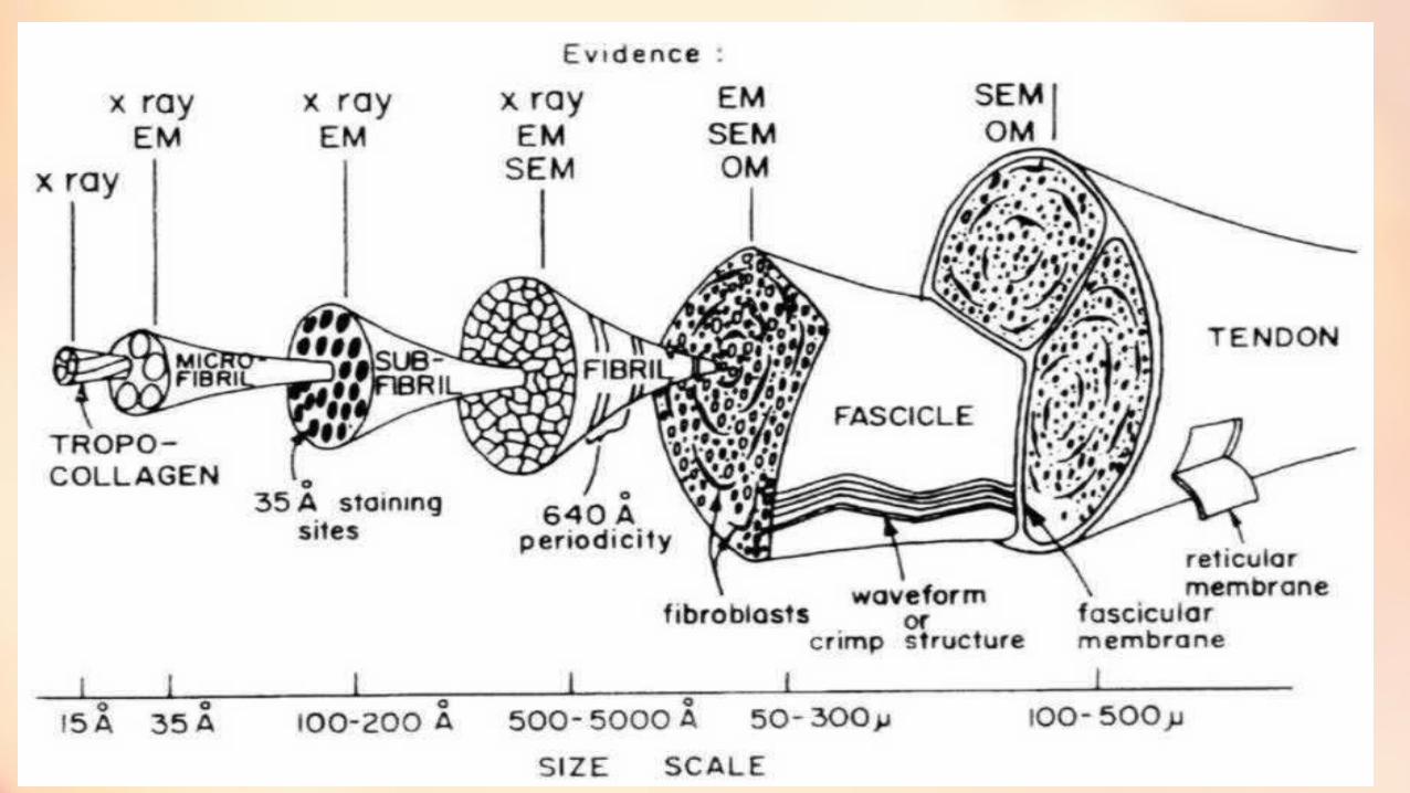
• Microfibril: 5 collagen molecules --Subfibril---- Fibril-- collagen fiber(1-20um in diameter)---
• Fascicle:….Closely-packed, longitudinal, parallel bundles of fibrils bound together by proteoglycans and glycoproteins in association with water incorporated in a matrix. Fibroblasts are aligned in rows between these bundles and are elongated along an axis in the direction of ligament or tendon function
• Tendon: Fascicles held together by loose connective tissue, the endotenon. Endotenon: Allows longitudinal fascicle movement, and
supports blood vessels, lymphatics and nervesTendon itself surrounded by epitenon
• The arrangement of collagen in tendons and ligaments varies as in accordance with the function.
• Parallel arrangement in tendons…. Uniaxial or Unidirectional tensile loads.
• In ligaments not all in parallel direction and suited according to the function.
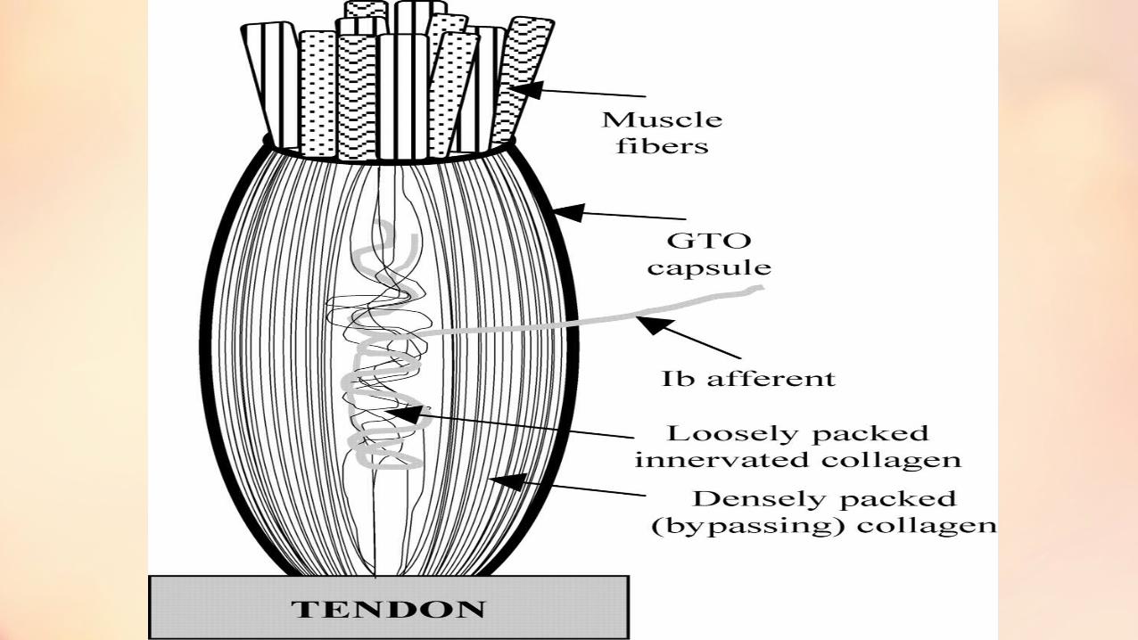
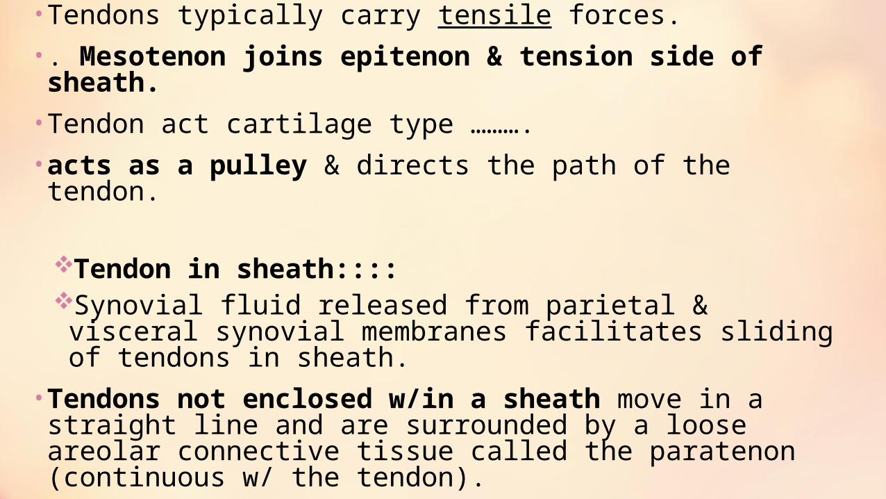
• Tendons typically carry tensile forces.• . Mesotenon joins epitenon & tension side of sheath.• Tendon act cartilage type ……….• acts as a pulley & directs the path of the tendon.
Tendon in sheath::::Synovial fluid released from parietal & visceral synovial
membranes facilitates sliding of tendons in sheath.• Tendons not enclosed w/in a sheath move in a straight line
and are surrounded by a loose areolar connective tissue called the paratenon (continuous w/ the tendon).
Elastin
•Mechanical properties…..
• present less in tendons and more ligaments.
• Ligamentum flavum: protection of spinal nerve roots, intrinsic stability to the spine, 2:1 ratio of Elastin to collagen.
•Ground substance::
• PGs, Same structures as in AC.
• Forms gel like material and binds ligaments or tendons.
• Cement like properties provide structural stability.
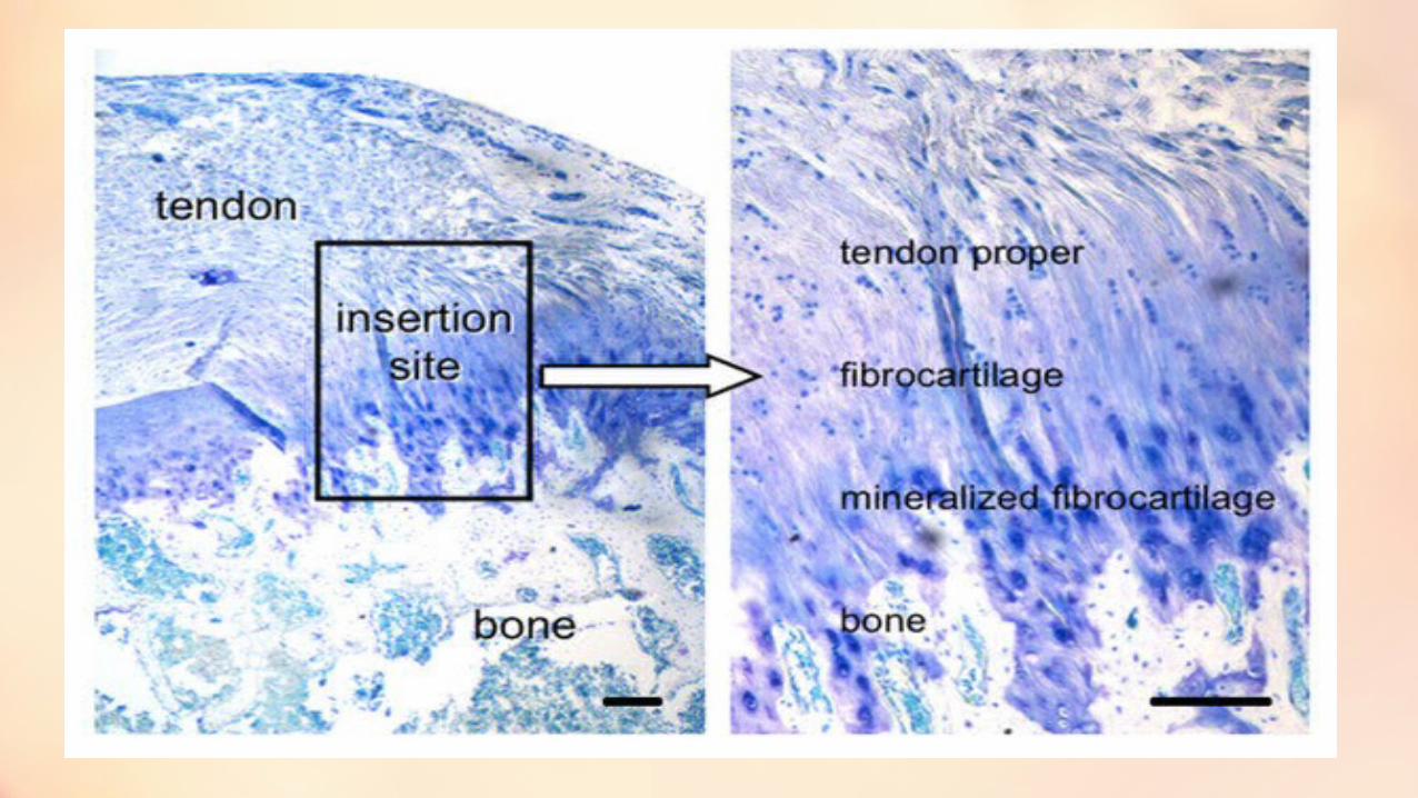
Insertion into Bone• At the tendo-osseus junction the collagen fibers of the
endotenon continue into the bone as Sharpey’s Fibers
• Then becomes continuous with the periosteum
• Zone I: end of tendon
• Zone II: collagen fibers intermesh with Fibrocartilage
• Zone III: fibrocartilage then gradually becomes mineralized fibrocartilage
• Zone IV: merge into cortical bone
Mechanical Behaviour of Tendon and ligaments
• Both are Viscoelastic tissues
• Visco - a small amount of load results in quite a bit of deformation (relative)
• Elastic - refers to Elastic Region
• Importance of Viscoelastic qualities?
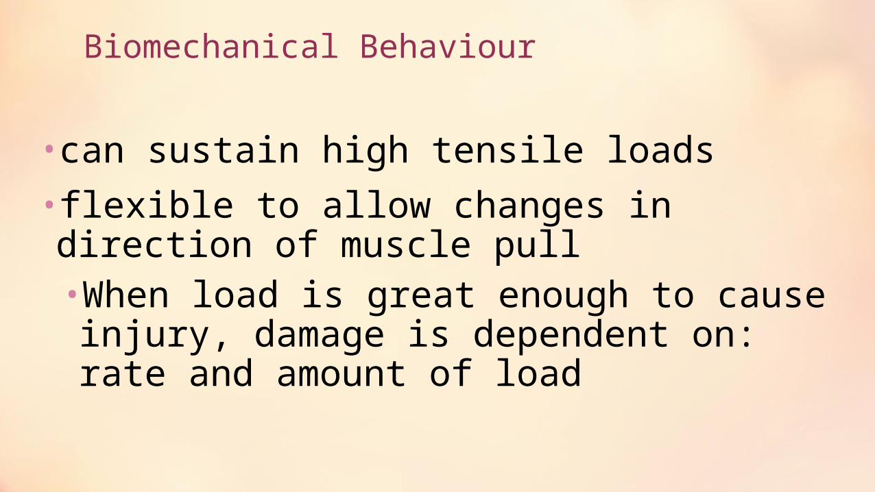
Biomechanical Behaviour
• can sustain high tensile loads• flexible to allow changes in direction of muscle pull•When load is great enough to cause injury,
damage is dependent on: rate and amount of load
Physiological loading of tendonsand ligaments
•Stress relaxation
•Creep
•Curves for creep and stress relaxation
•Load Deformation curve variation(stress strain curve)
•Hysteresis
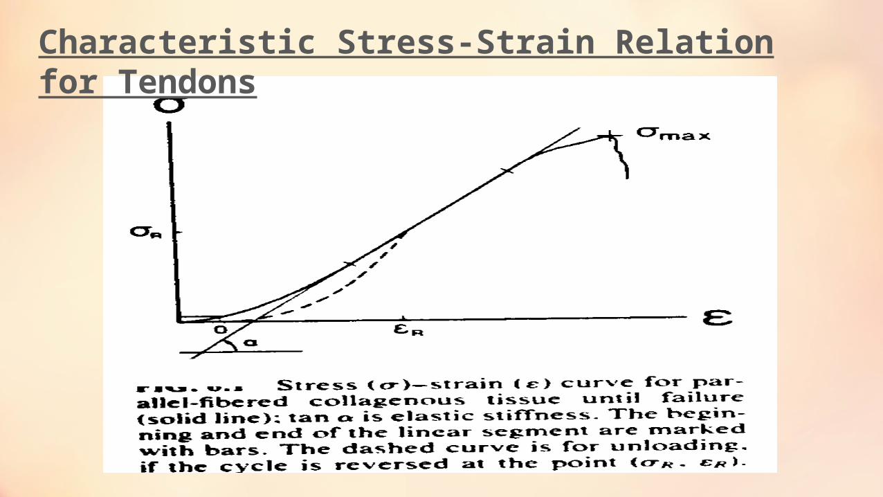
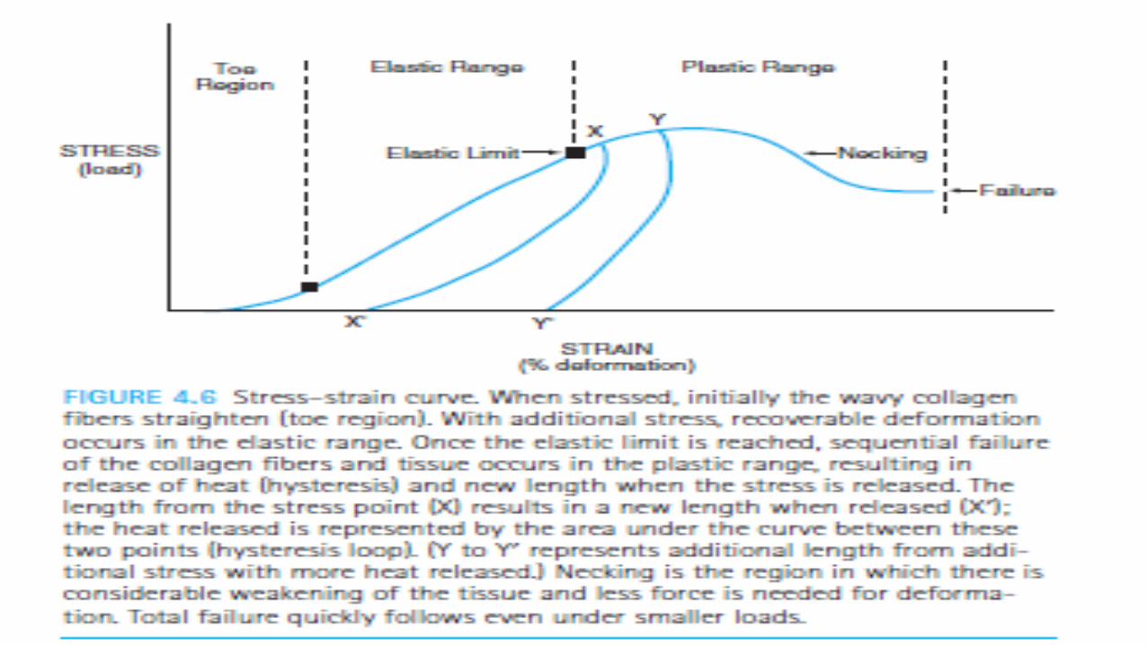
Characteristic Stress-Strain Relation for Tendons
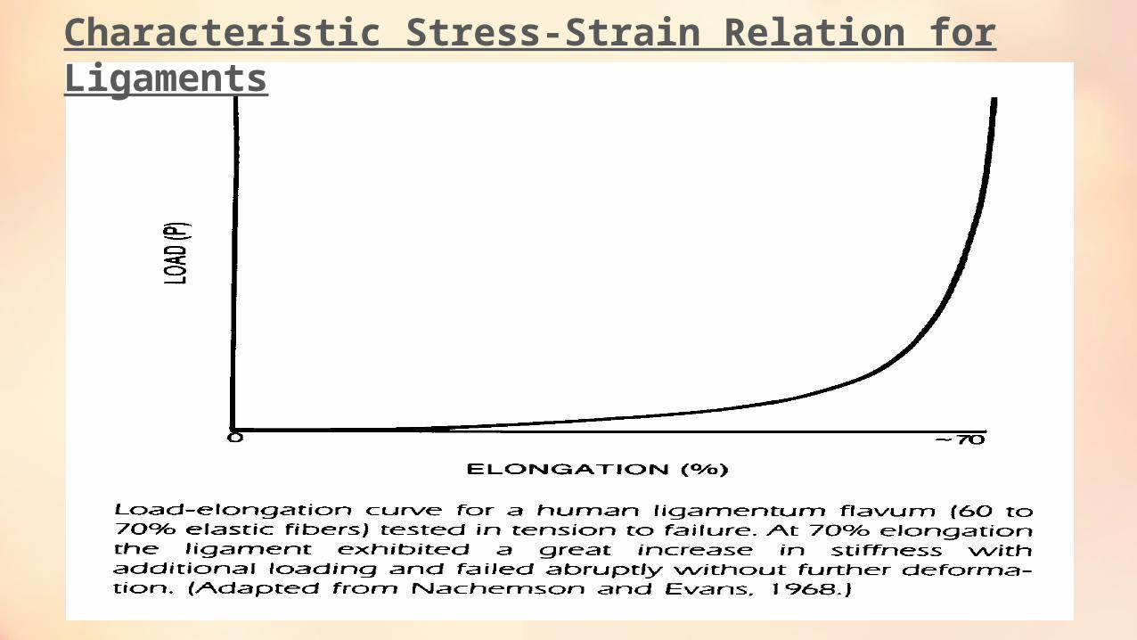
Characteristic Stress-Strain Relation for Ligaments
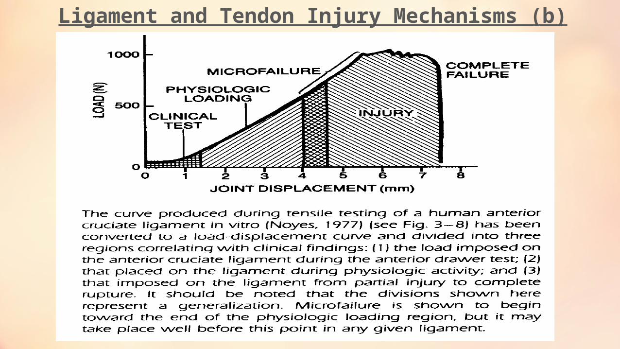
Ligament and Tendon Injury Mechanisms (b)
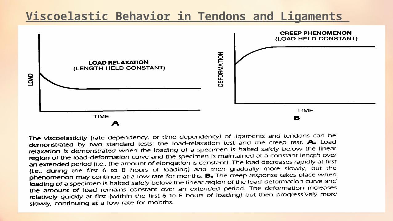
Viscoelastic Behavior in Tendons and Ligaments
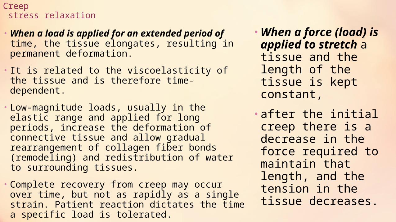
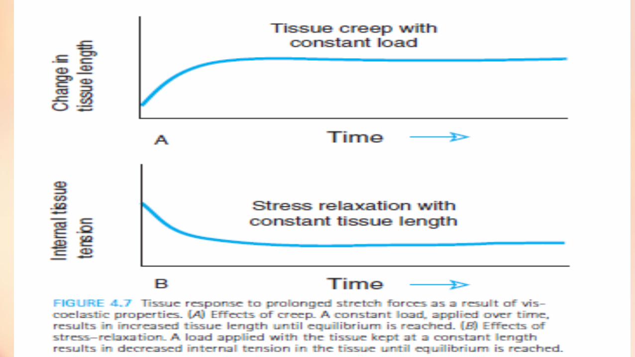
Creep stress relaxation• When a load is applied for an extended period of time,
the tissue elongates, resulting in permanent deformation.
• It is related to the viscoelasticity of the tissue and is therefore time-dependent.
• Low-magnitude loads, usually in the elastic range and applied for long periods, increase the deformation of connective tissue and allow gradual rearrangement of collagen fiber bonds (remodeling) and redistribution of water to surrounding tissues.
• Complete recovery from creep may occur over time, but not as rapidly as a single strain. Patient reaction dictates the time a specific load is tolerated.
• When a force (load) is applied to stretch a tissue and the length of the tissue is kept constant,
• after the initial creep there is a decrease in the force required to maintain that length, and the tension in the tissue decreases.

Tendon Vascularisation• Limited vascularisation...healing process & metabolic activity
• Tendons blood supply from vessels in perimysium, periosteal insertion, surrounding tissues vesseles in paratenon and mesotenon
• Tendons with in paratenon sheath(vascular tendon) Vessels enter from periphery and anastamose w/ longitudinal system of capillaries
• Tendons covered by tendon sheath as avascular tendon:
• Blood from Mesotenons vincula, & osseus insertions……avascularity in tendons adopt
• Dual pathway for nutrition : vascular and synovial, i.e. diffusion
• Diffusion Clinically important…--tendon healing
Ligamentous Vascularisation
• hypovascular compared to surrounding structures
•Uniform microvascularity originating from insertion sitesFlow is limited but essential to maintenance of cell
population, matrix synthesis & repair.
• Variety of specialized nerve endings & mechanoreceptors( proprioception & nociception--- joint function.
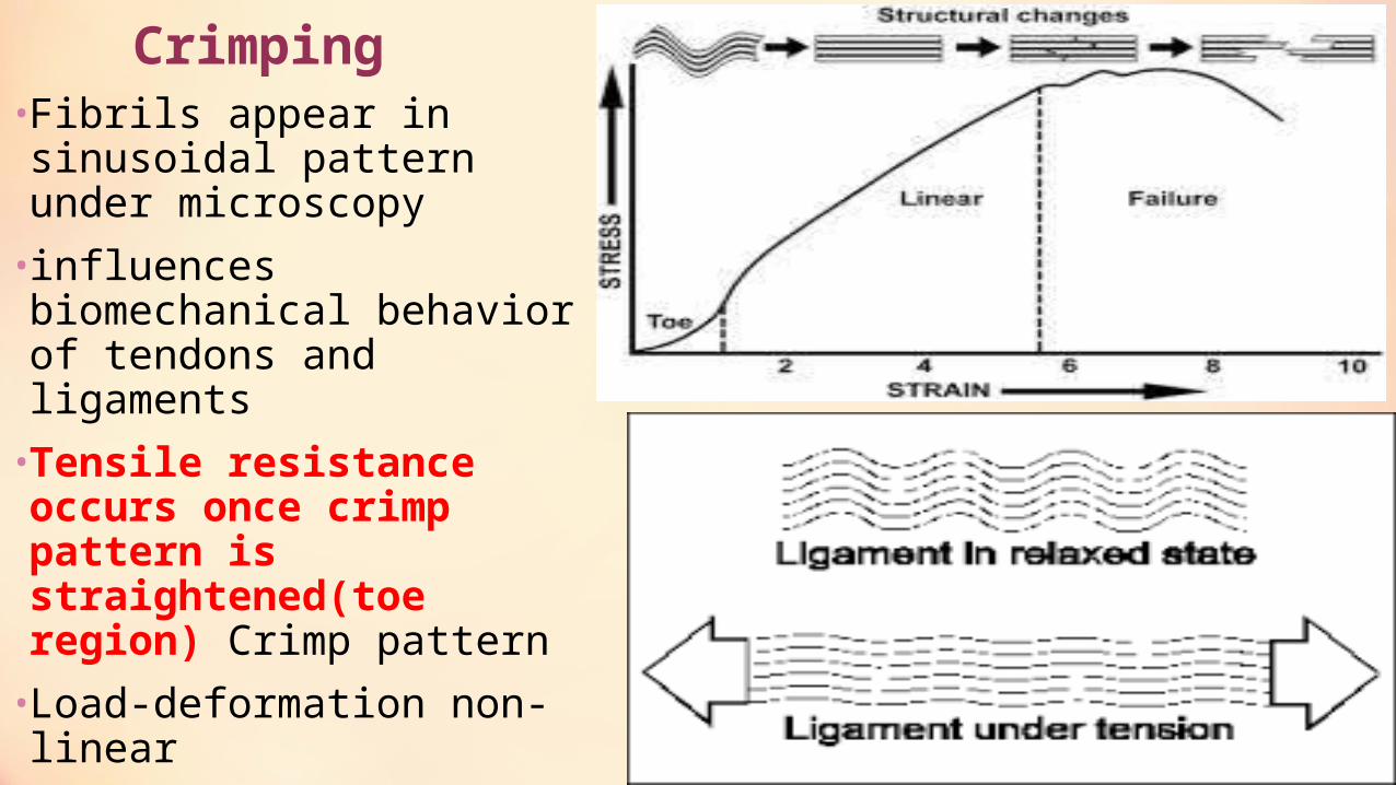
Crimping• Fibrils appear in
sinusoidal pattern under microscopy
• influences biomechanical behavior of tendons and ligaments
• Tensile resistance occurs once crimp pattern is straightened(toe region) Crimp pattern
• Load-deformation non-linear
Ligament Failure and Tendon Injury Mechanism
• Grade I injury:
• Negligible clinical symptoms… pain & swelling
• no joint instability
• Micro failure of the collagen fibers
Grade II injury:
• severe pain and some joint instability
• Progressive failure of the collagen fibers -- partial ligament rupture.
• The strength and stiffness of the ligament may have decreased by 50% or more, …………..
• The clinical test for joint stability is usually performed with the patient under anesthesia or without anesthesia
Grade III• Severe pain trauma time
• less pain after trauma time, the joint is found to be completely unstable.
• Most collagen fibers have ruptured, but a few still be intact, giving the ligament the appearance of continuity even though it is unable to Support any loads.
• Ligament or joint capsule rupture produces abnormally high stresses on the articular cartilage.
• early osteoarthritis in humans and in animals.
• Two additional factors become important in tendons because of their attachment to muscles:
1. The amount of force produced by contraction of the muscle to which the tendon is attached …A tendon is subjected to increasing stress as its muscle contracts
2. Cross-sectional area of the tendon in relation to that of its muscle.
• Tensile strength or health of tendon is more than muscles
• Muscle ruptures are more common than are ruptures through a tendon. (Muscle size and tendon size correlation.)
Factors That Affect the Biomechal1ical Properties of Tendons and Ligaments
• Maturation and aging
• Pregnancy and the postpartum period
• Mobilization and immobilization
• Diabetes mellitus
• Steroids
• Nonsteroidal anti-inflammatory drugs
• Hemodialysis
• Grafts