
第一 · 节 心脏的生物电现象及节律性 兴奋的产生与传导 ...
58
第一 · 第 第第第第第第第第第第第第 第第第第第第 第 传 第第第第第第第第第:第第第 第第 第第第 第第第 传 第第第第第第第: 第第第第 第第第第第 第 第第第第第第 第第第第第 ():, 第第第第 第第 第第第 第第第 第 第 第第第 第第第第第 ( ):(P), 传统 第第第第第第第 : 第第第 第第 第第第 第第第 传 第第第第 第 第 第 第 第第第第 第
-
Upload
ginger-olsen -
Category
Documents
-
view
653 -
download
3
description
第一 · 节 心脏的生物电现象及节律性 兴奋的产生与传导 心肌组织的生理特性: 兴奋性 传导性 自律性 收缩性 心肌细胞的分类: 工作细胞(普通心肌):心房肌细胞,心室肌细胞 自律细胞(特殊传导系统):起搏(P)细胞,浦肯野细胞. 二种细胞的比较 : 兴奋性 传导性 自律性 收缩性 工作细胞 有 有 无 有 - PowerPoint PPT Presentation
Transcript of 第一 · 节 心脏的生物电现象及节律性 兴奋的产生与传导 ...

第一 · 节 心脏的生物电现象及节律性兴奋的产生与传导
心肌组织的生理特性:兴奋性 传导性 自律性 收缩性 心肌细胞的分类: 工作细胞(普通心肌):心房肌细胞,心室肌细胞
自律细胞(特殊传导系统):起搏(P)细胞,浦肯野细胞
二种细胞的比较 :
兴奋性 传导性 自律性 收缩性
工作细胞 有 有 无 有
自律细胞 有 有 有 无

Electrical Activity of the Heart
OBJECTIVES
• Explain the types of cardiac action potentials.
• Define the ionic basis of cardiac action potentials.
• Explain the temporal changes in cardiac excitability.
• 心肌细胞的快反应动作电位和慢反应动作电位;• 心肌细胞的电生理特性(传导性和兴奋性)

Willem Einthoven
威廉 . 艾因特霍芬 ( Willem Einthoven ) 荷兰生理学家 威廉 . 艾因特霍芬 ( Willem Einthoven, 1860~1927 ) 因对心电图学 的 开创性工作和 无与伦比 的贡献 而 被誉为“心电图之父”,并于 1924 年获 诺贝尔生理学或医学奖。

Cardiac Transmembrane Potentials are Prolonged
The action potentials differ substantially among various type of cardiac cells depending on the function and location of those cells.
The electrical behavior of cardiac cells differs considerably from that of nerve cells or of smooth or skeletal muscle ceils. in general, the durations of action potentials are much longer in cardiac cells than in nerve cells or in smooth or skeletal muscle cells.

Action potential: Phase 0 : the rapid upstroke of the action potential. depolarizes from -90mV to +20 ~ 30 mV; Phase 1 : a brief period of partial repolarization +20 mV 0 mV,
Phase 2 : a plateau ( persists for about 0.2 second).
the potential charge recorded from ventricular muscle cell
Resting potential : - 90 mV (interior is lower than that of the surrounding medium)
It is main difference comparing with nerve and skeleton muscle cells.

Phase 3 : repolarization This proceeds more slowly than
dose depolarization (phase 0).
Phase 4 : The interval from the completion of repolarization
until the beginning of the next action potential.

Action potential occurs well before the contractile force
attains its peak, and repolarization is completed well befor
e the cell reaches its full resting value. The relaxation of th
e cardiac muscle takes place mainly during phase 4 of the
action potential.

• Fast-response action potentials: occur in atrial and ventricular myocardiac fibers and specialized conducting fibers (Purkinje fibers) that exist mainly in the endocardiac surfaces of the ventricles.
• Slow-response action potentials: occur in the sinoatrial (SA) node (SA), which is the natural pacemaker region of the heart;
and the atrioventricular (AV) node, which is the specialized tissue that conducts the cardiac impulse from the atria to the ventricles.
There Are Two Principal Types of Cardiac Action Potentials

Comparing with the fast-response action potential: The resting membrane of slower-response is considerable less negative, about -50 mV; The slop of the upstroke (phase 0), the amplitude and overshoot of the action potentials are less; ( amplitude of action potential and the rate of rise of the upstroke are important determinants of the conduction velocity).

Fast responses may change to slow responses under
certain pathological conditions, for example, in coronary
artery disease

The Cardiac Transmembrane Potential Dependends Mainly on K+, Na+, and Ca++
Each phase of the action potential is associated with a change in conductance to one or more ions.
The Resting Potential Is Determined by Ionic Diffusion

After raising [K]o, the measured value of Vm approximates that predicated by the Nernst equeation for Km (equilibrium potential). The measured values are slightly less negative than those predicted by Nernst equation because of the small but finite gNa

The Fast Response Depends on Na+
Phase 0 is genesis of the upstroke
Any stimulus that abruptly changes the resting membrane potential to a critical value (called the threshold 阈值 ) results in an action potential.
The rapid depolarization (phase 0) is related almost exclusively to the influx of Na+ into the myocyte due to a sudden increase in gNa.
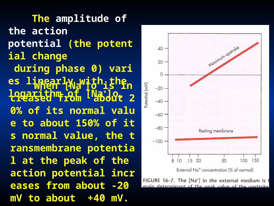
When [Na+]o is increased from about 20% of its normal value to about 150% of its normal value, the transmembrane potential at the peak of the action potential increases from about -20 mV to about +40 mV.
The amplitude of the action potential (the potential change during phase 0) varies linearly with the logarithm of [Na+]o,
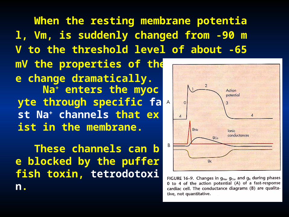
When the resting membrane potential, Vm, is suddenly changed from -90 mV to the threshold level of about -65 mV the properties of the cell membrane change dramatically.
Na+ enters the myocyte through specific fast Na+ channels that exist in the membrane.
These channels can be blocked by the puffer fish toxin, tetrodotoxin.
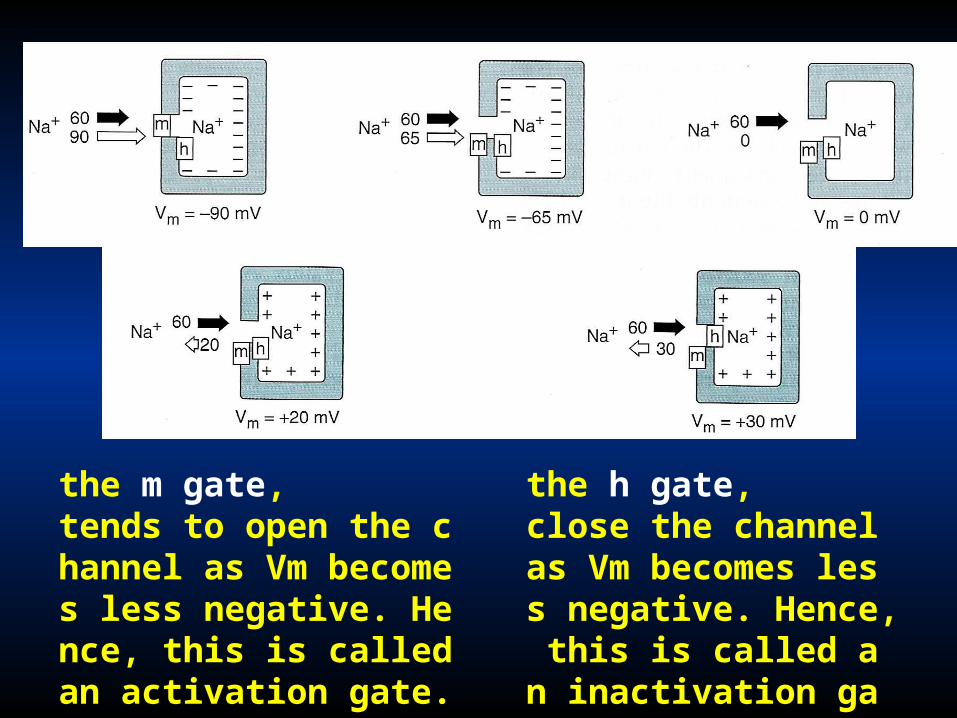
the m gate, tends to open the channel as Vm becomes less negative. Hence, this is called an activation gate.
the h gate, close the channel as Vm becomes less negative. Hence, this is called an inactivation gate.

The consequent change in Vm opens more m gates and augments the inward Na+ current. As Vm approaches about -65 mV, the remaining m gates rapidly swing open in the fast Na+ channels until virtually all the m gates are open
Regenerative
The h gates then remain closed until the cell has partially repolarized during phase 3, in this time the cell is in its effective refractory period, and it will not respond to further excitation.
Effective refractory period
Na+ 通道有三种状态,都是电压依赖性的: 备用 (-90 mV) 激活 失活
Recovered from inactivation

Phase 1 is genesis of early repolarization
• When a notch is not evidence, phase 1 reflects the inactivation of the fast Na+ channels.
Two mechanisms are responsible for phase 1:
• In cells characterized by a notch, phase 1 reflects not only the inactivation of Na+ channels, but also activation of a specific K+ current (transient outward current [Ito].

Roderick MacKinnon
罗德里克 . 麦金农
Rockefeller
University,
Howard Hughes
Medical Institute
New York, NY, USA


For inactivation to occur, a positively charged inactivation particle (ball) has to pass through one of the lateral windows and bind in the hydrophobic binding pocket of the pore’s central cavity. Kv1.4, 2001

• Plateau remains fairly constant for about 100 to 300 ms.
Phase 2 is genesis of the plateau
• The relevant Ca2+ channels is L-type Ca2+ channels. A substantial amount of Ca2+ enter the cardiac cells throughout the plateau as three reasons: gCa2+, Ca 2+ concentration, positive potential.
• The principal movement of cations across the cell membrane during phase 2 are a net efflux of K+ and a net influx of Ca 2+ .

• Various medications and neuro-transmetters may influence the Ca2+ current in cardiac cells.
• Epinephrine strengthen myocardial contraction by increasing Ca2+ influx.
• Ca2+ channels antagonist diminish the level of Vm during the plateau and abridge the duration of plateau by altering the balance between Ca2+ influx and K+ efflux.
地尔硫唑

Phase 3 is genesis of final repolarization
• when the efflux of K+ front the cardiac cell begins to exceed the influx of Ca2+ final repolarization stars.
• At least three outward K+ currents (ito, ik, and ik1) contribute to the final repolarization (phase 3) of the cardiac cell.
• fast-response action potentials consist of four components: an upstroke (phase 0); an early, partial repolarization (phase 1); a plateau (phase 2); and a final repolarization (phase 3).
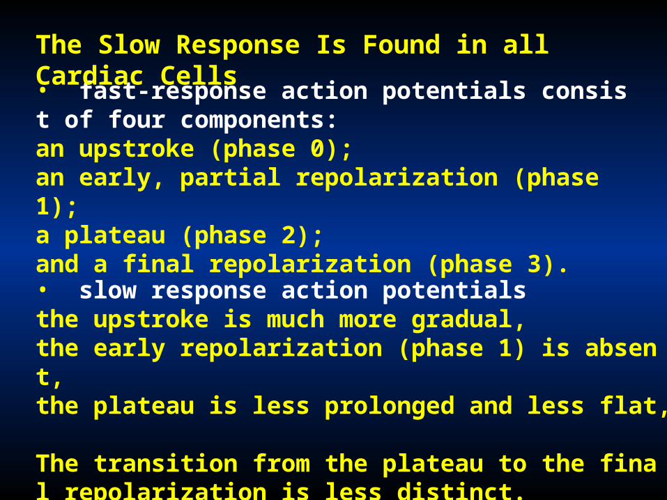
The Slow Response Is Found in all Cardiac Cells
• slow response action potentials the upstroke is much more gradual, the early repolarization (phase 1) is absent,the plateau is less prolonged and less flat, The transition from the plateau to the final repolarization is less distinct.
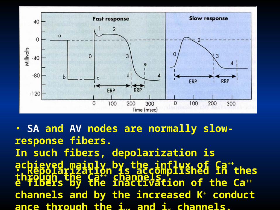
• SA and AV nodes are normally slow-response fibers.In such fibers, depolarization is achieved mainly by the influx of Ca++ through the Ca++ channels.
• Repolarization is accomplished in these fibers by the inactivation of the Ca++ channels and by the increased K+ conductance through the iK1 and iK channels.

The Fast Response Underlies Rapid Conduction of the Cardiac Impulse
The conduction velocity along the fiber varies directly with the amplitude of the action potential and with the rate of change of potential (dVm/dt) during phase 0.
important factor which determine the conduction velocity.
• The level of the resting membrane potential : This factor influences the amplitude of the action potential and the slope of the upstroke.
• the rate of change of potential (dVm/dt) during phase 0, • the amplitude of the action potential,
The Na+ Current Determines Conduction of the Fast Response

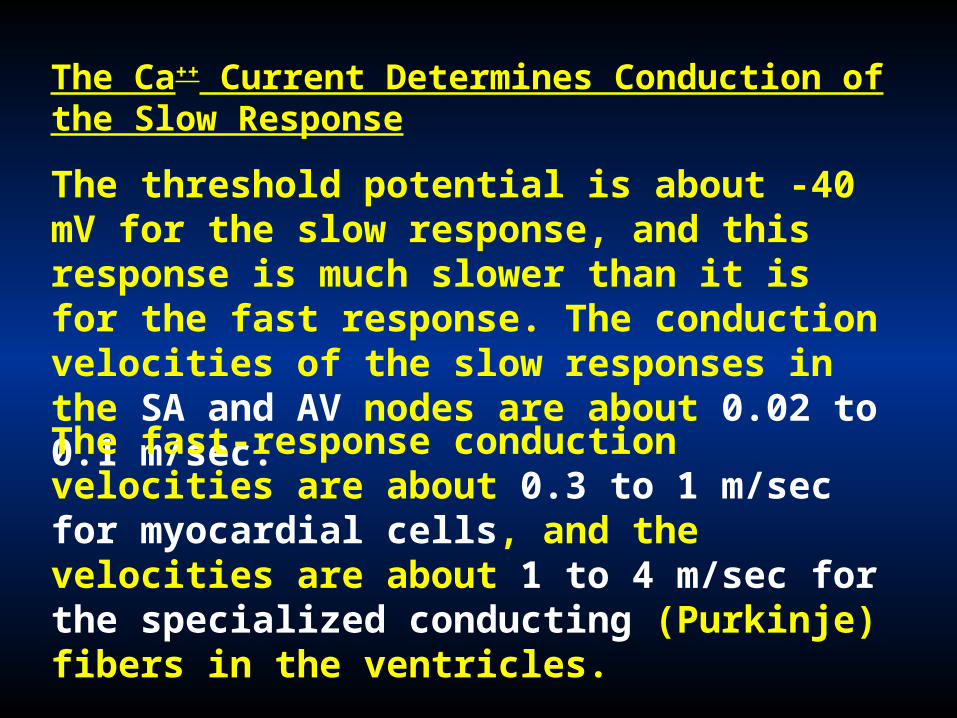
The Ca++ Current Determines Conduction of the Slow Response
The threshold potential is about -40 mV for the slow response, and this response is much slower than it is for the fast response. The conduction velocities of the slow responses in the SA and AV nodes are about 0.02 to 0.1 m/sec.
The fast-response conduction velocities are about 0.3 to 1 m/sec for myocardial cells, and the velocities are about 1 to 4 m/sec for the specialized conducting (Purkinje) fibers in the ventricles.
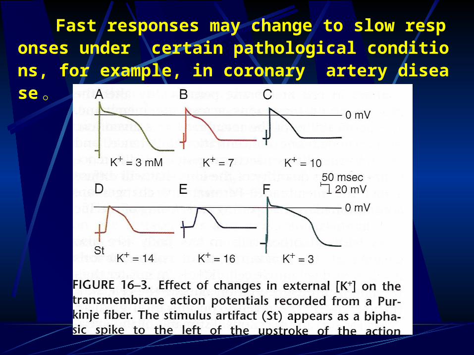
Fast responses may change to slow responses under certain pathological conditions, for example, in coronary artery disease 。

Cardiac Excitability Is Determined by the Availability of Na+ and Ca++ Currents
Effective refractory period: (有效不应期)
• from the beginning of phase 0 to the time in phase 3 at which Vm has reached about -50 mV (c to d).
Na+ channel activation and inactivation induced refractory period (不应期) :
The internal from the beginning of the action potential until the time the fiber can conduct another action potential.
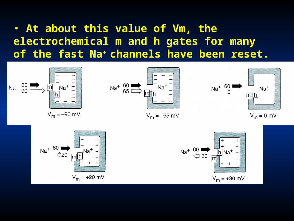
• At about this value of Vm, the electrochemical m and h gates for many of the fast Na+ channels have been reset.

图 4-11 心肌细胞动作电位与兴奋性的变化A. 在复极化的不同时期给予刺激所引起的反应
( a 为局部反应, b 、 c 和 d 为 0 期去极化速度和幅度都减小的动作电位)B. 用阈值变化曲线表示心肌细胞兴奋后兴奋性的变化
Relatively refractory period: during period (d to e), an action potential may be evoked but only when the stimulus is stronger than that which elicits a response during phase 4.
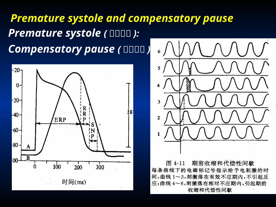
Premature systole and compensatory pause Premature systole ( 期前收缩 ):
Compensatory pause ( 代偿间隙 ):

In slow-response fibers, the relative refractory Period frequently extends well beyond phase 3 . Even after the cell has completely repolarized, it may be difficult to evoke a propagated response for some time.
Ca++ channel generates the slow response:
post-repolarization refractoriness:

Natural Excitation of the Heart
Objectives
• Discuss the basis of automaticity.
• Describe the spread of excitation of the heart.
• Describe the components of the electrocardiogram

Automaticity: the ability to initiate a heartbeat
Rhythmicity: the frequency and regularity of such
pacemaking activity are intrinsic to cardiac tissue
Natural pacemaker: the natural pacemaker cells are located in the SA node in the mammalian heart;
Ectopic pacemakers : Other regions of the heart that can
initiate beats under special circumstances are called
ectopic pacemakers.
Ectopic pacemakers may become dominant when (1) their own rhythmicity is enhanced, (2) The more rhythmic pacemakers are depressed, (3) all conduction pathways are blocked.

The Sinoatrial Node Is the Natural Pacemaker of the Heart
In adult humans, the SA node is about 8 mm long and 2 mm thick, and it lies posteriorly in the groove at the junction between the superior vena cava and the right atrium
• small, round cells , it are probably the pacenraker cells.
• slender, elongated cells, which are probably conduct the impulses to nodal margins
• transmembrane action potential of the pacemaker cell in the SA node are characteristic of the slow response.

• The principal distinguishing feature of an automatic fiber in the SA node (as in all automatic fibers) resides in phase 4.
• The automatic cell in the heart displays a gradual diastolic depolarization (pacemaker potential) during phase 4.
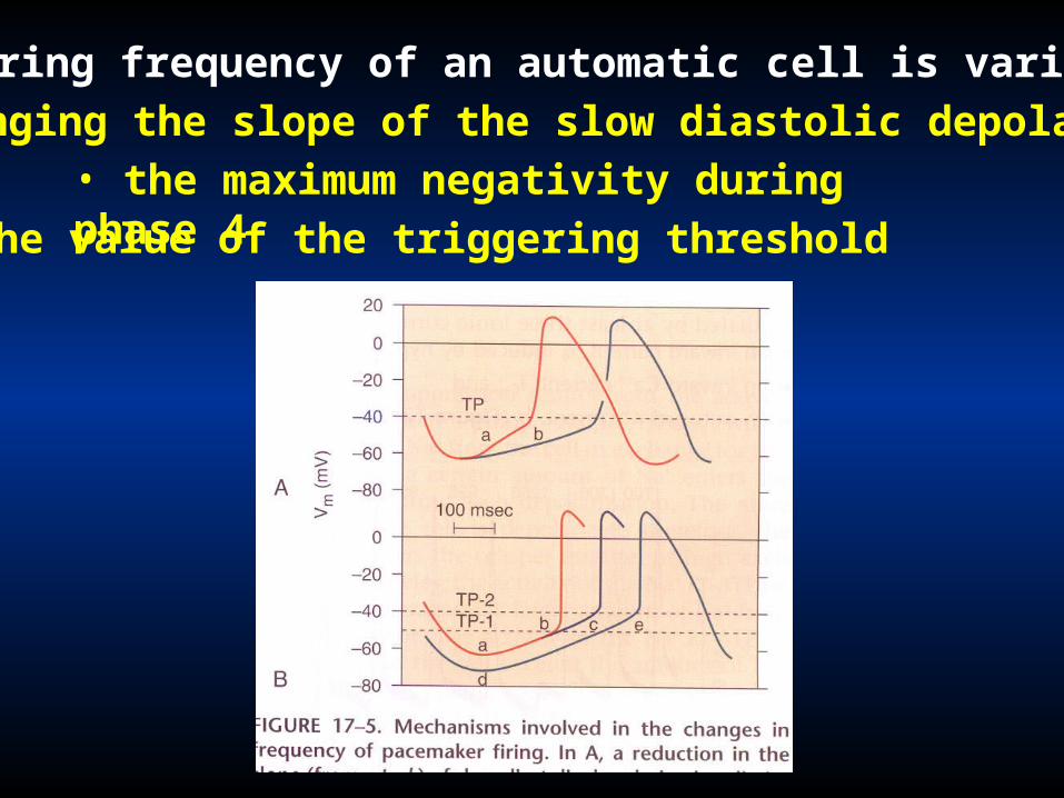
The firing frequency of an automatic cell is varied by• changing the slope of the slow diastolic depolarization• the maximum negativity during phase 4• the value of the triggering threshold

• Through the release of acetylcholine, increased parasympathetic activity diminishes the heart rate by reducing the slope and by increasing the maximum negativity of slow diastolic depolarization.
• Changes in the firing threshold occur in response to certain medications and to changes in the ionic composition of the myocardial interstitial fluid.

In the SA node, the slow diastolic depolarization is mediated by changes in at least three ionic currents:
fluxes of K+, Ca++ , and Na+ underlie automaticity
It is activated as the membrane potential becomes more negative than about -50 mV during repolarization
• an inward Ca++ current (iCa), • an inward funny" current (if),
• an outward K+ current (iK) .if is carried mainly by Na+.
The more negative the membrane potential, the greater of if .

iCa is activated toward the end of phase 4 (about -55 mV). The influx of Ca++ accelerates repolarization, which quickly leads to the upstroke of the action potential.
The efflux of K+ tends to oppose the depolarizing effects of if and iCa.
The outward current iK decays steadily throughout phase 4 because of its gradual inactivation. The diminishing of outward iK thus contributes to the slow diastolic depolarization.

• The ionic basis for automaticity in the AV node pacemaker cells resembles that in the SA node cells.
• For automaticity in Purkinje fibers, the Ca++ current does not contribute the slow diastolic depolarization .
• Hence the slow diastolic depolarization in Purkinje fibers is mediated by the balance between the if (x), and the gradually diminishing iK (y)•.

• The automaticity of pacemaker cells is suppressed temporarily after they have been driven at a critically high frequency. This phenomenon is known as overdrive suppression.
Overdrive suppresses automaticity
• The mechanism responsible for overdrive suppression involves the membrane pump (Na+-K+-ATPase) . This enhanced activity of the Na+ pump hyperpolarizes the cell membrane because there is a net loss of cation from the cell interior. (sick sinus syndrome )
The properties of automaticity in difference cardiac tissue
SA node > AV node > Purkinje fibers
90~100 次 / 分 40~60 次 / 分 15~40 次 / 分
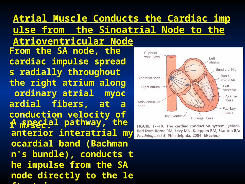
Atrial Muscle Conducts the Cardiac impulse from the Sinoatrial Node to the Atrioventricular Node
From the SA node, the cardiac impulse spreads radially throughout the right atrium along ordinary atrial myocardial fibers, at a conduction velocity of 1 m/sec.
A special pathway, the anterior interatrial myocardial band (Bachmann's bundle), conducts the impulse from the SA node directly to the left atrium.
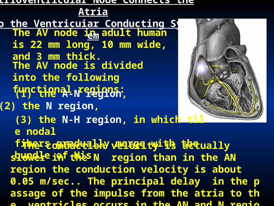
The AV node in adult human is 22 mm long, 10 mm wide, and 3 mm thick.
Atrioventricuiar Node Connects the Atriato the Ventricuiar Conducting System
The AV node is divided into the following functional regions:
(1) the A-N region, (2) the N region,
(3) the N-H region, in which tile nodal fibers gradually merge with the bundle of His 。
The conduction velocity is actually slower in the N region than in the AN region the conduction velocity is about 0.05 m/sec.. The principal delay in the passage of the impulse from the atria to the ventricles occurs in the AN and N regions of the AV node.

An abnormal prolongation of the AV conduction time is called first-degree AV block.

• The left bundle branch is thicker than the right. It arises almost perpendicularly from the bundle of His and perforates the interventricular septum.
Ventricular Conduction is Rapid
• The bundle of His is the beginning of the specialized conduction system for the ventricles .
• The right bundle branch, a direct continuation of the bundle of His, proceeds down the right side of the interventricular septum.

The bundle branches ultimately subdivide into a complex network of conducting fibers, called Purkinje fibers, which ramify over the subendocardial surfaces of both ventricles .
Purkinje fibers are tile broadest
cells in the heart, 70 to 80 um in
diameter. conduction velocity
are 1 to 4 m/sec.

(5) the influence of certain medications on the heart.
Electrocardiography Is an Important Clinical Tool
The electrocardiogram enables to infer the course of the cardiac impulse by recording the variations in electrical potential at various loci on the surface of the body.
By analyzing the details of these fluctuations, the physician gains valuable insight concerning:
(1) the anatomical orientation of the heart and the relative sizes of its chambers;
(2) various disturbances of rhythm and conduction;
(3) the extent, location, and progress of ischemic injury to the myocardium;
(4) the effects of altered electrolyte concentrations;

The cardiac impulse progresses through the heart in a complex three-dimensional pattern.
a lead is the electrical connection from the patient's skin to the recording device (an lectrocardiograph).
The electrocardiogram reflects the temporal changes in the electrical potential between pairs of points on the skin surface.


The configuration and amplitude of the QRS complex vary considerably among individuals. The duration is usually between 0.06 and 0.10 second.
The T wave reflects the repolarization of the ventricular myocardial cells. The T wave is usually deflected in the same direction from the isoelectric line as is the major component of the QRS complex.
P wave reflects the depolarization of the atrial myocardial cells.

The PR interval is the time from the beginning of atrial activation to the beginning ofventricular activation; it normally ranges from 0.12 to 0.20 second. Most of this conduction time involves thepassage of the impulse through the AV conduction system.
During the ST interval, the entire ventricular myocacdium is depolarized. Because all of the myocardial cells are at about the same electrical potential, the ST segment lies on the isoelectric line (which is the line that reflects that virtually all regions of the cardiac surface are at the same electrical potential).

The QT interval is sometimes referred to as the period of electrical systole of the ventricles; it reflects the action potential duration of the ventricular myocardial ceils. The duration of the QT interval is about 0.4 second, but it varies inversely with the heart rate, mainly because the action potential duration varies inversely with the heart rate.


![傳統性權的批判與性權宣言的發展3 傳統性權的批判與性權宣言的發展 -----代發刊詞 吳敏倫[1] 转载自《華人性權研究》创刊号 在人權內有關性方面的權便是性權。](https://static.fdocuments.net/doc/165x107/5e3a1a5f5f6e8f149c6d9763/cceecc-3-cceecc.jpg)


