-25579 - NASA · transplanted into the anterior temporalis bed after partial excision of this...
12
194 -25579 '" Cellular and Molecular Biology of Muscle Development, pages 15-26 © 1989 Alan R. Liss, Inc. THREE HI ERARCHIES IN SKELETAL MUSCLE FIBRE CLASSIFICATION ALLOTYPE , ISOTYPE AND PHENOTYPE l Joseph F . Y. Hoh , Suz a nne Hu ghes , Gr eg o ry Hugh and Ire ne Po zgaj Department of Physiol o gy , University of Sydney NSW , 2 006 , Au stralia ABSTRACT Immu noc y tochemic al an a lyses using specific a nti - myosin antibodies of mammalian mu scle fi bres during regener a tion , development a nd after denervat ion ha ve revealed two distinct myogenic co mponents determining fibre phenotype . The jaw- closing mu scles of the cat c ontain su perfast fibres which expr e ss a unique myosin not fo u nd in limb mu scles . W hen su per - fa st muscle is transplanted into a limb mu scle bed , regenerating myo tubes synthesize su perfast m yosin independent of innerva t ion . Reinnervati on by the nerve to a fast mu scle le a ds to t he expression of superf a st a nd not fast myosin , while reinnervation by the nerve to a sl ow mu scle leads to the expression of a slow myosin. When limb mu scle is tr ans planted into t he jaw muscle bed , o nly limb myo sin s are sy nthe sized . Th us jaw and limb mu scles bel o ng to distin ct a llotypes , ea ch with a u nique range of pheno typic options , the expression of whi c h ma y be mo d u lated by the nerve . Primary and s econd a ry myotubes in devel o ping jaw a nd limb mu s cles a re o bserved to be l ong to different categories ch ar ac terized by different pa tterns of my osin gene expressi on. By t a k i ng int o considera tion the patt e rn o f myo sins synthesized and the c hanges in fibre size after denervati on , 3 type s of prima ry (f a st , slow and intermedi a te) fibres and t wo t ypes of secondary (fast and slow) fibres can be distingu i shed in rat fas t limb muscles . All primaries synth e size slow myosin soon aft er the i r f o rmati on , but this is IThis work was su ppor t ed by grants from the Mu sc u lar Dy s troph y As so cia tion of Americ a, the Na tion al Hea l t h and Medi c al Rese a rch Counc il of Aus t ralia and the Muscul ar Dystroph y Associati o n o f New So u th Wales . Repriflled, wilh p crrnJ'ssion, from Ct!llula r and Molecular Biology of Muscle DevdopmU1J. Copyn'ghl e 1989 by Pergamolt Press, l nc. I Sec ti on I - Reprintsl https://ntrs.nasa.gov/search.jsp?R=19910016263 2018-09-16T22:05:54+00:00Z
Transcript of -25579 - NASA · transplanted into the anterior temporalis bed after partial excision of this...

194
-25579
'" Cellular and Molecular Biology of Muscle Development, pages 15-26 © 1989 Alan R. Liss, Inc.
THREE HI ERARCHIES IN SKELETAL MUSCLE FIBRE CLASSIFICATION ALLOTYPE , ISOTYPE AND PHENOTYPEl
Joseph F . Y. Hoh , Suza nne Hughes , Gr ego ry Hugh and I r e ne Pozgaj
Department of Physiolo gy , University of Sydney NSW , 2006 , Au stralia
ABSTRACT Immunocy tochemica l ana lyses using specific a nti- myosin antibodies o f mammalian muscle f i bres during regenera tion , development a nd after denervati o n h ave revealed two distinct myogenic c omponents determining fibre phenotype . The jaw- closing mu scles o f the cat contain s uperfast fibres which expre ss a unique myosin not fo und i n limb mu scles . When s uperf a st muscle is transplanted into a limb muscle bed , regenerating myo tubes synthesize s uperfast myosin independent of innervat ion . Reinnervation b y the nerve to a fast mu scle lea ds to t he expression of superfa st a nd no t fast myosin , while reinnervation by the nerve to a slow mu scle leads t o the expression of a slo w myosin. When limb muscle i s tra n s planted into t he jaw muscle bed , only limb myosins are s ynthe sized . Thus j aw and limb muscles belo ng to distinc t a llotypes , e a ch with a unique range o f pheno typic options , the expression of whic h ma y be mo du lated by the nerve . Primary and s econda ry myotubes in developing j aw a nd limb mus cles a re o bserved t o be l ong t o different categories cha r acterized by different p a tterns o f myosin gene expression . By t a k i ng into considerat i o n the patte rn o f myo sins synthesized and the c hanges in fibre size after denervati on , 3 type s of prima ry (fa st , slow and intermedia te) fibres and t wo t ypes of secondary (fast and slow) fibres can be distingui shed i n rat fas t limb muscles . All primaries synthe size slow myosin soo n a f t er thei r f o rmation , b u t this is
IThis work was s upport ed by grants from the Mu scu lar Dy s trophy As soc i a tion o f America , the Na tiona l Hea l t h and Medi cal Resea rch Cou n cil o f Aus t ralia and the Muscula r Dystrophy Associatio n o f New South Wales .
Repriflled, wilh p crrnJ'ssion, from Ct!llula r and Molecular Biology of Muscle D evdopmU1J. Copyn'ghl e 1989 by Pergamolt Press, lnc.
I
I Section I - Reprintsl
https://ntrs.nasa.gov/search.jsp?R=19910016263 2018-09-16T22:05:54+00:00Z

~'ection I - Reprints
16 Hoh et al.
withdrawn in fast and intermediate primaries at different times . After neonatal denervation , slow and intermediate primaries express slow myosin whereas fast primaries do not , and slow primaries hypertrophy while other fibres atrophy . In the mature rat, the number of slow fibres in the EDL is less than the number of slow primaries . Upon denervation, hypertrophic slow fibres matching the number and topographic distribution of slow primaries appear, s uggesting that a subpopulation of slow primaries acquire the fast phenotype during adult life , but reveal their original identity as slow primaries in response to denervation by hypertrophying and synthesizing slow myosin . It is proposed that within each muscle allotype, the various isotypes of primary and secondary fibres are myogenically determined , and are derived from different lineages of myoblasts .
INTRODUCTION
Fibres of limb and trunk muscles of mammals have been classified phenotypically into slow, fast- red and fastwhite t ypes , each ~ype containing a distinct type of myosin, and associated with a specific profile of metabolic enzymes . Consequently , these various types of fibres differ in intrinsic speed of contraction, power output and endurance. Such phenotypic diversity has been attributed to the myoregulatory function of the motor nerve supply. According to this hypothesis , muscle fibres are considered to be "plastic", and fibre types interconvertible according to the pattern of impulses received from the nerve (1, 2).
During myogenesis , myotubes are uniformly slow contracting and synthe s ize embryonic and foetal myosins before expressing adult fibre characteristics. I n view of the profound influence motor nerves have on mature muscle fibres, it has generally been considered, since the work of Buller, Eccles and Eccles (3 , 4) that the emergence of muscle fibre heterogeneity during myogenesis is also brought about by t he action of nerves on a common , undifferentiated myotube.
The experiments described in thi s paper were done to test the neural regulatory hypothesis for fibre type diversity . The jaw muscles of carnivores contain superfast muscle fibres which express a unique myosin not found in limb muscles. If the neural regulatory hypothesis is valid for these muscles , jaw muscles regenera ting in limb muscle
195
196
Myogenic Regulation of Fibre Types 17
beds should express limb muscle myosins and vice versa . During limb muscle development, mu scle fib res are polyneuronally innervated . Emerging myotubes would be expected to co- express a mixture of adult myosins. The results of these experiments obtained wi t h immunocytochemical techniques u sing monoclonal and polyclonal anti - myos i n heavy chain antibodies do not confirm these expectations . They reveal two hierarchically distinct levels of myogeni c influences affecting fibre phenotype . A hierarchical classification is proposed in which jaw and limb muscles belong t o different allotypes which define their phenotypic options. Within each allotype, myogenically distinct isotypes emerge during development.
RESULTS
Nerve Independent Intrinsic Differences Between Cat Jaw and Limb Muscles
Strips of posterior tempo ralis mu scle , a homogeneous s uperfast muscle , were treated with Ma rcaine t o destroy mature muscle fibres and transplanted into limb mu scle beds for r egeneration and reinnervation by the host nerve (5) . Early regenerates in the bed of eit her the fast extensor digitorum longu s (EDL) or the slow soleus muscle react with antibodi es against the heavy chain of foetal , slow or superfast myosins, but not with antibodies against fast myosin . In the long- term , regenerates innervated by the EDL nerve express only s uperfast myosin whereas in the regenerates innervated by the soleus nerve most fibres react on ly with the anti-slow myosin antibody , while some fibres react only against superfast myosin even afte r 213 days . In contrast , EDL and soleu s muscles regenera ting in their own beds express foetal , slow and fast myosins , but do not express superfast myosin . The isometric contraction times of the v a rious t ypes of regenerates reflect the types of myosin synthesized .
The ability of the regenerating superfast muscle to express the s uperfast myosin is independent of the nerve (6) . This i s shown in experiments in which reinnervation of the transplant in the EDL bed is prevented by cutting the common peroneal nerve and reflecting it back into the thigh . In these denervated beds the early temporalis regenerates are indistinguishable from innerva t ed regenerates in expres sing s uperfast myosin i n addition t o foetal and slow myosins .
Intrinsic d i fferences between jaw and limb muscle cells
Section I - Reprintsl

I ~Section I - Reprints
18 Hoh et aI .
have also been shown by the transplantation of limb muscles into the jaw muscle bed . Jaw muscle beds are less satisfactory from the point of view of defining the innervation of the regene rates . Limb mu scle strips were transplanted into the anterior temporalis bed after partial excision of this muscle . Fig . 1 shows the results of an EDL regenerated in the anterior temporalis muscle bed for 12 weeks . Staining for superfast (Fig . la) and foetal (Fig. lb ) myosins is negative , wher eas nearly all fibres stain for fas t myosin (Fig. Id), and some fibres also stain for slow myosin (Fig . lc ) . Although innervation of these fibres is not specifically demonstrated , their large size and the absence of staining for foetal myosin suggests that they are innervated .
It is concluded t hat jaw and limb mu scle cells are two dis"tinct types of mu scle cells , each having a distinct repertoire for the expression of adu lt isomyosins , and that the particular isomyosin expressed can be modu lated by the nerve.
Heterogeneity of P r i mary Fibres in Developing Limb Muscles
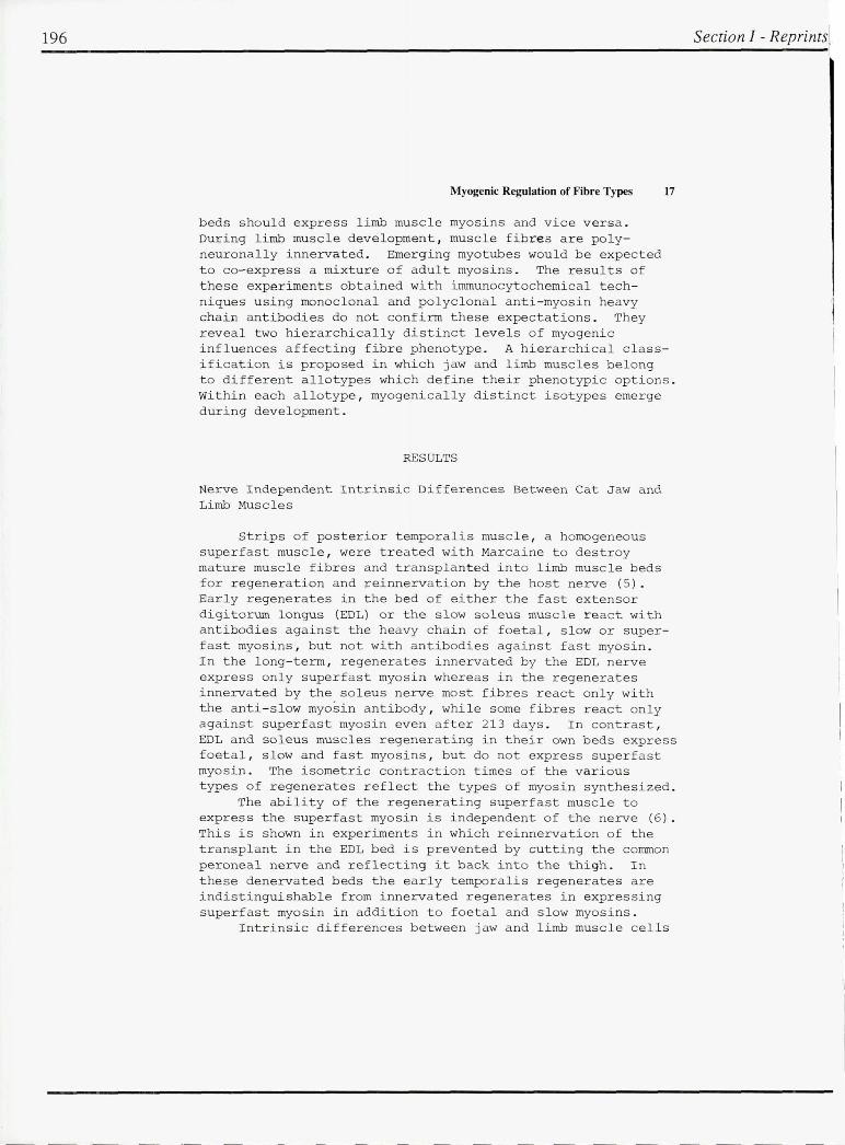
The postnatal development and t he effects of neonatal denervation on muscle fibres in the EDL and tibialis anterior (TA) muscles of the rat were stu died immunocytochemically using monoclonal antibodi es against myosin heavy chains. Three t ypes of primary myotubes (fast , intermediate and slow) with distinct topographic distributions can be distingu ished perinatally . All primaries synthesize slow myosin initially , but in fast and intermediate primaries , slow myosin is no longer detectable in the neonatal p e riod and a t 2 weeks of age respectively . The fast primaries are l ocalized principally in a superficial strip of the TA (Fig . 2B) where in the matured muscle slow fibres are absent . Slow primaries are located deep in the mu scle while intermediat e primari es lie in between . The distribution of slow and intermediate primaries at birth is shown in Fig . 2A.
Following neonatal denervation , the slow and intermediate primaries still express slow myosin , whereas the fast primaries do not stain for slow myosin . At three weeks after denervation , slow primaries are hypertrophic and intermediate primaries are a t rophic , both staining with anti - slow antibody . The topographi c distribution of these primaries is shown in Fig . 2C . These resu lts show that the three different types of primary myotubes respond
197

198
Myogenic Regulation of Fibre Types 19
ORIGINAL P GE BLACK A 0 WHITE PHOTOG~APH
Fig . 1 . Fluorescence photomicrographs of serial sections of c a t e x tens or digito rum longus muscle regenerated in the anterior tempora lis muscle bed f or 1 2 weeks stained for s uperfast (a), f oeta l (b), slow (c) , and fast/foetal (d) myosin heavy chains . The sca le represents 100~m.
i Section I - Reprints I
Section I - Reprints
20 Hoh et a1.
."
.. /f~lr~f~~t~*:::~·~:·: :
'. ' .. . -. -.
b .
... --. .. : ... ~ .
. ' :., .... ... : .. :.,' :,' ..... ~. : .. :.: .. ~ ·/:::.<~~~~.i ... :·'/;,:;:rj:iS;:{!}/:
Fig . 2 . Distribution of fibres which stain with anti- slow myosin heavy chain antibody in rat tibialis anterior (TA) muscle at birth (A , B) and TA three weeks after neonatal denervation (e). Fibres which stained strongly (slow and intermediate primaries) and those that stained faintly in the neonatal TA (fast primaries) are shown in (A) and (B) respectively . Note the h ypertrophy of s l ow primaries and atrophy of intermediate primaries following denervation (e ).
199

200
Myogenic Regulation of Fibre Types
differently to denervation .
Hetero geneity of Secondary Fibres in Developing Limb Muscles
Immunocytochemical analysis of neonatal cat EDL and soleus muscles (7) has revealed that there are a t least two distinct classes of secondary fibres. Both classes initia lly stain strongly for embryonic/foetal myosins . In one of these classes , developmental myosins are replaced by fast myosin . These fast secondaries do not stain for slow myosin nor react with anti - superfast myosin antibody
21
at any stage. The other class of secondaries , the slow secondaries , are prevalent in the slow soleus muscle . These fibres acquired staining for slow myosin but not for fast or superfast myosins .
The vast majori ty of neonatal secondary myotubes in rat EDL and TA muscles are fast secondaries and stain with an anti-foetal/fast- red myosin antibody . These myotubes diverge at 9 days into a superficial fast-white region and a deep fast-red region . The majority of superficial secondaries no longer stain for foetal/fast-red myosins , presumably expressing fast - white myosin , whereas seconda ries located in the region occupied by slow primaries predominantly express fast - red myosin . This topographical distribution of the two classes of secondaries is present in both the EDL and TA muscles, but is more conspicuous in the latter .
Following neonatal denervation in the rat , the divergence of fast- red and fast - white fibres in the EDL and TA muscles is not abolished , but delayed till three weeks post- operatively , s u ggesting that this divergence is neurally independent .
Effects of Denervation on Slow Primaries in Adult Rats
Immunocytochemical analyses of rat limb skeletal muscle fibres u sing specific anti-myosin antibodies have revealed that post-denervation changes of muscle fibres cannot be predicted by the fibre phenotype (8 ) . The number of slow fibres in the EDL of a three month old rat is about half the number of slow primaries seen during development . Upon denervation of this muscle , the number of slow fibres increases to match the number of slow primaries at birth . These fibres hypertrophy while other fibres suffer denervation atrophy . These observations suggest that about half
I
Section I - Reprints I

~ection I - Reprints
22 Hoh et al .
of the slow primaries in mature rats undergo fibre type transformation into phenotypically fast fibres. Upon denervation , all slow primaries express slow myosin and hypertrophy , just as they do after neonatal denervation, irrespective of their phenotype at the time of denervation .
Heterogeneity of Primary and Secondary Fibres in Developing Cat Jaw Muscles
There are two phenotypes in jaw- closing muscle fibres in the cat : superfast and slow. In the posterior temporalis muscle of the mature animal , all fibres are superfast . During late foetal life , sections of this muscle stained for slow myosin appear very similar to those of fast limb muscle : slow staining primary fibres surrounded by rosettes of secondary fibres . Later , both primary and secondary fibres synthesize superfast myosin and the slow myosin in primary fibres is withdrawn (9) . Primary fibres in the posterior temporalis a re therefore analogous to fast primaries in limb muscles , and may be termed superfast primaries . Slow fibres are present in the anterior temporalis and the masseter muscles of adult cats, and these developmentally are derived from both primary and secondary fibres . The jaw slow primary fibres are analogous to slow primaries in limb muscles in which slow myosin synthesis persists to adult life . The jaw slow secondaries appear in early postnatal life , and some of these fibres stain also for superfast myo s in during this period . At no time during the development of cat jaw muscle fibres does any fibre stain for fast myosin .
DISCUSSION
The results of these experiments reveal that the neural regulatory hypothesis cannot account for the difference between limb and j aw muscles . Each type of muscle has a specific repertoire for myosin gene expression, the limb muscles express slow , fast- red and fast- white myosins while jaw muscles expres s slow and superfast myosins .
The abili t y to express these myosins is intrinsic to the muscle type, and can occur during regeneration in the absence of innervation . Innervation by limb nerves does not induce jaw regenerates to express fast myosins , nor does innervation by jaw nerve fibres lead to the expression of superfast myosin in limb regenerates . However , the specific form of myosin expressed by jaw or limb muscles is subject
201

202
Myogenic Regulation of Fibre Types
to neu ral regulation within the constra int s of the repertoire . The limited repert oires for myosin gene expression for jaw and l i mb muscles is also seen dur ing developmental myogenesis .
23
It i s u sefu l to introdu ce the t erm allotype to d e scribe different classes o f skeletal mu scle f i b r es with disti nct intrinsic properties s u ch as limb and jaw mu scles . Jaw and limb allot ypes probably a rise from distinct lineages of myob l ast s committ ed t o d i fferent iate along differe nt paths . Extraocular muscles , which are isometrically faster t han limb and jaw mu scles (10 ) a nd whic h express a un ique myosin heavy chain (11 ) may be another skeletal mu scle allotype .
Immunocytochemical a nalyses of developing limb and jaw mu scles r eveal considerable het erogeneit y in the pattern of myosin gene expressi on in bot h pri mary a nd secondary fib r es . Such het ero geneity may be d ue to some ext rinsi c influ e n ce , s uch as innervation , acting upon a homoge neou s popu lat ion of myotubes . Alternatively , the myotubes may be intrinsica lly heterogeneous , being preprograrnrned to express different types of myosin during s ubsequent development .
Evidence aga i nst the s ugges t ion that fibre type diversity emerges as a result of neu ral regu l a tion is the observat ion that divergence o f fast and slow primaries i s alrea dy appa rent prena t a lly (1 2 ) whereas the impu lse p atterns of developing fast and s l ow motoneurons in t he neonata l r a t are very similar ; differences emerge only a t
3 weeks postnatally (13 ). Furthermore , the occu r rence of polyneuronal innervation o f mu scle fibres (14 ) in the early p ostnatal period also a rgues a gainst the neu r a l regulatory h ypo thesis .
In s upport of the notion tha t myotubes a re intrinsically heterogeneou s ma y be c ited the observations tha t clona l col onies of early myoblasts in chicken (15) a nd h uman embryos (1 6 ) a re not h omo geneous with respec t t o nutrient requirements and colo ny morpho l o gy . Miller a nd Stockda le (17) h ave i s olated three t ypes of c l o nes from early chicken
myoblas t s whic h expres s f a st , slow o r a mixture of both myosins . These clones provide a nerve- independent mechanism for the generation o f different musc le fibre types d uring myogenes i s (18 ).
We p r opose that the emergence o f diverse primary fibre s in mammalia n limb and j a w mu scles is due to variou s linea ges o f myoblasts with intrinsica l lydifferent properties . The cha racteristic responses t o neo na tal denervation of the three types of primaries in the rat limb c learly reveal their differences , the most s pectacu l a r fea t u re o f which being the
Section I - Reprint

I ~ection I - Reprints
24 Hoh et al.
hypertrophy of slow primaries. This property of slow primaries is retained in the adult even though some of the slow primaries had apparen tly undergone, through the neural regulatory influence, a phenotypic change to become fast fibres . Thus , the hypertrophic response of denervated adult muscle fibres cannot be predicted on the basis of fibre phenotype , but can be so predicted according to the developmental origin of t he fibres . Hence it is important to classify muscle fibres in accordance with their developmental origin in additi on to their allot ype and their phenotype . We propose to use the term isotype in this context . Thus there are at least three isotypes (slow, intermediate and fast ) of primary fibres in limb muscles and two isotypes (superfas t and slow ) of p r imary fibres in jaw muscle.
The emergence of phenotypic characteristics of secondary fibres occur s relatively late during myogenesis , making it possible for neural regulatory mechanisms to have an impact on it. However, t he possibility of there being various isotypes of secondary fibres cannot be di scounted . Our neonatal denervation study shows that the divergence of fast-red and fast -white fibres is neurally independent, raising the possibility of the existence of two distinct isotypes of fast secondary myo tubes. An interesting alternative mechanism for generating different phenotypes of secondary fibres is for primary fibres to influence the phenotype of the secondary fibres associated with them. The existence of gap junctions between primary and secondary fibres is well established (1 9 ). These junctions may provide the physical basis for the postu lated myogenic influence on secondary fibres. The co- localization of slow primaries and fast- red fibres in the deep region of TA is consistent with the no t ion that slow p r imaries induce the expression of the fas t-red phenotype in secondary myotubes associated with them .
The various myogenic and neurogenic influences on the phenotypic expression of myosin genes during myogenesis may now be s ummarized . The a llotype defines the various phenotypic options available: s uperfast and slow myosins for the jaw allotype and fast- red, fast-white and slow for the limb allotype. Very early in myogenesis, diverse isomyoblasts emerge within each allotype . These fuse to produce myotubes of corresponding isotypes , each destined to undergo a particular pattern of myosin gene expression within the options defined by the al l o type. Innervation may only play a trophic or permissive role on myogenesis up to this point.
203

204
Myogenic Regulation of Fibre Types
Neural regulatory influences may operate after polyneuronal innervation has been eliminated and phasic and t onic nerve impulse patterns established . These influences may change the fibre phenotype within the range of options defined by the allotype , but do not alter the fibre isotype , nor transform the fibre allotype.
ACKNOWLEDGEMENTS
We thank Drs . R. B. Fitzsimons a nd P . T . Hale for the monoclonal antibodies and Prof. J . Egerton , G. Tasler and C . Chow for help in producing the anti - superfast myosin antibody.
REFERENCES
1 . Jolesz F, Streter .FA (1981) . Development , innervation , and activity- pattern induced changes in skeletal muscle . Ann Rev Physiol 4 3 :531 .
2 . Pette 0 , Vrbova G (1985) . Invited review : neural control of phenotypic expression in mammalian muscle fibres . Muscle Nerve 8 : 676 .
25
3 . Buller AJ , Eccles JC , Eccles RM (1 960 ) . Differentiation of fast and slow muscles in the cat hindlimb . J . Physiol 150:399 .
4 . Buller AJ , Eccles JC , Eccles RM (1960) . Interactions between motoneurones and muscles in respect of the characteristic speeds of their responses . J Physiol 150:417.
5 . Hoh JFY , Hughes S (1 988) . Myogenic and neurogenic regulation of myosin gene expression in cat jaw-closing muscles regenerating in fast and slow limb muscle beds. J Musc Res Cell Motil 9 : in press .
6 . Hoh JFY, Hughes S (1986) . Myosin gene expression in cat temporalis muscle regenerating in the absence of a nerve . Proc Aust Physiol pharmacol Soc l7 : 142P .
7. Hoh JFY , Hughes S , Hale PT , Fitzsimons RB (1988). Immunocytochemical and electrophoretic analyses of changes in myosin gene expression in cat limb fast and slow muscles during postnatal development . J Musc Res Cell Motil 9 : in press .
8 . Hugh G, Hoh JFY (1987 ) . Immunocytochemical analysis of myosin isoenzymes in denervated rat fast and s l ow muscles . Proc Aust Physiol pharmacol Soc 18 : 45P .
Section I - Reprint]

Section I - Reprints
26 Hoh et al.
9 . Hoh JFY, Hughes S , Chow C , Hale PT , Fitzsimons RB (1988) . Immunocytochemical and electrophoretic analyses of changes in myosin gene expression in cat posterior temporalis muscle during postnatal development. J Musc Res Cell Motil 9 : in press .
10. Bach- y-Rita P , Ito F (1966). In vivo studies on fast and slow muscle fibres in cat extraocu lar muscles . J Gen Physiol 49:1177 .
11 . Wi eczorek DF , Periasamy M, Butler- Browne GS , Whalen RG , Nadal-Ginard B (1 985 ). Co- expression of mUltiple myosin heavy chain genes , in addition to a tissue-specific one, in extraocular musculature . J Cell Biol 101 : 618 .
12 . Dhoot GK (1986 ) . Selective synthesis and degradation of slow skeletal myosin heavy chains in developing muscle fibres . Mu scle Nerve 9: 155- 164 .
13 . Navarrete R, Vrbova G (1 983 ). Changes of activity p a tterns in slow and fast muscles during postnatal development. Dev Brain Res 8 :11-19 .
14 . Redfern PA (1970). Neuromuscular transmission in newborn rats. J Physiol 209 : 701.
15 . Bonner PH , Hauschka SD (1 974). Clonal analysis of vertebrate myogenesis . I . Early developmental events in the chick limb . Dev 37:317 .
16 . White NK , Bonner PH , Nelson DR , Hauschka SD (1 975 ) . Clonal analysis of vertebra te myogenesis . IV . Mediumdependent classification o f colony- forming cells . Dev Biol 44 : 346 .
17. Miller JB , Stockdale FE (1986) . Developmental origins of skeletal muscle fibres : clonal analysis of myogenic cell lineages based on expression of fast a nd slow myosin heavy chains . Proc Natl Acad Sci USA 83 : 3860 .
18 . Crow MT, Stockdale FE (1 986 ). Myosin expression and specialization a mong the earliest muscle fibres of the developing avian limb. Dev Biol 113 : 238 .
19 . Kelly AM , Zacks SI (1969 ) . The histogenesis of rat intercostal muscle . J Cell Biol 42 : 135 .
205