© 2013 Pearson Education, Inc. Digestive System Two groups of organs 1. Alimentary canal...
32
© 2013 Pearson Education, Inc. Digestive System • Two groups of organs 1. Alimentary canal (gastrointestinal or GI tract) • Mouth to anus • Digests food and absorbs fragments • Mouth, pharynx, esophagus, stomach, small intestine, and large intestine
-
Upload
leroy-ackley -
Category
Documents
-
view
214 -
download
0
Transcript of © 2013 Pearson Education, Inc. Digestive System Two groups of organs 1. Alimentary canal...

© 2013 Pearson Education, Inc.
Digestive System
• Two groups of organs1. Alimentary canal (gastrointestinal or GI
tract)• Mouth to anus• Digests food and absorbs fragments• Mouth, pharynx, esophagus, stomach,
small intestine, and large intestine

© 2013 Pearson Education, Inc.
Digestive System
2. Accessory digestive organs• Teeth, tongue, gallbladder• Digestive glands
– Salivary glands– Liver– Pancreas

© 2013 Pearson Education, Inc.
Figure 23.2 Gastrointestinal tract activities.
Ingestion
Mechanicalbreakdown
Digestion
Propulsion
Absorption
Defecation
Food
PharynxEsophagus• Chewing (mouth)
• Swallowing (oropharynx)• Peristalsis (esophagus, stomach, small intestine, large intestine)
Stomach
Lymphvessel
Small intestineLargeintestine
Bloodvessel
Mainly H2OFeces
Anus
• Churning (stomach)• Segmentation (small intestine)
© 2013 Pearson Education, Inc.
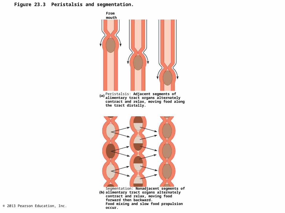
Figure 23.3 Peristalsis and segmentation.
Frommouth
Peristalsis: Adjacent segments of alimentary tract organs alternately contract and relax, moving food along the tract distally.
Segmentation: Nonadjacent segments of alimentary tract organs alternately contract and relax, moving food forward then backward.Food mixing and slow food propulsion occur.

© 2013 Pearson Education, Inc.
GI Tract Regulatory Mechanisms
1. Mechano and chemoreceptors – Respond to stretch, changes in osmolarity
and pH, and presence of substrate and end products of dig
– Initiate reflexes that• Activate or inhibit digestive glands • Stimulate smooth muscle

© 2013 Pearson Education, Inc.
Figure 23.4 Neural reflex pathways initiated by stimuli inside or outside the gastrointestinal tract.
External stimuli(sight, smell, taste,
thought of food)
Visceral afferents
Internal(GI tract)stimuli
Chemoreceptors,osmoreceptors, ormechanoreceptors
Long reflexes
Central nervous system
Local (intrinsic)nerve plexus("gut brain")
Effectors:Smooth muscle
or glands
Extrinsic visceral (autonomic)efferents
Short reflexes
Lumen of thealimentary canal
Gastrointestinalwall (site of shortreflexes)
Response:Change in
contractile orsecretory activity
© 2013 Pearson Education, Inc.
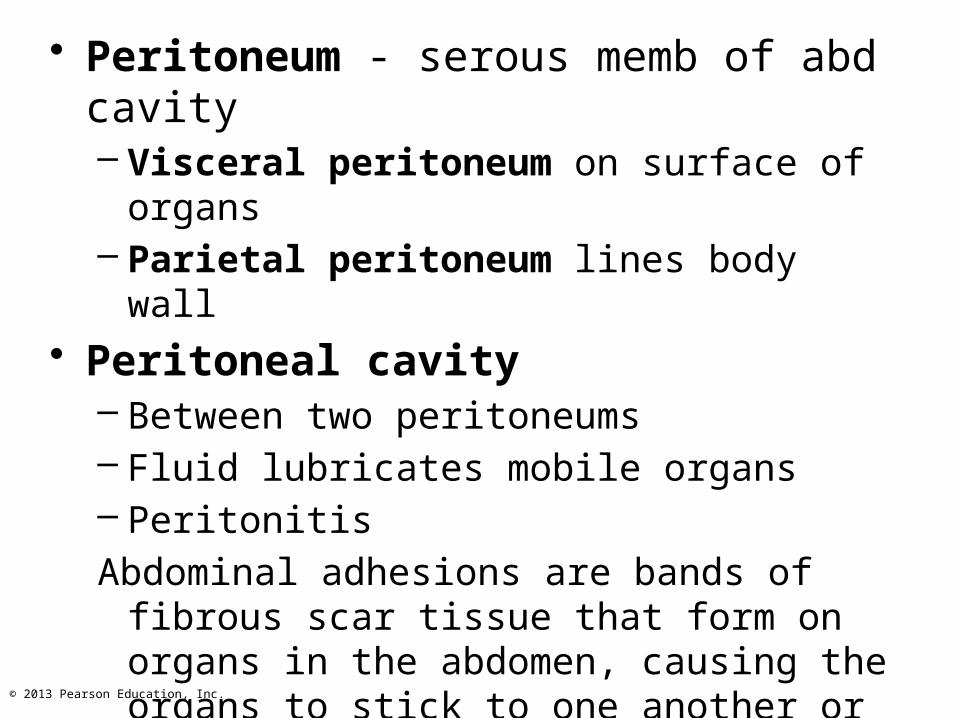
• Peritoneum - serous memb of abd cavity– Visceral peritoneum on surface of organs– Parietal peritoneum lines body wall
• Peritoneal cavity– Between two peritoneums– Fluid lubricates mobile organs – Peritonitis
Abdominal adhesions are bands of fibrous scar tissue that form on organs in the abdomen, causing the organs to stick to one another or to the wall of the abdomen.

© 2013 Pearson Education, Inc.
Figure 23.5a The peritoneum and the peritoneal cavity.
Abdominopelviccavity
Vertebra
Peritonealcavity
Alimentarycanal organ
Liver
Two schematic cross sections of abdominal cavity illustratethe peritoneums and mesenteries.
Ventralmesentery
Parietalperitoneum
Visceralperitoneum
Dorsalmesentery

© 2013 Pearson Education, Inc.
Peritoneum and Peritoneal Cavity
• Mesentery - double layer of peritoneum– Routes for BV, lymphatics, and nerves– Holds organs in place; stores fat

© 2013 Pearson Education, Inc.
Figure 23.5b The peritoneum and the peritoneal cavity.
Alimentarycanal organ
Alimentary canal organ ina retroperitoneal position
Some organs lose their mesentery and move,becoming retroperitoneal, during development.
Mesenteryresorbedand lost

© 2013 Pearson Education, Inc.
Blood Supply: Splanchnic Circulation
• Branches of aorta– Hepatic, splenic, gastric, mesenteric arteries
• Hepatic portal circulation
© 2013 Pearson Education, Inc.
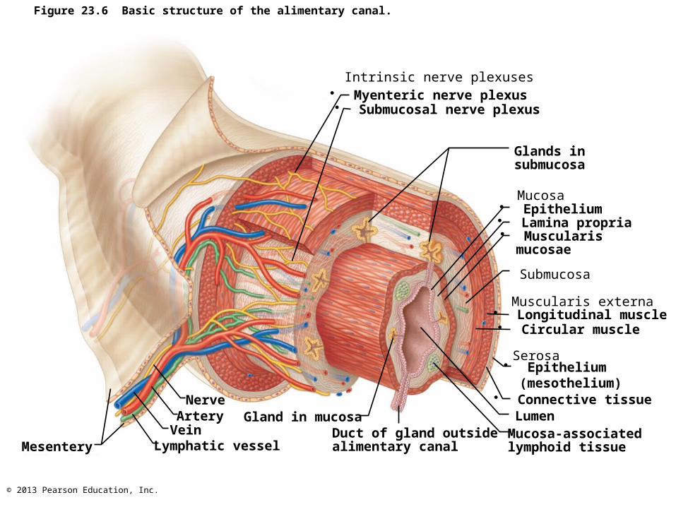
Figure 23.6 Basic structure of the alimentary canal.
Intrinsic nerve plexuses
Mucosa
Submucosa
Muscularis externa
Glands insubmucosa
Serosa
LumenMucosa-associatedlymphoid tissue
Duct of gland outside alimentary canal
Gland in mucosa
Lymphatic vesselVein
ArteryNerve
Mesentery
• Myenteric nerve plexus• Submucosal nerve plexus
• Epithelium• Lamina propria• Muscularis mucosae
• Longitudinal muscle• Circular muscle
• Connective tissue
• Epithelium (mesothelium)

© 2013 Pearson Education, Inc.
Enteric Nervous System
• Intrinsic nerve supply – enteric neurons• Major nerves to GI tract; control motility
– Submucosal nerve plexus• Regulates glands and smooth muscle in mucosa
– Myenteric nerve plexus• Controls GI tract motility
– Sympathetic inhibit dig activities– Parasympathetic stimulate dig activities
© 2013 Pearson Education, Inc.
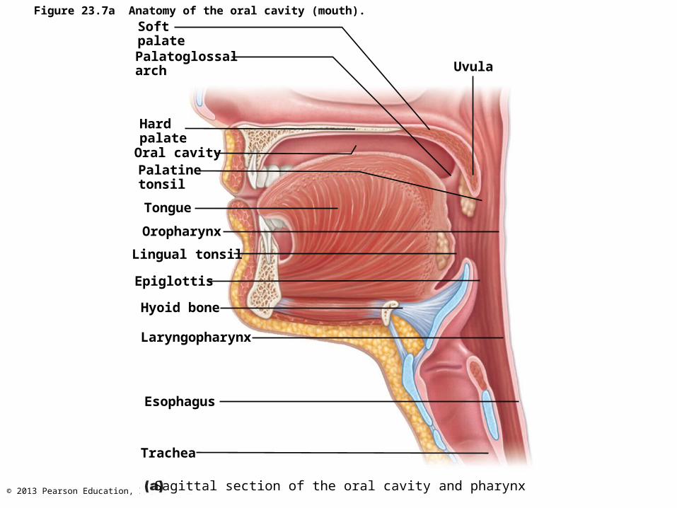
Figure 23.7a Anatomy of the oral cavity (mouth).
Palatoglossalarch
Softpalate
HardpalateOral cavityPalatinetonsil
Tongue
Oropharynx
Lingual tonsil
Epiglottis
Hyoid bone
Laryngopharynx
Esophagus
Trachea
Uvula
Sagittal section of the oral cavity and pharynx
© 2013 Pearson Education, Inc.
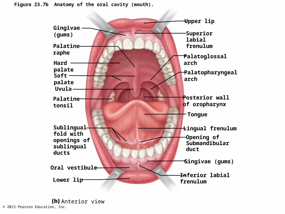
Figure 23.7b Anatomy of the oral cavity (mouth).
Gingivae (gums)
Palatineraphe
HardpalateSoftpalate
Palatinetonsil
Sublingualfold withopenings ofsublingualducts
Oral vestibule
Lower lip
Uvula
Upper lip
Superiorlabialfrenulum
Palatoglossalarch
Palatopharyngealarch
Posterior wallof oropharynx
Tongue
Lingual frenulum
Opening ofSubmandibularduct
Gingivae (gums)
Inferior labialfrenulum
Anterior view

© 2013 Pearson Education, Inc.
Figure 23.8 Dorsal surface of the tongue, and the tonsils.
Epiglottis
Palatopharyngealarch
Palatine tonsil
Lingual tonsil
Palatoglossal arch
Terminal sulcus
Foliate papillae
Vallate papilla
Medial sulcus of the tongue
Dorsum of tongue
Fungiform papilla
Filiform papilla

© 2013 Pearson Education, Inc.
Figure 23.9 The salivary glands.
Tongue
Teeth
Frenulumof tongue
Mylohyoidmuscle (cut)
Anterior belly ofdigastric muscle
Masseter muscle
Body of mandible(cut)
Posterior belly ofdigastric muscle
Serous cellsforming demilunes
Mucous cells
Parotid gland
Submandibularduct
Submandibulargland
Ducts ofsublingualgland
Sublingualgland
Parotid duct
© 2013 Pearson Education, Inc.
Salivary Glands
• Two types of secretory cells– Serous cells
• Watery, enzymes, ions, bit of mucin
– Mucous cells• Mucus
• Parotid, submandibular glands mostly serous; sublingual mostly mucous

© 2013 Pearson Education, Inc.
• 97–99.5% water, slightly acidic– Electrolytes—Na+, K+, Cl–, PO4 2–, HCO3–
– Salivary amylase and lingual lipase– Mucin– Metabolic wastes—urea and uric acid– Lysozyme, IgA, defensins, and a cyanide
compound protect against microorganismsa
PLAY Animation: Rotating head
Composition of Saliva

© 2013 Pearson Education, Inc.
Control of Salivation• 1500 ml/day• activated by parasympathetic when
– Ingested food stimulates chemoreceptors and mechanoreceptors in mouth
– Salivatory nuclei in brain stem send impulses along parasympathetic fibers in cranial nerves VII and IX
• Sympathetic inhibits salivation results xerostomia

© 2013 Pearson Education, Inc.

© 2013 Pearson Education, Inc.
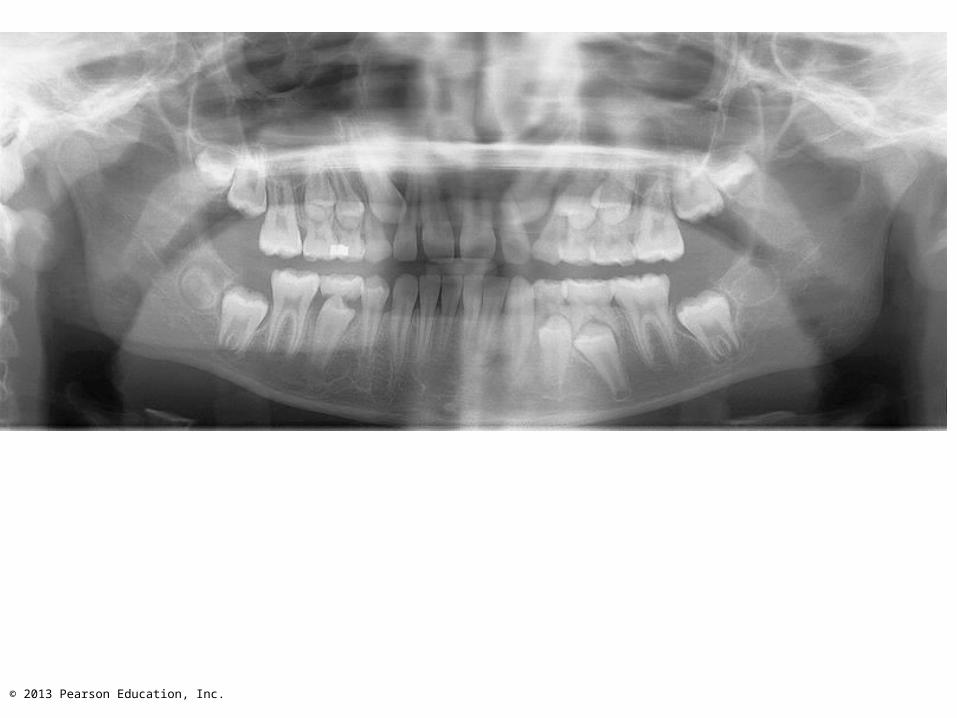
Teeth
• Primary and permanent dentitions formed by age 21
• 20 deciduous teeth erupt (6–24 months of age)– Roots resorbed, teeth fall out (6–12 years of
age) as permanent teeth develop• 32 permanent teeth
– All but third molars in by end of adolescence• Third molars at 17–25, or may not erupt
© 2013 Pearson Education, Inc.

© 2013 Pearson Education, Inc.
Figure 23.10 Human dentition.IncisorsCentral (6–8 mo)
Lateral (8–10 mo)
Canine (eyetooth)(16–20 mo)
MolarsFirst molar(10–15 mo)
Second molar(about 2 yr)
IncisorsCentral (7 yr)
Lateral (8 yr)
Canine (eyetooth)(11 yr)
Premolars(bicuspids)First premolar(11 yr)
Second premolar(12–13 yr)
MolarsFirst molar (6–7 yr)
Second molar(12–13 yr)
Third molar(wisdom tooth)(17–25 yr)
Permanentteeth
Deciduous(milk) teeth
© 2013 Pearson Education, Inc.
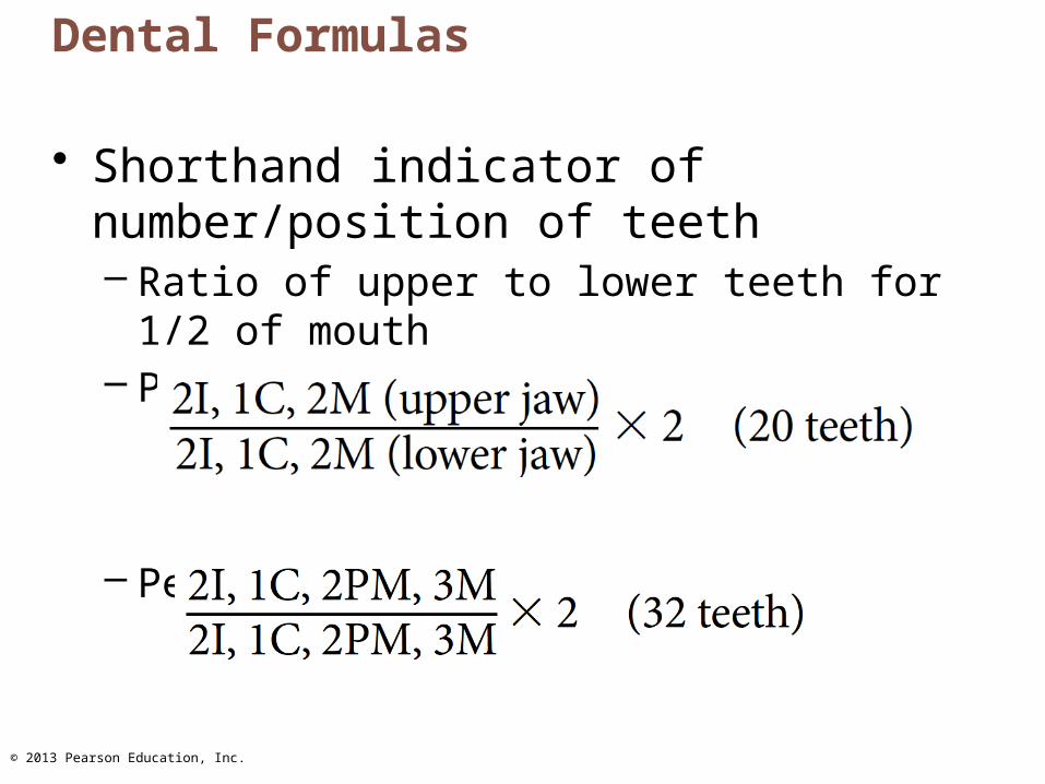
Dental Formulas
• Shorthand indicator of number/position of teeth– Ratio of upper to lower teeth for 1/2 of mouth– Primary:
– Permanent:

© 2013 Pearson Education, Inc.
Tooth Structure
• Crown - exposed part above gingiva (gum)– Covered by enamel—hardest substance in
body (calcium salts and hydroxyapatite crystals)• Enamel-producing cells degenerate when tooth
erupts no healing if decay or crack
• Root - portion embedded in jawbone– Connected to crown by neck

© 2013 Pearson Education, Inc.
Tooth Structure
• Cement - calcified connective tissue – Covers root; attaches to periodontal ligament
• Periodontal ligament– Fibrous joint called gomphosis– Anchors tooth in socket
• Gingival sulcus - groove where gingiva borders tooth

© 2013 Pearson Education, Inc.
Tooth Structure
• Dentin - bonelike material under enamel– Maintained by odontoblasts of pulp cavity
• Pulp cavity - surrounded by dentin – Pulp - connective tissue, BV, and nerves
• Root canal - as pulp cavity extends to root• Apical foramen at proximal end of root
– Entry for blood vessels, nerves, etc.

© 2013 Pearson Education, Inc.
Figure 23.11 Longitudinal section of a canine tooth within its bony socket (alveolus).
Enamel
Dentin
DentinaltubulesPulp cavity(containsblood vesselsand nerves)GingivalsulcusGingiva(gum)
Cement
Root canal
Periodontalligament
Apicalforamen
Bone
Crown
Neck
Root
© 2013 Pearson Education, Inc.
Tooth and Gum Disease
• Dental caries (cavities) - demineralization of enamel and dentin from bacterial action – Dental plaque (film of sugar, bacteria, and
debris) adheres to teeth– Acid from bacteria dissolves calcium salts– Proteolytic enzymes digest organic matter – Prevention: daily flossing and brushing

© 2013 Pearson Education, Inc.
Tooth and Gum Disease• Gingivitis
– Plaque calcifies to form calculus (tartar)– Calculus disrupts seal between gingivae and
teeth – Bacteria infect gums

© 2013 Pearson Education, Inc.
Tooth and Gum Disease• Periodontitis -Immune cells attack
• Destroy periodontal ligament• Activate osteoclasts dissolve bone
– Tooth loss; may promote atherosclerosis– Risk factors ?