© 2012 Pearson Education, Inc. Tissues A tissue is a group of cells with a common function Four...
43
© 2012 Pearson Education, Inc. Tissues • A tissue is a group of cells with a common function • Four primary tissues: – Epithelia – Connective tissues – Muscle – Nervous
-
Upload
virgil-lucas -
Category
Documents
-
view
215 -
download
0
Transcript of © 2012 Pearson Education, Inc. Tissues A tissue is a group of cells with a common function Four...

© 2012 Pearson Education, Inc.
Tissues
• A tissue is a group of cells with a common function
• Four primary tissues:– Epithelia– Connective tissues– Muscle– Nervous

© 2012 Pearson Education, Inc.
Epithelial Tissues
• Line body cavities, cover surfaces (i.e.: skin)
• Also are the interior lining of: blood & lymph vessels, heart, G.I. tract, respiratory tract, genital & urinary tracts.
• Everything that goes into the body will have to pass through epithelium.

© 2012 Pearson Education, Inc.
Glandular epitheliaEpithelial cells adapted to make up glands-Exocrine glands
Secrete into ducts to exterior of body
-Endocrine glandsSecrete into the blood to carry chemical messages throughout the body

© 2012 Pearson Education, Inc.
Epithelial Tissues: Classification
• Shape– Squamous
• Flattened cells• Line vessels, part of lungs, body surface
– Cuboidal• Cube shaped• Form lining of tubules, glandular tissue
– Columnar• Column shaped• Line respiratory, digestive, reproductive tracts

© 2012 Pearson Education, Inc.
Epithelial Tissues: Classification
• Number of layers– Simple/single–layered
• Adapted for diffusion across cell barriers• Line glands, and respiratory, digestive, urinary,
& reproductive systems
– Stratified/multiple–layered• Provide protection, as in the skin surface

© 2012 Pearson Education, Inc. Figure 4.1a
Simple squamous• Lines blood vessels and air sacs of lungs• Permits exchange of nutrients, wastes, and gases
Stratified squamous• Outer layer of skin, mouth, vagina• Protects against abrasion, drying out, infection
Stratified cuboidal• Lines ducts of sweat glands• Secretes water and ions
Simple cuboidal • Lines kidney tubules and glands • Secretes and reabsorbs water and small molecules
Simple columnar • Lines most digestive organs • Absorbs nutrients, produces mucus
Goblet cell Basement membrane
Stratified columnar• Lines epididymus, mammary glands, larynx• Secretes mucus
a) Most epithelial tissues line or cover surfaces or body cavities.

© 2012 Pearson Education, Inc. Figure 4.1b
Exocrine gland
Gland cells
Endocrine gland
Gland cellsBlood flow
b) Glandular epithelia secrete a product.

© 2012 Pearson Education, Inc.
The Basement Membrane Provides Structural Support for Epithelia
• Basement membrane– Provides structural support to overlying cells– Attaches epithelial layer to underlying tissues
• Junctions: hold epithelial cells together– Tight junctions
• Nothing passes
– Adhesion junctions/spot desmosomes• Some movement between cells
– Gap junctions• Protein channels

© 2012 Pearson Education, Inc. Figure 4.2a
Tight junction proteins
Intercellular space
a) Tight junctions form leak-proof seals between cells

© 2012 Pearson Education, Inc. Figure 4.2b
Protein filaments
Intercellular space
b) Adhesion junctions anchor two cells together, yet allow flexibility of movement

© 2012 Pearson Education, Inc. Figure 4.2c
Protein channel
Intercellular space
c) Gap junctions provide for the direct transfer of water and ions between adjacent cells

© 2012 Pearson Education, Inc.
Connective Tissue
• General functions– Supports softer organs of body– Connects parts of body– Stores fat– Produces blood cells
• Contains cells embedded in nonliving extracellular matrix
• Matrix provides the strength
• Two general types of connective tissue– Fibrous and special

© 2012 Pearson Education, Inc.
Fibrous Connective Tissue
• Function: provides strength and elasticity
• Contains fibers and cells embedded in gel-like ground substance (matrix)
• Matrix: intercellular material giving the CT its characteristics
• Cells: fibroblasts (function is to make the protein fibers & matrix), macrophages, lymphocytes, and neutrophils
• Fibers: collagen, elastic, and reticular

© 2012 Pearson Education, Inc.
Fibrous ConnectiveTissue
• Four general types– Loose: surrounds many organs, lines
cavities around blood vessels– Dense: forms tendons, ligaments, deeper
layers of skin– Elastic: surrounds stomach, bladder,
arteries, maintains shape– Reticular: makes up internal framework of
soft organs (liver) and the lymphatic system

© 2012 Pearson Education, Inc. Figure 4.3
Ground substance
Plasma cell
Neutrophil
Collagen fiber
Lymphocyte
Nerve fiber
Macrophage
Reticular fiber
Elastic fiber
Mast cell Fibroblast
Fat cell Capillary

© 2012 Pearson Education, Inc. Figure 4.4a
Elastin fibers
Fibroblast
Collagen fibers
a) Loose (areolar) connective tissue (X 160). In loose connective tissue the collagen and elastin fibers are arrayed in a random
pattern.

© 2012 Pearson Education, Inc. Figure 4.4b
Collagen fibers
Nuclei of fibroblasts
b) Dense connective tissue (X 160). In dense connective tissue the fibers are primarily collagen fibers. In tendons and ligaments the fibers are oriented all in the same direction, with fibroblasts occupying narrow spaces between adjacent fibers.

© 2012 Pearson Education, Inc.
Specialized Connective Tissues Serve Special Functions
• Cartilage: produced by chondroblasts (which are found in lacunae); no blood vessels; high collagen content
• Bone: inorganic matrix with calcium salts for hardness
• Blood: fluid matrix of plasma, red blood cells, white blood cells, and platelets
• Adipose tissue: fat cells; function in insulation, protection, and energy storage

© 2012 Pearson Education, Inc. Figure 4.5a
Chondrocyte in lacuna
Ground substance
a) Cartilage from the trachea (X 300). Mature cartilage cells, called chondrocytes, become trapped in chambers called lacunae within the hard, rubbery ground substance. Ground substance is composed of collagen fibers, polysaccharides, proteins, and water.

© 2012 Pearson Education, Inc. Figure 4.5b
Vacuole containing stored fat
Blood vessel
Nuclei of fat cells
b) Adipose tissue from the subcutaneous layer under the skin (X 140). Adipose tissue consists almost entirely of fat cells. The fat deposit within a fat cell can become so large that the nucleus is pushed to the side.

© 2012 Pearson Education, Inc.
Muscle Tissue Contracts to Produce Movement
• Skeletal muscle– Moves body parts– Voluntary, multinucleated
• Cardiac muscle– Functions in the heart– Involuntary, single nucleus
• Smooth muscle– Surrounds hollow structures– Involuntary, single nucleus

© 2012 Pearson Education, Inc. Figure 4.6a
Nuclei
Width of one muscle cell
a) Skeletal muscle (X 100). Skeletal muscle cells are very long and have many nuclei.

© 2012 Pearson Education, Inc. Figure 4.6b
Intercalated discNucleus
b) Cardiac muscle (X 225). Cardiac muscle cells interconnect with each other.
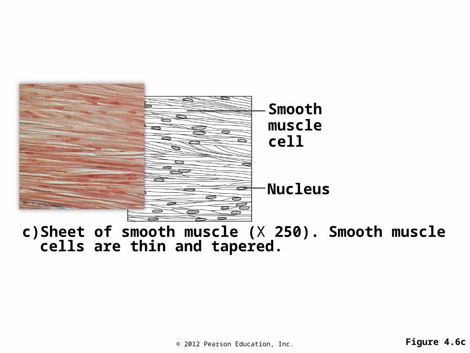
© 2012 Pearson Education, Inc. Figure 4.6c
Smooth muscle cell
Nucleus
c) Sheet of smooth muscle (X 250). Smooth muscle cells are thin and tapered.

© 2012 Pearson Education, Inc.
Nervous Tissues Transmit Impulses
• Neuron: specialized nervous system cell– Function: generate and transmit electrical
impulses– Structural components: cell body, dendrites,
axon
• Glial cells support neurons

© 2012 Pearson Education, Inc. Figure 4.7
Axon
Nuclei of glial cells
Cell body
Dendrites

© 2012 Pearson Education, Inc.
Organs and Organ Systems Perform Complex Functions
• Organs– Contain two or more tissue types joined
together; perform specific functions
• Organ systems– Groups of organs that perform a common
function– Examples
• Digestive system: mouth, throat, stomach, intestines, and liver
• Lymphatic system: lymph nodes, tonsils, and spleen

© 2012 Pearson Education, Inc.
Body Cavities
• Anterior cavity– Thoracic cavity
• Two pleural cavities (1 for each _________)• Pericardial cavity (for the __________)
– Abdominal cavity
• Posterior cavity– Cranial cavity (for the ____________)– Spinal cavity (for the ________ ______)
• Epithelial tissue membranes (called serous membranes) line anterior body cavities

© 2012 Pearson Education, Inc. Figure 4.8
Pelvic cavity
Abdominal cavity
Anterior cavity
Diaphragm separates thoracic and abdominal cavities
Pleural cavity
Pericardial cavityThoracic
cavity
Posterior cavity
Vertebral canal
Cranial cavity

© 2012 Pearson Education, Inc.
Tissue Membranes
• Serous membrane: reduces friction between organs
• Mucous membrane: lubricates surface, captures debris
• Synovial membrane: lines spaces in movable joints, lubricates the joint
• Cutaneous membrane: skin

© 2012 Pearson Education, Inc.
The Skin as an Organ System
• The proper name is integumentary system
• Includes skin, hair, nails, glands
• Functions – Prevents dehydration – Protects from injury – Serves as defense against microorganisms– Regulates body temperature – Makes vitamin D – Provides sensation

© 2012 Pearson Education, Inc. Figure 4.10ReceptorsNerveSweat gland
Adipose tissue
Smooth muscle
VeinArtery
Sebaceous gland
Hypodermis
Dermis
Epidermis
Small blood vessels
Hair shaft Free nerve endings
Hair root
Hair follicle

© 2012 Pearson Education, Inc.
Skin Consists of Epidermis and Dermis
• Epidermis– Outer layer – Stratified squamous epithelial cells – No blood vessels – Two major cell types
• Specialized keratinocytes• Melanocytes

© 2012 Pearson Education, Inc. Figure 4.11
Dead cells of epidermis
Living cells of epidermis
Dermis with blood vessel
Keratinocyte containing melanin
Melanocyte containing melanin granules
Dividing keratinocyte (basal cell)
Basement membrane
Blood vessel

© 2012 Pearson Education, Inc.
Skin Consists of Epidermis and Dermis
• Dermis– Lies underneath the epidermis– Supports tissues– Fibers
• Collagen• Elastic
– Cells• Fibroblasts• Mast cells• White blood cells• Fat cells

© 2012 Pearson Education, Inc.
Accessory Structures of Dermis
• Hair – Shaft– Follicle
• Smooth muscle– Attached to hair follicle, raises hair to
upright position
• Oil glands/sebaceous glands– Secretion moistens and softens skin

© 2012 Pearson Education, Inc.
Accessory Structures of Dermis
• Sweat glands– Secrete sweat, help in temperature regulation
• Blood vessels– Supply nutrients, remove waste, assist in
temperature regulation
• Sensory nerve endings– Detect heat, cold, touch, deep pressure,
vibration
© 2012 Pearson Education, Inc.
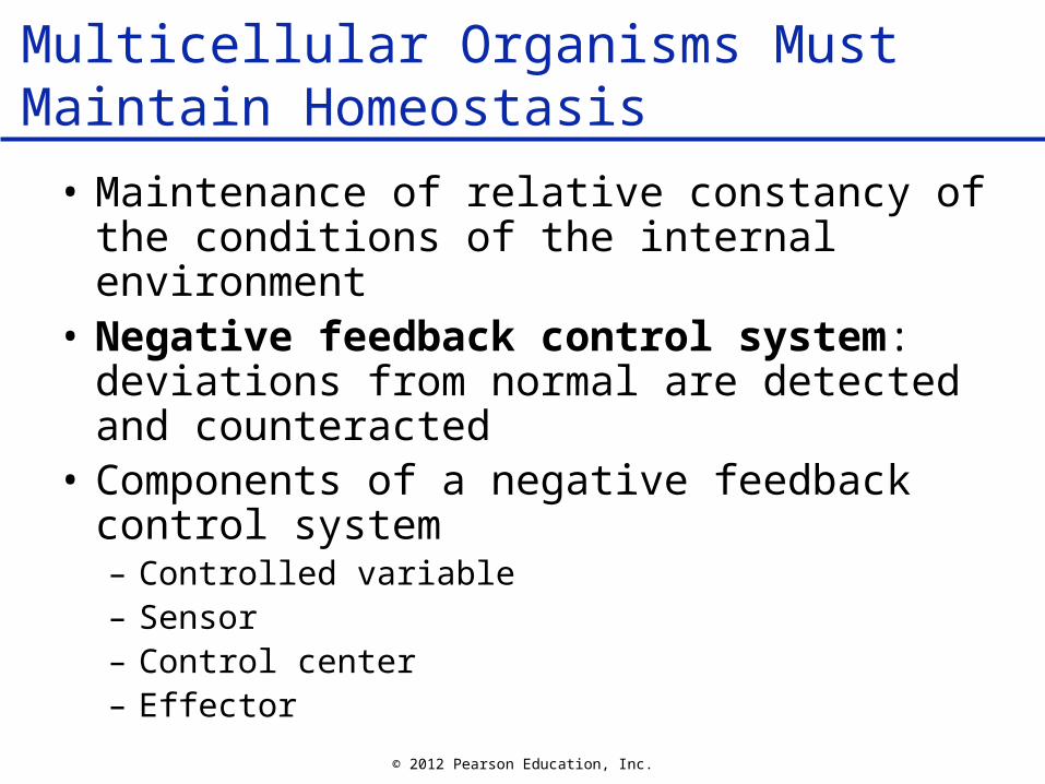
Multicellular Organisms Must Maintain Homeostasis
• Maintenance of relative constancy of the conditions of the internal environment
• Negative feedback control system: deviations from normal are detected and counteracted
• Components of a negative feedback control system – Controlled variable– Sensor– Control center– Effector

© 2012 Pearson Education, Inc. Figure 4.12a
SensorEffector
Control center
Controlled variable
Higher
Set point
Lower
a) An increase in the controlled variable causes events that lower the controlled variable toward its set point again.

© 2012 Pearson Education, Inc. Figure 4.12b
Controlled variable
Higher
Set point
Lower
SensorEffector
Control center
b) A decrease in the controlled variable causes events that raise the controlled variable toward its set point again.

© 2012 Pearson Education, Inc.
Negative Feedback Helps Maintain Core Body Temperature
• Controlled variable: body temperature
• Sensors: temperature sensors in skin and internal organs
• Control center: hypothalamus in the brain
• Effectors– Blood vessels– Sweat glands– Skeletal muscles

© 2012 Pearson Education, Inc. Figure 4.13
Core temperature Core temperature
Higher
Set point Set point
Higher
Lower Lower
Constriction of bloodvessels in skin(saves heat)
Dilation of bloodvessels in skin(promotes heat loss)
SensorsSensors
Shivering(generates heat) Sweating
Control center(hypothalamus)
Control center(hypothalamus)
to to
toto
© 2012 Pearson Education, Inc.
Homeostasis