© 2012 Pearson Education, Inc. Figure 7-3c The Adult Skull TEMPORAL BONE PARIETAL BONE OCCIPITAL...
17
© 2012 Pearson Education, Inc. Figure 7-3c The Adult Skull TEMPORAL BONE PARIETAL BONE OCCIPITAL BONE FRONTAL BONE Lateral view Squamous suture Lambdoid suture Squamous part of temporal bone External acoustic meatus Mastoid process Zygomatic arch Styloid process Zygomatic process of temporal bone Temporal process of zygomatic bone Coronal suture SPHENOID NASAL BONE LACRIMAL BONE ETHMOID MAXILLA ZYGOMATIC BONE MANDIBLE Supra-orbital foramen Infra-orbital foramen Mental foramen Mental protuberance
-
Upload
byron-palmer -
Category
Documents
-
view
518 -
download
2
Transcript of © 2012 Pearson Education, Inc. Figure 7-3c The Adult Skull TEMPORAL BONE PARIETAL BONE OCCIPITAL...

© 2012 Pearson Education, Inc.
Figure 7-3c The Adult Skull
TEMPORALBONE
PARIETALBONE
OCCIPITALBONE
FRONTALBONE
Lateral view
Squamous suture
Lambdoid suture
Squamous part oftemporal bone
External acousticmeatus
Mastoid process
Zygomaticarch
Styloid processZygomatic process
of temporal boneTemporal processof zygomatic bone
Coronal suture
SPHENOID
NASAL BONE
LACRIMAL BONE
ETHMOID
MAXILLA
ZYGOMATIC BONE
MANDIBLE
Supra-orbital foramen
Infra-orbital foramen
Mental foramen
Mental protuberance

© 2012 Pearson Education, Inc.
Figure 7-3a The Adult Skull
PARIETALBONE(left)
PARIETALBONE(right)
OCCIPITALBONE
MANDIBLE
TEMPORAL BONE
Sagittal suture
Lambdoidsuture
Squamoussuture
Mastoid processStyloid process
Occipital condyle
External occipitalprotuberance
Posterior view
© 2012 Pearson Education, Inc.
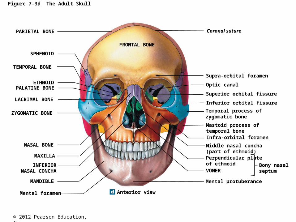
Figure 7-3d The Adult Skull
Coronal suture
SPHENOID
NASAL BONE
LACRIMAL BONE
ETHMOID
MAXILLA
Supra-orbital foramen
Infra-orbital foramen
Mental foramen
Mental protuberance
ZYGOMATIC BONE
Optic canal
Inferior orbital fissure
Temporal process ofzygomatic bone
Mastoid process oftemporal bone
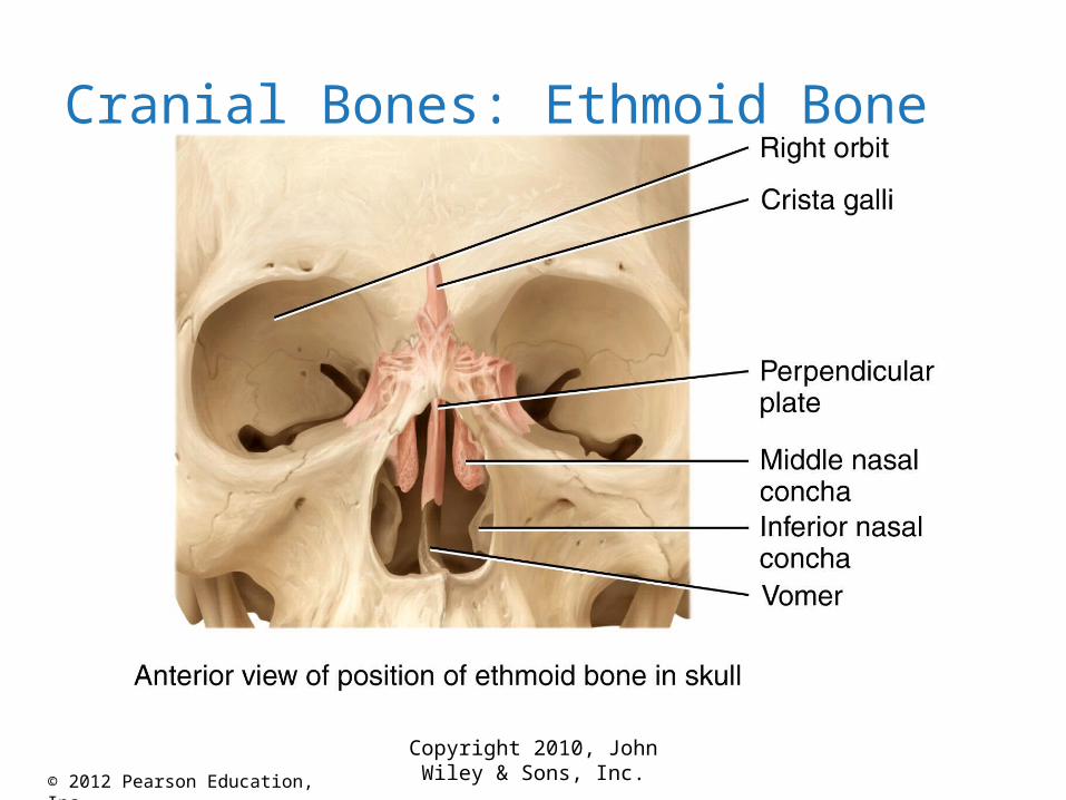
Middle nasal concha(part of ethmoid)Perpendicular plateof ethmoid Bony nasal
septumVOMER
Superior orbital fissure
PARIETAL BONE
TEMPORAL BONE
PALATINE BONE
INFERIORNASAL CONCHA
MANDIBLE
Anterior view
FRONTAL BONE

© 2012 Pearson Education, Inc.
Figure 7-3e The Adult Skull
ZYGOMATIC BONE MAXILLA
TEMPORAL BONE
PALATINE BONE
Zygomatic arch
Medial and lateralpterygoid processes
Foramen lacerum
Carotid canal
Mastoid process
Stylomastoid foramen
Occipital condyle
Foramen magnum
VOMER
SPHENOID
OCCIPITAL BONE
FRONTAL BONE
Jugular foramen
Foreman ovale
Styloid processMandibular fossa
Externalacoustic meatus
Lambdoid suture
External occipitalprotuberance
Inferior view

© 2012 Pearson Education, Inc.
Figure 7-4a The Sectional Anatomy of the Skull
PARIETAL BONE
Squamous suture
Lambdoid suture
Hypophyseal fossaof sella turcicaInternal acoustic meatus
Hypoglossal canalStyloid process
TEMPORAL BONE
OCCIPITAL BONE
Coronal suture
Sphenoidal sinus (right)
Frontal sinusCrista galli
FRONTAL BONE
SPHENOID
NASAL BONEETHMOID
VOMERPALATINE BONE
MANDIBLE
MAXILLA
Medial view of a sagittal section through the skull.

© 2012 Pearson Education, Inc.
Figure 7-4b The Sectional Anatomy of the Skull
Foramenmagnum
Carotid canal
Internal occipital crest
FRONTAL BONE
ETHMOID
SPHENOID
TEMPORAL BONE
PARIETAL BONE
OCCIPITAL BONE
Crista galli
Cribriform plate
Sella turcica
Foramen rotundum
Foramen lacerum
Foramen ovale
Foramen spinosum
Internalacoustic meatus
Jugular foramen
Hypoglossal canal
Superior view of a horizontal section through the skull, showing thefloor of the cranial cavity. Compare with part (a) and with Figure 7–3e.
© 2012 Pearson Education, Inc.
Copyright 2010, John Wiley & Sons, Inc.
Cranial Bones: Ethmoid Bone

© 2012 Pearson Education, Inc.
Figure 7-15a The Skull of an Infant
Sphenoidalfontanelle
NASAL BONE
MAXILLA
Coronalsuture
FRONTALBONE
PARIETALBONE
Squamous suture
Lambdoid suture
OCCIPITAL BONE
Mastoidfontanelle
TEMPORALBONE
SPHENOID
MANDIBLE
Lateral view

© 2012 Pearson Education, Inc.
Figure 7-15b The Skull of an Infant
PARIETALBONE
FRONTALBONE
Sagittal sutureAnteriorfontanelle
Frontal sutureCoronalsuture
FRONTALBONE PARIETAL
BONE
Lambdoidsuture
OCCIPITALBONE
OccipitalfontanelleSuperior view
© 2012 Pearson Education, Inc.
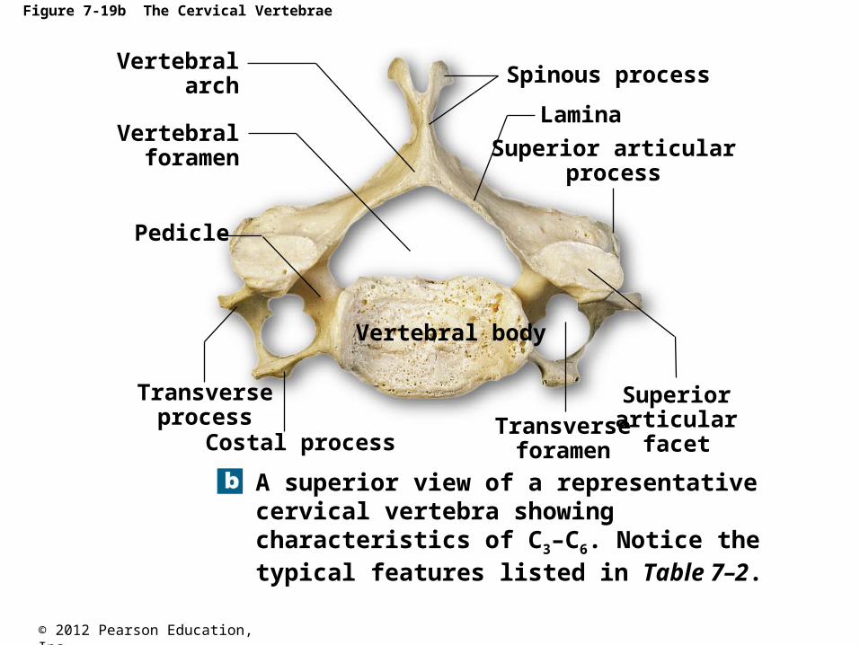
Figure 7-19b The Cervical Vertebrae
Pedicle
A superior view of a representativecervical vertebra showingcharacteristics of C3–C6. Notice thetypical features listed in Table 7–2.
Costal process
Transverseprocess
Vertebral body
Transverseforamen
Superiorarticular
facet
Superior articularprocess
Lamina
Spinous processVertebral
arch
Vertebralforamen

© 2012 Pearson Education, Inc.
Figure 7-19d The Cervical Vertebrae
Axis (C2)
Posteriorarch
Atlas (C1)
Dens ofaxis
Anteriorarch
Transverseligament
The atlas (C1) and axis (C2).

© 2012 Pearson Education, Inc.
Figure 7-20c The Thoracic Vertebrae
Spinousprocess
Transverseprocess
Transverse costalfacet for tubercle
of superior rib
Superiorarticular facet
Superior costal facetfor head of superior rib
Vertebralbody
Inferior costal facetfor head of inferior rib
Thoracic vertebra, lateral view.

© 2012 Pearson Education, Inc.
Figure 7-21b The Lumbar Vertebrae
Inferior articular process
Vertebralbody
PedicleSuperior articularprocess
Transverse process
Spinousprocess
Inferior articular facet
A lateral view of a typical lumbar vertebra

© 2012 Pearson Education, Inc.
Figure 7-22c The Sacrum and Coccyx
An anterior view
Apex
Transverselines
AlaAla
Base
Sacralforamina
Sacralpromontory
Coccyx
© 2012 Pearson Education, Inc.
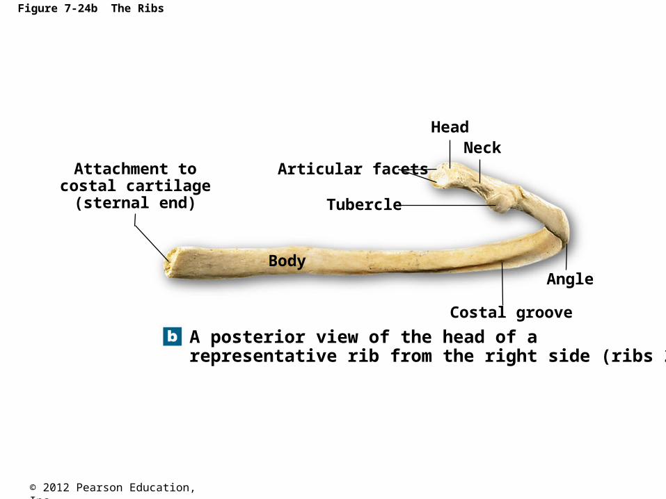
Figure 7-24b The Ribs
Attachment tocostal cartilage
(sternal end)
Articular facets
Body
Tubercle
HeadNeck
Angle
Costal groove
A posterior view of the head of a representative rib from the right side (ribs 2–9).

© 2012 Pearson Education, Inc.
Copyright 2010, John Wiley & Sons, Inc.
Vertebral Column: Ribs (Costal Bones)

© 2012 Pearson Education, Inc.
Figure 7-23a The Thoracic Cage
Jugular notch
Clavicular articulation
Manubrium
Sternum Body
Xiphoidprocess
Costalcartilages
An anterior view, showing thecostal cartilages and the sternum
True ribs(ribs 1–7)
False ribs(ribs 8–12)
Floating ribs(ribs 11–12)
Vertebrochondralribs
(ribs 8–10)
1
2
3
4
5
6
7
8
9
10
11
12
T12
T11
T1